Back to Journals » Clinical Ophthalmology » Volume 20
The Eight‑Chop Technique in Cataract Surgery: A Conceptual and Narrative Review of a Segmentation‑First Strategy
Authors Sato T ![]()
Received 12 May 2026
Accepted for publication 26 June 2026
Published 3 July 2026 Volume 2026:20 624182
DOI https://doi.org/10.2147/OPTH.S624182
Checked for plagiarism Yes
Review by Single anonymous peer review
Peer reviewer comments 4
Editor who approved publication: Dr Bharat Gurnani
Tsuyoshi Sato
Department of Ophthalmology, Sato Eye Clinic, Matsudo-shi, Chiba-ken, Japan
Correspondence: Tsuyoshi Sato, Sato Eye Clinic, Nemoto 3-3, Matsudo-shi, 271-0077, Chiba-ken, Japan, Tel +81-47-702-9822, Fax: +81-47-602-6081, Email [email protected]
Purpose: To review the conceptual evolution, mechanical principles, and clinical performance of the Eight-Chop Technique and to clarify its role within contemporary cataract surgery.
Methods: This narrative, concept‑driven review summarizes the historical development of nuclear fragmentation strategies, including sculpting-based techniques, divide-and-conquer, chop-based methods, femtosecond laser–assisted cataract surgery, and prechop techniques. A non‑systematic literature search of PubMed and Google Scholar was performed for articles published between 1991 and 2025 using combinations of the terms “cataract”, “phacoemulsification”, “nuclear fragmentation”, “phaco-chop”, “prechop”, and “eight-chop”. Adult human cataract surgery studies and key conceptual articles on nuclear segmentation and intraoperative fluidics were selectively included. Because most available clinical reports originate from the originating surgeon and closely affiliated groups, the evidence base is limited and heterogeneous, and formal systematic evidence synthesis was not attempted. Particular attention was directed toward the wedge-induced fracture mechanism, geometric optimization through eightfold nuclear division, and compatibility with modern fluidics systems. Published clinical studies together with the author’s clinical experience were reviewed across a broad spectrum of cataract subtypes.
Results: In both standard cataracts and challenging conditions—including hard nuclear cataracts, white cataracts, small pupils, shallow anterior chamber, microcornea, diabetic eyes, and pseudoexfoliation syndrome—the Eight-Chop Technique has been reported to be associated with reduced phaco time, cumulative dissipated energy, and irrigation volume compared with conventional techniques. Corneal endothelial cell density loss was generally limited to approximately 0.9– 6.7%, even in high-risk subgroups. Postoperative intraocular pressure also demonstrated sustained reduction during mid- to long-term follow-up. These findings are encouraging but preliminary and may partly reflect surgeon experience, case selection, and contemporary phacoemulsification platforms.
Conclusion: The Eight‑Chop Technique is a segmentation‑first nuclear fragmentation strategy based on complete in‑the‑bag prefragmentation using a wedge‑induced fracture mechanism. Its conceptual compatibility with modern fluidics systems, including active fluidics platforms, may contribute to improved anterior chamber stability and a minimally invasive surgical profile. However, because current evidence is derived largely from single‑surgeon and closely affiliated studies, the clinical advantages of the Eight‑Chop Technique should be regarded as provisional. Independent, multicenter validation and randomized comparative trials are required before definitive conclusions can be drawn regarding its overall clinical superiority and generalizability.
Keywords: cataract surgery, phacoemulsification, nuclear fragmentation, segmentation-first, eight-chop technique
Introduction
Phacoemulsification has become the standard procedure for modern cataract surgery worldwide, and its safety and efficiency have largely depended on strategies for crystalline lens nuclear fragmentation.1,2
Because the quality of nuclear fragmentation is closely associated with stabilization of intraoperative parameters, reduction of postoperative inflammation, preservation of corneal endothelial cell density (CECD), reduction of intraoperative complications, and earlier recovery of visual function,3–5 the evolution of nuclear fragmentation techniques has played a central role in improving the safety of cataract surgery.
Looking back at the history of nuclear fragmentation, early sculpting techniques required substantial amounts of ultrasound energy for nuclear removal and were associated with increased corneal endothelial damage.6 Subsequently, the introduction of divide-and-conquer by Gimbel established the concept of segmentation based on mechanical fracture.2 Thereafter, techniques such as stop-and-chop and phaco-chop were proposed and became widely adopted in contemporary practice.7–10 These conventional fragmentation techniques have enabled many surgeons to achieve excellent and reproducible outcomes, low complication rates, and favorable endothelial preservation in routine cataract surgery and even in many complex cases. In addition, recent advances in phacoemulsification platforms, including active-fluidics systems, have markedly improved anterior chamber stability across a broad range of surgical settings. Thus, these conventional methods currently represent well-established standard approaches in modern cataract surgery.
However, many conventional techniques do not presuppose complete mechanical nuclear division before the initiation of phacoemulsification and commonly rely on partial fragmentation within the anterior chamber. As a result, issues such as increased ultrasound energy, reduced aspiration efficiency, and unnecessary mechanical stress on the posterior capsule and zonular apparatus may still occur.11–13 Although such limitations do not preclude excellent outcomes with conventional methods, they may remain clinically relevant in selected high-risk situations such as hard nuclear cataracts, white cataracts, poor pupillary dilation, shallow anterior chamber, microcornea, diabetic eyes, and pseudoexfoliation syndrome.14–18
In contrast, recently introduced Active Fluidics Systems (AFS) have enabled phacoemulsification under low intraocular pressure (IOP) conditions and have markedly improved anterior chamber stability.19,20 However, under low IOP settings, fragment followability and occlusion stability become increasingly important determinants of surgical efficiency and chamber stability, making fragment size and geometry critical factors during phacoemulsification.21,22 In this context, a nuclear segmentation strategy compatible with modern fluidics technology may be increasingly important.
Against this background, the Eight-Chop Technique, originally described by the author, was developed as a distinct segmentation-first surgical strategy intended to complement, rather than replace, these established techniques. The Eight-Chop Technique was developed based on several fundamental surgical principles. First, technically demanding maneuvers should ideally be performed under conditions that maximize visibility and anterior chamber stability. Second, ultrasound energy should primarily be used for nuclear removal rather than nuclear division. Third, mechanical stress on the posterior capsule and zonules should ideally be minimized through surgical design. Fourth, maintaining a consistent surgical sequence, even in anatomically or pathologically challenging cases, may contribute to improved surgical safety.
Based on these principles, the Eight-Chop Technique was devised as a “segmentation-first” surgical concept in which complete intrabag nuclear division is achieved before phacoemulsification.23 The fragmentation principle of this technique applies concepts from fracture mechanics based on the wedge-induced fracture mechanism,24 while also aiming to optimize fragment controllability and aspiration efficiency through geometric division into eight segments. For routine nuclei (Emery-Little Grade I–III), a one-handed technique without the use of a second instrument is adopted as the standard, and subsequent phacoemulsification is completed using the phaco tip alone.23 Only in extremely hard nuclei (Emery-Little Grade IV or higher) may the Lance chopper or sustainer be used during the fragmentation process;23 however, this is limited to assisting segmentation, and the fundamental philosophy of the Eight-Chop Technique—namely, completing phacoemulsification without a second instrument after fragmentation—is consistently maintained.
In this review, we first provide an overview of the historical development of nuclear fragmentation techniques and then discuss the principles required to achieve safe and efficient nuclear segmentation from the perspectives of the wedge-induced fracture mechanism and intraoperative fluidics. We further describe the conceptual framework of the Eight-Chop Technique and discuss its clinical application and safety in various challenging cases, as well as its potential role in future cataract surgery. The purpose of this review is to present the Eight-Chop Technique not merely as a single surgical maneuver but as a clinically integrated approach to nuclear fragmentation in modern cataract surgery.
The author has performed more than 20,000 cataract surgeries over the past two decades, in the majority of which the Eight-Chop Technique has been predominantly used as the primary method of nuclear fragmentation. This long-term and consistent clinical experience has enabled evaluation of the technique across patient populations with diverse nuclear hardness and ocular comorbidities from a relatively unified surgical perspective. This narrative review aims to summarize the conceptual evolution of nuclear fragmentation strategies and to examine the mechanistic and clinical rationale of the Eight-Chop Technique in the context of contemporary phacoemulsification systems.
Therefore, a systematic synthesis of the clinical performance of the Eight-Chop Technique across a broad spectrum of cataract subtypes is needed to clarify its role among contemporary nucleofractis strategies and to guide its rational integration into modern cataract surgery.
The present narrative review aims to summarize the available clinical evidence on the Eight-Chop Technique, highlight its potential advantages and limitations relative to established fragmentation methods, and outline its significance as a segmentation-first approach in current phacoemulsification practice.
Methods
This narrative review aims to provide a clinically integrated overview of the conceptual evolution, mechanical principles, and clinical performance of the Eight-Chop Technique. Rather than serving as a systematic review or meta-analysis, this review focuses on the evolution of nuclear fragmentation strategies, fracture mechanics, geometric optimization, and compatibility with modern fluidics systems in contemporary cataract surgery.
Relevant literature related to phacoemulsification, nuclear fragmentation, fluidics, and cataract surgery was identified through searches of PubMed and Google Scholar. A non-exhaustive literature search was performed for articles published between 1991 and 2025 using combinations of the terms “cataract”, “phacoemulsification”, “nuclear fragmentation”, “phaco-chop”, “prechop”, and “eight-chop”. Adult human cataract surgery studies and key conceptual articles on nuclear segmentation and intraoperative fluidics were selectively included, while case reports or purely technical notes without clinical context were used only to clarify specific procedural aspects when appropriate. Published studies addressing chop-based techniques, prechop techniques, fluidics systems, and related clinical outcomes were considered together with the author’s long-term clinical experience using the Eight-Chop Technique.
No quantitative meta-analysis was performed. Instead, the selected literature was interpreted from the perspectives of fracture mechanics, nuclear segmentation geometry, and intraoperative fluidics in order to provide a conceptual and clinically oriented synthesis of the technique. In addition to the literature identified through the formal search up to 2025, the most recent author-derived publications on the Eight-Chop Technique published in 2026 were also considered to ensure that the conceptual and clinical discussion reflected the latest available data from the author’s series. This narrative, concept-driven approach may be subject to selection bias and limited reproducibility, and the findings should therefore be interpreted with appropriate caution.
Evolution of Nuclear Fragmentation Strategies
Sculpting-Based Techniques Before Segmentation
In the early era of phacoemulsification, sculpting techniques, in which the majority of the nucleus was removed using ultrasound energy, represented the dominant surgical approach.1,3 However, management of hard nuclei often required substantial ultrasound energy and was associated with increased corneal endothelial damage, anterior chamber instability, and prolonged surgical time.4–6 Because continuous sculpting removed the nucleus without fundamentally altering its morphology, technical limitations gradually became apparent, and the need for more efficient and less invasive nuclear management strategies became increasingly recognized.
Divide-and-Conquer: Establishment of the Segmentation Concept
The divide-and-conquer technique introduced by Gimbel represented a major advance by systematically introducing the concept of nuclear segmentation through groove creation and quadrant division.2 However, groove formation still required substantial ultrasound energy, and the technique remained partially dependent on sculpting maneuvers that may impose stress on the posterior capsule and zonular apparatus.4–6 In addition, nuclear division required simultaneous manipulation of the phaco tip and a second instrument, resulting in a relatively steep learning curve.
Because nuclear division in this method primarily relies on shear forces, fracture efficiency may be lower than that achieved using wedge-induced fracture mechanisms. Furthermore, because fragmentation is performed within the irrigated anterior chamber, unnecessary contact with the capsular bag, iris, or corneal endothelium must be carefully avoided in a potentially unstable surgical environment.11–13 During manipulation, traction through the main incision and side ports, as well as fluid leakage from the wounds, may further contribute to anterior chamber instability.
In hard nuclei, incomplete fracture of the posterior plate may occasionally occur, resulting in increased anterior chamber manipulation during emulsification and potentially increasing the risk of complications.13
Stop-and-Chop: A Hybrid Technique Combining Sculpting and Chopping
In 1994, Koch proposed the stop-and-chop technique as a modification of the chopping technique introduced by Nagahara.7 This technique is characterized by a stepwise approach in which the nucleus is first divided into two halves using groove creation and cracking, followed by further division of each hemi-nucleus using horizontal chopping.7–9
Stop-and-chop achieved wide clinical acceptance because it reduced the technical difficulty associated with pure chopping techniques. However, because it still depends on the space created by central sculpting and largely follows the workflow of divide-and-conquer, its ability to fully reduce ultrasound energy and maximize fragmentation efficiency may be limited compared with pure chop techniques.9,10 In particular, hard nuclei often require additional manipulation, making increased anterior chamber maneuvers difficult to avoid.10
Phaco-Chop: Advancement of Mechanical Fragmentation
Phaco-chop, introduced by Nagahara, represented an important transition from sculpting-based nuclear management to a technique emphasizing mechanical separation along the natural fiber arrangement of the nucleus.9 By approximating the tips of two instruments horizontally, the nucleus can be mechanically divided while reducing the ultrasound energy required for nuclear management.9,10 This energy efficiency is based on propagation of natural cleavage planes along the lamellar structure of lens nuclear fibers.
Nevertheless, certain limitations remain. Initial impalement of the phaco tip into the central nucleus still requires ultrasound energy, and dependence on ultrasound may increase as nuclear hardness rises.13 In addition, fragmentation requires coordinated manipulation of the phaco tip and an auxiliary instrument, and acquisition of the technique generally requires surgical experience. Furthermore, because the mechanism relies primarily on shear-dominant forces, incomplete fracture of the posterior plate may occur in hard nuclei, requiring additional lateral separation maneuvers.11–13 As a result, mechanical stress may be transmitted to the posterior capsule and zonular apparatus.
Phaco Prechop: Presentation of a Pre-Phaco Segmentation Concept and Its Limitations
Phaco prechop, proposed by Akahoshi, was developed as a technique to mechanically divide the lens nucleus prior to phacoemulsification.25 This prechop approach represented an important conceptual advance because it aimed to complete nuclear segmentation before phacoemulsification and may be regarded as one of the conceptual predecessors of the Eight-Chop Technique.
In cases with high nuclear hardness, equatorial support using a sustainer is often required to reduce transmission of force to the capsular bag and zonules. In addition, nuclear debris generated after initial division may reduce visibility during subsequent maneuvers, and because fragmentation is generally limited to four segments, fragment mobility and followability may remain insufficient. Furthermore, determination of insertion depth depends substantially on surgical experience, and both insufficient and excessive insertion may affect surgical safety. Maneuverability of the prechopper may also be challenging for less experienced surgeons, particularly in hard nuclei.
Thus, while phaco prechop introduced the important concept of intrabag nuclear segmentation prior to phacoemulsification, several procedural and technical factors may have limited broader adoption. Nevertheless, this concept shares conceptual similarities with the Eight-Chop Technique and can be viewed as an important step in the evolution of prefragmentation strategies.
Positioning and Limitations of Nuclear Pretreatment in Femtosecond Laser-Assisted Cataract Surgery
Nuclear pretreatment in femtosecond laser-assisted cataract surgery represents another strategy aimed at dividing the nucleus before phacoemulsification, and reductions in effective phaco time and cumulative dissipated energy (CDE) have been reported.18 However, consistent superiority over conventional manual cataract surgery has not been established for major outcomes such as postoperative visual acuity, refractive results, CECD preservation, or complication rates.18 This may reflect the fact that final nuclear removal still depends on phacoemulsification itself.
In addition, laser-pretreated fragments do not always achieve stable phaco tip occlusion, and reduced vacuum efficiency—particularly with peristaltic systems—has been reported.26 Therefore, while femtosecond laser-assisted cataract surgery offers rational advantages as a pretreatment strategy, several challenges related to nuclear management remain.
Common Procedural Characteristics and Limitations of Conventional Techniques
Conventional nuclear fragmentation techniques share several procedural characteristics and limitations. First, shear-force–dominant fragmentation may result in incomplete posterior plate separation and nonuniform fragment geometry. Consequently, fragment followability and occlusion stability may become suboptimal under modern fluidics conditions.20–22 Second, because fragmentation is performed within the irrigated anterior chamber, surgeons must avoid unnecessary contact with the capsule, iris, and corneal endothelium in an environment where chamber stability is not always fully secured. Traction through surgical incisions and fluid leakage may further contribute to chamber instability and intraoperative risk.19,21 Third, simultaneous manipulation of the phaco tip and a second instrument may increase procedural complexity and potentially transmit unnecessary mechanical stress to the zonules and posterior capsule.11,12 These issues may become more clinically relevant in high-risk eyes such as pseudoexfoliation syndrome, shallow anterior chamber, or microcornea.14–18
Positioning of the Eight-Chop Technique: A Segmentation Strategy Addressing Conventional Limitations
The Eight-Chop Technique was developed to address some of the limitations associated with conventional segmentation techniques. Its core principles include full-thickness fracture based on the wedge-induced fracture mechanism24 and geometric optimization through division into eight segments to improve fragment controllability and compatibility with modern fluidics systems. Complete prefragmentation facilitates early fragment mobilization, while reduced dependence on a second instrument may contribute to both surgical safety and procedural efficiency. In this respect, the Eight-Chop Technique can be viewed as part of the ongoing evolution of segmentation strategies in modern cataract surgery.23 Within the broader landscape of nucleofractis techniques, it represents a segmentation-first approach that conceptually extends earlier prechop and chop methods and is intended to complement, rather than replace, other established nuclear fragmentation strategies.
Conceptual Framework of the Eight-Chop Technique
Fundamental Philosophy of the Eight-Chop Technique: Strategic Shift Through Complete Prefragmentation
The most fundamental characteristic of the Eight-Chop Technique lies in its strategic concept of dividing the lens nucleus into eight independent small fragments prior to phacoemulsification. Through this complete prefragmentation strategy, the workflow of nuclear management becomes substantially different from that of conventional techniques. Whereas traditional methods such as divide-and-conquer, stop-and-chop, and phaco-chop generally involve simultaneous fragmentation and phacoemulsification within the anterior chamber, the Eight-Chop Technique completes nuclear segmentation before the phacoemulsification stage.2,7–10,23 This workflow modification may contribute to improved anterior chamber stability, reduced manipulation, enhanced safety margins, and greater procedural efficiency during phacoemulsification.23 The conceptual difference between conventional phaco-chop and the Eight-Chop Technique is illustrated in Figure 1.
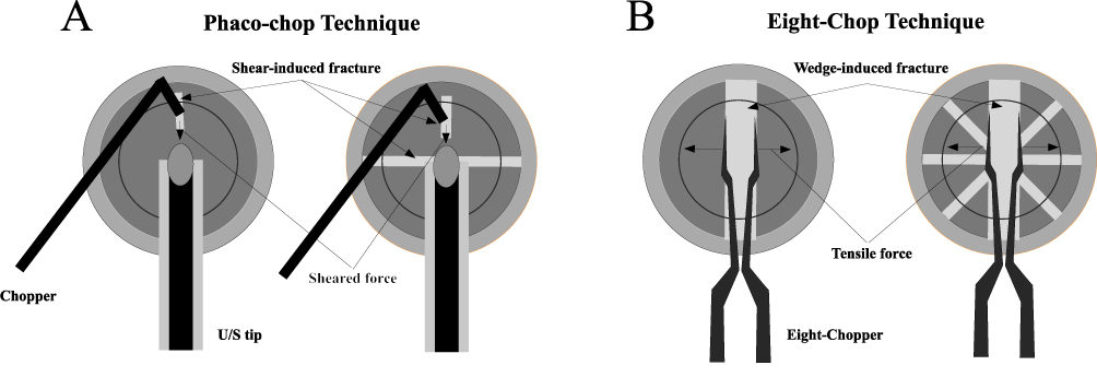 |
Figure 1 Conceptual comparison between the phaco-chop and Eight-Chop techniques. (A) In conventional phaco-chop, the ultrasound tip is embedded into the central lens nucleus to provide countertraction, and the chopper is advanced toward the tip to mechanically cleave the nucleus by shear-induced fracture, creating and extending a fracture plane while relatively large nuclear fragments are emulsified as they are generated. (B) In the Eight-Chop Technique, a specialized Eight-Chopper is inserted into the central nucleus and then opened to act as a wedge; this wedge-induced fracture mechanism propagates controlled radial cracks through the full thickness of the nucleus within the capsular bag, enabling complete prefragmentation into multiple small, independent segments before phacoemulsification. |
Wedge-Induced Fracture Mechanism: The Mechanical Foundation of the Eight-Chop Technique
Nuclear fragmentation in the Eight-Chop Technique is based on the wedge-induced fracture mechanism, which differs conceptually from the shear-dominant separation used in conventional chop techniques. The essence of this fracture mechanism lies in advancing the instrument tip into the nucleus and then opening it to generate a wedge effect, thereby producing tensile components within the nucleus and propagating these forces through the full thickness of the nuclear structure.24
In conventional divide-and-conquer, stop-and-chop, and phaco-chop techniques, successful nuclear division depends largely on shear or opposing forces generated by the second instrument and phaco tip, and these forces may be transmitted to the posterior capsule and zonular apparatus.2,7–10,23 In contrast, because the Eight-Chop Technique completes nuclear segmentation prior to phacoemulsification through wedge-induced fracture, there is less need to spread or tear the nucleus during emulsification. As a result, the technique may help reduce mechanical stress applied to the posterior capsule and zonules, even in nuclei of ordinary hardness, although this potential advantage has mainly been suggested by single-surgeon experience to date rather than by direct biomechanical measurements or randomized comparative studies. This characteristic may have particular relevance in eyes with pseudoexfoliation syndrome, shallow anterior chamber (SAC), or microcornea, where supporting structures are fragile or the working space is restricted.11,12
In cases with high nuclear hardness (Grade IV or higher), the presence of a posterior plate may impede fragmentation; however, the wedge-induced fracture mechanism may remain effective even under such conditions. With instruments such as the Eight-Chopper II or Lance-Chopper, improved tip design facilitates nuclear penetration, and the wedge effect generated by opening the instrument can be transmitted toward the posterior plate. Consequently, full-thickness fracture may be achieved in a relatively consistent manner even in hard nuclei that are often difficult to manage using conventional shear-based chop techniques.23,24,27
Through this mechanical framework, the Eight-Chop Technique may reduce the amount of manipulation required during both nuclear fragmentation and phacoemulsification, thereby contributing to improved intraoperative stability and a wider surgical safety margin. In particular, reduction of posterior capsule and zonular stress during routine cataract surgery may support the positioning of the Eight-Chop Technique as a broadly applicable nuclear fragmentation strategy.23
Segmentation Strategy of Eightfold Division in the Eight-Chop Technique: Geometric Rationale
The “eightfold division” employed in the Eight-Chop Technique may represent a practical and reproducible segmentation strategy derived from consideration of nuclear structure, operative space, and fluidics characteristics in modern phacoemulsification systems.
Importantly, the number eight does not represent a rigid requirement, but rather a practical balance between fragment size and intraoperative controllability.23 In the Eight-Chop Technique, the resulting nuclear fragments do not need to be perfectly uniform, and minor variability according to intraoperative conditions does not appear to compromise safety or efficiency. The essential requirement is that each fragment remains sufficiently small and controllable as an independent unit. The sequential intraoperative steps of achieving eightfold nuclear division in the Eight-Chop Technique are shown in Figure 2.
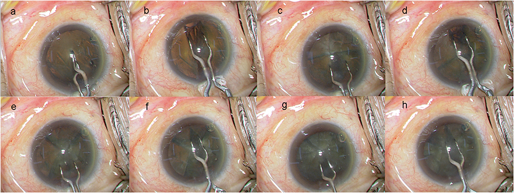 |
Figure 2 Sequential intraoperative photographs of the Eight-Chop Technique. (a) Introduction of the Eight-Chopper into the central lens nucleus. (b) Initiation of wedge-induced fracture within the nucleus. (c) Further division of the inferior heminucleus at the 6-o’clock position. (d) Further division of the superior heminucleus at the 12-o’clock position. (e) Secondary division of the inferior nuclear segment. (f) Secondary division of the superior nuclear segment. (g) Final subdivision of the remaining inferior segment. (h) Final subdivision of the remaining superior segment, completing eightfold division into small, independently mobile nuclear fragments within the capsular bag. |
In eyes with normal nuclear density (Emery-Little Grade I–III), nuclei divided into eight fragments exhibit high mobility within the anterior chamber, allowing aspiration to proceed while maintaining an adequate safety margin from the corneal endothelium, iris, and posterior capsule. Reduction in fragment volume improves followability and facilitates stable occlusion at the phaco tip, thereby enhancing emulsification and aspiration efficiency.21,22 As a result, the extent of instrument manipulation within the anterior chamber may be reduced, contributing to lower intraoperative fluid fluctuation and decreased risk of unintended tissue contact.
Small fragment size may also be important for surgical stability under low IOP conditions. With increasing adoption of active fluidics systems, anterior chamber stability has become highly dependent on the behavior of nuclear fragments. The small fragments generated through eightfold division are more easily controlled from a fluid-dynamic perspective, potentially suppressing surge events while contributing to reductions in irrigation volume and cumulative ultrasound energy.19–22
Even in cases with high nuclear hardness (Grade IV or higher), the eightfold division strategy may retain its effectiveness. By limiting the volume and thickness of each fragment, nuclear pieces may be more readily mobilized from within the capsular bag, and graspability with the phaco tip may improve. Consequently, emulsification and aspiration may be performed while minimizing unnecessary contact with the corneal endothelium, even in hard nuclei.23,27
From a practical standpoint, repeated bisection makes division into six fragments geometrically difficult, whereas subdivision beyond eight fragments may increase procedural complexity and reduce efficiency. Taken together, eightfold division may represent a practical balance among divisibility, maneuverability, and surgical efficiency, without requiring substantial modification according to nuclear hardness.23,28
In summary, the eightfold division employed in the Eight-Chop Technique is not limited to specific cataract subtypes or nuclear densities, but rather represents a geometrically designed segmentation strategy intended to support a stable operative environment and surgical safety in routine cataract surgery.
Minimization of Dependence on a second Instrument Through Complete Prefragmentation
One of the core elements supporting the clinical safety and reproducibility of the Eight-Chop Technique is the reduction in dependence on a second instrument achieved through complete prefragmentation. In conventional nuclear fragmentation techniques such as divide-and-conquer, stop-and-chop, and phaco-chop, simultaneous shearing maneuvers using a phaco tip and a chopper (or hook) are generally required, potentially imposing lateral and tractional stress on the posterior capsule and zonular apparatus during surgery.2,7–12
In contrast, in the Eight-Chop Technique, nuclear segmentation is completed before phacoemulsification begins, reducing the need for lateral separation or forceful auxiliary instrument manipulation during the emulsification phase. As a result, nuclear fragment removal can often be performed primarily using the phaco tip alone, potentially reducing inadvertent tractional or compressive forces transmitted to the lens-supporting structures.
Importantly, this characteristic may be advantageous regardless of nuclear hardness. Even in routine nuclei (Emery-Little Grade I–III), conventional techniques often require repeated chopper manipulation for nuclear division and fragment control, whereas in the Eight-Chop Technique, small and independent nuclear fragments become mobile at an earlier stage, allowing rotation, grasping, and aspiration to proceed more naturally. This may reduce the overall amount of intraoperative manipulation and contribute to improved surgical field stability.
Furthermore, reduced use of a second instrument may contribute to stabilization of wound architecture and anterior chamber fluidics. When side-port manipulation is minimized, wound deformation and irrigation leakage associated with instrument movement may also be reduced, thereby suppressing fluctuations in anterior chamber depth.19,21 This factor may be particularly relevant in surgery performed under low IOP conditions using active fluidics systems.
In high-risk eyes such as pseudoexfoliation syndrome, shallow anterior chamber, or microcornea, manipulation with a second instrument itself may constitute a risk factor for intraoperative complications.11,12,14–18 By adopting a workflow based on complete prefragmentation, the Eight-Chop Technique may reduce some of these structural risks and provide a potentially different safety profile from that of conventional techniques.
Taken together, reduced dependence on a second instrument in the Eight-Chop Technique should be regarded not merely as simplification of surgical maneuvers, but rather as a consequence of a design philosophy intended to reduce mechanical stress on lens-supporting structures.
Integration with Fluidics, Particularly Low–Intraocular-Pressure Active Fluidics Systems
In recent phacoemulsification systems, active fluidics has become increasingly widespread, and maintenance of anterior chamber stability under low IOP conditions has become an important surgical objective.19,20 The segmentation strategy of the Eight-Chop Technique appears highly compatible with modern fluidics systems, as smaller nuclear fragments may improve followability and facilitate more stable occlusion.21,22 As a result, surge events may become less frequent, while irrigation volume, postoperative inflammation, and corneal endothelial cell damage may also be reduced.4–6,20
In the modern surgical environment, in which nuclear fragment behavior has become an important determinant of surgical performance under low IOP conditions, the Eight-Chop Technique may represent a segmentation strategy with high compatibility for stabilizing fragment dynamics within low-pressure fluidics systems.29
The Eight-Chop Technique as a Comprehensive Segmentation Strategy Integrating Mechanics, Geometry, and Fluidics
The theoretical foundation of the Eight-Chop Technique is characterized by integration of fracture mechanics,24 geometric segmentation, and modern fluidics19–22 within a unified surgical workflow. Complete prefragmentation, wedge-induced fracture, eight-segment division, and compatibility with low-IOP fluidics collectively contribute to optimization of surgical stability and procedural efficiency.
The Eight-Chop Technique may therefore be regarded not merely as a new nuclear fragmentation maneuver, but as a clinically integrated segmentation strategy incorporating mechanics, geometry, and fluidics within contemporary cataract surgery.23
Clinical Evidence
Clinical Performance in Standard Cataract Surgery
The Eight-Chop Technique has demonstrated favorable efficiency and low invasiveness in standard cataract surgery, as reported in previously published studies and the author’s clinical data.23,27,29–36 Existing studies have shown that the Eight-Chop Technique is associated with lower intraoperative ultrasound usage, cumulative dissipated energy (CDE), shorter phaco time, and reduced irrigation volume, with operative times of approximately 3.7–5.4 minutes and CDE values around 5.0–9.2.23 These values appear to be generally lower than those reported for divide-and-conquer and phaco-chop techniques, suggesting that the segmentation characteristics of the Eight-Chop Technique, which divide the nucleus into small fragments, may contribute to intraoperative energy reduction.9,10,22,23
These findings suggest that the segmentation strategy of the Eight-Chop Technique—namely, completing nuclear fragmentation prior to phacoemulsification and reducing nuclear manipulation during ultrasound emulsification—may provide reproducibility and safety across a range of nuclear hardness levels.23 In particular, reductions in anterior chamber turbulence and excessive nuclear manipulation were associated with suppression of postoperative inflammation and endothelial stress.23,29,37 Collectively, evidence from multiple published studies and the author’s clinical data suggests that the Eight-Chop Technique may represent an efficient and minimally invasive fragmentation method for standard cataract surgery in terms of anterior chamber stability, energy efficiency, and endothelial protection.23,29,37
Hard Nuclear Cataracts
Hard nuclear cataracts corresponding to Emery-Little Grades IV–V have long been regarded as among the most technically challenging cases for lens nucleus fragmentation.38–41 Due to marked thickening and sclerosis of the posterior nuclear plate, achieving full-thickness fracture from the nuclear center to the posterior pole may be difficult using conventional techniques such as divide-and-conquer, stop-and-chop, and phaco-chop.9–11,13,38–41 Consequently, repeated chopping maneuvers and lateral separation within the anterior chamber are frequently required, potentially increasing mechanical stress on the posterior capsule and zonular apparatus.11,12 In addition, prolonged ultrasound use and increased irrigation volume in hard nuclear cases are major contributors to postoperative corneal endothelial damage.4–6,13,38–41
The Eight-Chop Technique was developed to help address these mechanical and geometric limitations.23,27 In this method, the Lance chopper is inserted obliquely into the central nucleus, and the tip is gradually opened to activate the wedge-induced fracture mechanism, thereby creating a continuous deep cleavage plane extending from the nuclear center toward the posterior nuclear plate.23,24,27 This principle may facilitate full-thickness nuclear fragmentation including the posterior plate while reducing excessive lateral traction forces often required in conventional techniques.23,27
The author has evaluated the efficacy and safety of the Eight-Chop Technique in clinical studies involving Emery-Little Grade IV–V nuclear cataracts, and the results have already been reported.23,27 In these studies, octasection of the nucleus prior to phacoemulsification was achieved in the majority of cases, even in extremely hard nuclei, and intraoperative parameters were favorable. Specifically, the mean operative time was 10.5 minutes, mean phaco time was 38.9 seconds, mean aspiration time was 135.6 seconds, mean CDE was 19.2, and mean irrigation volume was 53.0 mL.27 These findings suggest relatively low invasiveness compared with previously reported outcomes of conventional techniques for hard nuclear cataracts.27
Postoperative outcomes also demonstrated favorable endothelial preservation. At 19 weeks postoperatively, CECD loss was 0.2% in Grade IV, 6.8% in Grade IV plus, and 9.6% in Grade V cases, with an overall mean of 3.7%.27 CECD loss showed significant correlations with phaco time, CDE, and irrigation volume, suggesting that complete prefragmentation by the Eight-Chop Technique may reduce intraoperative energy load and contribute to endothelial protection.27
Because nuclear fragmentation is completed prior to phacoemulsification in the Eight-Chop Technique, additional fragmentation maneuvers within the anterior chamber are minimized. Ophthalmic viscosurgical device (OVD) injected before fragmentation not only protects the corneal endothelium but also forms a physical buffer between the nucleus and posterior capsule, potentially reducing direct transmission of stress to the posterior capsule during deep fracture.4–6 Even in Grade IV–V cases, the incidence of posterior capsule rupture was low, and nuclear drop or extensive zonular dehiscence was uncommon.23,27
Taken together, the Eight-Chop Technique may represent a safe and reproducible segmentation strategy for hard nuclear cataracts by combining geometric fracture strategy with reduction of intraoperative energy load.23,27
White Cataracts
White cataracts often present with high nuclear hardness and frequently lack epinucleus. As a result, the nucleus may come into direct contact with the posterior capsule, requiring conventional techniques such as divide-and-conquer, stop-and-chop, and phaco-chop to perform deep nuclear fragmentation in an unstable irrigation environment with extremely hard nuclei and absent cortical support.42–45 Consequently, the risks of posterior capsule rupture (PCR) and zonular apparatus damage may increase substantially.45,46 Moreover, maintaining an adequate distance between the nucleus and posterior capsule is inherently difficult, limiting the surgical safety margin during deep manipulation.42,43
Although white cataract remains a technically challenging condition even with the Eight-Chop Technique, the fundamental strategy of completing segmentation entirely prior to phacoemulsification remains unchanged.23 After achieving a stable capsulorhexis, a sufficient amount of OVD is injected into the anterior chamber and capsular bag to form a protective layer between the nucleus and posterior capsule.4–6 The Eight-Chopper is then inserted into the central nucleus, and continuous cracks are propagated toward the posterior plate using the wedge-induced fracture mechanism.23,24 By standardizing the insertion angle and opening width of the Eight-Chopper while maintaining posterior capsule protection with OVD, deep segmentation may be achieved with improved safety.23
The author has previously reported the efficacy and safety of the Eight-Chop Technique in white cataract cases.36 In that study, octasection of the nucleus was achieved at a high rate prior to phacoemulsification, and intraoperative parameters were favorable. Specifically, the mean operative time was 12.8 minutes, mean phaco time was 34.0 seconds, mean aspiration time was 154.0 seconds, mean CDE was 15.30, and mean irrigation volume was 74.0 mL.36,46,47
Postoperative endothelial preservation was also favorable, with CECD loss of 6.4% at 7 weeks and 6.7% at 19 weeks,36 even in the Grade IV group.36 The incidence of posterior capsule rupture was approximately 1.9%, and severe zonular dialysis or nuclear drop was rarely observed.36 These findings suggest that complication risk in white cataracts may be associated with deep nuclear fragmentation performed under unstable perfusion conditions within the anterior chamber.
After completion of octasection, each nuclear fragment is sequentially guided into the safety zone and emulsified while maintaining sufficient distance from the corneal endothelium, iris, and posterior capsule. At the start of phacoemulsification, liquefied cortical material has already been removed, and anterior chamber visibility is maintained.26 Because the fragments are small and mobile, and anterior chamber depth is stabilized by OVD and fluidics, phacoemulsification can be performed with maneuverability comparable to that in routine cases.
Thus, the Eight-Chop Technique may help address important risk factors in white cataracts—namely, deep nuclear fragmentation in an unstable irrigation environment and direct nucleus–posterior capsule contact—through the combined use of complete prefragmentation and posterior capsule protection with OVD.23
Small Pupil Eyes
Eyes with small pupils inherently carry increased surgical risk because the operative space itself is physically restricted, bringing nuclear fragmentation and phacoemulsification maneuvers into close proximity with the iris, corneal endothelium, and posterior capsule.48–50 In conventional techniques such as divide-and-conquer, phaco-chop, and stop-and-chop, nuclear fragmentation and emulsification proceed simultaneously, requiring complex instrument maneuvering within a limited visual field and working space. This may increase the likelihood of iris injury, posterior capsule damage, and excessive zonular stress.7–10,15
As with hard cataracts and white cataracts, the fundamental strategy of the Eight-Chop Technique is to complete nuclear segmentation within the capsular bag before initiating phacoemulsification.23 After stable completion of capsulorhexis, an adequate amount of OVD is injected into both the anterior chamber and capsular bag to stabilize the anterior chamber and protect the posterior capsule. Under these conditions, the Eight-Chopper is used to repeatedly create wedge-induced fractures from the central nucleus toward the posterior aspect, dividing the nucleus into eight fragments within the capsular bag.23,24 Through this complete pre-segmentation strategy, the most technically demanding fragmentation steps are completed within the capsular bag rather than in the anterior chamber, potentially improving reproducibility and safety even in small pupil eyes.
In small pupil eyes, conventional techniques often require forceful nuclear manipulation and lateral separation using a second instrument.7–10,20 In contrast, because the fragmentation process in the Eight-Chop Technique is completed within the capsular bag, the need for additional deep nuclear fragmentation within the anterior chamber is minimized.23 During the emulsification phase, the already miniaturized nuclear fragments can be sequentially guided to the center of the anterior chamber and aspirated using minimal tip manipulation under stable fluidics conditions.23,29 As a result, mechanical contact with the iris and unnecessary traction maneuvers may be reduced.
In previously reported data by the author, the Eight-Chop Technique performed in small pupil cases with iris hooks demonstrated favorable intraoperative parameters, including a mean operative time of 10.6 minutes, mean phaco time of 20.7 seconds, mean aspiration time of 101.1 seconds, mean CDE of 7.8, and mean fluid usage of 38.0 mL.51 Reduction in CECD was limited to 2.1% at 19 weeks postoperatively.51 Furthermore, IOP showed significant reduction at 7 and 19 weeks postoperatively, suggesting that this technique may be minimally invasive to the intraocular environment even in small pupil eyes.51
Thus, the Eight-Chop Technique may help address the inherent challenges of small pupil eyes—namely, restricted surgical space and excessive instrument maneuvering—through a workflow that separates segmentation from emulsification while using prefragmentation into small nuclear pieces as a dual safety mechanism.23 In this context, the Eight-Chop Technique may represent a practical alternative to conventional approaches in small pupil surgery.
Shallow Anterior Chamber Eyes
Eyes with a shallow anterior chamber (SAC) inherently present increased risk of corneal endothelial damage because the restricted operative space brings the phaco tip into close proximity with the corneal endothelium, increasing susceptibility to mechanical contact as well as thermal and fluidic stress.5,15,18 In conventional techniques such as divide-and-conquer and phaco-chop, nuclear fragmentation and emulsification are performed simultaneously within the anterior chamber, potentially limiting maneuverability in shallow chambers and increasing the risk of CECD loss.7–10,18
As with hard cataracts, white cataracts, and small pupil eyes, the fundamental principle of the Eight-Chop Technique is to complete nuclear segmentation within the capsular bag before initiating phacoemulsification.23 After stable completion of capsulorhexis, sufficient OVD is injected into both the anterior chamber and capsular bag to help maintain anterior chamber depth and create a protective buffer between the corneal endothelium and the nucleus.4–6 Subsequently, the Eight-Chopper is inserted into the central nucleus, and wedge-induced fracture mechanisms are used to divide the nucleus into eight fragments within the capsular bag.23,24 This approach allows the most technically demanding deep nuclear fragmentation steps to be completed within the capsular bag rather than in the anterior chamber.
Clinical data also support the effectiveness and safety of the Eight-Chop Technique in SAC eyes. In a study comparing 80 SAC eyes with anterior chamber depth <3.0 mm and 80 control eyes, the SAC group demonstrated a mean operative time of 4.7 ± 1.1 minutes, mean phaco time of 15.4 ± 6.1 seconds, mean aspiration time of 65.6 ± 17.3 seconds, mean CDE of 5.87 ± 2.01, and mean fluid usage of 26.6 ± 8.1 mL. Most of these parameters showed no significant differences compared with the control group with deeper anterior chambers.32
From the perspective of corneal endothelial protection, CECD loss in the SAC group remained limited, with reductions of 1.3% at 7 weeks, 1.1% at 19 weeks, and 0.9% at 1 year postoperatively.32 These values are lower than many previously reported outcomes in SAC cases treated with conventional techniques.4–6,32 Furthermore, no significant correlation was observed between anterior chamber depth and CECD loss, suggesting that anatomical shallowness alone may not be the primary determinant of endothelial damage.32
In contrast, a significant correlation was observed between CDE and CECD loss, indicating that endothelial damage may be more strongly associated with intraoperative energy load than with anatomical factors.4–6 These findings are consistent with the concept of the Eight-Chop Technique, in which complete prefragmentation may reduce ultrasound energy requirements and help maintain a stable surgical environment even under anatomically challenging SAC conditions.
Accordingly, the Eight-Chop Technique may represent a useful approach for SAC eyes by combining complete in-the-bag segmentation with spatial buffering using OVD.23 As with hard cataracts, white cataracts, and small pupil eyes, this workflow may help reduce intraoperative stress associated with restricted operative space and endothelial proximity.
Microcornea Eyes
Eyes with microcornea are regarded as particularly challenging in cataract surgery because the small corneal diameter restricts the anterior segment working space, bringing nuclear manipulation and phacoemulsification into close proximity with the corneal endothelium, iris, and posterior capsule.16,52,53 Even when axial length and anterior chamber depth are within normal ranges, the small corneal diameter alone may significantly limit intraoperative maneuverability, making conventional divide-and-conquer and phaco-chop techniques more difficult within the crowded anterior chamber environment.3,7–10
As with other challenging cataract subtypes, the fundamental principle of the Eight-Chop Technique is to complete nuclear segmentation within the capsular bag before phacoemulsification.23 After stable completion of continuous curvilinear capsulorhexis, sufficient OVD is injected into the anterior chamber and capsular bag to help maintain a safe distance from the corneal endothelium. The Eight-Chopper is then inserted into the central nucleus, and the nucleus is divided into eight fragments via the wedge-induced fracture mechanism, allowing deep nuclear fragmentation to be completed within the capsular bag rather than in the confined anterior chamber.23,24
The effectiveness of the Eight-Chop Technique in microcornea eyes is supported by clinical data. In a study of 52 microcornea eyes with corneal diameters ≤10 mm compared with 52 control eyes, the microcornea group showed a mean operative time of 5.9 ± 2.4 minutes, mean phaco time of 17.9 ± 8.9 seconds, mean aspiration time of 77.0 ± 27.2 seconds, mean CDE of 7.06 ± 3.97, and mean fluid usage of 31.1 ± 11.6 mL.34 Although operative time and fluid usage were significantly higher than in controls, there were no significant differences in phaco time, aspiration time, or CDE, suggesting preserved surgical efficiency despite anatomically restricted conditions.34
From the standpoint of corneal endothelial protection, postoperative CECD loss in the microcornea group was mild, at 3.6% at 7 weeks and 1.5% at 19 weeks, with no significant differences compared to controls (2.3% and 1.4%, respectively).34 These values are markedly lower than the 4.2–32.0% CECD loss reported for conventional techniques in normal eyes,54,55 suggesting that the structural disadvantage of small corneal diameter is effectively neutralized by the Eight-Chop Technique.
Moreover, despite significantly shallower anterior chamber depth and smaller pupil diameter in the microcornea group, no serious intraoperative complications such as posterior capsule rupture, zonular dialysis, or dropped nucleus were observed.34 These findings suggest that the segmentation-first workflow of the Eight-Chop Technique can be applied safely even in eyes with restricted anterior segment space.
In summary, the Eight-Chop Technique may represent a practical segmentation strategy for microcornea eyes by combining complete in-the-bag segmentation with OVD-assisted spatial protection.34 The core principles of the technique appear applicable even under anatomically challenging conditions, supporting its potential usefulness in safe and reproducible cataract surgery.
Diabetic Eyes
Diabetic eyes are known to carry increased risk of corneal endothelial damage during cataract surgery.17,56–58 Under hyperglycemic conditions, accumulation of transforming growth factor-β–induced proteins and advanced glycation end products may contribute to structural and functional impairment of corneal endothelial cells.17 In addition, increased deposition of extracellular matrix components and reduced Na⁺/K⁺-ATPase activity may compromise endothelial pump function, resulting in reduced endothelial reserve even before surgery.4,5 Consequently, diabetic patients may be more vulnerable to mechanical stress caused by ultrasound energy and irrigation fluid during cataract surgery.56–58
The Eight-Chop Technique is based on the principle of mechanically dividing the lens nucleus into eight fragments prior to phacoemulsification, thereby reducing ultrasound energy exposure and irrigation volume.23 In evaluations of this technique in diabetic eyes, intraoperative parameters included operative times of 4.6 minutes, phaco time of 15.5 seconds, CDE of approximately 6.5, and irrigation volume of 27.0 mL.31 CECD loss was 5.1% at 7 weeks postoperatively, 3.9% at 19 weeks, and 2.1% at 1 year, representing relatively favorable endothelial outcomes compared with previously reported diabetic cataract surgery data.31,56,57
When compared with control eyes, diabetic eyes showed slightly greater short-term reduction in CECD; however, no significant difference was observed in the long term. These findings suggest that reduction of ultrasound energy and irrigation load may contribute to protection of the vulnerable corneal endothelium in diabetic eyes.31
Furthermore, because diabetic patients have a higher prevalence of glaucoma, postoperative IOP changes are clinically important.14,56,57 With the Eight-Chop Technique, an 8.0% reduction in IOP was observed at 1 year postoperatively even in diabetic eyes.31 This IOP-lowering effect may partly reflect reduced irrigation volume and ultrasound exposure, potentially minimizing stress on aqueous outflow tissues such as the trabecular meshwork and Schlemm’s canal.14,19–21
In summary, although diabetic eyes present inherent vulnerability due to preexisting endothelial dysfunction, the Eight-Chop Technique may represent a useful surgical option for reducing intraoperative energy load while supporting both corneal endothelial protection and postoperative IOP stability.
Pseudoexfoliation Syndrome
Pseudoexfoliation (PEX) syndrome is a systemic fibrotic disorder characterized by deposition of abnormal fibrillar extracellular material in anterior segment tissues and lens-supporting structures, and it is regarded as a challenging condition in cataract surgery.11,12 Eyes with PEX frequently present with reduced CECD, small pupils, anterior chamber instability, and zonular weakness.11,12 Consequently, complications such as poor nuclear rotation, capsular instability, posterior capsule rupture, vitreous prolapse, and intraocular lens decentration may occur more frequently.11
The Eight-Chop Technique was designed with the principle of minimizing intraoperative zonular stress in such eyes.23 In conventional divide-and-conquer and phaco-chop techniques, nuclear manipulation and rotation are often required during fragmentation, potentially increasing tensile stress on weakened zonules.11–13 In contrast, in the Eight-Chop Technique, the nucleus is maintained in a stable position within the capsular bag, and deep nuclear fracture is induced from the nuclear center toward the posterior plate using a wedge-induced fracture mechanism, allowing octasection to be completed before phacoemulsification.23,24 Consequently, excessive nuclear rotation and lateral manipulation may be reduced.
Clinical data on the use of the Eight-Chop Technique in PEX eyes demonstrated favorable intraoperative parameters, including operative time of 6.7 minutes, phaco time of 17.4 seconds, aspiration time of 85.2 seconds, CDE of 6.91, and irrigation fluid use of 33.4 mL.30 Although some values differed significantly from controls, the overall intraoperative profile remained favorable even in PEX eyes.30
Notably, although intraoperative zonular weakness was identified in 13 of 75 PEX eyes (17.3%), insertion of a capsular tension ring (CTR) was not required in any case.30 This finding may reflect reduced stress on the capsule and zonules achieved through the segmentation-first workflow and minimized rotational manipulation.23 Postoperatively, no cases of clinically significant intraocular lens decentration or extensive zonular complications were observed.30
From the perspective of endothelial protection, CECD loss rates were 3.7% at 7 weeks and 2.7% at 19 weeks postoperatively,30 remaining lower than many previously reported outcomes in PEX cataract surgery.4,11 These findings may be associated with reduced ultrasound energy and irrigation volume.
Postoperative IOP management is also particularly important in PEX eyes. Significant IOP reductions of approximately 13% were observed postoperatively in both the PEX and control groups.30 This finding may reflect the relatively low-energy and low-irrigation characteristics of the Eight-Chop Technique and its compatibility with stable fluidics environments.
Taken together, the Eight-Chop Technique may represent a useful surgical approach for performing cataract surgery in PEX eyes while minimizing zonular stress and maintaining favorable intraoperative stability.
Glaucoma and Intraocular Pressure Changes
Changes in IOP after cataract surgery in glaucomatous eyes are closely associated with the degree of mechanical and fluid-dynamic stress applied to aqueous outflow pathways during surgery.59,60 Conventional techniques such as divide-and-conquer, stop-and-chop, and phaco-chop often involve relatively long ultrasound exposure times and large irrigation volumes, potentially increasing stress on the trabecular meshwork and Schlemm’s canal region.
The Eight-Chop Technique is characterized by completion of nuclear fragmentation before phacoemulsification, which may contribute to reductions in ultrasound energy use and irrigation volume.23 Clinical studies employing the Eight-Chop Technique demonstrated CDE values generally in the range of 5.0–9.2 and irrigation volumes of 22.9–33.3 mL.23
Long-term clinical studies demonstrated significant postoperative IOP reduction compared with preoperative values, with effects persisting for up to 5 years.33 Specifically, the IOP reduction rate at 1 year postoperatively ranged from 9.1% to 19.8%, and reductions of 10.6–17.4% were maintained even at 5 years.33
Notably, even in eyes with preoperative IOP values below 15 mmHg, significant postoperative IOP reduction was observed from the early postoperative period, with an average reduction of approximately 1.6 mmHg maintained at 5 years postoperatively.33 This differs from some previous reports of conventional cataract surgery, in which postoperative IOP reduction in low-IOP eyes was described as limited or transient.61,62
Analyses including eyes with primary open-angle glaucoma showed similar trends, suggesting that the minimally invasive characteristics of the Eight-Chop Technique may help preserve aqueous outflow function.35
In summary, the Eight-Chop Technique may represent a useful surgical approach for reducing intraoperative energy load and irrigation stress while contributing to stable postoperative IOP control in glaucomatous eyes. Further prospective comparative studies are warranted to clarify the precise mechanisms underlying the observed IOP-lowering effects.
Integration with Modern Fluidics Systems (Active Fluidics Systems and Low-IOP Surgery)
In recent years, cataract surgical platforms have evolved from conventional gravity-based irrigation systems to active fluidics systems (AFS), which enable real-time control of IOP.19,21 In traditional systems, irrigation pressure depends on bottle height, limiting responsiveness to abrupt changes in aspiration flow rate or vacuum pressure. Consequently, surge phenomena and transient anterior chamber instability may occur during surgery.19,21
AFS dynamically adjusts irrigation pressure based on a preset target IOP, thereby helping maintain stable anterior chamber volume and IOP even under changing aspiration loads.19,21 In particular, surgery under relatively low irrigation pressure conditions has attracted attention because of potential benefits for endothelial protection and reduction of pressure-related stress on intraocular tissues.20,63
The Eight-Chop Technique appears highly compatible with such modern fluidics environments. Because nuclear fragmentation is completed mechanically before phacoemulsification, ultrasound energy use and irrigation requirements may be intrinsically reduced.23,27 Under AFS conditions, improved anterior chamber stability may facilitate smooth fragment followability and stable phacoemulsification.
In contralateral-eye comparative studies using AFS, surgical conditions with target IOPs of 55 mmHg and 28 mmHg demonstrated no significant differences in operative time, CDE, or irrigation fluid usage.29 However, the lower-IOP group showed prolonged ultrasound time and aspiration time.29 Similar trends have also been reported in other AFS studies.19 These findings suggest that excessive reduction of irrigation pressure may impair fragment followability and vacuum efficiency.19,21
With respect to postoperative inflammatory response, no significant differences in anterior chamber flare values were observed between low-IOP and standard-IOP settings using AFS.29 In addition, CECD reduction remained limited, and no significant differences were observed in central corneal thickness or endothelial morphology indices.29
Overall, these findings suggest that the segmentation characteristics of the Eight-Chop Technique may be well suited to contemporary low-IOP fluidics environments.
Surgical Controllability and Reproducibility of the Eight-Chop Technique
A further characteristic of the Eight-Chop Technique lies in its consistent surgical workflow and procedural controllability. Because segmentation and phacoemulsification–aspiration are clearly separated into distinct phases, the procedure can be organized into a segmentation phase and a fragment removal phase, each with clearly defined objectives.
During the segmentation phase, the primary goal is complete octasection using the wedge-induced fracture mechanism within the capsular bag. During the subsequent aspiration–removal phase, the objective is safe and sequential removal of prefragmented nuclear pieces. This two-phase structure may facilitate recognition of intraoperative conditions and support procedural stability, particularly in challenging cases.
Furthermore, the one-handed approach without routine use of a second instrument allows the surgeon to focus continuously on phaco tip position, angle, and aspiration status. This feature may be advantageous in eyes with limited operative space, including SAC, fragile zonules, microcornea, and small pupils.
In addition, minimal wound distortion may help reduce irrigation fluid leakage and contribute to maintenance of a stable intraocular fluidics environment throughout surgery.
Taken together, the Eight-Chop Technique may be regarded not only as a nuclear fragmentation maneuver but also as a segmentation strategy based on a two-phase workflow separating nuclear segmentation from phacoemulsification–aspiration. This design may contribute to stable surgical workflow and reproducible outcomes across a wide spectrum of cataract subtypes. As noted in the table legends, these values are derived from separate observational series under different conditions and are primarily intended to illustrate intraoperative profiles and postoperative trends within the Eight-Chop framework, rather than to provide definitive comparative evidence against other nuclear fragmentation techniques. Importantly, the favorable intraoperative and postoperative outcomes reported in these series may reflect the combined influence of surgeon experience, case selection, contemporary phacoemulsification platforms, fluidics settings, and perioperative management, rather than the Eight-Chop Technique alone.
Limitations
This review has several limitations. First, because this article was designed as a narrative review, the literature selection process was intended to provide a conceptually integrated clinical overview rather than a systematic review or quantitative meta-analysis. Therefore, the findings should be interpreted within the context of a clinically oriented narrative synthesis.
Second, a substantial portion of the clinical evidence discussed in this review is derived from studies performed by the author and associated institutions. Although the reported findings were generally consistent across various cataract subtypes and surgical conditions, further multicenter investigations and independent validation may help clarify the broader reproducibility and generalizability of the technique.
This pattern reflects a broader structural issue in the field, in which many advanced fragmentation techniques have initially been evaluated primarily by their developers, and underscores the need for future multi-center, independently conducted studies to establish the external validity of the Eight-Chop Technique.
Third, comparisons with conventional fragmentation techniques were primarily based on previously published studies conducted under different surgical settings and clinical conditions. Variations in fluidics systems, surgeon experience, nuclear hardness, and study design may influence reported outcomes and should be considered when interpreting comparative findings. Consequently, any comparisons with conventional techniques should be regarded as indirect, and differences in surgical environment, case mix, and study design need to be carefully considered when evaluating the apparent advantages of the Eight-Chop Technique.
Accordingly, the observed clinical outcomes should not be attributed solely to the Eight-Chop Technique, but rather understood as the result of multiple interacting factors, including surgeon expertise, patient selection, device technology, fluidics parameters, and perioperative care.
Moreover, several mechanistic hypotheses discussed in this review—such as reductions in zonular stress, improvements in fragment followability under low-IOP active-fluidics conditions, and enhanced anterior chamber stability—are based primarily on fracture mechanics concepts and indirect clinical observations rather than on direct biomechanical measurements or randomized comparative trials, and therefore should be interpreted as conceptual rather than definitively proven effects.
In addition, aspects such as the learning curve, inter-surgeon reproducibility, and applicability across different surgical environments require further evaluation. Future prospective comparative studies may help further define the clinical positioning of the Eight-Chop Technique within modern cataract surgery.
Nevertheless, the currently available evidence suggests that the Eight-Chop Technique may represent a clinically useful segmentation strategy characterized by reduced ultrasound energy, lower irrigation volume, and favorable intraoperative stability across a broad range of cataract conditions.
Conclusion
The Eight-Chop Technique was developed as a segmentation-first nuclear fragmentation strategy designed to complete nuclear division within the capsular bag before phacoemulsification. Based on the currently available clinical data summarized in this review, predominantly from observational series including the author’s own cohort, the technique appears to provide encouraging outcomes in terms of surgical efficiency, intraoperative stability, and endothelial preservation in routine cataract surgery and selected challenging subtypes, but the overall level of evidence remains preliminary and limited.
Its compatibility with modern fluidics systems, together with reduction of ultrasound energy, irrigation volume, and instrument manipulation, may contribute to its minimally invasive surgical profile. The subtype-specific clinical findings summarized in Tables 1 and 2 collectively support the potential role of the Eight-Chop Technique as a clinically integrated segmentation strategy in modern phacoemulsification surgery; however, these findings are largely derived from single-center, single-surgeon observational data without randomized control arms and thus should be interpreted cautiously. Consequently, the generalizability of the reported outcomes and any implication of broad clinical superiority over established techniques remain uncertain at this time.
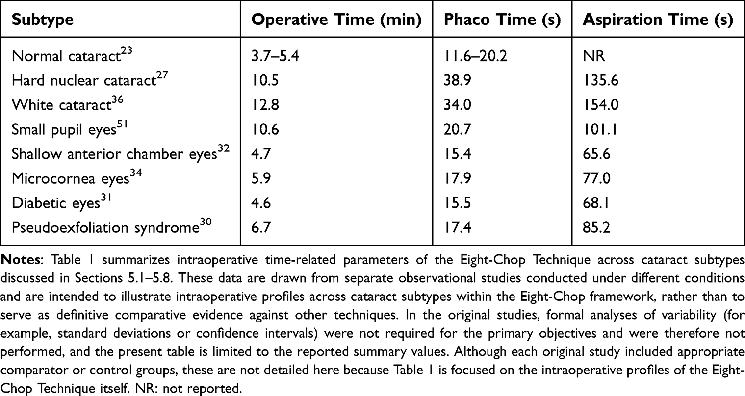 |
Table 1 Intraoperative Time-Related Parameters of the Eight-Chop Technique Across Cataract Subtypes |
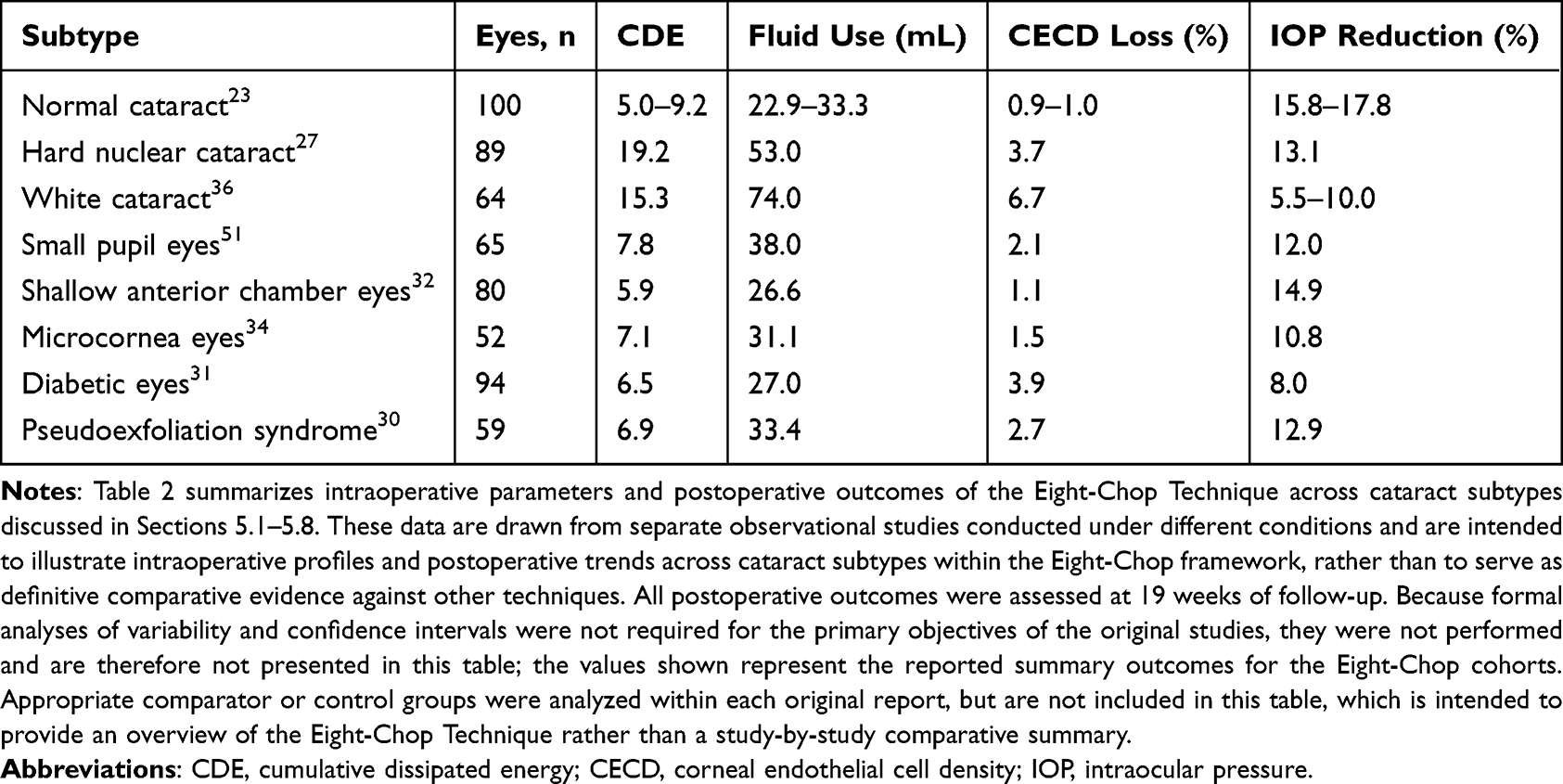 |
Table 2 Intraoperative Parameters and Postoperative Outcomes of the Eight-Chop Technique Across Cataract Subtypes |
With respect to postoperative outcomes, the Eight-Chop Technique has demonstrated relatively limited endothelial cell loss across multiple cataract subtypes, including hard nuclear cataracts and diabetic eyes, which are generally considered vulnerable to endothelial damage. Sustained IOP reduction during mid- to long-term follow-up has also been observed, potentially reflecting reduced intraoperative energy use and irrigation volume, as well as a lower impact on aqueous outflow tissues such as the trabecular meshwork and Schlemm’s canal. Postoperative CECD and IOP values in Table 2 were consistently evaluated at 19 weeks of follow-up across the included series. Comparisons with conventional nuclear fragmentation techniques in this review are therefore indirect, and differences in surgical setting, case mix, and study design should be carefully considered when interpreting the clinical relevance of the reported values. Accordingly, the present data should not be regarded as definitive evidence of comparative superiority over established techniques, but rather as preliminary observations that warrant further confirmation.
In summary, the Eight-Chop Technique may represent a clinically useful segmentation-first strategy in the evolution of nuclear fragmentation techniques. By integrating wedge-induced fracture, eightfold geometric division, and compatibility with modern fluidics systems, this technique may contribute to reductions in ultrasound energy, irrigation volume, and intraoperative mechanical stress across a wide range of cataract subtypes. Clinically, it may offer a stable and reproducible surgical workflow, particularly in high-risk eyes. Nonetheless, the current evidence base is dominated by author-derived, single-center observational studies, and independent external validation is still limited. With further validation through multicenter studies and independent surgeon evaluations, the Eight-Chop Technique may be further refined and more clearly positioned as a practical nuclear fragmentation strategy in modern cataract surgery.
Video Availability Statement
Publicly accessible surgical videos demonstrating the Eight-Chop Technique are available online. These videos include both routine and complex cataract cases and illustrate the surgical workflow and fragmentation principles described in this review.
Generative AI Statement
Generative AI statement: The author used Perplexity (Perplexity AI, model GPT‑5.1) solely for English language editing and editorial refinement. The author reviewed and edited the content generated by this tool and takes full responsibility for the content of the manuscript.
Abbreviations
AFS, Active Fluidics System; CCT, Central corneal thickness; CDE, Cumulative dissipated energy; CECD, Corneal endothelial cell density; CTR, Capsular tension ring; IOP, Intraocular pressure; OVD, Ophthalmic viscosurgical device; PEX, Pseudoexfoliation syndrome.
Acknowledgments
A previous version of this manuscript was uploaded to the following preprint servers under the title “The Eight-Chop Technique: Mechanistic Principles and Clinical Performance of a Segmentation-First Phacoemulsification Strategy”:
preprints.org: https://www.preprints.org/manuscript/202512.2212
sciety: https://sciety.org/articles/activity/10.20944/preprints202512.2212.v2
Author Contributions
All authors made a significant contribution to the work reported, whether that is in the conception, study design, execution, acquisition of data, analysis and interpretation, or in all these areas; took part in drafting, revising or critically reviewing the article; gave final approval of the version to be published; have agreed on the journal to which the article has been submitted; and agree to be accountable for all aspects of the work.
Funding
This research received no external funding.
Disclosure
The author declares no conflicts of interest.
References
1. Kelman CD. Phaco-emulsification and aspiration. A new technique of cataract removal. A preliminary report. Am J Ophthalmol. 1967;64(1):23–20. doi:10.1016/0002-9394(67)93340-5
2. Gimbel HV. Divide and conquer nucleofractis phacoemulsification: development and variations. J Cataract Refract Surg. 1991;17(3):281–291. doi:10.1016/S0886-3350(13)80824-3
3. Linebarger EJ, Hardten DR, Shah GK, Lindstrom RL. Phacoemulsification and modern cataract surgery. Surv Ophthalmol. 1999;44(2):123–147. doi:10.1016/s0039-6257(99)00085-5
4. Walkow T, Anders N, Klebe S. Endothelial cell loss after phacoemulsification: relation to preoperative and intraoperative parameters. J Cataract Refract Surg. 2000;26(5):727–732. doi:10.1016/S0886-3350(99)00462-9
5. Hwang HB, Lyu B, Yim HB, Lee NY. Endothelial cell loss after phacoemulsification according to different anterior chamber depths. J Ophthalmol. 2015;2015:210716. doi:10.1155/2015/210716
6. Pereira AC, Porfírio F Jr, Freitas LL, Belfort R Jr. Ultrasound energy and endothelial cell loss with stop-and-chop and nuclear preslice phacoemulsification. J Cataract Refract Surg. 2006;32(10):1661–1666. doi:10.1016/j.jcrs.2006.05.006
7. Koch PS, Katzen LE. Stop and chop phacoemulsification. J Cataract Refract Surg. 1994;20(5):566–570. doi:10.1016/S0886-3350(13)80239-8
8. Chang DF. Why Learn Chopping?
9. Vajpayee RB, Kumar A, Dada T, Titiyal JS, Sharma N, Dada VK. Phaco-chop versus stop-and-chop nucleotomy for phacoemulsification. J Cataract Refract Surg. 2000;26(11):1638–1641. doi:10.1016/S0886-3350(00)00544-7
10. Park J, Yum HR, Kim MS, Harrison AR, Kim EC. Comparison of phaco-chop, divide-and-conquer, and stop-and-chop phaco techniques in microincision coaxial cataract surgery. J Cataract Refract Surg. 2013;39(10):1463–1469. doi:10.1016/j.jcrs.2013.04.033
11. Shingleton BJ, Neo YN, Cvintal V, Shaikh AM, Liberman P, O’Donoghue MW. Outcome of phacoemulsification and intraocular lens implantation in eyes with pseudoexfoliation and weak zonules. Acta Ophthalmol. 2017;95(2):182–187. doi:10.1111/aos.13110
12. Hayashi K, Yoshida M, Manabe SI, Hirata A. High-risk factors for zonular complications during cataract surgery in eyes with pseudoexfoliation syndrome. Br J Ophthalmol. 2024;108(9):1193–1199. doi:10.1136/bjo-2023-324832
13. Fernández-Muñoz E, Chávez-Romero Y, Rivero-Gómez R, Aridjis R, Gonzalez-Salinas R. Cumulative dissipated energy (CDE) in three phaco-fragmentation techniques for dense cataract removal. Clin Ophthalmol. 2023;17:2405–2412. doi:10.2147/OPTH.S407705
14. Moghimi S, Johari M, Mahmoudi A, et al. Predictors of intraocular pressure change after phacoemulsification in patients with pseudoexfoliation syndrome. Br J Ophthalmol. 2017;101(3):283–289. doi:10.1136/bjophthalmol-2016-308601
15. Kopsachilis N, Carifi G. Phacoemulsification using 8 flexible iris hooks in a patient with a short eye, small pupil, and phacodonesis. J Cataract Refract Surg. 2014;40(9):1408–1411. doi:10.1016/j.jcrs.2014.07.009
16. Khokhar S, Gupta S, Tewari R, et al. Scleral tunnel phacoemulsification: approach for eyes with severe microcornea. Indian J Ophthalmol. 2016;64(4):320–322. doi:10.4103/0301-4738.182949
17. Zarei-Ghanavati S, Hadi Y, Habibi A, Ashraf Khorasani M, Yoo SH. Cataract and diabetes: review of the literature. J Cataract Refract Surg. 2024;50(12):1275–1283. doi:10.1097/j.jcrs.0000000000001547
18. Mencucci R, De Vitto C, Cennamo M, Vignapiano R, Buzzi M, Favuzza E. Femtosecond laser-assisted cataract surgery in eyes with shallow anterior chamber depth: comparison with conventional phacoemulsification. J Cataract Refract Surg. 2020;46(12):1604–1610. doi:10.1097/j.jcrs.0000000000000341
19. Rauen MP, Joiner H, Kohler RA, O’Connor S. Phacoemulsification using an active fluidics system at physiologic vs high intraocular pressure: impact on anterior and posterior segment physiology. J Cataract Refract Surg. 2024;50(8):822–827. doi:10.1097/j.jcrs.0000000000001457
20. Liu Y, Hong J, Chen X. Comparisons of the clinical outcomes of Centurion(®) active fluidics system with a low IOP setting and gravity fluidics system with a normal IOP setting for cataract patients with low corneal endothelial cell density. Front Med Lausanne. 2023;10(1):1294808. doi:10.3389/fmed.2023.1294808
21. Vasavada V, Vasavada AR, Vasavada VA, Vasavada SA, Bhojwani D. Real-time dynamic changes in intraocular pressure after occlusion break: comparing 2 phacoemulsification systems. J Cataract Refract Surg. 2021;47(9):1205–1209. doi:10.1097/j.jcrs.0000000000000666
22. Masuda Y, Iwaki H, Kato N, Takahashi G, Oki K, Tsuneoka H. Irrigation dynamic pressure-assisted hydrodissection during cataract surgery. Clin Ophthalmol. 2017;11:323–328. doi:10.2147/OPTH.S124528
23. Sato T. Efficacy and safety of the eight-chop technique in phacoemulsification for patients with cataract. J Cataract Refract Surg. 2023;49(5):479–484. doi:10.1097/j.jcrs.0000000000001141
24. Jacobsson L, Sandström J, Brander L, Flansbjer M. Tensile fracture initiation and propagation of granite and gneiss at wedge splitting tests: part 1—Effect of notch type on tensile crack initiation and fracture mechanics results. Int J Fract. 2025;249(3):45. doi:10.1007/s10704-025-00857-z
25. Akahoshi T. Phaco prechop: manual nucleofracture prior to phacoemulsification. Oper Tech Cataract Refract Surg. 1998;1(2):69–91.
26. Scott RA, Holtmeyer CJ, Parker TM, Willis JK, Scott WJ, Olson RJ. Efficiency of venturi vs peristaltic-based phacoemulsification in femtosecond laser cataract surgery. J Cataract Refract Surg. 2025;51(5):382–387. doi:10.1097/j.jcrs.0000000000001618
27. Sato T. Minimizing Endothelial Cell Loss in Hard Nucleus Cataract Surgery: efficacy of the Eight-Chop Technique. J Clin Med. 2025;14(8):2576. doi:10.3390/jcm14082576
28. Sato T. Reply: efficacy and safety of the eight-chop technique in phacoemulsification for patients with cataract. J Cataract Refract Surg. 2023;49(10):1078–1079. doi:10.1097/j.jcrs.0000000000001259
29. Sato T. Does further lowering intraoperative intraocular pressure reduce surgical invasiveness in active-fluidics eight-chop phacoemulsification? a fellow-eye comparative study. J Clin Med. 2026;15(1):366. doi:10.3390/jcm15010366
30. Sato T. Cataract surgery in pseudoexfoliation syndrome using the eight-chop technique. J Pers Med. 2025;15(9):396. doi:10.3390/jpm15090396
31. Sato T. Corneal endothelial changes after phacoemulsification using the eight-chop technique in diabetic eyes. J Pers Med. 2025;15(5):209. doi:10.3390/jpm15050209
32. Sato T. Corneal endothelial cell loss in shallow anterior chamber eyes after phacoemulsification using the eight-chop technique. J Clin Med. 2025;14(9):3045. doi:10.3390/jcm14093045
33. Sato T. Long-term effects of the eight-chop technique in phacoemulsification on intraocular pressure for cataract patients. 2025. doi:10.20944/preprints202512.201454.v202511
34. Sato T. Cataract surgery in microcornea eyes using the eight-chop technique. Preprints. 2025. doi:10.20944/preprints202510.201082.v202511
35. Sato T. Effects of the eight-chop technique in phacoemulsification on intraocular pressure in patients with primary open-angle glaucoma and controls. 2025. doi:10.20944/preprints202512.201427.v202511
36. Sato T. Clinical outcomes of the eight-chop technique in white cataract: a retrospective case series. BMC Ophthalmol. 2026;26(1):342. doi:10.1186/s12886-026-04859-w
37. Sato T. Postoperative flare and corneal endothelial cell loss after eight-chop technique phacoemulsification: a prospective observational study. J Clin Med. 2026;15(2):557. doi:10.3390/jcm15020557
38. Hwang HS, Kim EC, Kim MS. Drill-and-crack technique for nuclear disassembly of hard nucleus. J Cataract Refract Surg. 2010;36(10):1627–1630. doi:10.1016/j.jcrs.2010.08.003
39. He Y, Wang C, Zhou X, et al. Comparison of clinical outcomes between cystotome-assisted prechop phacoemulsification surgery and femtosecond laser-assisted cataract surgery for hard nucleus cataracts. Eye. 2023;37(2):235–241. doi:10.1038/s41433-021-01900-8
40. Chen D, Tang Q, Yu F, Cai X, Lu F. Consecutive drilling combined with phaco chop for full thickness segmentation of very hard nucleus in coaxial microincisional cataract surgery. BMC Ophthalmol. 2019;19(1):20. doi:10.1186/s12886-019-1033-1
41. Foster GJL, Allen QB, Ayres BD, et al. Phacoemulsification of the rock-hard dense nuclear cataract: options and recommendations. J Cataract Refract Surg. 2018;44(7):905–916. doi:10.1016/j.jcrs.2018.03.038
42. Arshinoff SA. Ophthalmic viscosurgical devices for intumescent cataracts: pressure-equalized cataract surgery. J Cataract Refract Surg. 2015;41(7):1549–1550. doi:10.1016/j.jcrs.2015.05.016
43. Balyan M, Jain AK, Malhotra C, Ram J, Dhingra D. Achieving successful capsulorhexis in intumescent white mature cataracts to prevent Argentinian flag sign - A new multifaceted approach to meet the challenge. Indian J Ophthalmol. 2021;69(6):1398–1403. doi:10.4103/ijo.IJO_1903_20
44. Titiyal JS, Kaur M, Singh A, Arora T, Sharma N. Comparative evaluation of femtosecond laser-assisted cataract surgery and conventional phacoemulsification in white cataract. Clin Ophthalmol. 2016;10:1357–1364. doi:10.2147/OPTH.S108243
45. Chakrabarti A, Singh S, Krishnadas R. Phacoemulsification in eyes with white cataract. J Cataract Refract Surg. 2000;26(7):1041–1047. doi:10.1016/S0886-3350(00)00525-3
46. Vasavada A, Singh R, Desai J. Phacoemulsification of white mature cataracts. J Cataract Refract Surg. 1998;24(2):270–277. doi:10.1016/S0886-3350(98)80210-1
47. Vajpayee RB, Bansal A, Sharma N, Dada T, Dada VK. Phacoemulsification of white hypermature cataract. J Cataract Refract Surg. 1999;25(8):1157–1160. doi:10.1016/S0886-3350(99)00118-2
48. Kim ES, Han SB, Lee SJ, Kim M. Cataract surgery through the small pupil. Clin Interv Aging. 2016;11:1387–1389. doi:10.2147/CIA.S119086
49. Hashemi H, Seyedian MA, Mohammadpour M. Small pupil and cataract surgery. Curr Opin Ophthalmol. 2015;26(1):3–9. doi:10.1097/ICU.0000000000000116
50. Papaconstantinou D, Kalantzis G, Brouzas D, et al. Safety and efficacy of phacoemulsification and intraocular lens implantation through a small pupil using minimal iris manipulation. Clin Interv Aging. 2016;11:651–657. doi:10.2147/CIA.S97254
51. Sato T. Eight-chop technique in phacoemulsification using iris hooks for patients with cataracts and small pupils. J Clin Med. 2024;13(23):7298. doi:10.3390/jcm13237298
52. Bayramlar H, Karadag R, Yildirim A, Cakici O, Sari U. Manual tunnel incision cataract surgery with sandwich technique in eyes with microcornea. J Cataract Refract Surg. 2014;40(6):1058. doi:10.1016/j.jcrs.2014.04.014
53. Ashok Kumar D, Agarwal A, Sivangnanam S, Chandrasekar R, Agarwal A. Implantation of glued intraocular lenses in eyes with microcornea. J Cataract Refract Surg. 2015;41(2):327–333. doi:10.1016/j.jcrs.2014.05.039
54. Altansukh A, Ma KS, Doyodmaa A, et al. Comparison of corneal endothelial cell loss in FLACS: impact on endothelial cells in different regions. J Cataract Refract Surg. 2024;50(9):912–918. doi:10.1097/j.jcrs.0000000000001474
55. Cruz JCG, Moreno CB, Soares P, et al. Comparison of endothelial cell loss in diabetic patients after conventional phacoemulsification and femtosecond laser-assisted cataract surgery. BMC Ophthalmol. 2023;23(1):181. doi:10.1186/s12886-023-02923-3
56. Salavat MC, Munteanu M, Chercotă V, et al. Corneal endothelial cell loss following cataract surgery in patients with type 2 diabetes mellitus: a comprehensive review. Biomedicines. 2025;13(7):1726. doi:10.3390/biomedicines13071726
57. Opala A, Kołodziejski Ł, Grabska-Liberek I. Impact of well-controlled type 2 diabetes on corneal endothelium following cataract surgery: a prospective longitudinal analysis. J Clin Med. 2025;14(10):3603. doi:10.3390/jcm14103603
58. Akansha YRS, Yadav RS. Comparative assessment of the corneal endothelium following phacoemulsification surgery in patients with type II diabetes and nondiabetes. Saudi J Ophthalmol. 2025;39(1):77–83. doi:10.4103/sjopt.sjopt_226_23
59. Gallardo MJ, Supnet RA, Ahmed IIK. Viscodilation of Schlemm’s canal for the reduction of IOP via an ab-interno approach. Clin Ophthalmol. 2018;12:2149–2155. doi:10.2147/OPTH.S177597
60. Guan H, Mick A, Porco T, Dolan BJ. Preoperative factors associated with IOP reduction after cataract surgery. Optom Vis Sci. 2013;90(2):179–184. doi:10.1097/OPX.0b013e31827ce224
61. Poley BJ, Lindstrom RL, Samuelson TW, Schulze R Jr. Intraocular pressure reduction after phacoemulsification with intraocular lens implantation in glaucomatous and nonglaucomatous eyes: evaluation of a causal relationship between the natural lens and open-angle glaucoma. J Cataract Refract Surg. 2009;35(11):1946–1955. doi:10.1016/j.jcrs.2009.05.061
62. Poley BJ, Lindstrom RL, Samuelson TW. Long-term effects of phacoemulsification with intraocular lens implantation in normotensive and ocular hypertensive eyes. J Cataract Refract Surg. 2008;34(5):735–742. doi:10.1016/j.jcrs.2007.12.045
63. Wang S, Tao J, Yu X, Diao W, Bai H, Yao L. Safety and prognosis of phacoemulsification using active sentry and active fluidics with different IOP settings - a randomized, controlled study. BMC Ophthalmol. 2024;24(1):350. doi:10.1186/s12886-024-03626-z
 © 2026 The Author(s). This work is published and licensed by Dove Medical Press Limited. The
full terms of this license are available at https://www.dovepress.com/terms
and incorporate the Creative Commons Attribution
- Non Commercial (unported, 4.0) License.
By accessing the work you hereby accept the Terms. Non-commercial uses of the work are permitted
without any further permission from Dove Medical Press Limited, provided the work is properly
attributed. For permission for commercial use of this work, please see paragraphs 4.2 and 5 of our Terms.
© 2026 The Author(s). This work is published and licensed by Dove Medical Press Limited. The
full terms of this license are available at https://www.dovepress.com/terms
and incorporate the Creative Commons Attribution
- Non Commercial (unported, 4.0) License.
By accessing the work you hereby accept the Terms. Non-commercial uses of the work are permitted
without any further permission from Dove Medical Press Limited, provided the work is properly
attributed. For permission for commercial use of this work, please see paragraphs 4.2 and 5 of our Terms.
Recommended articles
Correlations of Immediate Corneal Tomography Changes with Preoperative and the Elapsed Phaco Parameters
Haddad JS, Borges C, Daher ND, Mine A, Salomão M, Ambrósio Jr R
Clinical Ophthalmology 2022, 16:2421-2428
Published Date: 4 August 2022
Impact of Cataract Surgery on IOP and Ocular Structures in Normotensive Patients and Primary and Exfoliation Open-Angle Glaucoma Patients
Xirou V, Xirou T, Siganos C, Ntonti P, Georgakopoulos C, Stavrakas P, Makri OE, Kanakis M, Tsapardoni F, Fragkoulis I, Garnavou-Xirou C, Kozobolis V
Clinical Ophthalmology 2023, 17:1047-1055
Published Date: 5 April 2023
Twist Technique to Stop Fluid Leakage and Fixate the Eye During Bimanual Cortex Removal in Phacoemulsification

Khaled H, Haj Najeeb B
Clinical Ophthalmology 2023, 17:1147-1149
Published Date: 14 April 2023
Visual, Refractive, Functional, and Patient Satisfaction Outcomes After Implantation of a New Trifocal Diffractive Intraocular Lens
Mendicute J, Lauzirika G, Illarramendi I, Martínez-Soroa I
Clinical Ophthalmology 2024, 18:2785-2795
Published Date: 5 October 2024
Evaluation of the Occlusion Break Surge Volume in Five Different Phacoemulsification Systems
Yalamanchili S, Aboughaida A, Rohani OS, Dyk DW
Clinical Ophthalmology 2025, 19:1357-1364
Published Date: 23 April 2025