")
Back to Journals » Infection and Drug Resistance » Volume 16
The Effects of Holothurin and Caspofungin on the Vaginal Cell Inflammation Parameters of the Rattus norvegicus Strain Post Induction of Candida albicans
Authors Handayani S, Nurdiana N, Winarsih S, Tri Endharti A
Received 5 December 2022
Accepted for publication 1 April 2023
Published 27 April 2023 Volume 2023:16 Pages 2501—2509
DOI https://doi.org/10.2147/IDR.S400314
Checked for plagiarism Yes
Review by Single anonymous peer review
Peer reviewer comments 2
Editor who approved publication: Prof. Dr. Héctor Mora-Montes
Sri Handayani,1,2 Nurdiana Nurdiana,3 Sri Winarsih,4 Agustina Tri Endharti5
1Doctoral Program of Medical Science Department, Faculty of Medicine, Brawijaya University, Malang, Indonesia; 2Department of Midwifery of Aisyiyah, Institution of Surakarta University, Surakarta, Province of Middle Java, Indonesia; 3Department of Pharmacology, Faculty of Medicine, Brawijaya University, Malang, Province of East Java, Indonesia; 4Department of Pharmacy, Faculty of Medicine, Brawijaya University, Malang, Province of East Java, Indonesia; 5Department of Parasitology, Faculty of Medicine, Brawijaya University, Malang, Province of East Java, Indonesia
Correspondence: Sri Handayani, Department of Midwifery of Aisyiyah, Institution of Surakarta University, Surakarta, Middle Java State, 57146, Indonesia, Tel +62 816-4252-588, Email [email protected]
Purpose: Candida albicans (C. albicans) is a fungus that causes superficial and invasive candidiasis in its host. Caspofungin, has been widely used as a synthetic antifungal, whereas holothurin has been shown to have potential as a natural antifungal. The purpose of this study was to see how holothurin and caspofungin affected the number of C. albicans’s colonies, LDH levels, and the number of inflammatory cells in vagina of Rattus norvegicus.
Patients and Methods: Design of this research is using posttest only with control group design with 48 Rattus norvegicus Wistar strains used in this study were divided into six treatment groups. Each group was divided into three-time intervals of 12, 24, and 48 hours. LDH markers were tested using ELISA, inflammatory cells were counted manually, and the number of colonies was calculated using colonymetry before being diluted with NaCl 0.9% and planted in sabouraud dextrose agar (SDA).
Results: According to the findings, inflammatory cells in the treatment of holothurin (48-hours) had an OR of 1.68 CI (− 0.79– 4.16) P = 0.09 and caspofungin had an OR of 4.18 CI (1.26– 9.63) P = 0.09. Meanwhile, LDH in the holothurin (48-hour) treatment obtained OR 348, CI (286– 410), P=0.03, and Caspofungin OR 393, CI (277– 508), P=0.03. Colonies were obtained with zero numbers in the holothurin treatment (48 hours) and with Caspofungin OR 393, CI (273– 508) P=0.00.
Conclusion: Holothurin and caspofungin administration reduced the number of C. albicans colonies and the number of inflammatory cells (P 0.05), implying that holothurin and caspofungin could prevent C. albicans infection.
Keywords: Candida albicans, inflammatory cells and LDH, holothurin, caspofungin
Introduction
Candida albicans is a fungal pathogen that causes both superficial and invasive candidiasis. C. albicans causes vaginitis, which is characterized by itching, burning, pain, and redness of the vulva and vaginal mucosa, as well as vaginal discharge.1 Human infections caused by C. albicans in Indonesia are still relatively high while antifungal drugs are scarce. Treatment options for invasive fungal infections are limited as there are only a few classes of antifungal drugs available on the market. Polyene drugs can be fungicidal but are toxic to the host, while azoles are fungistatic, therefore fungi are more prone to develop resistance.2
C. albicans colonization occurs as a result of the fungus adhering to the epithelium, which then invades the vaginal epithelial cells, causing infection1,3 and candidiasis.4 Candidalysin is a fungal peptide poison that is important in mucosal infections because it directly damages epithelial membranes.5 Candidalysin is secreted by Candida albicans hyphae and enters the host epithelial membrane, damaging cells by causing pores to form, resulting in the loss of intracellular contents in the form of lactate dehydrogenase (LDH).6 C. albicans invasion causes necrosis, apoptosis, and pyroptosis in host cells.7
To overcome inflammation in vaginal epithelial tissue, safe drugs that actively collaborate with host cells are required, so that the inflammatory process can be resolved immediately. Human cells are extremely sensitive to antifungal drugs.8 Caspofungin, a common antifungal drug, has antifungal properties against Candida albicans. Caspofungin is a commonly used antifungal drug, often used as a first-line therapy that inhibits cell wall β-(1,3)-glucan synthesis.2 But despite its fungicidal effect, the drug resistance that occurs is a problem in the clinic due to the acquisition of GSC1 (FKS1) mutations, which decrease the affinity of the drug to the enzyme (Perlin 2007). If caspofungin is synthetic, then holothurin is a natural antifungal that has the potential to kill C. albicans and the active substance has the potential to overcome C. albicans invasion.
Caspofungin is non-toxic and effective against a variety of fungal systems.2 In addition to caspofungin, holothurin is a natural antifungal that has the potential to kill C. albicans. Holothurin is a saponin purification derived from one of the many marine animals found in Indonesia.9 Holothurin’s main active ingredients are triterpenoid glycosides.10,11 These active ingredients have anticancer, anticoagulant, antimicrobial, antioxidant, antiviral, and anti-inflammatory properties.12,13 The aim of this research was to see how holothurin affected the number of C. albicans colonies, inflammatory cells, and LDH levels in Rattus norvegicus Wistar vaginal epithelium.
Materials and Methods
Research Design
In this study, the research design was true experimental with a post-test only control group design approach. This research was conducted from January until March 2021.
Subject
A total of 48 female Rattus norvegicus Wistar were randomly assigned for acclimatization took place for one week prior to treatment. The overall sample was split randomly. Each group composed of 12 Rattus norvegicus Wistar Groups 1 (K1) is negative control, positive control (C. albicans) (P1), C. albicans + holothurin (P2), C. albicans + caspofungin (P3) and each group of treatment split by 3-time intervention groups (12, 24 and 48 hours). We excluded the rat born premature, and dead before given treatment. The positive control (P1) given topikal C. albicans after grown in yeast extract peptone dextrose (YPD) on vagina of Rattus norvegicus Wistar. Group P2 & P3 given Holothurin 3500 ug and caspofungin 140 ug treatment per topically to vagina of animal model. Furthermore, randomly selected for termination with intraperitoneally kethamine 100 mg before the tissue was taken. Colony counts were performed on vaginal organs, inflammatory cell analysis was performed using electron microscopy, and Lactat dehydrogenase (LDH) was measured using an ELISA.
Ethical Clearance
This research in accordance with ethical standards with the guidelines of the APA’s Guidelines for Ethical Conduct in the Care and Use of Animals. Ethics committee that approved this study is Health Research Ethics Committee (HREC) Medical Faculty of Brawijaya University number 337/EC/KEPK-S3/12/2019.
Settings
Preparation of C. albicans
Candida albicans culture preparation was carried out by referring to previous studies15 with the following steps. C. albicans isolate (4506547065307370) was obtained from the Microbiology Laboratory of Faculty of Medicine, Universitas Brawijaya and inoculated overnight on Sabouraud Dextrose Agar (SDA; Sabouraud Dextrose Broth (Ph 5.6)), Crystal violet, lugol, 96% alcohol, aguades, and Dimethyl Sulfoxide (DMSO)). C. albicans cultures were then propagated in Yeast extract Peptone Dextrose (YPD) media and incubated overnight at 37°C in an incubator. C. albicans was harvested and washed in sterile PBS and cell suspensions (Sigma Chemical Co., St. Louis, MO, USA). For subsequent analysis, cells were prepared in 0.9% NaCL at a cell density of 1.0105 CFU/mL. LDH levels were determined using ELISA. The supernatant from Rattus norvegicus vagina was centrifuged at 1500 g for 10 minutes at 40 °C). The LDH concentration in the centrifuged supernatant was determined using the ELISA method (Ready-SET-Go ELISA kit; eBioscience, USA) at 490 nm. The standard was bovine serum albumine. Haematoxylin and eosin (HE) staining of vaginal tissue. Fixation of a mouse vaginal sample Vaginal tissue was fixed in 10% formalin for 7 hours before being cut into 2–3 mm pieces. Tissue Processor Thermo Scientific STP 120 was used to process the samples for 16 hours.
Tissues blocking and cutting process using the fixed tissue was blocked with liquid paraffin and cut with a microtome (size 3–5 m).
Deparaffinization Process
The blocking result was deparaffinized in a water bath for 3 minutes before being incubated for 30 minutes at 70–80°C. The preparation was immersed in xylol for 20 minutes twice, hydrated with alcohol for 3 minutes four times, and rinsed with running water for 15 minutes to remove residual alcohol. After that, the preparations were dried at room temperature.
Haematoxylin Eosin Staining (HE)
Haematoxylin was used to incubate the preparations for 10–15 minutes. After 15 minutes of running water, the preparations were immersed in 1% acid alcohol (2–5 times) and ammonia lithium carbonate (3–5 times). The tissue preparations were then incubated in eosin solution for 10–15 minutes and graded alcohol for 3 minutes each: 70%, 80%, and 96%. The preparations were then cleared for 15 minutes with xylol.
Determination of the Number of Colonies of C. albicans
C. albicans cells were grown in yeast extract peptone dextrose (YPD) media after being inoculated for 24 hours on SDA agar medium (Becton Dickinson, Cockeysville, MD, USA). C. albicans was harvested and washed with sterile PBS after being incubated in an incubator at 37°C for 24 hours (Sigma Chemical Co., St. Louis, MO, USA). The cell suspension was prepared in 0.9% NaCL and struck at SDA (1.0105 CFU/Ml).
Data Analysis
Analysis of the average for numerical data using mean and standard deviations (SD) of each variable with the independent sample t-test to the data with univariate normal distribution. Statistical analysis using One Way ANOVA test on SPSS 23 software.
Results
Induction of C. albicans Colonies on LDH Level
LDH levels, the number of inflammatory cells, and the number of C. albicans colonies in the rat vagina can all be used to determine the degree of inflammation caused by exposure to C. albicans (Figure 1).
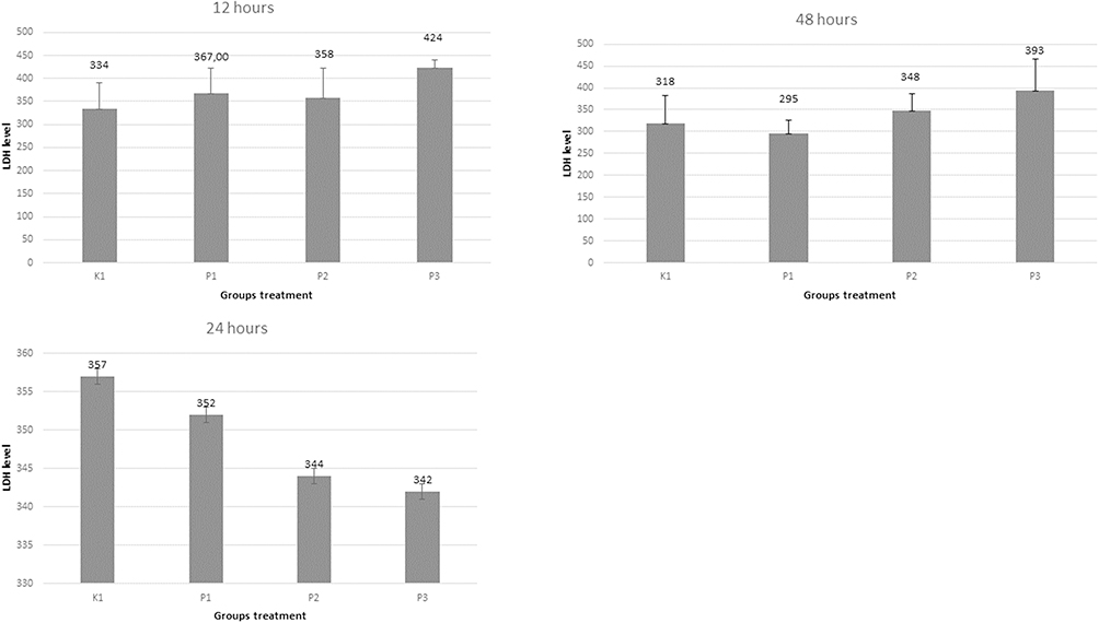 |
Figure 1 LDH levels at 12, 24 and 48 hours. LDH expression graphs in the fungal treatment and active ingredient treatment groups of Holothurin and Caspofungin at 12, 24, and 48 hours in Wistar rats. K: Control without any treatment (healthy). P1: C. albicans treatment. P2: Fungus/C. albicans treatment administered with Holothurin. P3: Fungus/C. albicans treatment administered with Caspofungin. |
Varying LDH levels have been seen in the vagina of rats after C. albicans induction. With the exception of sample P3 at 48 hours, practically every hour of treatment shows substantial variations. Group that received holothurin and caspofungin at 12 and 48 hours did not significantly reduce the LDH level. After receiving holothurin and caspofungin treatment, it can be assumed that the LDH levels of C. albicans -infected animals were close to those of control subjects, but there were still values that changed in LDH levels at 24 hours both after holothurin (P2) and caspofungin (P3) administration when compared to the positive control group (P1) (P=0.00).
The number of inflammatory cells in the negative control group at 12, 24 and 48 hours showed no significant expression (Figure 2). The positive treatment group showed significant values at 12, 24 and 48 hours, and showed valid values. The group with holothurin administration at 12 hours showed lower values compared to 24 and 48 hours (P<0.05), and was valid and significant when compared to the positive control group. The group with caspofungin administration at hour 48 showed lower values compared with hour 12 and 24 (P<0.05), and was valid and significant when compared with the positive control group. The comparison between holothurin and caspofungin was graphically different at 12, 24 and 48 hours, and concluded that holothurin has potential value against C. albicans (P=0.00).
 |
Figure 2 Inflammation cells at 12, 24 and 48 hours. Graph of the number of inflammatory cells in the fungal treatment group and the active ingredient treatment of Holothurin and Caspofungin at 12, 24 and 48 hours in Wistar rats. K: Control without any treatment (healthy). P1: C. albicans treatment. P2: Treatment of Fungus/C. albicans administered with Holothurin. P3: Fungus/C. albicans treatment administered with Caspofungin. |
Based on the results (Figure 2), the number of inflammatory cells in the vagina of rat varies greatly. When compared with other samples, the presence of C. albicans infection in the vagina of mice (P1) caused a significant increase in inflammatory cells. When compared to the caspofungin-treated sample, holothurin administration to sample P2 had a significantly lower value. This suggests that holothurin suppresses the number of inflammatory cells more effectively than caspofungin during and before infection. The histology image (Figure 3) shows the difference in inflammatory cells between the group that was only given C. albicans alone and compared with the holothurin (P2) and caspofungin (P3) treatment groups.
 |
Figure 3 Rattus norvegicus Wistar’s vaginal tissue. The number of inflammatory cells in the fungal treatment and active ingredient treatment groups of Holothurin and Caspofungin at 12, 24 and 48 hours in Wistar rats. K: Control without any treatment (healthy). P1: C. albicans treatment. P2: Fungal/C. albicans treatment administered with Holothurin. P3: Fungal/C. albicans treatment administered with Caspofungin. Red arrows indicate inflammatory cells. |
Induction C. albicans Influence on Epithelial Vagina of Rattus norvegicus Wistar After 12, 24 and 48 Hours
In this study, a trial groups with holothurin and caspofungin quantitatively lower (Figure 4), The study found that treatment with holothurin was effective in reducing the colonization of C. albicans in inflammatory cells compared to the positive control group. The reduction was statistically significant (p<0.05) and demonstrated the potential of holothurin to reduce the number of inflammatory cells. However, there was also a significant difference in the mean number of inflammatory cells between the positive control group and the caspofungin group, compared to the negative control. In the trial groups than the control groups with general results p<0.05, which shows the influence of C. albicans induced changes in the number of colonies after exposure on intervention time 12, 24 and 48 hours (Figure 5).
 |
Figure 4 Holothurin and caspofungin treatment affected the quantity of Candida albicans colonies in the vaginal Rattus norvegicus Wistar in the five groups. Graph of the number of colonies in the fungal treatment group and the active ingredient treatment of Holothurin and Caspofungin at 12, 24 and 48 hours in Wistar rats. K: Control without any treatment (healthy). P1: C. albicans treatment. P2: Fungus/C. albicans treatment administered with Holothurin. P3: Fungus/C. albicans treatment administered with Caspofungin. Red arrows show the expression of inflammatory cells in the cross section of vaginal epithelial tissue of Wistar Rattus norvegicus. |
 |
Figure 5 The number of C. albicans colonies at 12, 24 and 48 hours. The number of colonies in the fungal treatment group and the active ingredient treatment of Holothurin and Caspofungin at 12, 24 and 48 hours in Wistar rats. K: Control without any treatment (healthy). P1: C. albicans treatment. P2: Fungal/C. albicans treatment administered with Holothurin. P3: Fungal/C. albicans treatment administered with Caspofungin. |
Many studies have discussed the active substance holothurin and its various health benefits. However, there are still no studies that tested the active substance holothurin on vaginal tissue infected with C. albicans. This study shows LDH, and the number of inflammatory cells in vaginal epithelial tissue in experimental animals that have been infected with C. albicans colonies, which are then given antifungal therapy in the form of holothurin.
Holothurin in this study gave a satisfactory effect on C. albicans infection of vaginal epithelial tissue of Wistar Rattus norvegicus. Holothurin administration of 3.5 mg/mL was tried invagina in the rat group 12 hours, rat group 24 hours and rat group 48 hours after C. albicans infection. Inflammatory cells and LDH are parameters of the severity of infection in a tissue. Satisfactory results indicated a decrease in LDH and Inflammatory Cells after Holothurin was given which showed a decrease in the degree of epithelial infection at 12, 24 and 48 hours. So that the potential of Holothurin in suppressing C. albicans colonization can be suggested as an antifungal in preventing invasion and prolonged infection.
Discussion
Failure of antifungal therapy is a substantial clinical problem, due to the emergence of an increasing number of drug-resistant isolates. Caspofungin is a commonly used antifungal drug, often used as first-line therapy that inhibits cell wall β-(1,3)-glucan synthesis.16 Candida, has a major impact on keratitis cases in both tropical and subtropic regions.17 Caspofungin was one of the first echinocandins to be discovered and patented,18 and is recommended as a first-line treatment for Candida infections.19 Despite the fungicidal effect, the drug resistance that occurs is a problem in the clinic due to the acquisition of GSC1 (FKS1) mutations, which decrease the affinity of the drug to the enzyme (Perlin 2007). Candida biofilms are a well-organized form of planktonic yeast and mycelium, surrounded by extracellular polymeric substances; these structures are effective microbial protection and can produce well-known drug resistance.20
Resistance may be due to altered intracellular accumulation of drugs, changes in membrane sterol composition, changes in ERG11 (the gene encoding the enzyme lanosterol-14α-demethylase, the target of this drug), or changes in the function of efflux pumps.20. The external membrane is highly impermeable to drugs and has porin-dependent inhibition and efflux mechanisms that secrete different compounds.12 The use of synthetic antifungal caspofungin has been widely used as an alternative. In this study, holothurin was used as a natural antifungal whose effectiveness profile was to be tested together with caspofungin. Researchers use caspofungin as a gold standard antifungal material that has been tested to overcome C. albicans. However, this study only wants to confirm the degree of potential of caspofungin and holothurin in overcoming the growth of C. albicans colonies in the vaginal epithelial tissue of Wistar Rattus norvegicus as reference material and further studies on infection and inflammation parameters.
LDH levels in the negative and positive groups showed no significant differences after 12, 24, and 48 hours. Furthermore, when compared to the positive control group, the group that received C. albicans treatment as well as holothurin and caspofungin therapy showed no difference. These findings are consistent with the findings of Van den Bossche et al,21 who discovered that there is no difference in LDH levels with bacterial growth; when LDH bacteria attack, there are no changes, but when other types of bacteria invade cells, there are no changes. According to Van den Bossche et al,21 LDH activity at 12 hours indicated a change in bacteria when compared to the control group that did not have bacteria. This finding, however, contradicts the notion that when epithelial cells are damaged, they secrete LDH. LDH levels indicate epithelial cell damage caused by C. albicans in a monolayer of TR 146 cells.1 Both holothurin and caspofungin are expected to reduce inflammation caused by C. albicans infection.
However, after being given holothurin and caspofungin, the results of this study revealed no change in LDH. The observation of infection by measuring LDH activity due to C. albicans revealed that when C. albicans levels were low, LDH activity was also low. Furthermore, it was shown to be higher than the uninfected control.22
There was a significant difference in the number of inflammatory cells between the negative and positive control groups. The significant difference between the results of the holothurin treatment given directly to the Wistar vagina revealed that it was lower than the administration of caspofungin. There was a significant difference (Figure 2) in inflammatory cells after rat were given holothurin versus caspofungin in the C. albicans treatment group and therapy.
C. albicans causes inflammatory infiltration in vaginal tissue by generating inflammatory cells.23 This study sheds new light on the relationship between C. albicans-induced inflammation and inflammatory cells following holothurin therapy. It appears that inflammatory cells became more expensive during the purchase of holothurin. Therefore, it can be said that the use of holothurin has a protective effect against the C. albicans’ active invasion. Holothurin contains triterpene glycosides that have a variety of biological benefits, including the ability to cause lupus.24 Data comparing holothurin and caspofungin against inflammatory cells provide clear evidence that antijamur therapy has a noticeable effect in treating C. albicans infections, with holothurin being more effective than caspofungin. Regarding the size of the experimental animal samples, the mechanism of immunity, and indicators connected to intracellular and extracellular processes, the validity of this study is still constrained. To better understand how holothurin and caspofungin treat inflammatory cells, more study is required. According to this study, inflammation in vaginal cells is typically brought on by C. albicans infection. The presence of inflammatory cells is frequently linked to inflammation in tissue cells when correlated with LDH levels. At 12, 24 and 48 hours, the number of C. albicans colonies in this investigation displayed a change in colony activity (Figure 5).
Following holothurin administration, colony activity, which had peaked at 12 hours, decreased at 24 and 48 hours. However, these colonies demonstrated considerable infectious activity in the group of rats receiving C. albicans treatment when LDH levels were taken into account (Figure 1). Holothurin treatment has a good growth-inhibiting effect on C. albicans -infected rats. Saponins are one of the numerous active substances used in holothurin treatment. It is believed that the holothurin substance can interact with the chitin (CHS3) on the walls of C. albicans and inactivate the ergosterol in fungi, triggering the lysis of the cell membrane.13,25
Qualitative imaging was also used to demonstrate the association between the number of colonies, LDH levels, and the number of inflammatory cells following evaluation in this investigation (Figure 3). Prior to treating laboratory animals with the fungus, the C. albicans culture was validated (Figure 5). Inflammatory processes and immunological reactions play a significant role in C. albicans infection in experimental animals, host defense, and colony expansion. The host becomes infected and colonized by pathogens. This is demonstrated by the rapid C. albicans development in the vagina of the positive control group with LDH levels and inflammatory cells (Figure 1).
Previous research has shown that candidalysin induces the release of LDH and other alarmins5,6,26 in oral and vaginal epithelial cells, which are indicative of cell damage and membrane destabilization. There were differences and the effect of antifungal administration from both holothurin and caspofungin after observing the number of colonies at 12, 24, and 48 hours. The number of C. albicans colonies in this investigation displayed a change in colony activity (Figure 5). Holothurin administration resulted in a reduction in colony expansion, which had previously peaked at 12 hours, at 24 and 48 hours (Figure 5). LDH had a high infectious activity in rats treated with C. albicans (Figure 1). Holothurin was administered to the fungal treatment group after demonstrating a potential effect on C. albicans (Figures 1 and 3). Administration of holothurin inhibited the growth of C. albicans during the infection process. Saponins (triterpene glycosides with antioxidant, antimicrobial, anticancer, and anti-inflammatory properties) inhibit the growth of C. albicans.13 Saponins containing membrane secondary metabolites that inhibit the activity of ergosterol in fungal membranes. The interaction of saponins with fungal cells results in the lysis of C. albicans cell membrane.25
At 12 and 24 hours, there was no colony growth of C. albicans in the caspofungin-only group (Figure 3), but it began to appear at 48 hours. Caspofungin had a very effective effect at 12 and 24 hours, but its effectiveness began to decline at 12 hours. As the number of C. albicans colonies increased to 48, they began to dominate the vaginal epithelial tissue.
Comparing the two groups, those who received caspofungin alone displayed less inflammatory cells. This shows that there are less inflammatory cells in the tissue, whether from fungus infection or not. After giving the group C. albicans, the group experienced the potential effects of caspofungin, which were observed to diminish after 24 and 48 hours. The investigation of the impact of caspofungin administration on the suppression of inflammatory cells caused by C. albicans colony infection has received support from researchers.
Conclusion
Holothurin has antifungal potential against C. albicans invasion tested on Wistar Rattus norvegicus experimental animals. The holothurin in this investigation may have had an effect against C. albicans at 12, 24, and 48 hours presents low levels of LDH and inflammatory cells in vaginal tissues of experimental animals that have been administered holothurin indicates the efficacy of the agent as an antifungal.
Acknowledgments
Special thanks to Dr. Karyono and Lasmijan for their enlightening, encouraging discussions, and support. Ali Sabet as analyst from microbiology, Heny Endrawati from parasitology laboratory of Medical Faculty Brawijaya University.
Disclosure
The authors report no conflicts of interest in this work.
References
1. Wachtler W, Citiulo F, Jablonowski N, et al. Candida albicans-epithelial interactions: dissecting the roles of active penetration, induced endocytosis and host factors on the infection process. PLoS One. 2012;7(5):e36952. doi:10.1371/journal.pone.0036952
2. De Cesare GB, Hafez A, Stead D, Llorens C, Munro CA Biomarkers of caspofungin resistance in Candida albicans isolates: A proteomic approach. Virulence. 2022;13(1):1005–1018. doi:10.1080/21505594.2022.2081291.
3. d’Enfert C, Kaune AK, Alaban LR, et al. The impact of the Fungus-Host-Microbiota interplay upon Candida albicans infections: current knowledge and new perspectives. FEMS Microbiol Rev. 2021;45:1–55. doi:10.1093/femsre/fuaa060
4. Naglik JR. Candida immunity. N J Sci. 2014;2014:27.
5. Moyes DL, Wilson D, Richardson JP, et al. Candidalysin is a fungal peptide toxin critical for mucosal infection. Nature. 2016;532(7597):64–68. doi:10.1038/nature17625
6. Richardson JP, Moyes DL, Ho J, Naglik JR. Candida innate immunity at the mucosa. Semin Cell Dev Biol. 2018;89:58–70. doi:10.1016/j.semcdb.2018.02.026
7. Krysan DJ, Sutterwala FS, Wellington M. Catch fire: Candida albicans, macrophages and pyroptosis. PLoS Pathog. 2014;10(6):e1004139. doi:10.1371/journal.ppat.1004139
8. Wong SSW, Kao RYT, Yuen KY, et al. In vitro and in vivo activity of a novel antifungal small molecule against Candida infections. PLoS One. 2014;9(1):e85836. doi:10.1371/journal.pone.0085836
9. Pangkey H, Lantu S, Manuand L, Mokolensang J. Prospect of Sea Cucumber culture in Indonesia as potential food sources. J Coastal Dev. 2012;15(2):16.
10. Ozupek NM, Cavas L. Triterpene glycosides associated antifouling activity from Holothuria tubulosa and H. polii. Reg Stud Mar Sci. 2017;13:32–41. doi:10.1016/j.rsma.2017.04.003
11. Wargasetia TL, Ratnawati H, Widodo N. Anticancer potential of holothurin A, holothurin B, and holothurin B3 from the sea cucumber Holothuria scabra. AIP Conf Proc. 2020;2231(1):040084.
12. Bua A, Usai D, Donadu MG, et al. Antimicrobial activity of Austroeupatorium inulaefolium (H.B.K.) against intracellular and extracellular organisms. Natural Product Research. 2018;32(23):2869–2871). doi: 10.1080/14786419.2017.1385014
13. Handayani S, Nurdiana N, Winarsih S, Endharti AT. Holothurin compound from sea cucumber (Holothuria sp.) as antifungal alternative against Candida infections. Open Access Macedonian J Med Sci. 2022;10(A):470–474. doi:10.3889/oamjms.2022.8086
14. Uppuluri P, Singh S, Alqarihi A, et al. Human anti-Als3p antibodies are surrogate markers of NDV-3A vaccine efficacy against recurrent vulvovaginal candidiasis. Front Immunol. 2018. doi:10.3389/fimmu.2018.01349
15. Taff HT, Andes DR. Preparation of Candida albicans biofilms for transmission electron microscopy. Bio Protoc. 2013;3(14):e822. doi:10.21769/bioprotoc.822
16. Dongmo Fotsing LN, Bajaj TC. Caspofungin. StatPearls [Internet]. Treasure Island (FL): StatPearls Publishing; 2022.
17. Pinna A, Donadu MG, Usai D et al. In vitro antimicrobial activity of a new ophthalmic solution containing povidone‐iodine 0.6% (IODIM®). Acta Ophthalmol, 2020;98(2). doi: 10.1111/aos.14243
18. Balkovec JM, Clack RM, Fouffard FA, inventors; Merck & Co., Inc, assignee. AZA Cyclohexapeptide Compounds. United States patent USOO5378804A.
19. Cornely OA, Bassetti M, Calandra, T et al . ESCMID guideline for the diagnosis and management of Candida diseases 2012: non-neutropenic adult patients. Clinical Microbiology and Infection. 2012;18(Suppl 7):19–37. doi: 10.1111/1469-0691.12039
20. Donadu MG, Peralta-Ruiz, Y, Usai, D et al . Colombian Essential Oil of Ruta graveolens against Nosocomial Antifungal Resistant Candida Strains. J. Fungi (Basel).2021;7(5):383. doi: 10.3390/jof7050383
21. Van den Bossche s, Vandeplassche E, Ostyn L, Coenye T, Crabbé A . Bacterial Interference With Lactate Dehydrogenase Assay Leads to an Underestimation of Cytotoxicity. Front Cell Infect Microbiol. 2020;10:494. doi:10.3389/fcimb.2020.00494
22. Boros-Majewska J, Turczyk Ł, Wei X, Milewski S, Williams DW. A novel in vitro assay for assessing efficacy and toxicity of antifungals using human leukaemic cells infected with Candida albicans. J Appl Microbiol. 2015;119:177–187. doi:10.1111/jam.12817
23. Swidsinski A, Guschin A, Tang Q, et al. Vulvovaginal candidiasis: histologic lesions are primarily polymicrobial and invasive and do not contain biofilms. Am J Obstet Gynecol. 2019;220(1):91.e1–91. e8. doi:10.1016/j.ajog.2018.10.023
24. Bahrami Y, Zhang W, Franco C. Distribution of saponins in the sea cucumber Holothuria lessoni; the body wall versus the viscera, and their biological activities. Mar Drugs. 2018;16(11):423. doi:10.3390/md16110423
25. Wink M. Modes of action of herbal medicines and plant secondary metabolites. Medicines. 2015;2(3):251–286. doi:10.3390/medicines2030251
26. Ho J, Yang X, Nikou SA, et al. Candidalysin activates innate epithelial immune responses via epidermal growth factor receptor. Nat Commun. 2019;10:2297. doi:10.1038/s41467-019-09915-2
 © 2023 The Author(s). This work is published and licensed by Dove Medical Press Limited. The full terms of this license are available at https://www.dovepress.com/terms.php and incorporate the Creative Commons Attribution - Non Commercial (unported, v3.0) License.
By accessing the work you hereby accept the Terms. Non-commercial uses of the work are permitted without any further permission from Dove Medical Press Limited, provided the work is properly attributed. For permission for commercial use of this work, please see paragraphs 4.2 and 5 of our Terms.
© 2023 The Author(s). This work is published and licensed by Dove Medical Press Limited. The full terms of this license are available at https://www.dovepress.com/terms.php and incorporate the Creative Commons Attribution - Non Commercial (unported, v3.0) License.
By accessing the work you hereby accept the Terms. Non-commercial uses of the work are permitted without any further permission from Dove Medical Press Limited, provided the work is properly attributed. For permission for commercial use of this work, please see paragraphs 4.2 and 5 of our Terms.