")
Back to Journals » International Journal of Nanomedicine » Volume 13 » T-NANO 2014 Abstracts
Synthesis, docking, and preliminary in vitro/in vivo evaluation of MPP-dithiocarbamate-capped silver nanoparticle as dual-imaging agent for 5HT1A
Authors Chaturvedi S, Lal S, Sen P, Mishra AK
Received 18 October 2016
Accepted for publication 31 October 2016
Published 15 March 2018 Volume 2018:13(T-NANO 2014 Abstracts) Pages 19—23
DOI https://doi.org/10.2147/IJN.S125009
Checked for plagiarism Yes
Review by Single anonymous peer review
Peer reviewer comments 2
Editor who approved publication: Professor Lei Yang
Shubhra Chaturvedi,1 Sangeeta Lal,2 Prasenjit Sen,3 Anil K Mishra1
1Division of Cyclotron and Radiopharmaceutical Sciences, Institute of Nuclear Medicine and Allied Sciences, New Delhi, Delhi, 2Department of Physics, B.S. College, Magadh University, Patna, Bihar, 3School of Physical Sciences, Jawaharlal Nehru University, New Delhi, Delhi, India
Abstract: Methoxyphenyl piperazine is a versatile pharmacophore and has been exploited for targeting 5HT1A receptors. In the present study, silver nanoparticles were conjugated (capped) with methoxyphenyl piperazine-dithiocarbamate for application as targeted optical imaging agent at extremely low detection limits. Our results demonstrate an easy synthesis of the ligand methoxyphenyl piperazine-dithiocarbamate and silver nanoparticles and their conjugation was free from extraneous impurities.
Keywords: receptor imaging, optical imaging, targeted nanoparticles
Introduction
Neuroreceptor imaging is indispensable for understanding the pathophysiology of neuropsychiatric disorders and neurocognitive pathways. Selective and specific targeting of receptors in the central neural system enhances receptor imaging capability. Existing literature reports the specific and selective targeting of 5HT1A neuroreceptors using methoxyphenyl piperazine (MPP).1 MPP is a versatile pharmacophore that can be modified for enhanced targeting and conjugation with other ligands for applications, such as imaging. In this study, MPP scaffold has been functionalized as dithiocarbamate (MPP-DTC) because DTC ligands are capable of complexation with a gamut of metal ions, which can be used as a signaling component for imaging. MPP-DTC and Ag nanoparticles (NPs) are conjugated for application as targeted optical imaging agent at extremely low detection limits. An added advantage of the DTC functionalization is the ligand ability to cross the blood–brain barrier because the lipophilicity requirements are not compromised.2
Materials and methods
The reagents were purchased from Sigma Chemical Co. Ltd. (St Louis, MO, USA). The HEK cell line used for the experiments was procured from National Centre for Cell Science (NCCS; Pune, India), which is under the Government of India. NCCS distributes cell lines only to registered organizations, which have qualified investigators to use cell lines in their research.
Synthesis
MPP was DTC functionalized in a single step using carbon disulfide. Briefly, to a stirred solution of MPP (1 eq, 0.2 mmol) in dichloromethane (5 mL), triethylamine (3 eq, 0.6 mmol) was added. The solution was cooled in an ice-salt bath, and carbon disulfide (1 eq, 0.2 mmol) was added dropwise to the solution with continuous stirring over a period of 30 min. The reaction mixture was then brought to room temperature and stirred overnight. The pale yellow solution was evaporated, and the crude product was recrystallized from ethanol/diethyl ether. MPP-DTC was characterized using mass spectrometry and ultraviolet (UV) spectroscopy. Ag-NPs were synthesized by electro-explosion of wire (EEW technique).3 Briefly, a thin Ag wire is exploded in a thin Ag plate by passing a current density of ~1010 A/m2, in a time of 10−6 s. The Ag-NPs were then conjugated to MPP-DTC in the presence of sodium acetate in water:methanol (1:1) solvent at room temperature.
Particle characterization
The physicochemical parameters of the NPs were evaluated using X-ray diffraction (XRD), transmission electron microscopy (TEM), fluorescence spectroscopy, ultraviolet–visible (UV-vis) spectra, and infrared (IR) spectroscopy before and after conjugation.
Cytotoxicity
In vitro cellular studies include MTT assay for cytotoxicity assessment on HEK cells. Exponentially growing cells were seeded (3,000 cells/well for HEK) in a 96-well microtiter plate. After 24 h, media was replenished for all wells and cells were incubated with varying concentrations of the capped Ag-NPs at different time intervals (2, 6, 18, 24, and 48 h). After the specified time, standard MTT assay was performed. Optical density was measured at 570 nm with a reference filter at 630 nm. Percent surviving fraction was plotted as function of the treatment time for different doses of compound.
Results and discussion
MPP-DTC was synthesized in >95% yield and was characterized using mass spectrometry, wherein the molecular mass peak at m/z 267 in negative mode (Figure 1) was obtained. The UV spectra of MPP-DTC showed λmax at 255 and 285 nm corresponding to π–π* and n–π* transition. The contaminating Na2CS3 was not found at 330 nm.4 Ag-NPs were formed through EEW technique. This technique is a novel, physical, top-down approach, wherein the flow of the current through the Ag wire plate leads to heating at the point of contact, followed by melting. The melted Ag metal at the point of contact is further heated increasing the current density that leads to the evaporation of Ag metal and subsequent plasma formation. This plasma is contained by the self-induced magnetic field. When the vapor pressure of the plasma overwhelms the self-induced magnetic field, explosion occurs and the plasma products are dispersed in the medium with high speed. Synthesized Ag-NPs are free from extraneous impurities as no chemicals have been used. Ag-NPs and MPP-DTC conjugation was carried out in a single-step reaction. MPP-DTC when docked on the 5HT1A receptor homology model retained its binding with a glide score of −4.94.
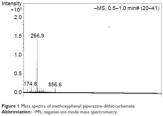 | Figure 1 Mass spectra of methoxyphenyl piperazine-dithiocarbamate. |
The physicochemical parameters are depicted in Figure 2. XRD pattern (Figure 2A) of the Ag-NPs corresponds to that of face centered cubic. On conjugation with MPP-DTC, only a slight distortion was observed. The capped MPP-DTC–Ag-NPs reflect peaks corresponding to (111), (200), (220), (311), and (222). Peaks in the region (0<2θ ≤35) match with the standard XRD data of Ag2CO3, the formation of which is attributed to carbon-dioxide in atmosphere.5 The UV-vis spectra (Figure 3A) showed a peak at ~400 nm characteristic of Ag-NPs and assigned to surface plasmon resonance (SPR). This peak, due to the SPR, is dependent on medium’s refractive index, size and shape of NPs, and absorption substance at the surface of the NPs. UV-vis spectra of MPP-DTC–Ag-NPs (Figure 3B) show two resonant peak absorption at ~250 and 300 nm (characteristic peaks of DTC), indicating conjugation although the peak at 400 nm got compromised because of capping. The broadening of the peak in 410–430 nm is due to the presence of DTC that acts as electron donor and changes the bonding pattern of the Ag-NPs.6–8 The fluorescence spectra of the Ag-NPs dispersed in water exhibit a single fluorescence emission at 300 nm when it is excited in the range of either 215–235 or 255–280 nm (λex), whereas the MPP-DTC–Ag-NPs fluorescence with a singular emission at 425 nm for λex is excited in the range of 230–250 nm (Figure 4A). The fluorescence emission peak intensity is maximum at 425 nm at λex 235 nm and decreases thereafter as λex increases. This indicates that resonant absorption/maximum transition probability is at λex 235 nm for capped Ag-NPs. The reason for the red shift could be attributed to the change in the environment of the MPP-DTC–Ag-NPs.
 | Figure 2 X-ray diffraction patterns of (A) Ag-NPs and (B) MPP-DTC–Ag-NPs. |
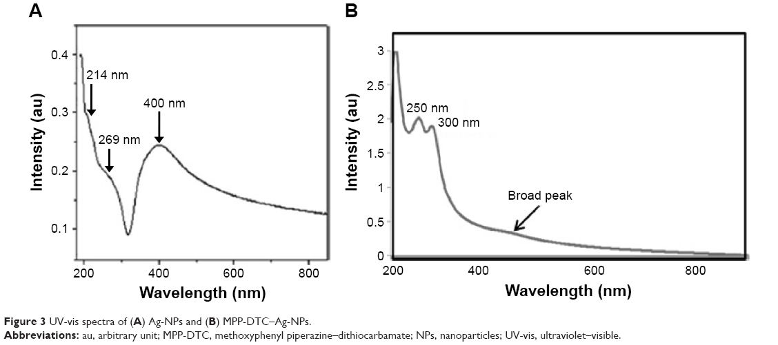 | Figure 3 UV-vis spectra of (A) Ag-NPs and (B) MPP-DTC–Ag-NPs. |
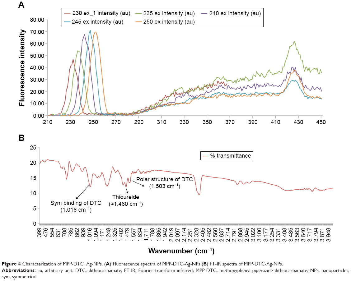 | Figure 4 Characterization of MPP-DTC–Ag-NPs. (A) Fluorescence spectra of MPP-DTC-Ag-NPs (B) FT-IR spectra of MPP-DTC-Ag-NPs. |
The IR spectra (Figure 4B) indicated the formation of silver-sulfide bonds by the disappearance of 2,550–2,600 cm−1 peak. The typical DTCs frequencies9 were 1) peak at 1,460 cm−1 associated primarily with the “thioureide”, 2) peak at 1,503 cm−1 indicating the polar structure of DTC, and 3) peak at 1,016 cm−1 indicating the symmetrical binding mode of DTC. The size of the synthesized Ag-NPs in TEM images was 10–20 nm. Conjugation with MPP-DTC resulted in NPs with similar size, but exact size could not be determined due to clustering. The cellular toxicity studies performed on HEK cell lines showed that the MPP-DTC–Ag-NPs were nontoxic up to a dose of 1 mM.
Conclusion
Our findings establish 1) easy synthesis of the ligand MPP-DTC, 2) easy synthesis of Ag-NPs free from extraneous impurities, 3) conjugation of Ag-NPs with MPP-DTC, and 4) evaluation of physicochemical parameters. Further work should concentrate on exact size and polydispersity index determination of the MPP-DTC–Ag-NPs and their application for optical imaging.
Acknowledgment
We thank Dr RP Tripathi, Director, Institute of Nuclear Medicine and Allied Sciences, and School of Physical Sciences, Jawaharlal Nehru University, for providing the necessary facilities.
Disclosure
The authors report no conflicts of interest in this work.
References
Yang W, Lin Y, Zhang X, Zhang J, Wang X. Synthesis of several MPP derivatives for 99mTc-labelling and evaluated as potential 5-HT1A receptor imaging agents. Sci China Chem. 2011;54(7):1148–1154. | ||
Chaturvedi S, Kaul A, Yadav N, Singh B, Mishra AK. Synthesis, docking and preliminary in vivo evaluation of serotonin dithiocarbamate as novel SPECT neuroimaging agent. MedChemComm. 2013;4(6):1006–1014. | ||
Sen P, Ghosh J, Abdullah A, Kumar P. Preparation of Cu, Ag, Fe and Al nanoparticles by the exploding wire technique. J Chem Sci. 2003;115(5–6):499–508. | ||
Shankaranarayana ML, Patel CC. The electronic spectra of some derivatives of xanthic, dithiocarbamic and trithiocarbonic acids. Acta Chem Stand. 1965;19:1113–1119. | ||
Liu C, Yang X, Yuan H, Zhou Z, Xiao D. Preparation of silver nanoparticle and its application to the determination of ct-DNA. Sensors. 2007;7(5):708–718. | ||
Yeshchenko OA, Dmitruk IM, Alexeenko AA, Kotko AV, Verdal J, Pinchuk AO. Size and temperature dependence of the surface plasmon resonance in silver nanoparticles. Ukr J Phys. 2012;57:266. | ||
Mankad V, Kumar RK, Jha PK. Investigation of blue-shifted plasmon resonance: an optical properties study of silver nanoparticles. Nanosci Nanotechnol Lett. 2013;5(8):889–894. | ||
Mogensen KB, Kneipp K. Size-dependent shifts of plasmon resonance in silver nanoparticle films using controlled dissolution: monitoring the onset of surface screening effects. J Phys Chem C. 2014;118(48):28075–28083. | ||
Mathew EJ, Studies on Some Metal Complexes of Dithio Ligands [doctoral thesis]. Cochin, India: Cochin University of Science and Technology; 1990. |
 © 2018 The Author(s). This work is published and licensed by Dove Medical Press Limited. The full terms of this license are available at https://www.dovepress.com/terms.php and incorporate the Creative Commons Attribution - Non Commercial (unported, v3.0) License.
By accessing the work you hereby accept the Terms. Non-commercial uses of the work are permitted without any further permission from Dove Medical Press Limited, provided the work is properly attributed. For permission for commercial use of this work, please see paragraphs 4.2 and 5 of our Terms.
© 2018 The Author(s). This work is published and licensed by Dove Medical Press Limited. The full terms of this license are available at https://www.dovepress.com/terms.php and incorporate the Creative Commons Attribution - Non Commercial (unported, v3.0) License.
By accessing the work you hereby accept the Terms. Non-commercial uses of the work are permitted without any further permission from Dove Medical Press Limited, provided the work is properly attributed. For permission for commercial use of this work, please see paragraphs 4.2 and 5 of our Terms.