Back to Journals » International Journal of Nanomedicine » Volume 20
Synergistic Wound Healing: Unraveling the Multi-Target Effects of Traditional Chinese Medicine and Its Biomaterials on Chronic Wound Pathways
Authors Shen J, Tong Z, Han B, Zhang Z, Xian Z, Yuan Y, Duan X, Han S, Liu P, Wang Z ![]()
Received 16 January 2025
Accepted for publication 29 May 2025
Published 23 October 2025 Volume 2025:20 Pages 12889—12912
DOI https://doi.org/10.2147/IJN.S513585
Checked for plagiarism Yes
Review by Single anonymous peer review
Peer reviewer comments 3
Editor who approved publication: Professor Eng San Thian
Jiayu Shen,1,2,* Zhuo Tong,1,2,* Bing Han,1,2 Ziyin Zhang,1,2 Zudan Xian,1,2 Yao Yuan,1,2 Xinliang Duan,1,2 Sichen Han,1,2 Peng Liu,2,3 Zilin Wang1,2
1Department of Oral and Maxillofacial Surgery, School and Hospital of Stomatology, Jilin University, Changchun, 130021, People’s Republic of China; 2Jilin Provincial Key Laboratory of Tooth Development and Bone Remodeling, Jilin University, Changchun, People’s Republic of China; 3VIP Department, Hospital of Stomatology, Jilin University, Changchun, People’s Republic of China
*These authors contributed equally to this work
Correspondence: Zilin Wang, Department of Oral and Maxillofacial Surgery, School and Hospital of Stomatology, Jilin University, Changchun, 130021, People’s Republic of China, Tel +18744014751, Email [email protected] Peng Liu, Department of Oral and Maxillofacial Surgery, School and Hospital of Stomatology, Jilin University, Changchun, 130021, People’s Republic of China, Tel +18744014751, Email [email protected]
Abstract: Chronic wounds, including diabetic ulcers (DUs), radiation-induced ulcers, and burns, present significant clinical challenges due to their distinct pathological mechanisms, necessitating tailored therapeutic strategies. Diabetic ulcers, characterized by impaired angiogenesis, persistent inflammation, and hyperglycemia, require dual modulation of inflammatory (NF-κB) and pro-repair (PI3K/Akt, Nrf2) pathways. Radiation ulcers involve DNA damage, NLRP3-driven inflammation, and TGF-β/Smad-mediated fibrosis, while burns trigger acute inflammation via DAMPs/PAMPs-TLR/NLR activation. Traditional Chinese medicine (TCM) and its bioactive components, such as Scutellaria baicalensis, curcumin, and Panax notoginseng, exhibit multi-target therapeutic effects by regulating oxidative stress, inflammation, angiogenesis, and extracellular matrix remodeling through key pathways, including Nrf2/ARE, MAPK, NF-κB, PI3K/Akt, HIF-1α/VEGF, Wnt/β-catenin, and TGF-β/Smad. Emerging supramolecular self-assembled biomaterials—nanofibrous scaffolds, hydrogels, and microneedles—address the hydrophobicity and low bioavailability of natural plant-derived macromolecules (NPHMs), enabling spatiotemporally controlled drug delivery. Innovative formulations, such as curcumin-loaded hydrogels and exosome-based systems, enhance antioxidant, anti-inflammatory, and pro-angiogenic activities, accelerating wound closure. Despite progress, challenges remain in optimizing multifunctional co-assembly systems, elucidating NPHM self-assembly mechanisms, and developing smart biomaterials responsive to dynamic wound microenvironments. Future research should focus on clinical translation by improving material stability, refining stimulus-responsive release systems, and integrating interdisciplinary insights from herbal medicine, nanotechnology, and regenerative biology. This review systematically summarizes the mechanistic roles of TCM in wound healing, highlights advancements in bioactive material design, and outlines future directions to bridge traditional knowledge with modern therapeutic innovations, offering a scientific foundation for advancing chronic wound management.
Keywords: chronic wounds, traditional Chinese medicine, biomaterials, signaling pathways, wound healing
Introduction
It is reported that approximately 1–2% of the global population, particularly patients over the age of 65, are prone to chronic non-healing wounds.1 Chronic wounds, such as diabetic ulcers (DU), have a global incidence rate of 0.02% to 10% among diabetic patients.2–7 The presence of wounds severely affects people’s physical functions and capabilities,8 mental health,9 and creates economic10 and social burdens.11 Wound healing has always been a challenge for clinicians, and there is an urgent need for new materials and methods. In recent years, extensive animal studies have focused on the impact of herbal compounds and active ingredients on the healing of diabetic wounds. The main advantage of TCM is that it usually contains a variety of effective components that can act on multiple targets simultaneously, producing synergistic effects. For example, TCM such as Scutellaria baicalensis, curcumin, Panax notoginseng, and Astragalus membranaceus, and their derived biomedical materials play roles including inhibiting the release of inflammatory factors, regulating oxidative stress, promoting macrophage polarization, inhibiting cell apoptosis, inducing autophagy, and involve multiple signaling pathways such as nuclear factor kappa-B (NF-κB), mitogen-activated protein kinase (MAPK), and phosphoinositide 3-kinase (PI3K)/protein kinase B (AKT). Additionally, research on composite reagents and new materials made from TCM components to promote healing is emerging. This article aims to review the mechanisms of TCM in regulating wound healing and summarizes reports, as shown in Figure 1, on the related signaling pathways of TCM reagents on wound healing, providing a scientific reference for further research on TCM treatment of wound healing and rational clinical application.
 |
Figure 1 Figure 1 TCM and its derived biomaterials in Chronic Woud Healing. (a) Classification of Wounds. (b) TCM for wound healing. (c) TCM derived biomaterials. (d) Advantages of TCM derived biomaterials. (e) The mechanism of action of TCM. |
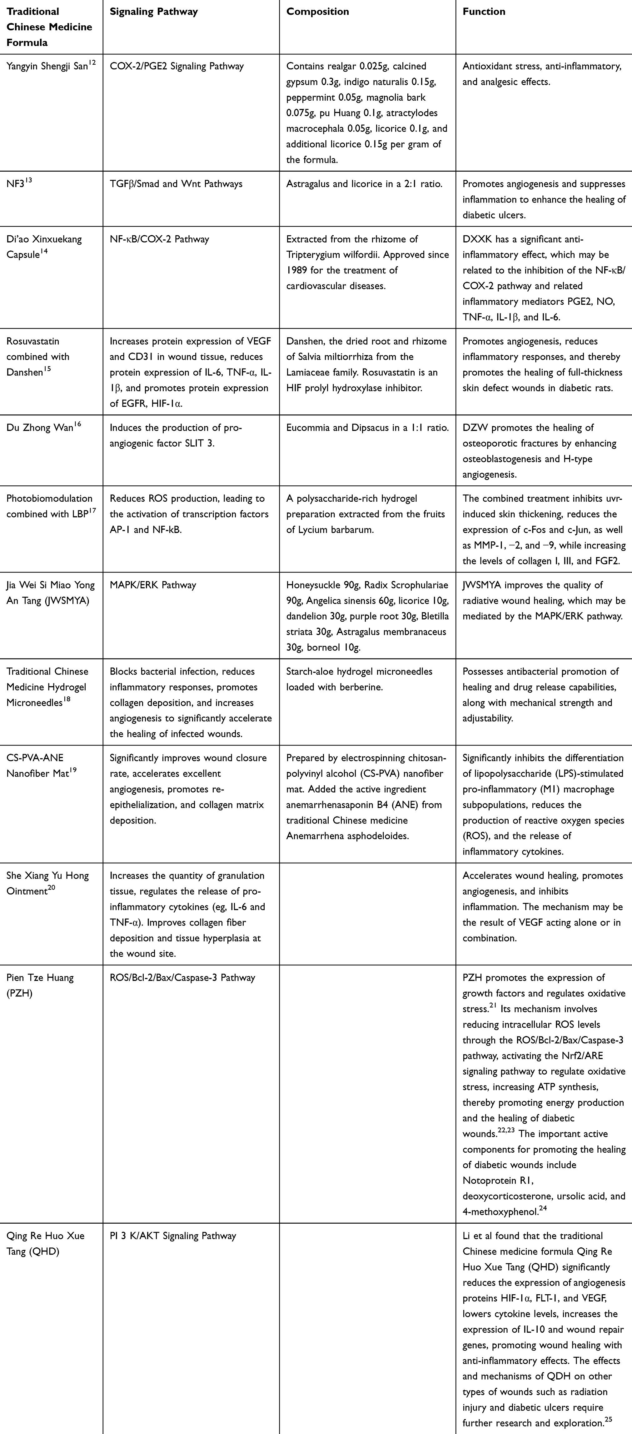 |
Table 1 Traditional Chinese Medicine Compound Preparation Promotes Wound Healing |
The Basic Process of Wound Healing
Wound healing requires the processes of blood clotting to stop bleeding, inflammation, epithelial regeneration, granulation tissue formation, and tissue remodeling.26,27 Wound healing is a complex physiological process involving multiple stages and interactions of various cell types. The entire process of wound healing is depicted in Figure 2.
 |
Figure 2 Wound healing occurs through five dynamically interconnected stages—clot formation, inflammation, angiogenesis, cell migration and proliferation, and tissue remodeling—collaboratively repairing damage and restoring tissue functionality. |
Coagulation (Hemostatic Phase)
After skin injury, platelet function is activated, and platelets release granules. Concurrently, the coagulation cascade is activated, with various serine proteases being activated, ultimately leading to the conversion of fibrinogen to fibrin by thrombin, forming fibrin fibers.28 The initiation of these cascades is crucially dependent on factor XI binding to negatively charged surfaces (such as collagen).29 In this phase, platelets are not only involved in hemostasis but also release multiple effector factors through degranulation, activating the innate immune inflammatory response.
Inflammatory Response Phase
The inflammatory phase follows the coagulation phase and lasts from hours to days. In this stage, neutrophils migrate to the wound interstitium under the influence of inflammatory signals,30,31 releasing Monocyte Chemoattractant Protein-1 (MCP1), Leukotriene B4 (LBT4), gasdermin, elastase, and myeloperoxidase, recruiting more inflammatory cells,32,33 and dying after killing microbes. Subsequently, macrophages phagocytose neutrophils, promoting the transformation of macrophage phenotype and function, and inducing cell differentiation to ensure the restoration of vascular integrity,34–36 preparing for the subsequent repair phase.
Cellular Proliferation (Granulation Proliferation Phase)
In the late inflammatory response, fibroblasts and endothelial cells begin to proliferate, forming granulation tissue. In this stage, neovascularization, fibroblast migration, and new collagen matrix deposition are involved in the formation of granulation tissue.
Re-Epithelialization
Concurrently with granulation tissue formation, the release from platelet degranulation, wound edges will enhance the migratory activity under the action of fibroblast growth factor 7 (FGF 7) heparin-binding epidermal growth factor (EGF) or hepatocyte growth factor (HGF), achieving re-epithelialization of the wound.37,38 In this process, the level of ephrin B1 expressed by wound epidermal cells increases, activating the adjacent Eph receptors, allowing cells to move flexibly with loose desmosomal connections, achieving epidermal cell coverage of the wound and restoring the skin’s barrier function.39
Tissue Remodeling and Maturation (Scar Maturation/Remodeling Phase)
The final stage of wound healing includes the remodeling of collagen fibers and the reorganization of tissues, often leaving a scar. Scar formation is related to the deposition of new collagen by fibroblasts and myofibroblasts, and inflammation plays a fundamental role in driving the reaction of scarring and fibrosis. Throughout the healing process, multiple signaling pathways are involved in regulating to promote wound healing. For example, the PI3K/AKT signaling pathway affects the repair process with the activity of mTOR and GSK3β. Additionally, wound healing involves various growth factors such as Platelet-Derived Growth Factor (PDGF), EGF, TGF-β, etc., which coordinate intercellular and intracellular signal transmission, promoting cell proliferation, differentiation, migration, and protein synthesis.
Classification of Wounds
Wounds are classified based on their etiology into DU, radiation ulcers (RU), and burn ulcers.40
Diabetic Ulcers
DU are a serious complication in diabetic patients, mainly characterized by infection, ulceration, tissue necrosis, and clinical features such as lower limb vascular and neurological diseases. DU is one of the most dangerous complications of diabetes, severely affecting the quality of life and survival rate of patients. Its pathogenesis involves peripheral vascular disease, neuropathy, and infection, with molecular signaling pathways crucial to healing, including the PI3K-Akt signaling pathway, nuclear factor erythroid 2-related factor 2 (Nrf2)/antioxidant response element (ARE) signaling pathway, NF-κB signaling pathway.41,42 Additionally, diabetes reduces the level of pro-angiogenic factors, increases the expression of anti-angiogenic factors, leads to apoptosis of vascular endothelial cells,43 causes skin ischemia and hypoxia, and lacks nerve growth factors.
Radiation-Induced Ulcers
Radiotherapy is an indispensable part of cancer treatment but also carries the risk of radiation-induced skin injury. Approximately 95% of patients undergoing radiotherapy will experience radiation-induced skin injury, with 85% of patients exhibiting moderate to severe skin injury reactions.44,45 The clinical manifestations of RU include ulcers, erythema, desquamation, and these chronic wounds can persist for years, causing significant physical and psychological impact on patients.46–48 The reason why RU are difficult to heal is that tissue fibrosis caused by radiation disrupts lymphatic and vascular drainage,49 reducing tissue perfusion, leading to tissue hypoxia, which is prone to ulceration and slows down wound healing.50,51 In addition, radiation directly acts on nuclear DNA and mitochondrial DNA, causing double-strand breaks, blocking the cell cycle of cell mitosis, inducing cellular senescence and damage. At the same time, DAMP molecules released by tissues and apoptosis interrupt skin tissue regeneration, produce inflammatory reactions, and the release of pro-inflammatory cytokines and inflammatory bodies such as tumor necrosis factor-alpha (TNF-α), Interleukin-1 (IL-1), Interleukin-6 (IL-6), and nucleotide-binding oligomerization domain, leucine-rich repeat and pyrin domain-containing 3 (NLRP3) further inhibits DNA repair, forming a vicious cycle. In addition, radiation injury is associated with changes in the expression of proteins such as tumor protein 53 (P53), bcl2-associated X protein (Bax), and B-cell lymphoma (Bcl-2). These complex biological processes work together to cause the development and persistence of RU, posing a serious threat to patient health and quality of life.
Burn Ulcers
Burn ulcers are caused by friction, cold, heat, radiation, chemicals, or electricity, involving some energy transfer that leads to tissue structure destruction and cell death.52 The clinical manifestations of burns include coagulative necrosis of the local skin, stagnant ischemia, and thermal inflammation. In severe burns,53 DAMPs and their exogenous counterparts, pathogen-associated molecular pattern molecules (PAMPs), are recognized by pattern recognition receptors, namely Toll-like receptors (TLR) and NOD-like receptors (NLR). TLR and NLR activate NF-κB, and the transcription of various inflammatory mediators (such as IL-1, IL-6, IL-8) promotes the inflammatory cycle, leading to systemic inflammatory response syndrome.54,55 It is worth noting that in this process, processes such as macrophage antigen presentation, neutrophil killing of pathogens,56,57 T-cell proliferation, and IL-2 production are all inhibited.58–60
Mechanisms of Wound Healing Promotion by TCM
Nrf2/ARE Signaling Pathway
The Nrf2/ARE is one of the crucial intracellular signaling pathways that maintain cellular redox homeostasis.61–63 Nrf2 is a key factor in antioxidant stress, capable of regulating the expression of cellular protective genes and antioxidants such as heme oxygenase 1 (HO-1), superoxide dismutase (SOD), and glutathione peroxidase (GSH-Px), to eliminate free radicals.64 The disruption of the body’s antioxidant defense mechanisms is one of the factors delaying wound healing, and an increasing number of studies have shown that the activation of Nrf2 can regulate inflammatory factors, reduce oxidative stress damage, and promote the healing of diabetic wounds. After skin irradiation, the upregulation of NRF2 expression leads to persistent DNA damage and cellular senescence, inducing the occurrence of RU. The entire process of wound healing through the Nrf2/ARE pathway, MAPK pathway, and PI3K-Akt pathway is depicted in Figure 3. There are many Nrf2 activators in natural medicines. For example, curcumin,65–68 baicalin,69–71 berberine, and paeoniflorin(PF) all act on the Nrf2 pathway, involving the upregulation of Nrf2, inhibiting pro-inflammatory molecules matrix metalloproteinase 9 (MMP-9) and matrix metalloproteinase 3 (MMP-3), regulating inflammatory factors TNF-1, IL-1, IL-8, promoting macrophage polarization, reducing the production of reactive oxygen species (ROS), alleviating inflammatory responses, and thus promoting skin healing. Unfortunately, the low solubility, bioavailability, and in vivo stability of curcumin hinder its efficacy as a clinical therapeutic drug. Curcumin is a hydrophobic molecule with low solubility in aqueous solutions. When taken orally, curcumin is degraded and rapidly metabolized under the acidic pH of the intestines. Similarly, after intravenous administration, it degrades within minutes and undergoes rapid first-pass metabolism, resulting in a short circulating half-life. Curcumin belongs to class IV of the biopharmaceutical classification system, characterized by low solubility and low permeability. These characteristics lead to reduced absorption, bioavailability, and stability.68 Various curcumin composite preparations have been optimized, such as Bhattacharya optimizing a curcumin hydrogel scaffold with cerium oxide nanoparticles (CNP).72 Liu C et al prepared a nanofiber wound dressing with a silver core and a curcumin surface using chitosan (CS) as the raw material, CS/Cur@β-CD/AgNPs,73 which is highly biodegradable and allows rapid release of curcumin within 2 days. This dressing has a synergistic effect on wound healing while achieving antibacterial properties.74 Gong et al combined curcumin with Mg2+ to develop an exudate-absorbing and antibacterial hydrogel with a curcumin-loaded magnesium polyphenol network (Cur-Mg@PP), which has strong stability, high bioavailability, and high adaptability to complex-shaped wounds.75 Curcumin, while achieving antioxidant and antibacterial effects, Mg2+ promotes macrophage polarization, thus promoting the healing of burn wounds. Tan W et al used curcumin and metformin loaded onto carboxymethyl chitosan (CMC) and oxidized hyaluronic acid (HA) to create a dual-drug loaded nanocomposite polysaccharide-based self-healing hydrogels (OCM@P) hydrogel for promoting diabetic wound healing, utilizing the principles of dynamic imine bonds and electrostatic interactions to form a porous structure, showing rapid release of Met and long-term sustained release of Cur, effectively inhibiting free radicals and significantly promoting re-epithelialization.76 Li et al used exosomes with good biocompatibility, strong drug loading capacity, and immune inertia as an innovative fulcrum to construct macrophage exosomes loaded with curcumin (Exos-cur).65 The study found that Exos-cur upregulated the expression of vascular endothelial growth factor (VEGF), platelet endothelial cell adhesion molecule 1 (PECAM-1, also known as CD31), and SMA proteins, repaired the mitochondrial membrane potential of human umbilical vein endothelial cells (HUVECs) and reduced oxidative damage, and accelerated wound healing in diabetic rats by activating the Nrf2/ARE pathway. Based on this, Yerneni SS et al further hypothesized that using dissolvable microneedle arrays (dMNA) to locally deliver curcumin-albumin-EV (CA-EV) can effectively control skin inflammation in vivo. In rat and mouse models, dMNA-delivered CA-EVs significantly reduced inflammation triggered by lipopolysaccharide and imiquimod by acting on inflammatory signal cascades such as NF-κβ, MAPK, and janus tyrosine kinase (JAK)/signal transducer and activator of transcription (STAT).77
 |
Figure 3 The entire process of wound healing through the Nrf2/ARE pathway, MAPK pathway, and PI3K-Akt pathway. Targeting the Nrf2/ARE, MAPK, and PI3K-Akt signaling pathways modulates autophagy, antioxidant responses, and anti-inflammatory reactions to promote wound healing. |
In terms of the application of baicalin, Masoud et al prepared a collagen hydrogel using fish skin extract and improved its mechanical properties through the PC method, incorporating baicalin into the luteolin hydrogel scaffold in an alkaline solution, increasing the Young’s modulus to 15.24 ± 0.59 kPa, achieving a wound closure rate of 94.01 ± 2.1%. Compared with the swelling loading technology to incorporate baicalin into the hydrogel matrix, the new scaffold is more accurate and has a high incorporation efficiency, solving the problem of loading baicalin into collagen gels. In contrast, A Rang Kim et al reported incorporating baicalin into a HA/PNIPAM IPN hydrogel after synthesis with a baicalin solution, with a loading efficiency of 42.80%,78 while the hydrogel treated with PC initially achieved a loading efficiency of up to 100%.
Berberine, the main chemical active components of PA, are flavonoids and alkaloids. Studies have confirmed that berberine ointment promotes wound healing, reduces advanced glycation end products (AGEs) and inflammatory factors such as IL-1β, while increasing the secretion of growth factors.79,80 The compound berberine liquid dressing is currently well-researched and clinically applied. Xu et al found that compared with the clinically commonly used alginate calcium silver fiber dressing, the external application of the compound berberine liquid dressing is more conducive to solving the problem of multidrug resistance in diabetic foot. In addition, combining with Centella asiatica can improve wound healing and reduce scar formation. An anti-acne double-layer patch loaded with PA and Centella CA extracts was created. The experiment adopted the construction of a GC double-layer patch at −80°C/room temperature, adding 1 mg/mL PA and 2.4 mg/mL CA, and found that after soaking for 150 hours, it had a good water retention rate of up to 3%. While improving the growth vitality of skin fibroblasts, it can absorb exudate from open acne wounds.81
PF has great potential in regulating M1 to M2 macrophages and can also accelerate wound healing through the Nrf2 pathway, reducing oxidative stress, increasing cell proliferation and migration, reducing apoptosis, and increasing the expression of VEGF and TGF-β1. Zhang et al designed a self-adaptive stimulus-responsive OBPG hydrogel, then added paeoniflorin-loaded micelles (MIC@Pae) to construct the OBPG&MP hydrogel drug delivery system, mainly acting on M2 macrophage polarization and extracellular matrix (ECM) remodeling processes.82 Due to the dynamic reversibility and pH sensitivity of the Schiff base bond, the OBPG&MP hydrogel showed excellent injectability, self-healing ability, and responsiveness. It can also achieve sustained release of Pae for at least 72 hours, with scavenging rates of 68.1%, 74.6%, and 71.8% for ·OH, O2−, and H2O2, respectively. On the 14th day, the OBPG&MP hydrogel group’s wound was almost completely healed, with a wound contraction rate of 98.1%. It has various properties, including protecting cells from oxidative damage. Hao et al developed a paeoniflorin hydrogel HA-PF (8%), and it was determined that signal transducer and STAT 1 were significantly inactivated, the expression of downstream protein iNOS was significantly inhibited, while STAT 6 was significantly activated, and the expression of downstream protein Arg-1 was significantly enhanced, confirming the great ability of PF to regulate the STAT signaling pathway (a classic pathway for macrophage polarization).83
Gynura divaricata (L). DC. (GD), known as “Bai Bei San Qi” in China, is used in TCM for its various pharmacological effects, including blood sugar-lowering effects, insulin sensitization, and blood pressure reduction. GD has been proven to enhance antioxidant activity in the body. Xu C et al showed that GD can activate Nrf2, prevent oxidative stress damage, promote angiogenesis, and thus promote wound healing in diabetic patients.84
MAPK Signaling Pathway
The MAPK signaling pathway plays a crucial role in skin wound healing by finely regulating processes such as cell migration, proliferation, and differentiation, which promote tissue repair.85 The MAPK family mainly includes extracellular signal-regulated kinase (ERK), c-Jun N-terminal kinase (JNK), and p38 MAPK. These kinases transmit signals through a phosphorylation cascade, affecting gene expression and cellular behavior.86 During wound healing, the activation of the MAPK signaling pathway not only promotes angiogenesis but also regulates inflammatory responses and the reconstruction of the ECM. The ERK pathway is primarily associated with cell proliferation and differentiation, while JNK and p38 MAPK are closely related to cellular stress, inflammation, and apoptosis.87
Emu oil, extracted from the subcutaneous and retroperitoneal fat of emus, possesses strong anti-inflammatory properties. It may promote wound healing by fostering the growth of keratinocytes and re-epithelialization. Additionally, Ruyi Fan et al found in in vitro and in vivo experiments that emu oil enhances the secretion of IL-10, suppresses the secretion of IL-1β, promotes M2 macrophage polarization, and significantly reduces the phosphorylation of JNK and p38, thereby strengthening the anti-inflammatory response and promoting skin healing.88
Aloe vera is recognized as a wound healing promoter and has been shown to upregulate growth factors such as keratinocyte growth factor-1 (KGF-1), TGF-β, cyclin D1, insulin-like growth factor 1 (IGF-1), VEGF, basic fibroblast growth factor (bFGF), and microfibril-associated glycoprotein 4 (MFAP4), as well as collagen, fibrillin, elastin α-smooth muscle actin (α-SMA), CD31, while downregulating the expression of MMPs. Aloe vera treatment for 48 hours shows increased expression of phosphorylated ERK (p-ERK) and increased cell proliferation.89 Similarly, fibroblasts isolated from diabetic rats treated with aloe vera for 48 hours show increased JNK-1 mRNA expression. Aloeresin, an aromatic C-glucosyl-5-methylchromone found in aloe vera, possesses antioxidant activity. Iosageanu A et al found that aloesin promotes wound healing by participating in multiple processes, including cell migration, tissue development, angiogenesis, and cytokine release. Moreover, aloesin increases the phosphorylation of ERK and JNK in a concentration-dependent manner, leading to the activation of the MAPK signaling pathway, but has little effect on p38. In addition, aloesin strongly activates the effector proteins of TGF-β1 signaling, significantly increasing the phosphorylation of Smad2 and Smad3 in mouse skin samples, with little impact on Smad7 expression.90 Furthermore, aloe emodin and β-sitosterol can bind to other MAPK signaling molecules, such as Myc and Jun, as well as Akt-1 from the PI3K/Akt pathway.91 A silk protein/aloe vera gel wound dressing developed using silk protein and aloe vera, two natural extracts, enhances cell vitality, proliferation, migration, regulates VEGF secretion, inhibits cellular senescence, and activates the MAPK signaling pathway, particularly ERK1/2, thereby promoting the healing of DU.
NF-κB Signaling Pathway
The NF-κB family of transcription factors plays a pivotal role in controlling gene expression involved in immune responses, inflammation, and cell survival, and is central to the regulation of wound healing.92 Comprising five main components, these proteins form dimers that control a variety of genes associated with inflammatory responses. Typically, the NF-κB pathway is suppressed by inhibitors of the IκB family, but upon stimulation by inflammatory signals such as TNF-α or lipopolysaccharide (LPS), the IκB kinase (IKK) complex is activated, leading to the phosphorylation and degradation of IκB, thereby allowing NF-κB to translocate to the nucleus and induce the transcription of inflammatory cytokines.93 In diabetic wounds, the upregulation of NF-κB into active transcription factors results in prolonged inflammation, hindering wound healing. The activation of NF-κB is also associated with the activation of the NLRP3 inflammasome, which further impedes the wound healing process in diabetic patients.94 Additionally, overexpression of NF-κB can alter the gene expression of angiogenesis-related factors, affecting vascular cell damage and neovascularization.95
Studies have shown that Scutellaria baicalensis, also known as Huang Qin, modulates macrophage polarization induced by oxidative stress through pathways such as NF-κB, PI3K/Akt/NRF2, and HO-1, thereby mediating inflammatory responses.96,97 An ethanol composite of baicalin has been found to alleviate inflammatory pain induced by complete Freund’s adjuvant (CFA) by suppressing the P2X3 receptor.98 The combination of baicalin with SGC can activate the MAPK signaling pathway, promoting the proliferation of keratinocytes at the wound site to accelerate wound healing. A 3 μM concentration of baicalin significantly enhances the effect of VEGF on the migration of keratinocytes, increasing the expression of MMP 9 induced by VEGF by about 30%. The optimal concentration of SGC is 0.5%, and its use in conjunction with 3 μM baicalin increases the migration rate of keratinocytes by about 80%.99 Innovative research from a chemical structure and molecular biology perspective has utilized non-covalent interactions between baicalin, which has similar scaffold and phenolic hydroxyl groups, and supramolecular hydrogel factors to construct co-assemblies. A new type of hydrogel dressing, CFPL, has been developed through chain-chain non-covalent interactions.100 On the 11th day of treatment, the hydrogel continues to slowly release baicalin, with IL-6, IL-1β, and TNF-α levels dropping to 0.29%, 0.41%, and 0.94%, respectively. On the 7th and 14th days after wound induction, there is almost no distance between the epithelial tips in the acute wound tissue, indicating that the hydrogel containing chiral LBF has good anti-inflammatory properties, induces re-epithelialization, and promotes wound contraction. A hydrogel wound dressing prepared by Zhu Y et al, carboxymethyl chitosan-Sesbania gum@sesbania gum (CMCS−OSG@SE), promotes the healing of diabetic wounds by promoting epidermal regeneration, collagen deposition, and clearing ROS.101 Liu X et al prepared ROS-sensitive nanoparticles loaded with baicalin polysaccharides (APS) based on the effective components of Scutellaria baicalensis (SB) and Coptis chinensis (CC), berberine (Bai) and berberine (Ber), and constructed a multifunctional TCM composite microneedle (C/B@APB@Ber). This microneedle downregulates the ROS-activated NF-κB pathway, thereby blocking the expression of pro-inflammatory cytokines and promoting the repolarization of M1 to M2 macrophages.102
Panax notoginseng, also known as San Qi, has the effect of promoting wound healing and reducing scar formation. Panax notoginseng and Astragalus membranaceus have been proven to promote the polarization of macrophages to the M2 phenotype by upregulating β-catenin and downregulating NF-κB, reducing inflammatory responses, and promoting wound healing in diabetic rats.103,104 Rb1 significantly promotes skin wound healing in a second-degree burn rat model, with its potential mechanism related to the upregulation of FGF-2 by modulating the PDGF-BB/PDGF receptor beta (PDGFR-β) signaling pathway in wound tissue.105 A review published in 2022 detailed the different signaling pathways affected by different active components of Panax notoginseng,106 including the NF-κB pathway,107 AMPK and endothelial nitric oxide synthase (eNOS) dependent signaling pathways,108 hypoxia-inducible factor-1α (HIF-1α) dependent pathways,109 and Ang 2/Tie 2 signaling pathways.110,111 Notoginsenoside (PNS), the main active component of Panax notoginseng (Burkill) F.H.Chen (PN) leaves, has been found to upregulate the expression of miR-146-5p in the NF-κB signaling pathway and downregulate the expression of IL-1β, IL-6, TNF-α, IRAK1, TRAF6, and p65, thereby reducing inflammatory responses and promoting skin healing.112
Jianting Li et al prepared ginger carbon dots (GCDs) from dried ginger solution using microwave-assisted hydrothermal method. GCDs effectively block the TLR4/NF-κB inflammatory trigger pathway, reducing the damage caused by inflammatory mediators, thus accelerating wound healing.113
Ginsenoside Rg5 can inhibit the activation of NF-κB p65 in keratinocytes and macrophages. NF-κB p65 is a transcriptional activator of SLC7A11. In this study, we confirmed that ginsenoside Rg5 dose-dependently reduced the expression of NF-κB p65, SLC7A11, as well as NRF2 and STAT3 in the wounded area of diabetic mice.114
Encapsulating Pae in microspheres and combining it with oxidized hyaluronic acid hydrogel to prepare a hydrogel loaded with Pae microspheres (Pae-MPs@OHA) promotes the healing of acute wounds in rats. Pae-MPs@OHA can reduce the number of M1 macrophages and increase the number of M2 macrophages, thereby regulating the release of inflammatory factors TNF-α and IL-1β. Molecular docking revealed that Pae can bind through various covalent bonds, reducing the expression of pro-caspase-1 and inhibiting the activation of caspase-1, thus reducing the degree of inflammatory response. Western blot results further confirmed that Pae-MPs@OHA can reduce the expression of NF-κB, pNF-κB, NLRP3, ASC, and pro-caspase-1.115
Xia et al prepared Cur-GelMA hydrogels using 3D printing technology, which have good biocompatibility and promote tissue repair by alleviating ROS generation and ADSC cell apoptosis induced by AGE/AGER/NF-κB p65.116
Hu Y et al developed a novel 3D-printed dermal scaffold composed of decellularized ECM and copper-epigallocatechin gallate (Cu-EGCG), with a porous structure similar to that of natural human dermal matrix. This scaffold drives the expression of inflammatory-related genes in the HIF-1α/VEGF pathway and the TNF-α/NF-κB/MMP9 pathway, promoting the healing of diabetic wounds and restoring the appearance, epidermal thickness, dermal thickness, number of skin appendages, and the arrangement and deposition of collagen in the dermis to a state similar to that of normal, undamaged skin, thereby reversing the scarring of diabetic wounds.117
Zhang P et al developed a novel berberine nanocolloidal hydrogel (BNH) that can inhibit the expression of NF-κB, TNF-α, and IL-6, and by activating Sirt1, increase the expression of VEGF, CD31, and SMA, which are beneficial for the wound healing of diabetic rats.118,119
Honeysuckle is an effective free radical scavenger that can promote wound healing. Baicalein, an active component of honeysuckle, can promote the mRNA levels of inflammatory cytokines IL-6, IL-1β, and TNF-α, which can be transcriptionally regulated by NF-κB and STAT3 to alleviate skin inflammation.120 Lonicera japonica Thunb, from which chlorogenic acid is isolated, is a type of flavonoid glycoside. Silent information regulator 1 (Sirt1) is a highly conserved nicotinamide adenine dinucleotide (NAD)-dependent histone deacetylase, which is related to various intracellular signals such as aging, inflammation, apoptosis, and autophagy. Honeysuckle extract increases the autophagy flux in HUVECs and alleviates oxidative stress and reduces the ability to induce cell apoptosis by promoting the Sirt1-autophagy pathway, thereby accelerating the healing process of wounds both in vitro and in vivo.121 TCM, such as Ma Nian, promotes the proliferation of HaCaT cells and promotes healing through active components like linoleic acid glycerides, betulin, L-lysine, and morin, acting on targets such as TNF, SRC, and TP3. Liu et al used network pharmacology methods to predict the targets of Ma Nian acting on wound healing and found that TNF induces autophagy flux and the expression of autophagy genes in epidermal keratinocytes through NFKB, and other targets are associated with various validated signaling pathways such as the JAK/STAT pathway and the NF-κB pathway.122
PI3K/AKT Signaling Pathway
The PI3K/AKT signaling pathway plays a central role in skin wound healing, activated by growth factors and cytokines such as PDGF and EGF through their interaction with receptor tyrosine kinases (RTKs).123 The autophosphorylation of RTKs recruits PI3K to the cell membrane, converting phosphatidylinositol-4,5-bisphosphate (PIP2) to phosphatidylinositol-3,4,5-trisphosphate (PIP3), which in turn activates AKT. The activation of AKT promotes mTORC1, influencing biological activities such as proliferation, growth, and survival, while also regulating downstream proteins like mTOR and GSK3.124 In patients with diabetes mellitus (DM), impaired PI3K/AKT signaling is associated with the progression of DM and its complications, including lower limb ulcers. Abnormal PI3K/AKT signaling may lead to abnormalities in cell migration, proliferation, differentiation, apoptosis, and excessive degradation of the ECM. (Pentaerythritol tetranitrate) PTEN, a negative regulator of the PI3K/AKT pathway, when absent, can increase epithelial cell proliferation and migration, accelerating wound healing.125 Therefore, the precise regulation of the PI3K/AKT signaling pathway is crucial for maintaining tissue homeostasis and promoting the healing of acute wounds, and its balanced activation in skin wound healing has potential clinical significance for the treatment of chronic wounds related to diabetes.126
Chen L et al found that periplocin can induce the sequential phosphorylation of Src, PI3K, Akt, and ERK1/2 in a dose- and time-dependent manner, mediated by Na/K-ATPase, leading to the activation of Src/ERK1/2 and PI3K/Akt pathways and accelerated wound repair.127 Scholars have synthesized a multifunctional chitosan/sodium alginate/velvet antler blood peptide (VBPs) hydrogel (CAVBPH). Treatment with this hydrogel activates the PI3K/AKT/mTOR and SIRT1/NF-κB pathways, thereby enhancing cell proliferation, angiogenesis, and ECM secretion, and alleviating inflammation at the wound site.128
A hyaluronic acid hydrogel containing Poria (Schw). Wolf terpenoid compounds can significantly accelerate the healing of diabetic wounds, with anti-inflammatory effects and enhanced angiogenesis. Using network pharmacology and molecular docking, Ding X et al identified 74 potential target genes (JUN, MAPK1, STAT3, AKT1, and CTNNB1) related to the treatment of diabetic ulcers with Poria (Schw). Wolf, showing significant enrichment in the PI3K-AKT signaling pathway. In vitro experiments showed that Poria polysaccharides (10 mg/L) promote the migration of HUVECs and increase the expression of CD31 and VEGF mRNA, while also activating the expression of p-PI3K and p-AKT. Additionally, PTE reduces the expression of IL-1β, IL-6, TNF-α, and NF-κB mRNA in THP-1 cells and promotes M2 macrophage polarization, indicating the potential therapeutic effect of PTE in the treatment of diabetic ulcers.129
Danmu can promote the proliferation of HUVECs, upregulate the expression levels of VEGF in HUVECs, and regulate the PI3K/AKT/mTOR pathway to promote cell repair and wound healing.130 Xu Y constructed a magnetically attracted thermosensitive hydrogel (FDA-TA) based on polyether F127 diacrylate (F127) and tannic acid (TA). Under pre-iron enrichment conditions, the PI3K-AKT pathway is activated to inhibit the autophagy mediated by nuclear receptor coactivator 4, promoting tissue healing.131 Furthermore, a composite hydrogel (GBTF)3+ was synthesized through one-step photopolymerization of gelatin methacrylate (GelMA), Bletilla striata polysaccharide (BSP), and tannic acid/iron(III) complex (TA/Fe), which rapidly promotes wound healing through anti-inflammatory and pro-angiogenic effects.132
Ginseng-derived nanoparticles (GDNPs) possess the basic characteristics of exosomes, enhancing the migration of HaCaT cells and HUVECs, as well as angiogenesis in HUVECs, and promoting skin wound healing and reducing inflammation in mice through the ERK and AKT/mTOR pathways.133,134
Moist Exposed Burn Ointment (MEBO), an effective drug for treating diabetic trauma, has been shown to improve microcirculation, clear necrotic material, reduce inflammation, and increase the expression of vascular endothelial factors. Zheng et al studied its mechanism and found that in a rat ulcer wound model, the MEBO group first enhanced and then weakened the regulatory effect on autophagosomes. The expression levels of key factors in the mTOR signaling pathway, such as PI3K, Akt, and mTOR, were relatively low, increased and then decreased, and were significantly higher than those in the model group. It is speculated that MEBT/MEBO may activate the PI3K-Akt-mTOR signaling pathway, activate autophagy inhibition, reverse the expression levels of PI3K, Akt, and mTOR, and participate in the repair and healing of ulcer wounds.135
HIF-1α/VEGF Signaling Pathway
VEGF plays a central role in wound healing, not only promoting angiogenesis but also regulating vascular permeability and endothelial cell function.136 The expression of VEGF is regulated by HIF-1α, which activates receptors such as VEGFR-2 and downstream signaling pathways, including PLC-γ/PIP2/DAG/IP3, MAPK/ERK, PI3K/AKT, and FAK/Paxillin, to promote cell proliferation and migration.137 The dysfunction of the HIF-1α/VEGF axis is a primary mechanism of impaired vascularization in diabetes. Additionally, HIF-1α is widely recognized as a major regulator of oxygen homeostasis. The entire process of wound healing through the HIF-1α/VEGF pathway is depicted in Figure 4.138
The active component of Asperosaponin VI (ASA VI) from Dipsacus asperoides, a traditional Chinese medicinal plant, promotes cell proliferation, migration, and enhances angiogenic capacity by upregulating the HIF-1α/VEGF pathway, ultimately facilitating wound healing.139 Jamun honey (JH) (0.1% v/v) significantly improves wound closure, migration, and the expression of α-SMA. Local application of JH in a diabetic mouse model promotes re-epithelialization, collagen deposition (I/III), and balanced myofibroblast formation, while also upregulating angiogenic markers (HIF-1α, VEGF, VEGF R-II).140
 |
Figure 4 HIF-1α/VEGF Signaling Axis in Wound Healing-Associated Angiogenesis. (a) Promoting angiogenesis by targeting the HIF-1α/VEGF pathway facilitates wound healing. (b) Angiogenesis is a complex and highly orchestrated process that relies on elaborate intercellular and intracellular signaling networks between endothelial cells (ECs) and pro-angiogenic cells. |
Ji M et al reported the synthesis of dandelion-shaped mesoporous strontium-gallium particles (GE@SrTPP) through dopamine-mediated biomineralization and self-assembly, followed by functionalization with a gallium metal polyphenol network. GE@SrTPP can release bioactive ions in a spatiotemporal manner akin to dandelion seeds. In the early stages of wound healing, it exhibits rapid and effective hemostasis and antibacterial properties. During the inflammatory phase, GE@SrTPP promotes M2 polarization of macrophages, suppresses the expression of pro-inflammatory factors, and reduces oxidative stress in the wound. In the proliferation and tissue remodeling stages, GE@SrTPP promotes angiogenesis by activating the HIF-1α/VEGF pathway.141
Pae, a major active component found in Paeonia lactiflora, has demonstrated angiogenic properties in endothelial cells with vascular dysfunction and zebrafish models. Pae reportedly enhances the secretion of angiogenic growth factors and promotes the proliferation, migration, and tube formation of endothelial cells. He J et al developed a multifunctional sponge dressing based on copper, Bletilla striata polysaccharide (BSP), and Peony leaf extract (PLE), known as BSP-Cu-PLE sponge. The BSP-Cu-PLE sponge exhibits excellent antimicrobial activity and hemostatic properties, and significantly (p < 0.05) accelerates the healing process of skin wounds by stimulating collagen deposition, promoting angiogenesis, and reducing inflammatory cells.142 A stimulus-responsive sugar-peptide hydrogel, termed OBPG&MP, was constructed from oxidized Bletilla striata polysaccharide (OBSP), tannic acid-grafted ε-polylysine (PLY-GA), and Pae-loaded micelles (MIC@Pae). This hydrogel mimics the glycoprotein fiber structure of the ECM, demonstrating excellent injectability, self-healing ability, and biocompatibility. It adaptively responds to the inflammatory microenvironment of chronic wounds, sequentially releasing therapeutic agents to eradicate bacterial infections, neutralize ROS, modulate macrophage polarization, suppress inflammation, and promote angiogenesis and ECM remodeling, playing a key role in the inflammatory, proliferative, and remodeling phases of wound healing.82
As recorded in the “Compendium of Materia Medica” the fruit of the Chinese herb CFI can be moistened with wine, baked, mashed (flesh and kernel), and ground into a fine powder with borneol to treat persistent oral ulcers.143 As a flavonoid-rich traditional Chinese medicinal plant, CFI has been found to possess anti-ulcer, antioxidant defense enhancement, protection against UV-induced skin damage, prevention of endothelial cell dysfunction, antibacterial, and wound healing properties, and can alleviate ischemia/reperfusion injury in mouse intestines by activating PPARα and inhibiting the NF-κB signaling pathway.144–146 Feng et al validated through research that CFIE may promote the healing of diabetic wounds through the PI3K/AKT and HIF-1α pathways.
Wnt Signaling Pathway
The Wnt/β-catenin signaling pathway is a widely conserved cellular signaling network present in both invertebrate and vertebrate animals, playing crucial roles in various physiological processes such as embryonic development, tumorigenesis, and wound healing.147 This pathway includes core components such as Wnt proteins, Frizzled family receptors, LRP5/6, and GSK-3β, which control cell proliferation, differentiation, and tissue homeostasis by regulating the stability and nuclear localization of β-catenin.148 In wound healing, the Wnt/β-catenin signaling pathway is involved in the regulation of inflammatory responses, cell proliferation and migration, angiogenesis, hair follicle regeneration, and skin fibrosis, with its activity changes being closely related to the healing outcome.149 Activation and inhibition factors of the Wnt signaling pathway play significant roles at different stages of wound healing, affecting inflammation, proliferation, differentiation, and tissue regeneration, indicating the complex regulatory role of the Wnt signaling pathway in wound healing and tissue regeneration, making it an important area of research and application in wound therapy and tissue engineering.150
In skin wounds treated with quercetin, the expression levels of VEGF, fibroblast growth factor, and α-SMA are positively correlated in a dose-dependent manner. Molecular docking analysis also reveals that hydrogen bonds formed between quercetin and residues Ala195, Gln308, Asn369, and Lys372 of TERT suggest that the telomerase RNA component catalytic element (TERT) plays an indispensable role in enhancing wound healing capabilities. Quercetin can stimulate the proliferation of human epidermal stem cells and promote wound healing through the estrogen receptor (ERα)/β-catenin/c-Myc signaling pathway.151–153 Studies have also shown that quercetin can regulate the PI3K-AKT-mTOR signaling pathway to accelerate wound healing.154–156 Compared to the direct application of quercetin, Xing et al developed a polysaccharide-based injectable hydrogel (PECE) loaded with quercetin. A 1% concentration of PECE showed the maximum release within the first 12 hours, with the best antibacterial effect, and almost no bacteria present by the fourth day. By the 20th day, the narrowest wound measured only 0.976 mm, with the best epidermal recovery and the observation of new hair follicle formation. The clearance rate for DPPH was over 99%, and there was a significant reduction in IL-6 and TNF-α.157–159 The hydrogel exhibited high antioxidant properties and excellent antibacterial performance. Another microneedle system, QPS MN, released about 47% in vitro within the first hour, with a scratch migration rate of 61.4 ± 3.25%, ten times that of the control group. Expression of IL-6 and TNF-α was reduced, while VEGF protein expression increased. Quercetin chitosan tripolyphosphate nanoparticles can achieve better healing quality and maturity of granulation tissue,160 with the optimal quercetin nanoparticle concentrations identified as 0.3% and 0.1%.161,162 Additionally, a composite of polycaprolactone (PCL) and chitosan oligosaccharides was prepared and mixed with quercetin to construct a PCL-COS-Qe membrane.163 The loading efficiency was 97.0%, and it had high water vapor permeability, which is beneficial for wound drying. The application of quercetin and its derived biomaterials in wound healing is depicted in Figure 5.
 |
Figure 5 Investigations on the Effects of Quercetin and Related Treatments on Wound Healing. (a) Detection of β-catenin and c-Myc in human embryonic stem cells (EpSCs) treated with 1 μM quercetin for 24 hours after pre-treatment with ICI 182780 for 30 minutes. Reprinted from Wang Z, Zhang G, Le Y, et al. Quercetin promotes human epidermal stem cell proliferation through the estrogen receptor/beta-catenin/c-Myc/cyclin A2 signaling pathway. Acta Biochim Biophys Sin. 2020;52(10):1102–1110.151 (b) Western blot (WB) analysis to detect changes in proteins related to the PI 3-kinase-AKT-mTOR signaling pathway after QCT treatment. Reprinted from Zhang Z, Wang L, Li X, Miao Y, Li D. Integrating network pharmacology, molecular docking and experimental validation to explore the pharmacological mechanisms of quercetin against diabetic wound. Int J Med Sci. 2024;21(14):2837–2850. Published under Creative Commons 4.0, https://creativecommons.org/licenses/by/4.0/.156 (c) Hematoxylin and eosin (HE) staining to observe the degree of inflammatory cell infiltration and the condition of granulation tissue formation. Reprinted with permission from Xing D, Du Y, Dai K, Lang S, Bai Y, Liu G. Polysaccharide-based injectable hydrogel loaded with quercetin promotes scarless healing of burn wounds by reducing inflammation. Biomacromolecules. 2024;25(11):7529–7542. Copyright 2024, American Chemical Society.158 (d) Scratch assay to test the ability of DMN to promote the migration and repair of HUVECs.159 (e) Wound maturity evaluation when quercetin ointment is applied directly. Reprinted from Kant V, Jangir BL, Sharma M, Kumar V, Joshi VG. Topical application of quercetin improves wound repair and regeneration in diabetic rats. Immunopharmacol Immunotoxicol. 2021;43(5):536–553. Reprinted by permission of the publisher (Taylor & Francis Ltd, http://www.tandfonline.com).157 (f) Wound maturity scoring for quercetin chitosan tripolyphosphate nanoparticles at different drug concentrations. Reprinted from European Journal of Pharmacology, 880, Choudhary A, Kant V, Jangir BL, Joshi VG. Quercetin loaded chitosan tripolyphosphate nanoparticles accelerated cutaneous wound healing in Wistar rats, 173172, Copyright 2020, with permission from Elsevier.160 (g) Expression of VEGF and FGF in HUVEC cells exposed to 10, 20, and 320 nM doses of quercetin. Reproduced from Kartal B, Alimogullari E, Elci P, et al. The effects of Quercetin on wound healing in the human umbilical vein endothelial cells[J]. Cell Tissue Bank. 2024;25(3):851–860. Springer Nature, https://link.springer.com/article/10.1007/s10561-024-10144-1.152 (h) Molecular docking analysis. Quercetin forms hydrogen bonds with the Ala195, Gln308, Asn369, and Lys372 residues of TERT. Reprinted from Journal of Ethnopharmacol, 290, Mi Y, Zhong L, Lu S, et al. Quercetin promotes cutaneous wound healing in mice through Wnt/β-catenin signaling pathway. 115066, Copyright 2022 with permission from Elsevier.153 |
Lucidone can effectively heal skin wounds in mice. The activation of Wnt/β-catenin signaling by GSK3β leads to an increase in c-Myc and cyclin D1 gene activation. Lucidone increases cell cycle regulatory proteins to accelerate keratinocyte proliferation and triggers EMT/MMP activation, promoting the loosening of tight junctions and facilitating migration/invasion. Furthermore, lucidone also participates in the inflammatory phase of wound healing through NF-κB-mediated iNOS/COX-2/NO production and VEGF expression in macrophages.164
Tan D et al developed a multifunctional hydrogel wound dressing composed of royal jelly-derived extracellular vesicles (RJ-EV) and methacryloyl sericin (SerMA). RNA sequencing results indicate that the SerMA/RJ-EVs hydrogel dressing mainly accelerates wound healing by inhibiting inflammation and promoting vascular repair through Wnt signaling and Hippo signaling pathways.165
TGF-β/Smad Signaling Pathway
TGF-β/mothers against decapentaplegic (Smad) signaling pathway plays a central role in skin wound healing. TGF-β is a key cytokine synthesized by damaged tissue, fibroblasts, and M2 macrophages, and it is crucial in the generation of myofibroblasts, cells responsible for substantial collagen deposition and wound contraction.166 Growth factors such as VEGF, PDGF, and TGF-β released by M2 macrophages promote the proliferation, migration, and differentiation of fibroblasts, which is essential for capillary growth and granulation tissue formation in the reparative dermis.167 TGF-β initiates signaling by binding to cell surface type I and II receptors, leading to the phosphorylation of Smad2 and Smad3, which then mediate gene expression, promote collagen deposition, and lead to fibrosis.168 However, the abnormal regulation of the TGF-β signaling pathway can lead to scarring and fibrosis, characterized by abnormal synthesis and deposition of collagen, as well as an increased ratio of type I to type III collagen.169 Therefore, modulating the TGF-β/Smad signaling pathway is of significant importance for controlling abnormal skin scarring and promoting wound healing. The entire process of wound healing through the TGF-β/Smad pathway is depicted in Figure 6.
 |
Figure 6 The entire process of wound healing through the TGF-β/Smad pathway. |
Astragalus (AR) and Rehmannia (RR) in a 2:1 ratio form NF3, which affects the migration of skin fibroblasts and the activation of selected genes and proteins related to wound healing. A concentration of 4 mg/mL of NF3 promotes the migration of human skin fibroblasts and upregulates the synthesis of TGF-β1 and BMP-6. qRT-PCR analysis shows that NF3 regulates genes including TGF-β superfamily ligands and downstream effector genes, as well as genes involved in the TGF/Smad pathway and Ras/MAPK (non-Smad) pathways. Additionally, in ECM-related molecules, NF3 upregulates the expression of type I and III collagen, fibronectin, and TIMP-1, and downregulates the expression of MMP-9 in skin fibroblasts.167
Salvia miltiorrhiza, a commonly used medicinal plant, is reported to have anti-inflammatory effects. Studies have found that the water extract of Salvia miltiorrhiza (RSM) increases the levels of serum epidermal growth factor (EGF), transforming growth factor (TGF)-β, and hydroxyproline (Hyp), and increases the protein expression of bFGF, TGF-β1, and EGF, playing anti-inflammatory roles, improving local wound microcirculation, and accelerating wound surface metabolism to promote wound healing (WH).170 Tanshinone IIA (TSA), a bioactive component of Salvia miltiorrhiza, has been shown by Zhan Y et al to prepare TSA self-dissolving microneedles (TSA-MN) that inhibit the proliferation and migration of HSF cells in a dose-dependent manner, significantly reduce the secretion of TGF-β1, and increase the expression of Smad7.171 Luan X et al prepared microneedles with Tripterygium wilfordii hook F (TP)/Pae tips and BP-hydrogel bases. The combined administration of TP and Pae not only provides anti-inflammatory, detoxifying, and immunomodulatory effects but also, with the addition of BPs, exhibits good biocompatibility and near-infrared (NIR) responsiveness, endowing MN with photothermal controlled drug release capabilities. TP/Pae loaded responsive MNs can effectively improve skin fibrosis and capillary dilation, reduce collagen deposition, and decrease epidermal thickness.172 Alpinia galanga has been shown to enhance TGF-β1-mediated growth inhibition by inhibiting the phosphorylation of the Smad3 linker region threonine 179 residue and can be used locally for the treatment of abnormal skin scars.173
Conclusion
Natural products, including herbs, are among the most precious treasures left to us by nature and have played an indelible role in the history of human health and development. Our research elucidates the mechanisms and potential of TCM in promoting wound healing from the perspective of signaling pathways. This work is relatively novel and holds significance for in-depth study. The unclear composition and mechanisms of action of TCM are one of the reasons it is rarely used in large-scale clinical treatments, yet the regulation of signals in skin regeneration and wound healing by TCM is still in its infancy. Further research and a deeper understanding of the mechanisms involved in healing are required. However, the mechanisms by which TCM and derived biomaterials promote healing vary, and there is still a lack of systematic summarization of this system. Our goal is to bridge this gap by contributing to the field. This article focuses on categorizing the mechanisms involved in the promotion of wound healing by Chinese medicine and its derived biomaterials into the following seven pathways: I) Nrf2/ARE, II) MAPK signaling pathway, III) NF-κB pathway, IV) PI3K/AKT signaling pathway, V) HIF-1α/VEGF signaling pathway, VI) Wnt pathway, and VII) TGF-Smad. It systematically summarizes the specific mechanisms by which these seven pathways play a role in wound healing and how Chinese medicine and its derived materials regulate different pathways to promote wound healing. Additionally, it summarizes the etiological classification of wounds and the pathogenesis of different types of wounds. In the final part, it also briefly outlines several basic stages involved in the wound healing process. In summary, the promotion of wound healing based on natural products and their derived biomaterials is an emerging system worth further development. Next, we will discuss the current bottlenecks in the field and potential exploration directions that could further advance our understanding and application of this area.
Future Directions and Exploration
Chronic wounds, including DU, radiation-induced ulcers, and burns, exhibit distinct pathological mechanisms, necessitating tailored therapeutic interventions. DU are characterized by impaired angiogenesis, persistent inflammation, and a hyperglycemic microenvironment, requiring dual modulation of inflammatory (NF-κB) and pro-repair (PI3K-Akt, Nrf2) pathways. Combinatorial therapies employing Astragalus membranaceus and Salvia miltiorrhiza to regulate macrophage polarization and suppress inflammation show exceptional therapeutic promise. Radiation ulcers involve DNA damage, chronic NLRP3-driven inflammation, and TGF-β/Smad-mediated fibrosis, where interventions such as curcumin (activating Nrf2/ARE signaling) combined with hyperbaric oxygen therapy target inflammatory pathways to promote healing. Burns trigger acute inflammatory cascades via DAMPs/PAMPs-TLR/NLR activation (elevated IL-6, TNF-α), necessitating immediate anti-inflammatory/antimicrobial dressings followed by epidermal regenerative enhancers like Panax notoginseng (NF-κB pathway inhibition and keratinocyte migration mediation). Beyond single herbs or their simple combinations, compound TCM formulations represent a significant therapeutic approach in wound management (Table 1). These formulations are meticulously designed by integrating multiple herbs according to TCM principles—such as achieving synergistic effects and attenuation of toxicity—to enable comprehensive regulation of the complex wound microenvironment through simultaneous multi-target and multi-pathway actions.
The unique value of natural plant-derived herbal macromolecules (NPHMs) in skin repair is increasingly recognized, yet their clinical translation is hindered by hydrophobicity and low bioavailability. Supramolecular self-assembled bioactive materials—nanofibrous scaffolds, hydrogels, vesicles, and microneedles—offer innovative solutions. Nanofibrous scaffolds mimic the extracellular matrix, providing high surface area for cell adhesion and growth factor delivery, while hydrogel microneedles enable painless, efficient transdermal drug administration. Future challenges include elucidating NPHM self-assembly mechanisms, optimizing multifunctional co-assembly systems for synergistic multi-target therapy, and designing smart biomaterials responsive to spatiotemporal cues in the wound microenvironment. Clinically oriented research must enhance material stability, develop stimulus-responsive release systems, and refine precision drug delivery. Targeting cytoskeletal dynamics (eg, tissue fluidity) and signaling pathways (JAK/STAT, TLR) may further modulate healing.174,175 Notably, NPHM self-assembly principles may bridge traditional herbal medicine and modern science, offering novel insights into regenerative potential through microenvironmental modulation. Although challenges persist, advanced delivery strategies hold promise to unlock host-driven healing and improve therapeutic outcomes in chronic wound management.
Data Sharing Statement
Data will be made available on request from the corresponding author, Peng Liu and Zilin Wang.
Acknowledgements
Both Peng Liu and Zilin Wang are corresponding authors for this paper.
Funding
This research was funded by Natural Science Fund of China, grant number 82301026; Jilin Provincial Department of Finance Science and Technology Program, grant number JCSZ2023481-12; Graduate Education Teaching Reform Research Project, grant number 2025RGZNZ010; Innovation Research Initiative, grant number2025CX325; AI-Enhanced Undergraduate Teaching Reform Special Project, grant number 24AI109Z; College Student Innovation and Entrepreneurship Training Program, grant number S202510183727 and X202510183467.
Disclosure
The authors declare that they have no known competing financial interests or personal relationships that could have appeared to influence the work reported in this paper.
References
1. Investigation on wound healing effect of Mediterranean medicinal plants and some related phenolic compounds: a review. J Ethnopharmacol. 2022;298:Article115663.
2. Armstrong DG, Tan TW, Boulton AJM, Bus SA. Diabetic foot ulcers: a review. JAMA. 2023;30(1):62–75. doi:10.1001/jama.2023.10578
3. Berenguer Perez M, Lopez-Casanova P, Sarabia Lavin R, Gonzalez de la Torre H, Verdu-Soriano J. Epidemiology of venous leg ulcers in primary health care: incidence and prevalence in a health centre-A time series study (2010–2014). Int Wound J. 2019;16(1):256–265. doi:10.1111/iwj.13026
4. Heyer K, Herberger K, Protz K, Glaeske G, Augustin M. Epidemiology of chronic wounds in Germany: analysis of statutory health insurance data. Wound Repair Regen. 2016;24(2):434–442. doi:10.1111/wrr.12387
5. Wang F, Zhang X, Zhang J, et al. Recent advances in the adjunctive management of diabetic foot ulcer: focus on noninvasive technologies. Med Res Rev. 2024;44(4):1501–1544. doi:10.1002/med.22020
6. Villa F, Marchandin H, Lavigne J-P, et al. Anaerobes in diabetic foot infections: pathophysiology, epidemiology, virulence, and management. Clin Microbiol Rev. 2024;37(3):e0014323. doi:10.1128/cmr.00143-23
7. Zhang W, Wu W, Wang T, et al. Novel supramolecular hydrogel for infected diabetic foot ulcer treatment. Adv Healthc Mater. 2024;13(31):e2402092. doi:10.1002/adhm.202402092
8. Olsson M, Jarbrink K, Divakar U, et al. The humanistic and economic burden of chronic wounds: a systematic review. Wound Repair Regen. 2019;27(1):114–125. doi:10.1111/wrr.12683
9. Zhou K, Jia P. Depressive symptoms in patients with wounds: a cross-sectional study. Wound Repair Regen. 2016;24(6):1059–1065. doi:10.1111/wrr.12484
10. Kapp S, Miller C, Santamaria N. The quality of life of people who have chronic wounds and who self-treat. J Clin Nurs. 2018;27(1–2):182–192. doi:10.1111/jocn.13870
11. Kapp S, Santamaria N. The financial and quality-of-life cost to patients living with a chronic wound in the community. Int Wound J. 2017;14(6):1108–1119. doi:10.1111/iwj.12767
12. Liu Y, Ren S, Ji H, Yan D. Study on the inhibition of inflammation by the cyclooxygenase-2 (COX-2)/prostaglandin E2 (PGE2) pathway and the promotion of wound healing of oral ulcer of Yangyin Shengji powder after chemotherapy. Ann Palliat Med. 2021;10(12):12716–12726. doi:10.21037/apm-21-3496
13. Ma H, Siu WS, Koon CM, et al. The Application Of Adipose Tissue-Derived Mesenchymal Stem Cells (ADMSCs) and a twin-herb formula to the rodent wound healing model: use alone or together? Int J Mol Sci. 2023;24(2):1372. doi:10.3390/ijms24021372
14. Yu Y, Li X, Qu L, et al. DXXK exerts anti-inflammatory effects by inhibiting the lipopolysaccharide-induced NF-κB/COX-2 signalling pathway and the expression of inflammatory mediators. J Ethnopharmacol. 2016;178:199–208. doi:10.1016/j.jep.2015.11.016
15. Xia RY, Tang D, Yang B. Effect of salvia miltiorrhiza combined with roxadustat on wound healing of full-thickness skin defects in diabetic rats and its mechanism. Zhonghua Shao Shang Yu Chuang Mian Xiu Fu Za Zhi. 2024;40(4):380–388. doi:10.3760/cma.j.cn501225-20231020-00124
16. Tian S, Zou Y, Wang J, Li Y, An BZ, Liu YQ. Protective effect of Du-Zhong-Wan against osteoporotic fracture by targeting the osteoblastogenesis and angiogenesis couple factor SLIT3. J Ethnopharmacol. 2022;295:115399. doi:10.1016/j.jep.2022.115399
17. Neves LMG, Parizotto NA, Tim CR, et al. Polysaccharide-rich hydrogel formulation combined with photobiomodulation repairs UV-induced photodamage in mice skin. Wound Repair Regen. 2020;28(5):645–655. doi:10.1111/wrr.12826
18. Luan Q, Qiao R, Wu X, et al. Plant-derived Chinese herbal hydrogel microneedle patches for wound healing. Small. 2024;20(45):e2404850. doi:10.1002/smll.202404850
19. Zhang H, Zhang M, Wang X, et al. Electrospun multifunctional nanofibrous mats loaded with bioactive anemoside B4 for accelerated wound healing in diabetic mice. Drug Deliv. 2022;29(1):174–185. doi:10.1080/10717544.2021.2021319
20. Li Q, Liu X, Yang S, Li C, Jin W, Hou W. Effects of the Chinese herb medicine formula “she-xiang-yu-hong” ointment on wound healing promotion in diabetic mice. Evid Based Complement Alternat Med. 2022;2022:1062261. doi:10.1155/2022/1062261
21. Yan Y, Liu X, Zhuang Y, et al. Pien Tze Huang accelerated wound healing by inhibition of abnormal fibroblast apoptosis in Streptozotocin induced diabetic mice. J Ethnopharmacol. 2020;261:113203. doi:10.1016/j.jep.2020.113203
22. Liu Y, Mo J, Liang F, et al. Pien-tze-huang promotes wound healing in streptozotocin-induced diabetes models associated with improving oxidative stress via the Nrf2/ARE pathway. Front Pharmacol. 2023;14:1062664. doi:10.3389/fphar.2023.1062664
23. Zhang J, Cao G, Tian L, et al. Intragastric administration of Pien Tze Huang enhanced wound healing in diabetes by inhibiting inflammation and improving energy generation. Phytomedicine. 2023;109:154578. doi:10.1016/j.phymed.2022.154578
24. Cao GZ, Tian LL, Hou JY, et al. Integrating RNA-sequencing and network analysis to explore the mechanism of topical Pien Tze Huang treatment on diabetic wounds. Front Pharmacol. 2024;14:1288406. doi:10.3389/fphar.2023.1288406
25. Li W, Chen Y, Cai Z, et al. Traditional Chinese medicine Qingre Huoxue decoction enhances wound healing in through modulation of angiogenic and inflammatory pathways. Int Wound J. 2024;21(3):e14724. doi:10.1111/iwj.14724
26. Yu FX, Lee PSY, Yang L, et al. The impact of sensory neuropathy and inflammation on epithelial wound healing in diabetic corneas. Prog Retin Eye Res. 2022;89:101039. doi:10.1016/j.preteyeres.2021.101039
27. Martin P, Pardo-Pastor C, Jenkins RG, Rosenblatt J. Imperfect wound healing sets the stage for chronic diseases. Science. 2024;386(6726):eadp2974. doi:10.1126/science.adp2974
28. Golebiewska EM, Poole AW. Platelet secretion: from haemostasis to wound healing and beyond. Blood Rev. 2015;29:153–162. doi:10.1016/j.blre.2014.10.003
29. de Bont CM, Boelens WC, Pruijn GJM. NETosis, complement, and coagulation: a triangular relationship. Cell Mol Immunol. 2019;16:19–27. doi:10.1038/s41423-018-0024-0
30. Wong SL, Demers M, Martinod K, et al. Diabetes primes neutrophils to undergo NETosis, which impairs wound healing. Nat Med. 2015;21(7):815–819. doi:10.1038/nm.3887
31. De Oliveira S, Rosowski EE, Huttenlocher A. Neutrophil migration in infection and wound repair: going forward in reverse. Nat Rev Immunol. 2016;16:378–391. doi:10.1038/nri.2016.49
32. Lämmermann T, Afonso PV, Angermann BR, et al. Neutrophil swarms require LTB4 and integrins at sites of cell death in vivo. Nature. 2013;498(7454):371–375. doi:10.1038/nature12175
33. Isles HM, Loynes CA, Alasmari S, et al. Pioneer neutrophils release chromatin within in vivo swarms. eLife. 2021;10:e68755. doi:10.7554/eLife.68755
34. Medeiros AI, Serezani CH, Lee SP, Peters-Golden M. Efferocytosis impairs pulmonary macrophage and lung antibacterial function via PGE2/EP2 signaling. J Exp Med. 2009;206:61–68. doi:10.1084/jem.20082058
35. Bellingan GJ, Caldwell H, Howie SE, Dransfield I, Haslett C. In vivo fate of the inflammatory macrophage during the resolution of inflammation: inflammatory macrophages do not die locally, but emigrate to the draining lymph nodes. J Immunol. 1996;157(157):2577–2585. doi:10.4049/jimmunol.157.6.2577
36. Minutti CM, Modak RV, Macdonald F, et al. A macrophage-pericyte axis directs tissue restoration via amphiregulin-induced transforming growth factor beta activation. Immunity. 2019;50:645–654.e6. doi:10.1016/j.immuni.2019.01.008
37. Kang NW, Jang K, Song E, et al. In situ-forming, bioorthogonally cross-linked, nanocluster-reinforced hydrogel for the regeneration of corneal defects. ACS Nano. 2024;18(33):21925–21938. doi:10.1021/acsnano.4c02345
38. Meyer M, Müller A-K, Yang J, et al. FGF receptors 1 and 2 are key regulators of keratinocyte migration in vitro and in wounded skin. J Cell Sci. 2012;125:5690–5701. doi:10.1242/jcs.108167
39. Nunan R, Campbell J, Mori R, et al. Ephrin-Bs drive junctional downregulation and actin stress fiber disassembly to enable wound re-epithelialization. Cell Rep. 2015;13(7):1380–1395. doi:10.1016/j.celrep.2015.09.085
40. Chen Z, Zhao X, Lin L, et al. CaGA nanozymes with multienzyme activity realize multifunctional repair of acute wounds by alleviating oxidative stress and inhibiting cell apoptosis. Biomater Sci. 2024.
41. Chang M, Nguyen TT. Strategy for treatment of infected diabetic foot ulcers. Acc Chem Res. 2021;54(5):1080–1093. doi:10.1021/acs.accounts.0c00864
42. Morgenstern J, Groener JB, Jende JME, et al. Neuron-specific biomarkers predict hypo- and hyperalgesia in individuals with diabetic peripheral neuropathy. Diabetologia. 2021;64(12):2843–2855. doi:10.1007/s00125-021-05557-6
43. Volmer-Thole M, Lobmann R. Neuropathy and diabetic foot syndrome. Int J Mol Sci. 2016;17(6):917. doi:10.3390/ijms17060917
44. Sas N, Lacroix JB, Dedieu V, Boyer L. Optimized radiological alert thresholds based on device-dosimetric information to predict peak skin dose between 2 and 4 Gy during vascular fluoroscopically guided intervention. Eur Radiol. 2023;33(8):5707–5716. doi:10.1007/s00330-023-09538-8
45. Moloudi K, Azariasl S, Abrahamse H, George BP, Yasuda H. Expected role of photodynamic therapy to relieve skin damage in nuclear or radiological emergency: review. Environ Toxicol Pharmacol. 2024;110:104517. doi:10.1016/j.etap.2024.104517
46. Feldmeier JJ, Heimbach RD, Davolt DA, et al. Hyperbaric oxygen in the treatment of delayed radiation injuries of the extremities. Undersea Hyperb Med. 2000;27:15–19.
47. Li Z, Maitz P. Cell therapy for severe burn wound healing. Burns Trauma. 2018;6:13. doi:10.1186/s41038-018-0117-0
48. Yang H, Wang W-S, Tan Y, et al. Investigation and analysis of the characteristics and drug sensitivity of bacteria in skin ulcer infections. Chin J Traumatol =Zhonghua chuang shang za zhi. 2017;20:194–197. doi:10.1016/j.cjtee.2016.09.005
49. Rodemann HP, Bamberg M. Bamberg M Cellular basis of radiation-induced fibrosis. Radiother Oncol. 1995;35(2):83–90. doi:10.1016/0167-8140(95)01540-W
50. Koerdt S, Rohleder NH, Rommel N, et al. An expression analysis of markers of radiation-induced skin fibrosis and angiogenesis in wound healing disorders of the head and neck. Radiat Oncol. 2015;10(1):202. doi:10.1186/s13014-015-0508-3
51. Wang J, Boerma M, Fu Q, Hauer-Jensen M. Hauer-Jensen M Radiation responses in skin and connective tissues: effect on wound healing and surgical outcome. Hernia. 2006;10(6):502–506. doi:10.1007/s10029-006-0150-y
52. r c L. Injury by electrical forces: pathophysiology, manifestations, and therapy[J]. CurrentProblems Surg. 1997;34(9):677–764.
53. Jeschke MG, van Baar ME, Choudhry MA, Chung KK, Gibran NS, Logsetty S. Burn injury. Nat Rev Dis Primers. 2020;6(1):11. doi:10.1038/s41572-020-0145-5
54. Singer M, Deutschman CS, Seymour CW, et al. The third international consensus definitions for sepsis and septic shock (sepsis-3. JAMA. 2016;315:801–810. doi:10.1001/jama.2016.0287
55. Kupper TS, Green DR, Durum SK, Baker CC. Defective antigen presentation to a cloned T helper cell by macrophages from burned mice can be restored with interleukin-1. Surgery. 1985;98:199–206.
56. Miyazaki H, Kinoshita M, Ono S, Seki S, Saitoh D. Burn-evoked reactive oxygen species immediately after injury are crucial to restore the neutrophil function against postburn infection in mice. Shock. 2015;44:252–257. doi:10.1097/SHK.0000000000000404
57. Hampson P, Dinsdale RJ, Wearn CM, et al. Neutrophil dysfunction, immature granulocytes, and cell-free DNA are early biomarkers of sepsis in burn-injured patients: a prospective observational cohort study. Ann Surg. 2017;265:1241–1249. doi:10.1097/SLA.0000000000001807
58. Gao S, Leng Y, Qiu Z, et al. burn-induced gut microbiota dysbiosis aggravates skeletal muscle atrophy by tryptophan-kynurenine mediated AHR Pathway Activation. Adv Sci. 2025;12(14):e2409296. doi:10.1002/advs.202409296
59. Cai W, Shen K, Ji P, et al. The Notch pathway attenuates burn-induced acute lung injury in rats by repressing reactive oxygen species. Burns Trauma. 2022;10:tkac008. doi:10.1093/burnst/tkac008
60. Murphy TJ, Paterson HM, Mannick JA, Lederer JA. Injury, sepsis, and the regulation of Toll-like receptor responses. J Leukoc Biol. 2003;75:400–407. doi:10.1189/jlb.0503233
61. Melvin WJ, Bauer TM, Mangum KD, et al. The histone methyltransferase Mixed-lineage-leukemia-1 drives T cell phenotype via Notch signaling in diabetic tissue repair. JCI Insight. 2024;9(19):e179012. doi:10.1172/jci.insight.179012
62. Bray SJ. Notch signalling in context. Nat Rev Mol Cell Biol. 2016;17(11):722–735. doi:10.1038/nrm.2016.94
63. Zhou B, Lin W, Long Y, et al. Notch signaling pathway: architecture, disease, and therapeutics. Signal Transduct Target Ther. 2022;7(1):95. doi:10.1038/s41392-022-00934-y
64. Guo W, Li Z, Anagnostopoulos G, et al. Notch signaling regulates macrophage-mediated inflammation in metabolic dysfunction-associated steatotic liver disease. Immunity. 2024;57(10):2310–2327.e6. doi:10.1016/j.immuni.2024.08.016
65. Li D, Zhang C, Gao Z. Curcumin-loaded macrophage-derived exosomes effectively improve wound healing. Mol Pharmaceut. 2023;20(9):4453–4467. doi:10.1021/acs.molpharmaceut.3c00062
66. Sadeghi M, Dehnavi S, Asadirad A, et al. Curcumin and chemokines: mechanism of action and therapeutic potential in inflammatory diseases. Inflammopharmacology. 2023;31(3):1069–1093. doi:10.1007/s10787-023-01136-w
67. Marton LT, Pescinini-E-Salzedas LM, Camargo MEC, et al. The effects of curcumin on diabetes mellitus: a systematic review. Front Endocrinol. 2021;12:669448. doi:10.3389/fendo.2021.669448
68. Kumari A, Raina N, Wahi A, et al. Wound-healing effects of curcumin and its nanoformulations: a comprehensive review. Pharmaceutics. 2022;14(11):2288. doi:10.3390/pharmaceutics14112288
69. Calabrese EJ, Agathokleous E, Kapoor R, Dhawan G, Calabrese V. Luteolin and hormesis. Mech Ageing Dev. 2021;199:111559. doi:10.1016/j.mad.2021.111559
70. Chen LY, Cheng HL, Kuan YH, Liang TJ, Chao YY, Lin HC. Therapeutic potential of luteolin on impaired wound healing in streptozotocin-induced rats. Biomedicines. 2021;9(7):761. doi:10.3390/biomedicines9070761
71. Gendrisch F, Esser PR, Schempp CM, Wölfle U. Luteolin as a modulator of skin aging and inflammation. Biofactors. 2021;47(2):170–180. doi:10.1002/biof.1699
72. Bhattacharya D, Tiwari R, Bhatia T, et al. Accelerated and scarless wound repair by a multicomponent hydrogel through simultaneous activation of multiple pathways. Drug Delivery Trans Res. 2019;9:1143–1158. doi:10.1007/s13346-019-00660-z
73. Xiang J, Shen L, Hong Y. Status and future scope of hydrogels in wound healing: synthesis, materials and evaluation. Eur Polym J. 2020;130:109609. doi:10.1016/j.eurpolymj.2020.109609
74. Liu C, Zhu Y, Lun X, Sheng H, Yan A. Effects of wound dressing based on the combination of silver@curcumin nanoparticles and electrospun chitosan nanofibers on wound healing. Bioengineered. 2022;13(2):4328–4339. doi:10.1080/21655979.2022.2031415
75. Gong Y, Wang P, Cao R, et al. Exudate absorbing and antimicrobial hydrogel integrated with multifunctional curcumin-loaded magnesium polyphenol network for facilitating burn wound healing. ACS Nano. 2023;17(22):22355–22370. doi:10.1021/acsnano.3c04556
76. Tan W, Long T, Wan Y, et al. Dual-drug loaded polysaccharide-based self-healing hydrogels with multifunctionality for promoting diabetic wound healing. Carbohydr Polym. 2023;312:120824. doi:10.1016/j.carbpol.2023.120824
77. Yerneni SS, Yalcintas EP, Smith JD, Averick S, Campbell PG, Ozdoganlar OB. Skin-targeted delivery of extracellular vesicle-encapsulated curcumin using dissolvable microneedle arrays. Acta Biomater. 2022;149:198–212. doi:10.1016/j.actbio.2022.06.046
78. Kim AR, Lee SL, Park SN. Properties and in vitro drug release of pH- and temperature-sensitive double cross-linked interpenetrating polymer network hydrogels based on hyaluronic acid/poly (N-isopropylacrylamide) for transdermal delivery of luteolinInt. J Biol Macromol. 2018;118:731–740. doi:10.1016/j.ijbiomac.2018.06.061
79. Xue M, Qian Q, Adaikalakoteswari A, Rabbani N, Babaei-Jadidi R, Thornalley PJ. Activation of NF-E2-related factor-2 reverses biochemical dysfunction of endothelial cells induced by hyperglycemia linked to vascular disease. Diabetes. 2008;57:2809–2817. doi:10.2337/db06-1003
80. Zheng H, Whitman SA, Wu W, et al. Therapeutic potential of Nrf2 activators in streptozotocin-induced diabetic nephropathy. Diabetes. 2011;60(11):3055–3066. doi:10.2337/db11-0807
81. Kuo C-W, Chiu Y-F, Wu M-H. Gelatin/chitosan bilayer patches loaded with cortex phellodendron amurense/centella asiatica extracts for anti-acne application. Polymers. 2021;13(4):579. doi:10.3390/polym13040579
82. Zhang X, Wu Y, Gong H, et al. A multifunctional herb-derived glycopeptide hydrogel for chronic wound healing. Small. 2024;20:2400516. doi:10.1002/smll.202400516
83. Yang H, Song L, Sun B, et al. Modulation of macrophages by a paeoniflorin-loaded hyaluronic acid-based hydrogel promotes diabetic wound healing. Mater Today Bio. 2021;12:100139. doi:10.1016/j.mtbio.2021.100139
84. Xu C, Hu L, Zeng J, et al. Gynura divaricata (L.) DC. promotes diabetic wound healing by activating Nrf2 signaling in diabetic rats. J Ethnopharmacol. 2024;323:117638. doi:10.1016/j.jep.2023.117638
85. Moustafa K, AbuQamar S, Jarrar M, Al-Rajab AJ, Trémouillaux-Guiller J. MAPK cascades and major abiotic stresses. Plant Cell Rep. 2014;33(8):1217–1225. doi:10.1007/s00299-014-1629-0
86. Liu Y, He C. A review of redox signaling and the control of MAP kinase pathway in plants. Redox Biol. 2017;11:Pages192–204. doi:10.1016/j.redox.2016.12.009
87. Clerk A, Sugden PH. Untangling the Web: specific signaling from PKC isoforms to MAPK cascades. Circ Res. 2001;89(10):847–849. doi:10.1161/res.89.10.847
88. Fan R, Zhang Y, Zhang M. Emu oil enhances cutaneous wound healing by modulating inflammation through promoting macrophage M2 polarization via MAPK signaling pathway. J Functional Foods. 2024;Volume 119:106283. doi:10.1016/j.jff.2024.106283
89. Wang Z, Tao H, Ma Y, et al. Aloin induces apoptosis via regulating the activation of MAPKs signaling pathway in human gastric cancer cells in vitro. Nan Fang Yi Ke Da Xue Xue Bao. 2018;38(9):1025–1031. doi:10.12122/j.issn.1673-4254.2018.09.01
90. Iosageanu A, Mihai E, Seciu-Grama AM, et al. In vitro wound-healing potential of phenolic and polysaccharide extracts of aloe vera gel. J Funct Biomater. 2024;15(9):266. doi:10.3390/jfb15090266
91. Lee ZM, Goh BH, Khaw KY. Aloe vera and the proliferative phase of cutaneous wound healing: status quo report on active principles, mechanisms, and applications. Planta Med. 2024;91:4–18. doi:10.1055/a-2446-2146
92. Hayden MS, Ghosh S. Shared principles in NF-kappaB signaling. Cell. 2008;132(3):344–362. doi:10.1016/j.cell.2008.01.020
93. Sun SC. Non-canonical NF-κB signaling pathway. Cell Res. 2011;21(1):71–85. doi:10.1038/cr.2010.177
94. Oeckinghaus A, Hayden MS, Ghosh S. Crosstalk in NF-κB signaling pathways. Nat Immunol. 2011;12(8):695–708. doi:10.1038/ni.2065
95. Morgan MJ, Liu ZG. Crosstalk of reactive oxygen species and NF-κB signaling. Cell Res. 2011;21(1):103–115. doi:10.1038/cr.2010.178
96. Liao H, Ye J, Gao L, Liu Y. The main bioactive compounds of Scutellaria baicalensis Georgi. for alleviation of inflammatory cytokines: a comprehensive review. Biomed Pharmacother. 2021;133:110917. doi:10.1016/j.biopha.2020.110917
97. Bevilacqua M, Concheri G, Concheri S, Fanti G. Commentary on: Borrini M, Garlaschelli L. A BPA approach to the Shroud of Turin. J Forensic Sci. 2019;64(1):329–332. doi:10.1111/1556-4029.13943
98. Gao L, Zhao JX, Qin XM, Zhao J. The ethanol extract of Scutellaria baicalensis Georgi attenuates complete Freund’s adjuvant (CFA)-induced inflammatory pain by suppression of P2X3 receptor. J Ethnopharmacol. 2023;317:116762. doi:10.1016/j.jep.2023.116762
99. Guo MS, Gao X, Hu W, Wang X, Dong TT, Tsim KWK. Scutellarin potentiates the skin regenerative function of self growth colony, an optimized platelet-rich plasma extract, in cultured keratinocytes through VEGF receptor and MAPK signaling. J Cosmet Dermatol. 2022;21:4836–4845. doi:10.1111/jocd.14800
100. Sun M, Peng S, Zhao C, et al. Herb-functionalized chronic wound dressings for enhancing biological functions: multiple flavonoids coordination driven strategy. Adv Funct Mater. 2022;32,2204291.
101. Zhu Y, Li F, Wang S, et al. Composite polysaccharide hydrogel loaded with Scutellaria baicalensis extract for diabetic wound treatment. Gels. 2024;10(9):605. doi:10.3390/gels10090605
102. Liu X, Guo C, Yang W, et al. Composite microneedles loaded with Astragalus membranaceus polysaccharide nanoparticles promote wound healing by curbing the ROS/NF-κB pathway to regulate macrophage polarization. Carbohydr Polym. 2024;345:122574. doi:10.1016/j.carbpol.2024.122574
103. Men SY, Huo QL, Shi L, et al. Panax notoginseng saponins promotes cutaneous wound healing and suppresses scar formation in mice. J Cosmet Dermatol. 2020;19(2):529–534. doi:10.1111/jocd.13042
104. Zhen Z, Wei S, Yunfei W, et al. Astragalus polysaccharide improves diabetic ulcers by promoting M2-polarization of macrophages to reduce excessive inflammation via the β-catenin/ NF-κB axis at the late phase of wound-healing. Heliyon. 2024;10(4):e24644. doi:10.1016/j.heliyon.2024.e24644
105. Zhang L, Hu Q, Jin H, et al. Effects of ginsenoside Rb1 on second-degree burn wound healing and FGF-2/PDGF-BB/PDGFR-β pathway modulation. Chin Med. 2021;16(1):45. doi:10.1186/s13020-021-00455-w
106. Li J, Li R, Wu X, et al. An update on the potential application of herbal medicine in promoting angiogenesis. Front Pharmacol. 2022;13:928817. doi:10.3389/fphar.2022.928817
107. Lei T, Gao Y, Duan Y, Cui C, Zhang L, Si M. Panax notoginseng saponins improves healing of high glucose-induced wound through the GSK-3β/β-catenin pathway. Environ Toxicol. 2022;37(8):1867–1877. doi:10.1002/tox.23533
108. Wang D, Jie Q, Liu B, et al. Saponin extract from Panax notoginseng promotesangiogenesis through AMPK- and eNOS-dependent pathways in HUVECs. Mol Med Rep. 2017;16(4):5211–5218. doi:10.3892/mmr.2017.7280
109. Shen K, Ji L, Gong C, et al. Notoginsenoside Ft1 promotes angiogenesis via HIF-1α mediated VEGF secretion and the regulation of PI3K/AKT and Raf/MEK/ERK signaling pathways. Biochem Pharmacol. 2012;84(6):784–792. doi:10.1016/j.bcp.2012.05.024
110. Zhu T, Xie WJ, Wang L, et al. Notoginsenoside R1 Activates the NAMPT-NAD+-SIRT1 cascade to promote postischemic angiogenesis by modulating notch signaling. Biomed Pharmacother. 2021;140:111693. doi:10.1016/j.biopha.2021.111693
111. Zhong J, Lu W, Zhang J, et al. Notoginsenoside R1 activates the ang2/Tie2 pathway to promote angiogenesis. Phytomedicine. 2020;78:153302. doi:10.1016/j.phymed.2020.153302
112. Du Y, Chen W, Li Y, Liang D, Liu G. Study on the regulatory effect of Panax notoginseng saponins combined with bone mesenchymal stem cell transplantation on IRAK1/TRAF6-NF-κB pathway in patients with diabetic cutaneous ulcers. J Orthop Surg Res. 2023;18(1):80. doi:10.1186/s13018-022-03467-w
113. Zhu D, Fu W, Zhang X. Green preparation of ginger-derived carbon dots accelerates wound healing. Carbon. 2023;Volume 208:Pages208–215. doi:10.1016/j.carbon.2023.03.039
114. Xia W, Zhu Z, Xiang S, Yang Y. Ginsenoside Rg5 promotes wound healing in diabetes by reducing the negative regulation of SLC7A11 on the efferocytosis of dendritic cells. J Ginseng Res. 2023;47(6):784–794. doi:10.1016/j.jgr.2023.06.006
115. Liu J, Chen S, Zhang Z, et al. The oxidized hyaluronic acid hydrogels containing paeoniflorin microspheres regulates the polarization of M1/M2 macrophages to promote wound healing. Int J Biol Macromol. 2024;282(Pt 5):137107. doi:10.1016/j.ijbiomac.2024.137107
116. Xia S, Weng T, Jin R, et al. Curcumin-incorporated 3D bioprinting gelatin methacryloyl hydrogel reduces reactive oxygen species-induced adipose-derived stem cell apoptosis and improves implanting survival in diabetic wounds. Burns Trauma. 2022;10:tkac001. doi:10.1093/burnst/tkac001
117. Hu Y, Xiong Y, Zhu Y, et al. Copper-epigallocatechin gallate enhances therapeutic effects of 3d-printed dermal scaffolds in mitigating diabetic wound scarring. ACS Appl Mater Interfaces. 2023;15(32):38230–38246. doi:10.1021/acsami.3c04733
118. Zhang P, He L, Zhang J, et al. Preparation of novel berberine nano-colloids for improving wound healing of diabetic rats by acting Sirt1/NF-κB pathway. Colloids Surf B Biointerfaces. 2020;187:110647. doi:10.1016/j.colsurfb.2019.110647
119. Caprioli G, Iannarelli R, Innocenti M, et al. Blue honeysuckle fruit (Lonicera caerulea L.) from eastern Russia: phenolic composition, nutritional value and biological activities of its polar extracts. Food Funct. 2016;7(4):1892–1903. doi:10.1039/C6FO00203J
120. Law SK, Wu XX, Jiang Z, Tong CWS, Chow WYL, Au DCT. Pharmacological activities of lonicerae japonicae flos and its derivative-”chrysoeriol” in skin diseases. Molecules. 2024;29(9):1972. doi:10.3390/molecules29091972
121. Lin Z, Li LY, Chen L, et al. Lonicerin promotes wound healing in diabetic rats by enhancing blood vessel regeneration through Sirt1-mediated autophagy. Acta Pharmacol Sin. 2024;45(4):815–830. doi:10.1038/s41401-023-01193-5
122. Liu S, Tian G, Chen J, et al. Traditional Chinese Medicine for bradyarrhythmia: evidence and potential mechanisms. Front Pharmacol. 2018;9:324. doi:10.3389/fphar.2018.00324
123. Zheng W, Ao D, Cao Q, et al. Porcine TLR8 signaling and its anti-infection function are disturbed by immune checkpoint receptor TIM-3 via inhibition of P13K-AKT pathway. Int J Biol Macromol. 2024;269(Pt 1):132018. doi:10.1016/j.ijbiomac.2024.132018
124. Pan L, Cheng Y, Yang W, et al. Nintedanib ameliorates bleomycin-induced pulmonary fibrosis, inflammation, apoptosis, and oxidative stress by modulating PI3K/Akt/mTOR pathway in mice. Inflammation. 2023;46(4):1531–1542. doi:10.1007/s10753-023-01825-2
125. Zhang SY, Li XB, Hou SG, Sun Y, Shi YR, Lin SS. Cedrol induces autophagy and apoptotic cell death in A549 non-small cell lung carcinoma cells through the P13K/Akt signaling pathway, the loss of mitochondrial transmembrane potential and the generation of ROS. Int J Mol Med. 2016;38(1):291–299. doi:10.3892/ijmm.2016.2585
126. Cheng Y, He Q, Li N, Luo M. Activation of PTEN/P13K/AKT signaling pathway by miRNA-124-3p-loaded nanoparticles to regulate oxidative stress attenuates cardiomyocyte regulation and myocardial injury. Oxid Med Cell Longev. 2022;2022:8428596. doi:10.1155/2022/8428596
127. Chen L, Jiang P, Li J, et al. Periplocin promotes wound healing through the activation of Src/ERK and PI3K/Akt pathways mediated by Na/K-ATPase. Phytomedicine. 2019;57:72–83. doi:10.1016/j.phymed.2018.12.015
128. Hao M, Peng X, Sun S, Ding C, Liu W. Chitosan/sodium alginate/velvet antler blood peptides hydrogel promoted wound healing by regulating PI3K/AKT/mTOR and SIRT1/NF-κB pathways. Front Pharmacol. 2022;13:913408. doi:10.3389/fphar.2022.913408
129. Ding X, Li S, Huang H, et al. Bioactive triterpenoid compounds of Poria cocos (Schw.) Wolf in the treatment of diabetic ulcers via regulating the PI3K-AKT signaling pathway. J Ethnopharmacol. 2024;325:117812. doi:10.1016/j.jep.2024.117812
130. Ming GX, Liu JY, Wu YH, et al. Strictosamide promotes wound healing through activation of the PI3K/AKT pathway. Heliyon. 2024;10(9):e30169. doi:10.1016/j.heliyon.2024.e30169
131. Xu Y, Cai F, Zhou Y, et al. Magnetically attracting hydrogel reshapes iron metabolism for tissue repair. Sci Adv. 2024;10(33):eado7249. doi:10.1126/sciadv.ado7249
132. Xu N, Gao Y, Li Z. Immunoregulatory hydrogel decorated with Tannic acid/Ferric ion accelerates diabetic wound healing via regulating Macrophage polarization. Chem Eng J. 2023;Volume 466:143173. doi:10.1016/j.cej.2023.143173
133. Yang S, Lu S, Ren L, et al. Ginseng-derived nanoparticles induce skin cell proliferation and promote wound healing. J Ginseng Res. 2023;47(1):133–143. doi:10.1016/j.jgr.2022.07.005
134. Zhao J, Duan Z, Ma X, Liu Y, Fan D. Recent advances in systemic and local delivery of ginsenosides using nanoparticles and nanofibers. Chin J Chem Eng. 2021;30:Pages291–300. doi:10.1016/j.cjche.2020.11.012
135. Zheng A, Ma H, Liu X, et al. Effects of Moist Exposed Burn Therapy and Ointment (MEBT/MEBO) on the autophagy mTOR signalling pathway in diabetic ulcer wounds. Pharm Biol. 2020;58(1):124–130. doi:10.1080/13880209.2019.1711430
136. Li Y, Xu T, Tu Z, et al. Bioactive antibacterial silica-based nanocomposites hydrogel scaffolds with high angiogenesis for promoting diabetic wound healing and skin repair. Theranostics. 2020;10(11):4929–4943. doi:10.7150/thno.41839
137. Palazon A, Tyrakis PA, Macias D, et al. An HIF-1α/VEGF-A axis in cytotoxic t cells regulates tumor progression. Cancer Cell. 2017;32(5):669–683.e5. doi:10.1016/j.ccell.2017.10.003
138. Huang X, Zheng L, Zhou Y, et al. Controllable adaptive molybdate-oligosaccharide nanoparticles regulate m2 macrophage mitochondrial function and promote angiogenesis via PI3K/HIF-1α/VEGF Pathway to accelerate diabetic wound healing. Adv Healthc Mater. 2024;13(3):e2302256. doi:10.1002/adhm.202302256
139. Wang CG, Lou YT, Tong MJ, et al. Asperosaponin VI promotes angiogenesis and accelerates wound healing in rats via up-regulating HIF-1α/VEGF signaling. Acta Pharmacol Sin. 2018;39(3):393–404. doi:10.1038/aps.2017.161
140. Chaudhary A, Bag S, Banerjee P, Chatterjee J. Wound healing efficacy of Jamun honey in diabetic mice model through reepithelialization, collagen deposition and angiogenesis. J Tradit Complement Med. 2019;10(6):529–543. doi:10.1016/j.jtcme.2019.10.002
141. Ji M, Yuan Z, Ma H, et al. Dandelion-shaped strontium-gallium microparticles for the hierarchical stimulation and comprehensive regulation of wound healing. Regen Biomater. 2024;11:rbae121. doi:10.1093/rb/rbae121
142. He J, Ye G, Ma H, et al. Multifunctional Bletilla striata polysaccharide/copper/peony leaf sponge for the full-stage wound healing. Int J Biol Macromol. 2023;240:124487. doi:10.1016/j.ijbiomac.2023.124487
143. Jia Y, Wang M, Lambers PH, van Andel T. The catalogue of the Westhoff collection of Chinese materia medica (c. 1870): evidence of interaction between a Chinese medicine practitioner and the Dutch in Indonesia. J Ethnopharmacol. 2024;318(Pt B):116987. doi:10.1016/j.jep.2023.116987
144. Qiu F, Fan S, Diao Y, et al. The mechanism of Chebulae Fructus Immaturus promote diabetic wound healing based on network pharmacology and experimental verification. J Ethnopharmacol. 2024;322:117579. doi:10.1016/j.jep.2023.117579
145. Li N, Li B, Zhang J, et al. Protective effect of phenolic acids from Chebulae Fructus immaturus on carbon tetrachloride induced acute liver injury via suppressing oxidative stress, inflammation and apoptosis in mouse. Nat Prod Res. 2020;34(22):3249–3252. doi:10.1080/14786419.2018.1553174
146. Liu J, Li B, Liu J, et al. Phenolic acids from fructus chebulae immaturus alleviate intestinal ischemia-reperfusion injury in mice through the PPARα/NF-κB pathway. Molecules. 2022;27(16):5227. doi:10.3390/molecules27165227
147. Liu J, Xiao Q, Xiao J, et al. Wnt/β-catenin signalling: function, biological mechanisms, and therapeutic opportunities. Signal Transduct Target Ther. 2022;7(1):3. doi:10.1038/s41392-021-00762-6
148. Nusse R, Clevers H. Wnt/β-catenin signaling, disease, and emerging therapeutic modalities. Cell. 2017;169(6):985–999. doi:10.1016/j.cell.2017.05.016
149. Clevers H, Nusse R. Wnt/β-catenin signaling and disease. Cell. 2012;149(6):1192–1205. doi:10.1016/j.cell.2012.05.012
150. Huang P, Yan R, Zhang X, Wang L, Ke X, Qu Y. Activating Wnt/β-catenin signaling pathway for disease therapy: challenges and opportunities. Pharmacol Ther. 2019;196:79–90. doi:10.1016/j.pharmthera.2018.11.008
151. Wang Z, Zhang G, Le Y, et al. Quercetin promotes human epidermal stem cell proliferation through the estrogen receptor/beta-catenin/c-Myc/cyclin A2 signaling pathway. Acta Biochim Biophys Sin. 2020;52(10):1102–1110. doi:10.1093/abbs/gmaa091
152. Kartal B, Alimogullari E, Elci P, et al. The effects of Quercetin on wound healing in the human umbilical vein endothelial cells[J]. Cell Tissue Bank. 2024;25(3):851–860. doi:10.1007/s10561-024-10144-1
153. Mi Y, Zhong L, Lu S, et al. Quercetin promotes cutaneous wound healing in mice through Wnt/β-catenin signaling pathway. J Ethnopharmacol. 2022;290:115066. doi:10.1016/j.jep.2022.115066
154. Kant V, Jangir BL, Kumar V, et al. Quercetin accelerated cutaneous wound healing in rats by modulation of different cytokines and growth factors[J]. Growth Factors. 2020;38(2):105–119. doi:10.1080/08977194.2020.1822830
155. Ren M, Zhao Y, He Z, et al. Baicalein inhibits inflammatory response and promotes osteogenic activity in periodontal ligament cells challenged with lipopolysaccharides. BMC Complement Med Ther. 2021;21(1):43. doi:10.1186/s12906-021-03213-5
156. Zhang Z, Wang L, Li X, Miao Y, Li D. Integrating network pharmacology, molecular docking and experimental validation to explore the pharmacological mechanisms of quercetin against diabetic wound. Int J Med Sci. 2024;21(14):2837–2850. doi:10.7150/ijms.100468
157. Kant V, Jangir BL, Sharma M, Kumar V, Joshi VG. Topical application of quercetin improves wound repair and regeneration in diabetic rats. Immunopharmacol Immunotoxicol. 2021;43(5):536–553. doi:10.1080/08923973.2021.1950758
158. Xing D, Du Y, Dai K, Lang S, Bai Y, Liu G. Polysaccharide-based injectable hydrogel loaded with quercetin promotes scarless healing of burn wounds by reducing inflammation. Biomacromolecules. 2024;25(11):7529–7542. doi:10.1021/acs.biomac.4c01276
159. Wang A, Ruan X, Wang X, et al. A one-stop integrated natural antimicrobial microneedles with anti-inflammatory, pro-angiogenic and long-term moisturizing properties to accelerate diabetic wound healing. Eur J Pharm Biopharm. 2024;Volume 203:114448. [ISSN 0939-6411]. doi:10.1016/j.ejpb.2024.114448
160. Choudhary A, Kant V, Jangir BL, Joshi VG. Quercetin loaded chitosan tripolyphosphate nanoparticles accelerated cutaneous wound healing in Wistar rats. Eur J Pharmacol. 2020;880:173172. doi:10.1016/j.ejphar.2020.173172
161. Escobar-Chávez JJ, López-Cervantes M, Naïk A, Kalia YN, Quintanar-Guerrero D, Ganem-Quintanar A. Applications of thermo-reversible pluronic F-127 gels in pharmaceutical formulations. J Pharm Pharm Sci. 2006;9(3):339–358.
162. Albanese A, Tang PS, Chan WCW. The effect of nanoparticle size, shape, and surface chemistry on biological systems. Annu Rev Biomed Eng. 2012;14:1–16. doi:10.1146/annurev-bioeng-071811-150124
163. Zhou L, Cai L, Ruan H, et al. Electrospun chitosan oligosaccharide/polycaprolactone nanofibers loaded with wound-healing compounds of Rutin and Quercetin as antibacterial dressings. Int J Biol Macromol. 2021;183:1145–1154. doi:10.1016/j.ijbiomac.2021.05.031
164. Yang HL, Tsai YC, Korivi M, Chang CT, Hseu YC. Lucidone promotes the cutaneous wound healing process via activation of the PIK/AKT, Wnt/β-catenin and NF-κB Signaling Pathways. Biochim Biophys Acta Mol Cell Res. 2017;1864(1):151–168. doi:10.1016/j.bbamcr.2016.10.021
165. Tan D, Zhu W, Liu L, et al. In situ formed scaffold with royal jelly-derived extracellular vesicles for wound healing. Theranostics. 2023;13(9):2811–2824. doi:10.7150/thno.84665
166. Hong L, Li F, Tang C, et al. Semaphorin 7A promotes endothelial to mesenchymal transition through ATF3 mediated TGF-β2/Smad signaling. Cell Death Dis. 2020;11(8):695. doi:10.1038/s41419-020-02818-x
167. Zhang Q, Fong CC, Yu WK, et al. Herbal formula astragali radix and rehmanniae radix exerted wound healing effect on human skin fibroblast cell line Hs27 via the activation of transformation growth factor (TGF-β) pathway and promoting extracellular matrix (ECM) deposition. Phytomedicine. 2012;20(1):9–16. doi:10.1016/j.phymed.2012.09.006
168. Farooqi AA, de la Roche M, Djamgoz MBA, Siddik ZH. Overview of the oncogenic signaling pathways in colorectal cancer: mechanistic insights. Semin Cancer Biol. 2019;58:65–79. doi:10.1016/j.semcancer.2019.01.001
169. Farooqi AA, Naureen H, Attar R. Regulation of cell signaling pathways by circular RNAs and microRNAs in different cancers: spotlight on Wnt/β-catenin, JAK/STAT, TGF/SMAD, SHH/GLI, NOTCH and Hippo pathways. Semin Cell Dev Biol. 2022;124:72–81. doi:10.1016/j.semcdb.2021.04.002
170. Tian S, Guo L, Song Y, et al. Radix salvia miltiorrhiza ameliorates burn injuries by reducing inflammation and promoting wound healing. J Inflamm Res. 2023;16:4251–4263. doi:10.2147/JIR.S427024
171. Zhan Y, Xu X, Luo X, et al. Preparation of tanshinone II self-soluble microneedles and its inhibition on proliferation of human skin fibroblasts. Chin Herb Med. 2023;15(2):251–262. doi:10.1016/j.chmed.2022.10.002
172. Luan X, Zhang X, Nie M, Zhao Y. Traditional Chinese Medicine integrated responsive microneedles for systemic sclerosis treatment. Research. 2023;6:0141. doi:10.34133/research.0141
173. Kwak MK, Yang KM, Park J, et al. Galangin enhances TGF-β1-mediated growth inhibition by suppressing phosphorylation of threonine 179 residue in Smad3 linker region. Biochem Biophys Res Commun. 2017;494(3–4):706–713. doi:10.1016/j.bbrc.2017.10.138
174. Yin J, Shi D, Sun Y, et al. IRF7 and CTSS are pivotal for cutaneous wound healing and may serve as therapeutic targets. Signal Transduct Target Ther. 2023;8(1):322. doi:10.1038/s41392-023-01517-1
175. Mascharak S, desJardins-Park HE, Davitt MF, et al. Longaker MT.Preventing Engrailed-1 activation in fibroblasts yields wound regeneration without scarring. Science. 2021;372(6540):eaba2374. doi:10.1126/science.aba2374
176. Jin Z, Zhang W, Luo Y, et al. Protective effect of Qingre Huoxue decoction against myocardial infarction via PI3K/Akt autophagy pathway based on UPLC-MS, network pharmacology, and in vivo evidence. Pharm Biol. 2021;59(1):1607–1618. doi:10.1080/13880209.2021.2001542
 © 2025 The Author(s). This work is published and licensed by Dove Medical Press Limited. The
full terms of this license are available at https://www.dovepress.com/terms
and incorporate the Creative Commons Attribution
- Non Commercial (unported, 4.0) License.
By accessing the work you hereby accept the Terms. Non-commercial uses of the work are permitted
without any further permission from Dove Medical Press Limited, provided the work is properly
attributed. For permission for commercial use of this work, please see paragraphs 4.2 and 5 of our Terms.
© 2025 The Author(s). This work is published and licensed by Dove Medical Press Limited. The
full terms of this license are available at https://www.dovepress.com/terms
and incorporate the Creative Commons Attribution
- Non Commercial (unported, 4.0) License.
By accessing the work you hereby accept the Terms. Non-commercial uses of the work are permitted
without any further permission from Dove Medical Press Limited, provided the work is properly
attributed. For permission for commercial use of this work, please see paragraphs 4.2 and 5 of our Terms.
Recommended articles
Recent Research Progress of Wound Healing Biomaterials Containing Platelet-Rich Plasma

Huang S, Li Q, Li X, Ye H, Zhang L, Zhu X
International Journal of Nanomedicine 2025, 20:3961-3976
Published Date: 31 March 2025
Exosomes in Diabetic Wound Healing: Mechanisms, Applications, and Perspectives

Yi J, Tang Q, Sun S, Xie H, Wang L, Yin X
Diabetes, Metabolic Syndrome and Obesity 2025, 18:2955-2976
Published Date: 21 August 2025