Back to Journals » Neuropsychiatric Disease and Treatment » Volume 19
Ru360 Alleviates Postoperative Cognitive Dysfunction in Aged Mice by Inhibiting MCU-Mediated Mitochondrial Dysfunction
Authors Xu X, Zhou B, Liu J, Ma Q, Zhang T, Wu X
Received 9 March 2023
Accepted for publication 22 May 2023
Published 4 July 2023 Volume 2023:19 Pages 1531—1542
DOI https://doi.org/10.2147/NDT.S409568
Checked for plagiarism Yes
Review by Single anonymous peer review
Peer reviewer comments 2
Editor who approved publication: Dr Yu-Ping Ning
Xiaoxiao Xu, Bin Zhou, Jun Liu, Qianli Ma, Tengyu Zhang, Xiang Wu
The First Hospital of Ningbo University, Ningbo, 315211, People’s Republic of China
Correspondence: Xiang Wu, The First Hospital of Ningbo University, Ningbo, 315211, People’s Republic of China, Email [email protected]
Purpose: Ru360, a selective inhibitor of mitochondrial calcium uptake, maintains mitochondrial calcium homeostasis. To evaluate whether mitochondrial calcium uniporter (MCU)-mediated mitochondrial function is associated with the pathological process of Postoperative cognitive dysfunction (POCD), elucidate its relationship with neuroinflammation, and observe whether the relevant pathological process can be improved with Ru360.
Methods: Aged mice underwent experimental open abdominal surgery after anesthesia. Open field tests, Novel object recognition tests and Y Maze Tests were used to conduct behavioral experiments. The reactive oxygen species (ROS) content, the levels of inflammatory cytokines interleukin-1β (IL-1β), interleukin-6 (IL-6) and tumor necrosis factor-α (TNF-α), intra-mitochondrial calcium, mitochondrial membrane potential (MMP) and the activity of antioxidant superoxide dismutase (SOD) in the hippocampus of mice were detected using kits. The expression of proteins was detected using Western blot.
Results: After treatment with Ru360, MCU-mediated mitochondrial dysfunction was inhibited, neuroinflammation was reduced, and the learning ability of the mice was improved after surgery.
Conclusion: Our study demonstrated that mitochondrial function plays a crucial role in the pathology of POCD, and using Ru360 to improve mitochondrial function may be a new and necessary direction for the treatment of POCD.
Keywords: postoperative cognitive dysfunction, mitochondria, Ru360, mitochondrial calcium uniporter, neuroinflammation
Introduction
Postoperative cognitive dysfunction (POCD) is a disorder of the central nervous system that occurs after anesthesia and surgery and is an adverse outcome that impacts patients’ quality of life. It is likely to occur in the elderly.1 Mitochondrial dysfunction and postoperative neuroinflammation are the leading causes of neuronal degeneration.2 However, the exact mechanism of postoperative cognitive dysfunction remains unclear. Evidence suggests that mitochondrial calcium accumulation accelerates mitochondrial damage and increases the release of inflammatory cytokines.3 Mitochondrial calcium is critical for the production of adenosine triphosphate (ATP). However, calcium overload can lead to ATP imbalance and further cause mitochondrial dysfunction. Increased mitochondrial calcium ions have been reported to be associated with the accumulation of ROS, resulting in the opening of cyclosporine-sensitive permeable transit pores, causing rapid changes in mitochondrial membrane potential (MMP) and mitochondrial deformation, ultimately leading to mitochondrial dysfunction.4 Several uniporters and channels have been found to mediate the mitochondria to complete calcium influx and efflux. Among them, the mitochondrial calcium uniporter (MCU) is a high selectivity for calcium and thus is one of the most important calcium transport complexes involved in mitochondrial calcium uptake.5 It is situated on the inner mitochondrial membrane.6 In addition, there is demonstrated evidence that MCU is pivotal in the activation of inflammation. The MCU-mediated increase in mitochondrial calcium is critical for the activation of NLRP3 (NOD-like receptor thermal protein domain associated protein 3, NLRP3) and interleukin-1 β (IL-1β), interleukin-6 (IL-6) and tumor necrosis factor-α (TNF-a) processing.7 The MCU can converge proinflammatory signals and transmit signals to NLRP3 by increasing mitochondrial calcium uptake and leading to mitochondrial dysfunction.8 Given the critical role of MCU in ROS generation and inflammatory activation, MCU inhibition could rescue the dysfunction of hippocampal mitochondria. These studies reveal that inhibition of calcium overload to attenuate mitochondrial dysfunction could be a target for alleviating postoperative cognitive dysfunction.9
Ru360, a particular MCU channel inhibitor, is a binuclear oxo-bridge ruthenium compound, so named because of its strong UV-visible absorbance characteristics at 360nm.10 The axial formate ligand of Ru360 is exchanged for a coordinated water molecule to selectively inhibit MCU-mediated mitochondrial Ca2+ uptake at a nanomolar potency in permeabilized cells without affecting the activity of Na+/Ca2+ channels.11 Evidence has been presented for its dose-dependent pharmacodynamic activity in the brain working in vivo. In addition, Ru360 was demonstrated to have measurable neurochemical effects over a 3-day treatment regimen without significant inhibitory effect on the central nervous system.12 Ischemic brain cells will be in a state of severe calcium overload, and Ru360 can protect brain cells from necrosis in an ischemic state.13 Ru360 maintains calcium homeostasis by regulating mitochondrial calcium channels. It has a protective effect on the nervous system. In addition, it can also protect the myocardium in myocardial reperfusion injury and protect the brain in rats with traumatic brain injury. Can Ru360 improve postoperative cognition in some aspects? Therefore, we hypothesized that Ru360 treatment could reduce postoperative neuroinflammation, alleviate mitochondrial dysfunction, maintain cell viability, and further reduce the occurrence of postoperative cognitive dysfunction.
Materials and Methods
Chemicals
Ru360 was purchased from Sigma-Aldeich (557440, St. Louis, USA,). Droperidol (2mL:5mg) was purchased from Xudong Haipu Pharmaceutical (Shanghai, China). Fentanyl (2mL:0.1mg) was gained from Yichang human well Pharmaceutical (Hubei, China).
Animals
Male Institute of Cancer Research (ICR) mice weighing 35–45 g, 12 months old, were purchased from Hangzhou Ziyuan Experimental Animal Technology Co., Ltd (Zhejiang, China). A specific pathogen free (SPF) barrier environment was provided to mice with standard temperature, humidity, light, free access to food and water. All experiments were approved by the Animal Care and Use Committee of Ningbo University with the National Institution of Health (NIH) Guide for the Care and Use of Laboratory Animals (NIH Publications NO. 80–23, revised 1996).
Experimental Grouping
The mice were divided into four groups randomly (n = 15): control (Con), surgery (Sur), surgery + saline (a solvent for dissolving Ru360), surgery + 120 μg/kg Ru360. Ten animals in each group were subjected to behavioral tests, while others were sacrificed for biochemical examination (Figure 1).
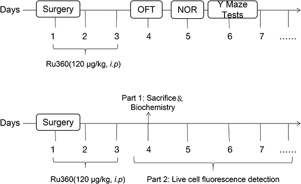 |
Figure 1 The experimental procedures were demonstrated. Behavioral tests were performed three days after surgery (n=10). Part 1: the animals were sacrificed, and hippocampus were collected to detect biochemical indicators three days after surgery (n=3–5). Part 2: experiments requiring live cell assays required mice to be sacrificed until testing (n=3). |
Surgery and Treatment
The mice were anesthetized intraperitoneally with a mixture of fentanyl and droperidol (0.01mL/mg). 2.5mg droperidol and 0.1mg fentanyl were diluted to 15mL using saline. The effects of anesthesia were evaluated by skin pinch reaction. In brief, the fur in the operative site was shaved. A laparotomy was performed through a 1.5-cm median incision. A 5 cm piece of the small intestine was removed and covered with clean, moist gauze for 10 min, and the abdominal wall was sutured. The whole procedure took about 20 min. Animals in surgery + Ru360 group was treated with 120 μg/kg Ru360 by intraperitoneal injection for three consecutive days, surgery + saline group was treated with saline by intraperitoneal injection.14 At a dose of 120 μg/kg, extracellular recordings from the somatosensory cortex failed to alter spontaneous and evoked neuronal electrical activity. However, at a dose of 240 μg/kg, Ru360 significantly reduced stimulation-evoked neuronal electrical activity and cerebral blood flow (CBF) responses and associated blood oxygen level dependent (BOLD).13 Low dose Ru360 treatments (120 μg/kg), which mostly spares neuronal electrical activity while inhibiting neurovascular and neurometabolic activity, will improve brain injury recovery.15 Tetracaine hydrochloride gel was applied to the abdomen of mice to relieve pain on the first postoperative day. The experimental procedures were exhibited in Figure 1.
Open Field Tests
The open field tests were performed to evaluate the behavioral characteristics of the autonomous activity and spatial exploration of mice. A black box (50×50×50 cm3) makes up the open field in a quiet room, installed a camera directly above the open field box. Recorded their behavior on the video for 5 minutes, and then used Any maze behavior tracking software (Stoelting Co., USA) for analysis. The box was cleaned with 75% alcohol and dried between each experiment to remove odor. Thirty min before the experiment, mice in each group were sent to a quiet test room for dark adaptation. Speed: the ratio of the total distance and time of the mice in the open field experiment, which can indicate the autonomous movement ability of mice. Number of crossing lines-crossing a line once means that all four feet of the mouse are within the range of the adjacent squares. This index is used to reflect the activity ability of the mouse, and the number of crosses reflects the degree of spontaneous activity and excitement of the mouse.
Novel Object Recognition Tests (NOR)
The novel object recognition test was performed in a black box (50×50×50 cm3) with objects placed on box. Video recording of the test was used for behavior reading. To evaluate the object recognition memory ability of rodents by using the characteristics of animals like to explore the new environment to detect their memory of the objects they have touched. The test consisted of training and recognition sessions.
During training and testing, the mice were placed in the center of a black box, and recorded exploring the two objects for 5 minutes. Between trials, the room was washed with 75% alcohol. The training period involved placing the mice in front of two identical objects. One of the objects was replaced by a new object during the test phase and used to test the recognition process one and a half hours later. The training session was performed in the presence of two identical objects. A new object was substituted for one of the old objects during the test phase and used to test the recognition process after one and a half hours. Touching and sniffing objects were considered exploratory behaviors. A trained researcher records the time spent exploring each object. Recognition index: [Time at the novel object / (Time at the novel object + Time at the familiar object)] x 100 (%).16 Mice with poor cognitive ability showed no difference in the exploration of new and old objects. Mice with normal cognitive ability showed longer exploration of new objects than those with old objects.
Y Maze Tests
The Y-maze consisted of a black box (30×8×30 cm3) with three identical arms. The test was used to study the discriminative learning and working memory ability of mice. The angle of the three arms was 120°. Different geometric pictures were randomly attached to the end of each arm. A video camera recorded the time the mice spent on each arm. In the training session, mice explored freely for 5 min when a new arm was blocked. Between tests, the maze was washed with 75% ethanol to eliminate odor interference. In the exploring session, mice explored for 5 min with three arms open. Time in novel arm: record time spent on the new arm as a proportion of time spent on the three arms.17 Because rodents have the instinct to explore new things, the number, time and distance of exploration in the new arm will be shortened when memory is impaired.
Western Blotting Analysis
Mice were anesthetized and sacrificed, and the hippocampus was isolated. The hippocampal tissue was lysed and the protein content was determined by BCA protein assay kit. Electrophoresis was performed using the PAGE Gel Fast Preparation Kit (Shanghai Epizyme Biomedical Technology Co., Ltd, Shanghai China), and proteins were then transferred to polyvinylidene fluoride. The polyvinylidene fluoride was sealed with 5% skim milk powder and incubated overnight with primary antibody in a 4° C room. MCU (A16281, rabbit polyclonal; 1:1000, ABclonal, Wuhan, China), caspase-1 (ab138483, rabbit polyclonal; 1:1000, Abcam, Cambridge, UK), NLRP3 (15101S, rabbit monoclonal; 1:2000, Cell Signaling Technology, USA), and β-actin (AC038, rabbit monoclonal, 1:5000, ABclonal, Wuhan, China). The polyvinylidene fluoride was washed and incubated with HRP Goat Anti-Rabbit IgG (1:1,0000, ABclonal, Wuhan, China) for 1 h at room temperature. Blots were imaged with an enhanced chemiluminescence imaging system (Tanon, Shanghai, China). Quantitative analysis of images using Image J software.
Enzyme-Linked Immunosorbent Assay (ELISA)
The concentrations of IL-1β, IL-6, and TNF-α were determined from pre-prepared hippocampal tissues. Samples were homogenized in PBS. The mixture was centrifuged at 2000 rpm for 15 min after sonication at 4 °C. The concentrations of IL-1β, IL-6, and TNF-α were measured by ELISA kits (Jiangsu Meimian Industrial Co., Ltd, Jiangsu, China). The absorbance was determined using a microplate reader (Thermo Fisher Scientific, Massachusetts, USA).
Anti-Oxidative Enzyme Activity Assay
The activity of SOD was analyzed using kits according to the protocols from manufacturers (A001-3, Nanjing Jiancheng Biotechnology Institute, Jiangsu, China). Activation of SOD in mice’s hippocampus was detected.
Measurement of the Intracellular ROS Levels and the Mitochondrial ROS (mtROS) Levels
The intracellular ROS levels were detected using a fluorescent redox-sensitive-fluorescent probe-DCFH-DA (E004-1-1, Nanjing Jiancheng Bioengineering Institute, China). The stories of mtROS were measured using MitoSOX™ red mitochondrial superoxide indicator (M36007, Thermo Fisher, USA).18 In brief, fresh mouse hippocampus was isolated, cut to 1mm3 with ophthalmic scissors, ground, and filtered through a 300-mesh nylon mesh—the entire operation on ice. The final single-cell suspension obtained was incubated with 10 μM DCFH-DA at 37°C for 60 min. The fluorescence of DCF was measured at 488/525 nm wavelengths by flow cytometry. 1mL working solution of MitoSOX™ red mitochondrial superoxide indicator was added to the single-cell suspension and incubated at 37°C for 10 min. The fluorescence was measured at a wavelength of 510/580 nm by flow cytometer (Beckman Coulter, USA). Compare the average fluorescence intensity of each group.19 And the percentage of cells with high fluorescence value was marked in the figure.
Detection of MMP and Mitochondrial Calcium
The mitochondrial membrane potential detection kit (C2006, Beyotime, Shanghai, China) is a rapid detection kit for mitochondrial membrane potential changes using JC-1 as a fluorescent probe. Briefly, the single-cell suspensions were stained with 0.5mL working solution of JC-1 at 37 °C for 40 minutes and analyzed using a flow cytometry (Beckman Coulter, USA). Single-cell suspensions were prepared as above. Low MMP was indicated by cells with green fluorescence (JC-1 monomers) and high MMP was indicated by cells with red fluorescence (JC-1 aggregates). Mitochondrial calcium levels were measured using Rhod-2 (R1244, Thermo Fisher, USA). Calcium in the cytoplasm was quenched using CoCl2.20 Compare the average fluorescence intensity of each group.
Data Analysis
SPSS 27.0 (Chicago, IL, USA) was used for statistical analysis. All data are presented as mean ± standard derivation (SD). One-way ANOVA was used to analyze the data. The difference was considered statistically significant at P < 0.05 between the two groups.
Results
Ru360 Alleviates the Cognitive Impairment After Surgery
To further examine the effects of inhibiting MCU expression on cognitive performance, open field tests were used to measure the impacts of Ru360 on locomotion ability. The speed (Figure 2A) did not differ among groups, demonstrating that Ru360 did not significantly alter the motor function of aged mice. The number of crossing lines differed among the groups (Figure 2B). There was a decrease in the number of crossing lines in the Sur group and Sur+ saline group compared with the Con group. Compared with the Sur group, the number of crossing lines increased in the Sur+Ru360 group, reflecting that the surgery reduced the degree of spontaneous activity and excitability of the mice, but it could be rescued by Ru360. The action trajectories of mice in the OFT also confirmed the above conclusions (Figure 2E).
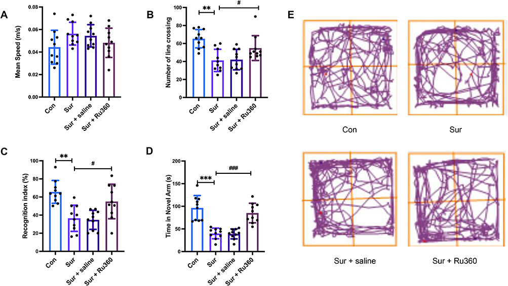 |
Figure 2 Ru360 can alleviate postoperative cognitive dysfunction in aged mice. (A) Surgery and Ru360 did not significantly alter mouse movement speed in the open field test. (B) Ru360 significantly rescued the surgery-induced reduction in the number of crossing lines in aged mice. (C) Ru360 significantly rescued the surgery-induced reduction of recognition index in aged mice during the retention session of NOR tests. (D) Ru360 significantly rescued the surgery-induced time reduction in closed arms during Y maze tests in aged mice. (E) Representative paths of mice in open field experiments. Data are expressed as mean ± SD (n = 10/group). One-way ANOVA was used for statistical analysis. **p<0.01, ***p<0.001, versus Con; #p<0.05, ###p<0.001, versus Sur. Abbreviation: Sur, surgery. |
NOR tests were employed to evaluate the effects of Ru360 on cognitive function. As expected, the Sur + Ru360 group clearly preferred the novel object in the NOR test session. In contrast, the ability of Sur group and Sur + saline group to recognize the new object decreased, and the time spent in exploring familiar and new objects was equal (Figure 2C). Results showed that Ru360 could improve the object recognition ability of mice. In the Y-maze tests, the animals in the Sur group spent less time in the new arm than those in the Con group; this time, the Sur+Ru360 group increased significantly (Figure 2D). The study demonstrated that Ru360 could improve the exploration, discrimination and memory ability of mice.
Ru360 Inhibited the Overexpression of MCU in the Hippocampus After Surgery
Hippocampal neuronal damage is the primary pathological feature of cognitive dysfunction caused by surgery.21 To assess the effect of surgery and Ru360 on MCU expression in the hippocampal region, we examined MCU expression by Western blot assay (Figure 3A). MCU expression was increased in the Sur group compared to the Con group (Figure 3B). MCU levels were significantly lower in the Sur+Ru360 group compared to the Sur group, and there was no difference between the Sur and Sur+ saline groups (Figure 3B). The above results show that MCU expression is increased after anesthesia and surgery, and the use of Ru360 can indeed inhibit its overexpression. The overexpression of MCU can break the balance of calcium ion, which may further lead to mitochondrial dysfunction. Therefore, we next tested whether the inhibition of MCU could alleviate calcium overload.
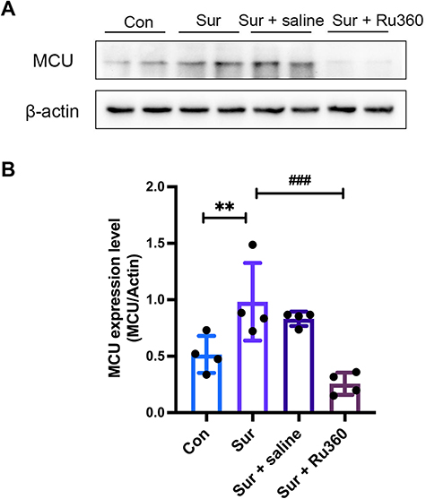 |
Figure 3 Ru360 suppresses MCU overexpression in the postoperative hippocampus. (A) Western blotting analysis was employed to measure the expression of MCU and β-actin in the hippocampal regions of aged mice. (B) Quantitative analysis of MCU expression level in hippocampus. Data are expressed as mean ± SD (n = 4/group). One-way ANOVA was used for statistical analysis. **p<0.01, versus Con; ###p<0.001, versus Sur. Abbreviation: Sur, surgery. |
Ru360 Reduces Calcium Overload in Hippocampal Mitochondria After Surgery
Excess calcium causes mitochondrial function. Ru360 decreased the mitochondrial calcium level by inhibiting the expression of MCU. Flow cytometry measured calcium levels in hippocampal mitochondria of postoperative aged mice. Calcium levels were found to accumulate in the hippocampal mitochondria of mice after surgery, which was reduced after Ru360 treatment (Figure 4A). There was no difference in calcium levels between the Sur group and the Sur +saline group (Figure 4A). The fluorescence intensity of rhod-2 in each group was quantitatively analyzed (Figure 4B). The results demonstrate that Ru360 can inhibit the entry of excess calcium into mitochondria.
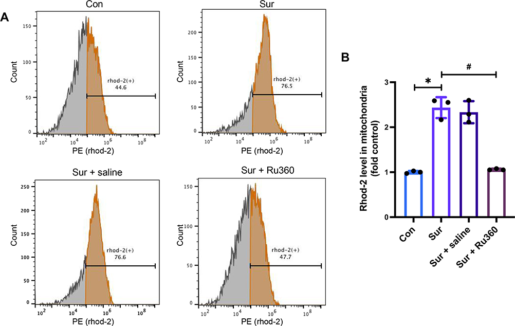 |
Figure 4 Ru360 reduces calcium overload in hippocampal mitochondria after surgery. (A) The cells stained with rhod-2 fluorescent dye were analyzed with the flow cytometer. The proportion of total cells with hyperfluorescence in each group. (B) Quantitative data on calcium accumulation in hippocampal mitochondria. Flow cytometry plots are all representative. Data are expressed as mean ± SD (n = 3/group). One-way ANOVA was used for statistical analysis. *p<0.05, versus Con; #p<0.05, versus Sur. Abbreviation: Sur, surgery. |
Ru360 Improved Mitochondrial Function in the Hippocampus After Surgery
Postoperative oxidative damage of hippocampal neurons in mice was due to decreased mitochondrial function. The increase of MCU in the hippocampus of mice after surgery mediated the entry of a large amount of calcium into cells, leading to mitochondrial damage. Mitochondrial damage could influence the accumulation of ROS22 and mitochondrial calcium, as well as the activities of SOD and the reduction of MMP. Thus, the amount of ROS and the activities of SOD, and the MMP, were detected. As shown in (Figures 5A and B), these results indicate that Ru360 treatment reduced surgery-induced accumulation of intracellular ROS levels. Meanwhile, the mitochondrial ROS was also decreased by Ru360 treatment (Figures 5C and D). Calcium overload within mitochondria fails to maintain average mitochondrial membrane potential, leading to mitochondrial dysfunction.23 Flow cytometry analysis revealed a significant increase in MMP in the Sur+Ru360 group compared with the Sur group and Sur+ saline group (Figures 5E and F). Surgery resulted in a decrease in mitochondrial membrane potential in hippocampal neurons (Figures 5E and F). Furthermore, the SOD activity in Sur group was lower than that in Con group. The activity of SOD in the Sur+Ru360 group was improved (Figure 5G). The results demonstrate that Ru360 played a positive role in improving mitochondrial function, which further proves that the occurrence of postoperative cognitive dysfunction may be due to the imbalance of mitochondrial function.
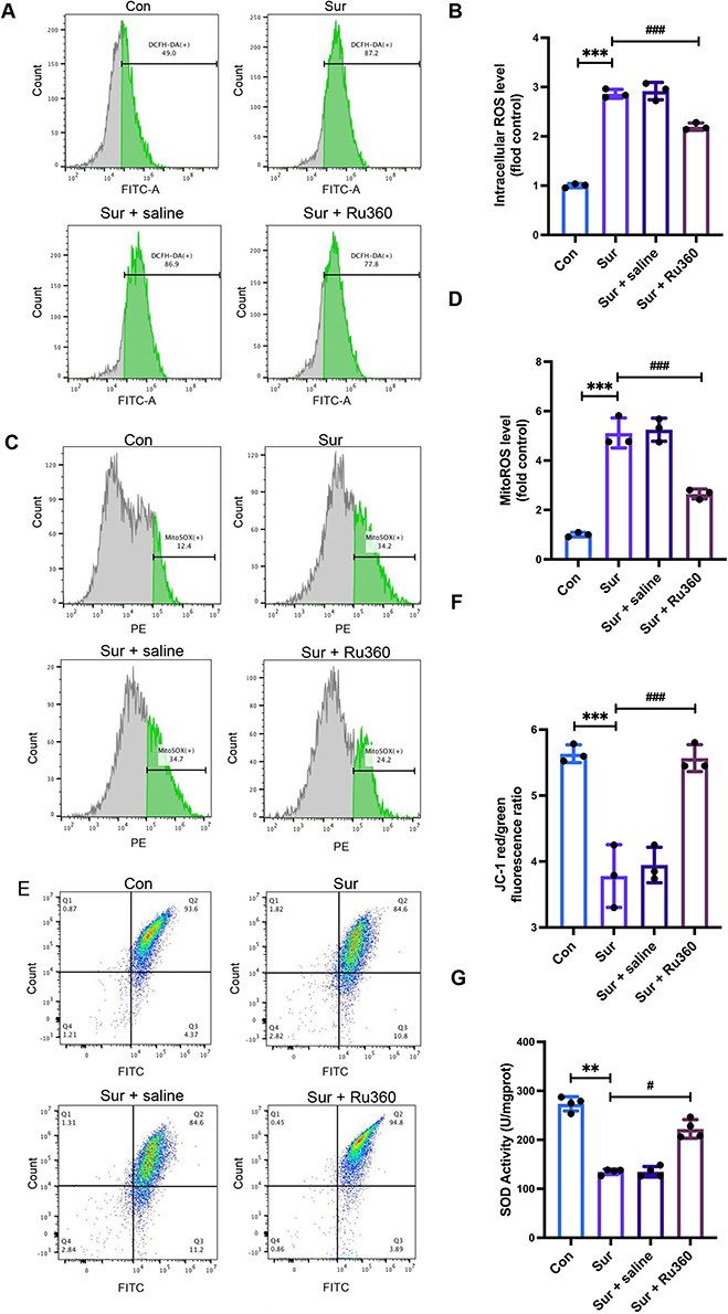 |
Figure 5 Ru360 improved mitochondrial function in the hippocampus after surgery in aged mice. (A) DCFH-DA stained cells were analyzed by FITC channel of flow cytometer. The proportion of total cells with hyperfluorescence in each group. (B) Quantitative analysis of mean fluorescence intensity in hippocampal neurons. (C) MitoSOX stained cells were analyzed by PE channel of flow cytometer. The proportion of total cells with hyperfluorescence in each group. (D) Quantitative analysis of mean fluorescence intensity in hippocampal neurons. (E) The JC-1-positive cells were counted by using a flow cytometer. The proportion of total cells with hyperfluorescence in each group. (F) The ratio of JC-1 aggregate(red)/ JC-1 monomer(green) fluorescence intensity shows abnormal changes in mitochondrial membrane potential. Decreased mitochondrial membrane potential indicates decreased mitochondrial function. (G) SOD activity in the hippocampus of aged mice in each group. Flow cytometry plots are all representative. Data are expressed as mean ± SD (n = 3/group). One-way ANOVA was used for statistical analysis. **p<0.01, ***p<0.001, versus Con; #p<0.05, ###p<0.001, versus Sur. Abbreviations: Sur, surgery; mtROS, mitochondrial ROS. |
Ru360 Inhibited the Release of Inflammatory Cytokines in the Hippocampus After Surgery
To further assess inflammation in the hippocampus after surgery, we examined NLRP3 and caspase-1 levels in the hippocampus by Western blot. (Figure 6A). The level of inflammation in the Sur group was markedly higher than that in the Con group (Figures 6B and C). And Ru360 significantly reduced the level of inflammation in aged mice after surgery (Figures 6B and C). Meanwhile, the concentrations of IL-1β, TNF-α and IL-6 were detected by ELISA to further evaluate the postoperative inflammatory response in the hippocampus (Figures 6D–F). The results indicate that the concentrations of inflammatory cytokines in the Sur group and Sur + saline group were increased compared with the Con group. The level of inflammation was reduced after Ru360 treatment. The reduction of inflammation level is beneficial to the recovery of mitochondrial function, and also protects neurons from the attack of inflammatory factors and protects cognitive function.
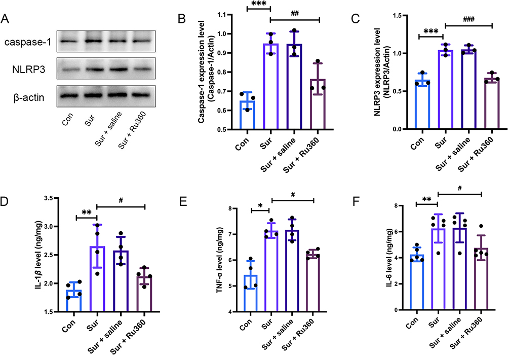 |
Figure 6 Ru360 inhibited the release of inflammatory cytokines in the hippocampus after surgery in aged mice. (A) The effect of surgery and Ru360 on the expression of NLRP3 in the hippocampus was detected by Western blot analysis. Quantitative analysis of (B) caspase-1 and (C) NLRP3 expression level in the hippocampus after surgery and after Ru360 treatment. The concentrations of IL-1β (D), TNF-a (E), and IL-6 (F) in the hippocampus among groups detected by ELISA kit. Data are expressed as mean ± SD (n = 4–5/group). One-way ANOVA was used for statistical analysis. *p<0.05, **p<0.01, ***p<0.001, versus Con; #p<0.05, ##p<0.01, ###p<0.001, versus Sur. Abbreviation: Sur, surgery. |
Discussion
In this study, we found that mitochondrial functional stability and cell viability were significantly improved after Ru360 treatment, further corroborating the notion that inhibition of MCU can protect nerve cells from surgical injury. Our results showed that excessive mitochondrial Ca2+ uptake via MCU results in a decrease in mitochondrial membrane potential, ROS generation, and decreased SOD activity, which produces toxic effects on hippocampal neurons in mice. Postoperative administration of Ru360 improved cognition, rescued mitochondrial function, and reduced inflammatory cytokines in the hippocampus of mice.
Mitochondria are highly metabolically active organelles that play an essential role in Ca2+ release signals from the endoplasmic reticulum (ER).24 Under normal physiological conditions, the majority of Ca2+ resides in the lumen of the ER, and a small fraction crosses the outer mitochondrial membrane when Ca2+ signaling is required. Under pathological conditions, such as endoplasmic reticulum stress, drug action and surgery, the increase of calcium released from endoplasmic reticulum leads to long-term mitochondrial calcium overload.25 It has been reported that mitochondrial calcium overload can induce oxidative stress by increasing ROS production.26 MCU overexpression can lead to higher sensitivity of cells to oxidative stress. The results of in vitro experiments showed that MCU inhibitor RU360 could inhibit MCU activity to protect cells against oxidative stress.27 Oxidative stress, in turn, is thought to be associated with inflammatory responses.28 Our results confirmed that MCU could induce mitochondrial calcium accumulation in the hippocampus of aged mice, which activated the NLRP3 inflammasome and released IL-1β, IL-6 and TNF-a. The activated NLRP3 inflammasome triggers the cleavage of the pro-caspase-1 to caspase-1, which is then released into the cytoplasm to further induce the conversion of the pro- IL-1β to active IL-1β (Figure 7).29 There is a high concentration of inflammatory cytokine receptors in regions related to learning and memory, especially in the hippocampal region, so that the neuroinflammation in the brain induced by surgery may mainly destroy the memory and learning dependent on the hippocampus.30 It has been shown that the hippocampus contains the largest number of IL-1β receptors and is associated with the best memory and learning processes.31 This brain area is also sensitive to aging damage, and in animal models of aging, excessive inflammatory cytokines such as IL-1 β related to cognitive impairment.32 In addition to IL-1β, it can promote the secretion of pro-inflammatory cytokines such as IL-6 and TNF-α that do not require NLRP3 inflammasome processing. More importantly, oxidative stress can cause a decrease in mitochondrial membrane potential, leading to mitochondrial dysfunction, which in turn aggravates oxidative stress and forms a vicious cycle.33 Maintaining a healthy mitochondrial calcium pool is essential to protect neuronal function.34 Behavioral tests and biochemical experiments confirmed our speculation. Postoperative cognitive dysfunction in aged mice is closely related to inflammation and mitochondrial dysfunction in the hippocampus.
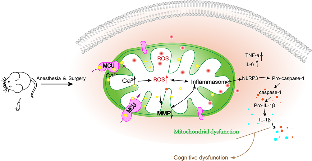 |
Figure 7 MCU regulates mitochondrial calcium inflow, leading to mitochondrial dysfunction and inflammatory outburst. After anesthesia and surgery, the intracellular environment was disturbed and MCU expression was increased in elderly mice. Overexpressed MCU mediates calcium transport to mitochondria, resulting in mitochondrial dysfunction, which is manifested as decreased mitochondrial membrane potential, increased ROS in mitochondria, enhanced oxidative stress. Mitochondrial dysfunction further triggers a burst of inflammatory cytokines (NLRP3, IL-6 and TNF-a). The release of Inflammatory cytokines, in turn, further aggravate mitochondrial dysfunction, which together led to cognitive impairment in the elderly mice. |
Understanding the MCU-controlled transmission of calcium signals inside and outside the mitochondria may provide a new insight for research into the pathogenesis of many neuroinflammatory diseases. The present study has limitations in that it only confirmed that Ru360 improved postoperative cognitive function. However, the effects of Ru360 itself on the animals were not studied and the extent to which different doses of Ru360 improved postoperative cognitive function was not investigated.
In conclusion, we have shown that Ru360 can improve postoperative cognitive function in aged mice. Biochemical information analysis confirmed that Ru360 enhanced mitochondrial function and reduced neuroinflammation. Improving mitochondrial function by inhibiting MCU may be a target for preventing and treating POCD.
Data Sharing Statement
The data supporting this study’s findings are available from the corresponding author upon reasonable request.
Funding
This study was funded by Zhejiang Provincial Natural Science Foundation of China (LY19H25001), Medical Health Science and Technology Project of Zhejiang Provincial Health Commission (2019RC266 and 2021KY1043), Ningbo Medical Science and Technology Project (2019Y16), Ningbo Science and Technology Innovation 2025 major special Project (2019B10035), and the Affiliated Hospital of Medical School of Ningbo University Youth Talent Cultivation Program (FYQM-LC-202002).
Disclosure
The authors report no conflict of interest in this work.
References
1. Xin J, Shan W, Li J, Yu H, Zuo Z. Activation of the lateral habenula-ventral tegmental area neural circuit contributes to postoperative cognitive dysfunction in mice. Adv Sci. 2022;9(22):e2202228. doi:10.1002/advs.202202228
2. Cai H, Qiao J, Chen S, et al. MCU knockdown in hippocampal neurons improves memory performance of an Alzheimer’s disease mouse model. Acta Biochim Biophys Sin. 2022;54(10):1528–1539. doi:10.3724/abbs.2022138
3. Dong H, Zhao B, Chen J, et al. Mitochondrial calcium uniporter promotes phagocytosis-dependent activation of the NLRP3 inflammasome. Proc Natl Acad Sci U S A. 2022;119(26):e2123247119. doi:10.1073/pnas.2123247119
4. Orrenius S, Zhivotovsky B, Nicotera P. Regulation of cell death: the calcium-apoptosis link. Nat Rev Mol Cell Biol. 2003;4(7):552–565. doi:10.1038/nrm1150
5. Delgado B, Long S. Mechanisms of ion selectivity and throughput in the mitochondrial calcium uniporter. Sci Adv. 2022;8(50):eade1516. doi:10.1126/sciadv.ade1516
6. Kirichok Y, Krapivinsky G, Clapham DE. The mitochondrial calcium uniporter is a highly selective ion channel. Nature. 2004;427(6972):360–364. doi:10.1038/nature02246
7. Rimessi A, Bezzerri V, Patergnani S, et al. Mitochondrial Ca2+-dependent NLRP3 activation exacerbates the Pseudomonas aeruginosa-driven inflammatory response in cystic fibrosis. Nat Commun. 2015;6(1):6201. doi:10.1038/ncomms7201
8. Panahi G, Pasalar P, Zare M, et al. MCU-knockdown attenuates high glucose-induced inflammation through regulating MAPKs/NF-κB pathways and ROS production in HepG2 cells. PLoS One. 2018;13(4):e0196580. doi:10.1371/journal.pone.0196580
9. Murakami T, Ockinger J, Yu J, et al. Critical role for calcium mobilization in activation of the NLRP3 inflammasome. Proc Natl Acad Sci U S A. 2012;109(28):11282–11287. doi:10.1073/pnas.1117765109
10. Ying WL, Emerson J, Clarke MJ, Sanadi DR. Inhibition of mitochondrial calcium ion transport by an oxo-bridged dinuclear ruthenium ammine complex. Biochemistry. 1991;30(20):4949–4952. doi:10.1021/bi00234a016
11. Matlib MA, Zhou Z, Knight S, et al. Oxygen-bridged dinuclear ruthenium amine complex specifically inhibits Ca2+ uptake into mitochondria in vitro and in situ in single cardiac myocytes. J Biol Chem. 1998;273(17):10223–10231. doi:10.1074/jbc.273.17.10223
12. Yu S, Zheng S, Leng J, Wang S, Zhao T, Liu J. Inhibition of mitochondrial calcium uniporter protects neurocytes from ischemia/reperfusion injury via the inhibition of excessive mitophagy. Neurosci Lett. 2016;628:24–29. doi:10.1016/j.neulet.2016.06.012
13. Sanganahalli BG, Herman P, Hyder F, Kannurpatti SS. Mitochondrial calcium uptake capacity modulates neocortical excitability. J Cereb Blood Flow Metab. 2013;33(7):1115–1126. doi:10.1038/jcbfm.2013.61
14. Chitturi J, Santhakumar V, Kannurpatti S. Traumatic brain injury metabolome and mitochondrial impact after early stage Ru360 treatment. Mitochondrion. 2021;57:192–204. doi:10.1016/j.mito.2021.01.003
15. Sanganahalli BG, Herman P, Hyder F, Kannurpatti SS, Gasman S. Mitochondrial functional state impacts spontaneous neocortical activity and resting state FMRI. PLoS One. 2013;8(5):e63317. doi:10.1371/journal.pone.0063317
16. Iban-Arias R, Trageser K, Yang E, et al. Exposure to world trade center dust exacerbates cognitive impairment and evokes a central and peripheral pro-inflammatory transcriptional profile in an animal model of Alzheimer’s disease. J Alzheimer’s Dis. 2022. doi:10.3233/jad-221046
17. Kraeuter AK, Guest PC, Sarnyai Z. The Y-Maze for assessment of spatial working and reference memory in mice. Methods Mol Biol. 2019;105–111. doi:10.1007/978-1-4939-8994-2_10
18. Wu T, Liang X, Liu X, et al. Induction of ferroptosis in response to graphene quantum dots through mitochondrial oxidative stress in microglia. Part Fibre Toxicol. 2020;17(1):30. doi:10.1186/s12989-020-00363-1
19. Xiang D, Yang W, Fang Z, et al. Agrimol B inhibits colon carcinoma progression by blocking mitochondrial function through the PGC-1α/NRF1/TFAM signaling pathway. Front Oncol. 2022;12:1055126. doi:10.3389/fonc.2022.1055126
20. Lee HJ, Jung YH, Choi GE, et al. Urolithin A suppresses high glucose-induced neuronal amyloidogenesis by modulating TGM2-dependent ER-mitochondria contacts and calcium homeostasis. Cell Death Differ. 2021;28(1):184–202. doi:10.1038/s41418-020-0593-1
21. Venkat P, Chopp M, Chen J. Models and mechanisms of vascular dementia. Exp Neurol. 2015;272:97–108. doi:10.1016/j.expneurol.2015.05.006
22. Cai Y, Xiao R, Zhang Y, et al. DHPA protects SH-SY5Y cells from oxidative stress-induced apoptosis via mitochondria apoptosis and the Keap1/Nrf2/HO-1 signaling pathway. Antioxidants. 2022;11(9):1794. doi:10.3390/antiox11091794
23. Yu X, Wang T, Li Y, et al. Apoptin causes apoptosis in HepG-2 cells via Ca imbalance and activation of the mitochondrial apoptotic pathway. Cancer Med. 2022. doi:10.1002/cam4.5528
24. Malhotra JD, Kaufman RJ. ER stress and its functional link to mitochondria: role in cell survival and death. Cold Spring Harb Perspect Biol. 2011;3(9):a004424. doi:10.1101/cshperspect.a004424
25. Romagnoli A, Aguiari P, De Stefani D, et al. Endoplasmic reticulum/mitochondria calcium cross-talk. Novartis Found Symp. 2007;287:122–131; discussion 131–129.
26. Peng TI, Jou MJ. Oxidative stress caused by mitochondrial calcium overload. Ann N Y Acad Sci. 2010;1201(1):183–188. doi:10.1111/j.1749-6632.2010.05634.x
27. Liao Y, Hao Y, Chen H, He Q, Yuan Z, Cheng J. Mitochondrial calcium uniporter protein MCU is involved in oxidative stress-induced cell death. Protein Cell. 2015;6(6):434–442. doi:10.1007/s13238-015-0144-6
28. Park GB, Choi Y, Kim YS, et al. ROS-mediated JNK/p38-MAPK activation regulates Bax translocation in Sorafenib-induced apoptosis of EBV-transformed B cells. Int J Oncol. 2014;44(3):977–985. doi:10.3892/ijo.2014.2252
29. Shao BZ, Xu ZQ, Han BZ, Su DF, Liu C. NLRP3 inflammasome and its inhibitors: a review. Front Pharmacol. 2015;6:262. doi:10.3389/fphar.2015.00262
30. Zhang X, Dong H, Li N, et al. Activated brain mast cells contribute to postoperative cognitive dysfunction by evoking microglia activation and neuronal apoptosis. J Neuroinflammation. 2016;13(1):127. doi:10.1186/s12974-016-0592-9
31. Gemma C, Fister M, Hudson C, Bickford PC. Improvement of memory for context by inhibition of caspase-1 in aged rats. Eur J Neurosci. 2005;22(7):1751–1756. doi:10.1111/j.1460-9568.2005.04334.x
32. Chen J, Buchanan JB, Sparkman NL, Godbout JP, Freund GG, Johnson RW. Neuroinflammation and disruption in working memory in aged mice after acute stimulation of the peripheral innate immune system. Brain Behav Immun. 2008;22(3):301–311. doi:10.1016/j.bbi.2007.08.014
33. Pires M, Rego A. Apoe4 and Alzheimer’s disease pathogenesis-mitochondrial deregulation and targeted therapeutic strategies. Int J Mol Sci. 2023;24(1):778. doi:10.3390/ijms24010778
34. Ben-Kasus Nissim T, Zhang X, Elazar A, et al. Mitochondria control store-operated Ca(2+) entry through Na(+) and redox signals. EMBO J. 2017;36(6):797–815. doi:10.15252/embj.201592481
 © 2023 The Author(s). This work is published and licensed by Dove Medical Press Limited. The
full terms of this license are available at https://www.dovepress.com/terms
and incorporate the Creative Commons Attribution
- Non Commercial (unported, 3.0) License.
By accessing the work you hereby accept the Terms. Non-commercial uses of the work are permitted
without any further permission from Dove Medical Press Limited, provided the work is properly
attributed. For permission for commercial use of this work, please see paragraphs 4.2 and 5 of our Terms.
© 2023 The Author(s). This work is published and licensed by Dove Medical Press Limited. The
full terms of this license are available at https://www.dovepress.com/terms
and incorporate the Creative Commons Attribution
- Non Commercial (unported, 3.0) License.
By accessing the work you hereby accept the Terms. Non-commercial uses of the work are permitted
without any further permission from Dove Medical Press Limited, provided the work is properly
attributed. For permission for commercial use of this work, please see paragraphs 4.2 and 5 of our Terms.