Back to Journals » Cancer Management and Research » Volume 12
Risk Stratification Based on Synchronous Neoplasia and Clinical Physicochemical Characteristics Predicts a Higher Incidence of Metachronous Advanced Neoplasia in Patients Undergoing Colorectal Resection for Colorectal Cancer
Received 10 July 2020
Accepted for publication 22 September 2020
Published 5 November 2020 Volume 2020:12 Pages 11295—11307
DOI https://doi.org/10.2147/CMAR.S271614
Checked for plagiarism Yes
Review by Single anonymous peer review
Peer reviewer comments 2
Editor who approved publication: Professor Harikrishna Nakshatri
Yanan Tian,1 Yu Xin,2 Shuai Li2
1Department of Gastroenterology, Shandong Provincial Hospital, Cheeloo College of Medicine, Shandong University, Jinan, Shandong, People’s Republic of China; 2Department of Gastroenterology, The Second Hospital, Cheeloo College of Medicine, Shandong University, Jinan, Shandong, People’s Republic of China
Correspondence: Shuai Li
Department of Gastroenterology, The Second Hospital, Cheeloo College of Medicine, Shandong University, Jinan, Shandong, People’s Republic of China
Tel +86 158 6663 0635
Fax +86 531 8587 5454
Email [email protected]
Purpose: Patients who undergo primary colorectal cancer (CRC) resection remain at increased risk for metachronous advanced neoplasia (MAN) in the remnant colorectum. This study aimed to investigate the incidence and clinicopathological characteristics predictive of MAN development in the residual colon after surgery.
Patients and Methods: We retrospectively reviewed 450 primary CRC cases referred to our hospital during a 4-year period. Univariate and multivariate analyses were performed to identify risk factors for MAN. The cumulative incidence of MAN was evaluated by the Cox proportional hazards model.
Results: MAN development was confirmed in 78 of the 450 patients (17.3%). Overall 1-, 2-, 3-, and 5-year cumulative probabilities were 0.9%, 4.8%, 9.8%, and 16.1%, respectively, for MAN. Among the clinical and colonoscopic factors at baseline, the independent factors that were significantly associated with MAN were synchronous neoplasia, carcinoembryonic antigen (CEA) level ≥ 10.0 ng/mL, and index cancer size ≥ 50 mm. The cumulative probability of MAN was significantly higher for patients with synchronous advanced neoplasia (SAN) than for those without synchronous neoplasia (P = 0.000). A subgroup analysis of patients based on the CEA level and index cancer size indicated that CEA ≥ 10 ng/mL and index cancer ≥ 50 mm resulted in a significantly higher cumulative probability of MAN (P = 0.039).
Conclusion: Patients with SAN or high preoperative serum CEA levels and large index cancer are at increased risk for early-onset MAN. More intensive surveillance strategies may be appropriate for these groups. Risk stratification based on synchronous neoplasia and clinical physicochemical characteristics requires further investigations involving modified appropriate postoperative colonoscopic surveillance schedules.
Keywords: primary colorectal cancer, remnant colorectum, advanced neoplasia, colonoscopics surveillance, risk stratification
Introduction
Colorectal cancer (CRC) is a common neoplasm that accounts for approximately 1.2 million new cases and 600,000 deaths per year worldwide.1 Over the course of the past decade, significant progress has been made in the treatment of localized CRC due to advances in surgical and endoscopic resection. Furthermore, the prognosis of patients with CRC has slowly but steadily improved in many countries. However, patients who undergo primary CRC resection remain at increased risk for metachronous CRC in the remnant colorectum; their risk is 1.5-fold to 2-fold that of the general population, which corresponds to a 1% to 2% long-term risk.2–4 Therefore, early diagnosis of recurrence during a curable stage, timely detection and removal of metachronous precursor lesions, especially metachronous advanced neoplasia (MAN), and new prevention strategies are needed to reduce the burden of this disease. Colonoscopy has a pivotal role in this setting because it is able to detect both early intraluminal recurrence and metachronous neoplasia.5 However, the optimal timing of endoscopic surveillance remains unclear because there is little information regarding the long-term occurrence of metachronous lesions. The recently published recommendations of the Japanese Society for Cancer of the Colon and Rectum (JSCCR) suggested endoscopic examinations at 12, 24, 36, and 60 months after surgery.6 The United States Multi-Society Task Force, European Society of Gastrointestinal Endoscopy (ESGE), European Society of Digestive Oncology (ESDO), and Chinese Society of Clinical Oncology (CSCO) noted that patients who undergo curative resection for CRC should undergo colonoscopy 1, 3, and 5 years after the initial surgery regardless of the preoperative colonoscopic findings.4,7,8 They do not recommend an intensive endoscopic surveillance strategy (eg, annual colonoscopy) because it has failed to show significant survival benefits compared to standard follow-up.9,10 This may be attributable to the diverse risk of metachronous neoplasia development within the population. Therefore, we focused on a subpopulation at higher risk for advanced adenomas in the residual colon.
Previous studies have indicated that synchronous neoplasia and cancer are risk factors for metachronous lesions.11,12 It has not yet been determined whether clinical and baseline colonoscopic characteristics associated with the presence of synchronous neoplasia or cancer are related to the development of MAN. Identification of these risk factors may help clinicians establish appropriate surveillance schedules for their patients based on their individual characteristics. Therefore, the primary aim of this study was to investigate the clinical and baseline preoperative colonoscopic characteristics predictive of MAN in the residual colon after surgery. The secondary aim was to evaluate whether these risk factors could contribute to informing the surveillance schedule.
Materials and Methods
Study Population
A retrospective study involving 582 consecutive patients who underwent primary CRC resection at the Second Hospital of Shandong University (Shandong, China) between January 2012 and December 2015 was performed. The medical ethics committee of the Second Hospital of Shandong University approved this study on June 16, 2018. The methods were performed in accordance with the relevant guidelines and regulations. All personal information had already been anonymized and de-identified to protect patient privacy. Therefore, the study protocol was exempted from the requirement for informed consent from its participants. This study was conducted in accordance with the Declaration of Helsinki. All pathological specimens were evaluated by a single experienced pathologist to avoid inter-observer differences, and the histological type, location, tumor differentiation, depth of invasion, lymph node metastasis, and lymphovascular invasion were reviewed. Histopathological diagnoses were based on the World Health Organization classification of tumors of the digestive system.13 The exclusion criteria were as follows: CRC in the context of familial adenomatous polyposis, inflammatory bowel disease or Lynch syndrome; TNM stage IV14 at presentation; and total proctocolectomy or local recurrence at the anastomosis site. Patients were also excluded if they had undergone one or fewer surveillance colonoscopies.
Perioperative Colonoscopy and Colonoscopic Surveillance Protocol After Surgery
All patients with CRC enrolled in the study underwent a clearing perioperative colonoscopy to detect synchronous lesions and complete resection of precancerous polyps. Individuals with obstructive CRC underwent perioperative colonoscopic examinations within 3 to 6 months after surgery, and the findings of these examinations were included as a part of the baseline colonoscopic findings. Surveillance colonoscopies were generally performed 1, 2, 3, and 5 years after surgical or endoscopic resection (or after the clearing perioperative colonoscopy in the case of obstructive CRC) according to our institutional protocol.
Colonoscopies were completed with a standard colonoscope (CF H260AI, CF Q260AI, PCF-Q260AZI, PCF-H290ZI, or CF-HQ290ZI; Olympus, Tokyo, Japan) by one of three endoscopists who had experience performing more than 500 colonoscopies. We excised all adenomas detected during preoperative and postoperative colonoscopies.
Definitions and Data Collection
Synchronous neoplasia was defined as adenoma/cancer detected by preoperative colonoscopy in the case of non-obstructive CRC and adenoma resected within 3 to 6 months after surgery in the case of obstructive CRC. Particularly, for patients with synchronous colorectal cancer (more than one primary colorectal tumor detected at diagnosis), the most advanced/deepest tumor was considered the index cancer and the other lower-stage tumors were labeled as synchronous lesions. Metachronous neoplasia was defined as a lesion at a site other than that of the anastomosis detected during the surveillance colonoscopy. Advanced neoplasia was defined as any advanced adenoma (ie, an adenoma with a diameter ≥10 mm, high-grade dysplasia, or ≥25% villous features) or carcinoma.
Patient demographics, operative method (for the index CRC), pathological findings, and perioperative and surveillance endoscopic observations were obtained from the electronic medical records. Patients were classified into three categories according to the presence or absence of synchronous neoplasia or synchronous advanced neoplasia (SAN) during the perioperative colonoscopic examinations. Patients who had smoked seven or more cigarettes weekly for at least 1 year were considered current smokers,15 patients who consumed alcohol daily were considered current drinkers,16 and those who used oral aspirin two times or more weekly for at least 1 year were considered current users of aspirin. The serum carcinoembryonic antigen (CEA) and cancer antigen 19-9 (CA19-9) concentrations were measured by electro-chemiluminescence immunoassay (ECLI) kit (Roche Diagnostics GmbH) following the manufacturer’s instructions. Tumor markers higher than the upper limits of normal range (10.0 ng/mL for CEA and 39.0 U/mL for CA19-9 at our institution) were defined as positive markers. Fecal occult blood tests were performed using immunohistochemical methods. A positive history of a first-degree relative with CRC was defined as having one or more first-degree relatives with CRC. The size of index cancer was according to the maximum diameter of the tumor which, referred to prior study, were grouped into < 50 mm and ≥ 50 mm.17 The location of index cancer was classified into left-sided colon or right-sided colon, which was defined as being distal (left-sided) or proximal (right-sided) to the splenic flexure. The tumor stage was according to the Union for International Cancer Control tumor-node-metastasis classification.14
Outcomes
The primary endpoint of this study was the incidence of MAN at the first or later colonoscopic surveillance according to the presence or absence of synchronous neoplasia or SAN. Secondary outcomes were other factors related to MAN, such as age at diagnosis, sex, and index cancer size. Follow-up time was calculated from the date of surgery in the case of non-obstructive CRC or the date of perioperative colonoscopy after surgery in the case of obstructive CRC until the date of the MAN diagnosis or the date of the last available colonoscopy before the endpoint date (December 2019), whichever came first.
Statistical Analysis
Statistical analyses were performed using SPSS version 25 (IBM, New York, NY). Continuous variables are expressed as means ± standard deviations, whereas categorical variables are presented as numbers and percentages. Comparisons were performed to determine the significance of the association between clinical or baseline colonoscopic characteristics and the development of MAN. During the univariate analysis, the chi-square test or Fisher exact test was used for categoric variables. Because sex might be a confounding factor in the comparison of the association between smoking or drinking and the development of MAN, the Cochran-Mantel-Haenszel test was performed to eliminate confounding factor disturbance. To determine the independent risk factors associated with the development of MAN, significant variables (P < 0.2) in the univariate analysis were entered in a multiple logistic regression analysis. The Omnibus test was used to assess the overall fit of the model (P = 0.000). We calculated the odds ratio (OR) and 95% confidence interval (CI) for each factor of interest. The incidence of MAN over time was determined based on the cumulative distribution function estimated using the multivariate Cox proportional hazards regression model. We also calculated the hazard ratio (HR) and 95% CI of the incidence of MAN to determine the independent factors significantly associated with MAN. The Omnibus test was used to assess the overall fit of the model (P = 0.000). A two-sided test was used for all analyses and P < 0.05 was considered statistically significant.
Results
Baseline Characteristics of the Patients
A total of 582 consecutive patients who underwent primary CRC resection between January 2012 and December 2015 were identified in the database. After reviewing the patient characteristics and surveillance colonoscopy data, 132 patients were excluded (Figure 1). Therefore, 450 patients (283 males and 167 females) comprised this study. The characteristics of the study population are summarized in Table 1. The median age was 62.7 ± 11.5 years (range, 27 to 89 years), and the median follow-up time was 54.9 ± 11.4 months (range, 11 to 77 months). MAN development was confirmed in 78 patients (17.3%). In all patients, 48 (10.7%) underwent colonoscopic resection, 85 (18.9%) underwent laparoscopic surgery, and 317 (70.4%) underwent laparotomy.
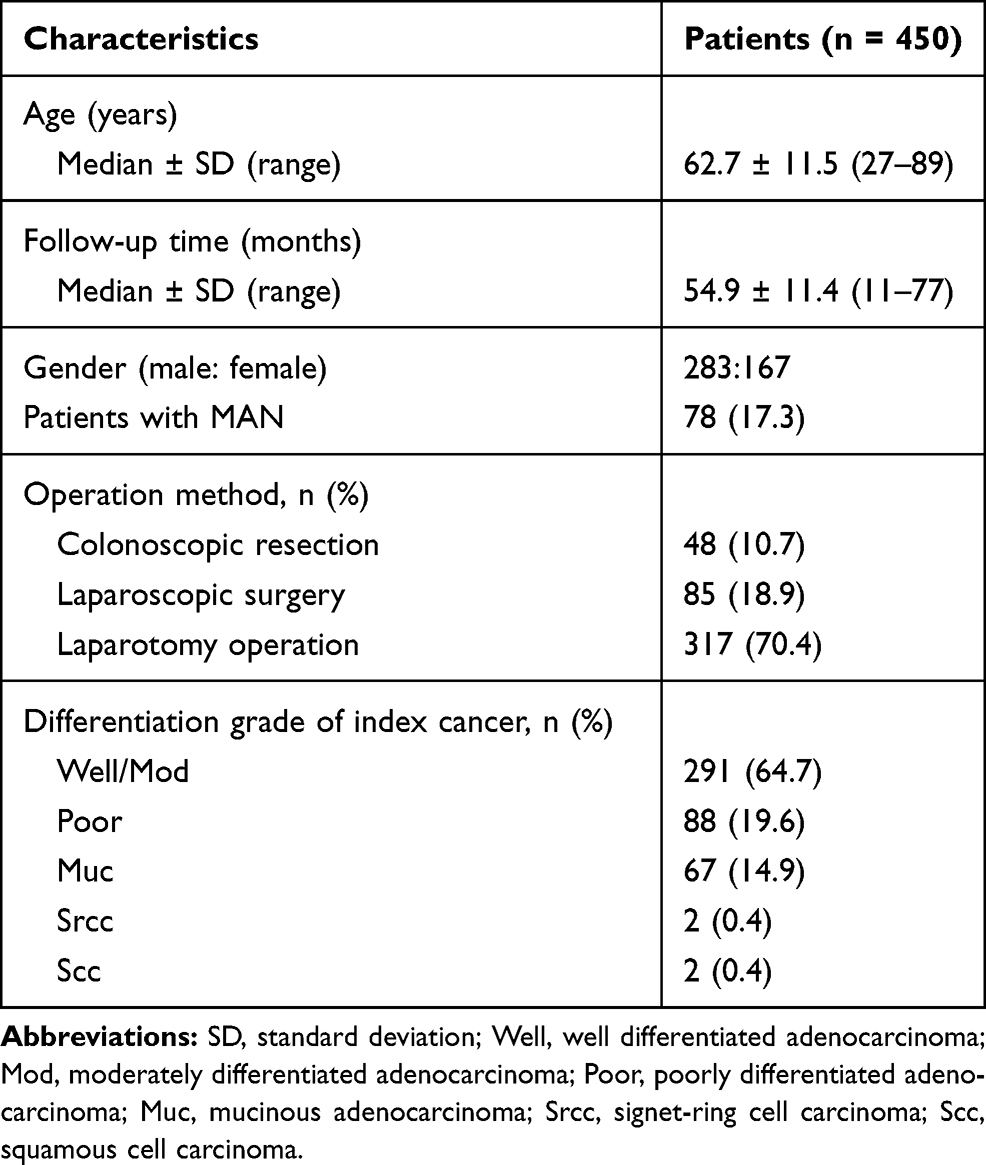 |
Table 1 Clinicopathological Characteristics of Colorectal Cancer Patients Included in the Study |
 |
Figure 1 Flow chart of study participants. |
Identification of Risk Factors for MAN
In the univariate analysis, synchronous neoplasia, especially SAN, was strongly associated with the development of MAN (P = 0.000). Moreover, male sex (P = 0.005), CEA level ≥10.0 ng/mL (P = 0.001), and positive fecal occult blood test results (P=0.010) were related to the risk of MAN. We included these four variables in the multivariate analysis to identify independent risk factors for MAN. In addition, drinking (P = 0.186), the use of aspirin (P = 0.141), the index cancer size (P = 0.067), location (P = 0.115), pathologic T stage (P = 0.058), pathologic N stage (P = 0.122) were also added to the multivariate analysis. Finally, the independent factors that were significantly associated with MAN were synchronous neoplasia (with synchronous non-advanced neoplasia: OR, 2.12 and 95% CI, 1.11–3.59; with SAN: OR, 5.21 and 95% CI, 2.43–10.15), CEA level ≥10.0 ng/mL (OR, 2.58; 95% CI, 1.31–5.41), and index cancer ≥50 mm (OR, 1.59; 95% CI, 0.98–2.97). However, male sex (P = 0.092), positive fecal occult blood test results (P = 0.061), and other factors (All P > 0.005) were not found to increase the risk of MAN (Table 2). During the subgroup analysis of patients with SAN at baseline, neither the histopathology (P = 0.190) nor the number (P = 0.599) of SAN predicted a higher incidence of MAN (Table 3).
 |
Table 2 Univariate and Multivariate Analysis of Clinical and Baseline Colonoscopic Characteristics Associated with Metachronous Advanced Neoplasia |
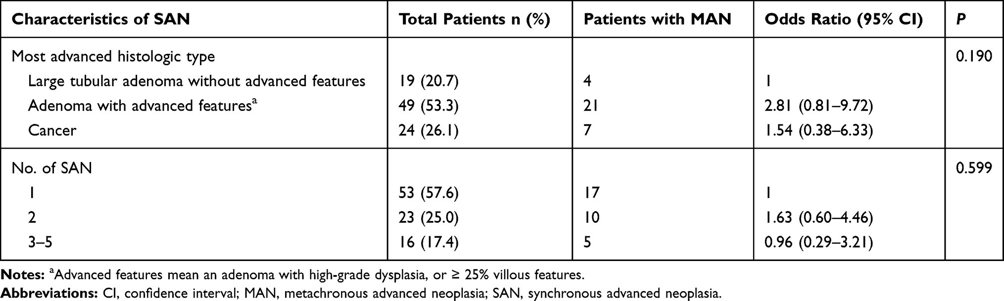 |
Table 3 Comparison of Metachronous Advanced Neoplasia According to the Characteristics of Synchronous Advanced Neoplasia at Baseline |
Incidence and Cumulative Probabilities of MAN
The overall 1-, 2-, 3-, and 5-year cumulative probabilities were 0.9%, 4.8%, 9.8%, and 16.1% for MAN (Figure 2). It is noteworthy that the cumulative probability of MAN was significantly higher for patients with SAN than for those without synchronous neoplasia (P = 0.000) (Table 4). The cumulative probabilities of MAN at 2, 3, and 5 years were 9.7%, 19.8%, and 31.9% for patients with SAN compared to 3.6%, 5.5%, and 10.0% for those without synchronous neoplasia (HR, 3.31; 95% CI, 1.81–6.07) (Figure 3). There was no statistically significant difference among patients with different CEA levels or index cancer sizes in terms of the cumulative probability of MAN. However, in the subgroup analysis of patients based on CEA level and index cancer size, patients with CEA ≥10 ng/mL and index cancer ≥50 mm had a significantly higher cumulative probability of MAN than those with CEA <10 ng/mL and index cancer <50 mm (P = 0.039). The 2-, 3-, and 5-year cumulative probabilities of MAN were 12.5%, 24.7%, and 35.2% for patients with CEA ≥10 ng/mL and index cancer ≥50 mm and 4.0%, 7.4%, and 13.6% for patients with CEA <10 ng/mL and index cancer <50 mm (HR, 2.87; 95% CI, 1.38–5.61) (Figure 4). No significant differences were observed with other variables, such as age and sex, for the cumulative incidence of MAN.
 |
Table 4 Multivariate Analysis of Variables Associated with Metachronous Advanced Neoplasia After Combining CEA Level with the Size of Index Cancer |
 |
Figure 2 The overall cumulative probabilities of metachronous advanced neoplasia. |
 |
Figure 3 Cumulative probabilities of metachronous advanced neoplasia according to the presence or absence of synchronous neoplasia (P = 0.000). |
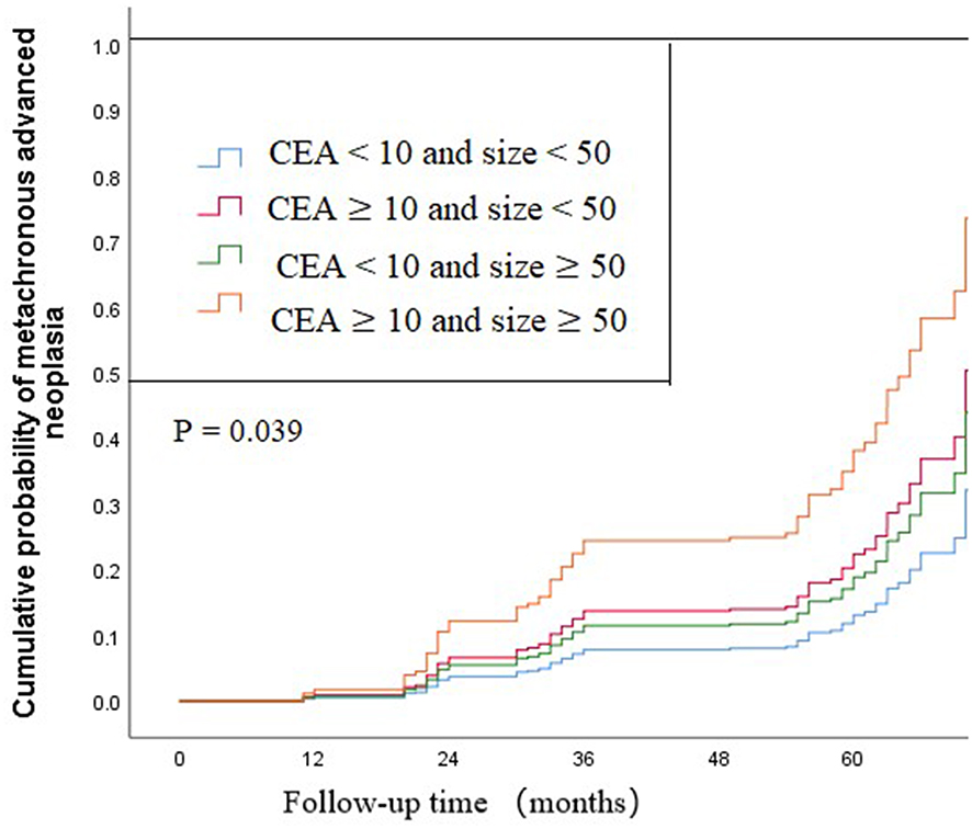 |
Figure 4 Cumulative probabilities of metachronous advanced neoplasia according to CEA level (ng/mL) combining with the size of index cancer (mm). CEA, carcinoembryonic antigen (P = 0.039). |
Discussion
This study investigated the incidence and clinicopathological characteristics predictive of MAN in the residual colon of CRC patients after surgery. We analyzed several variables obtained from the medical records, identified several risk factors for MAN, and calculated the cumulative incidence of MAN for various subpopulations of patients. The results are of significance to the development of individualized surveillance schedules for patients with resected CRC.
The incidence of MAN (17.3%) in our study during a 5-year follow-up period was higher than that found by prior studies (7.8–13.1%).11,12,18 This could be attributable to the different methods of computation used. We defined the incidence of advanced neoplasia detected during the first surveillance colonoscopy 1 year after surgery (only three cases) as MAN according to most studies and guidelines.4,7,11,19 In contrast, a previous study defined lesions that were detected during the first postoperative colonoscopy as synchronous lesions.12 Patients with early CRC who underwent endoscopic submucosal dissection were also included. To our knowledge, the present study is the first investigation to specifically enroll patients with early CRC to evaluate risk factors for MAN. Furthermore, the longer follow-up time (median, 54.9 ± 11.4 months) might have contributed to higher incidence rates. The TNM stage IV patients were not included in the study because of poor prognosis (11.7% for 5-year relative survival) and irregular endoscopic follow-up schedules.
The incidence of synchronous advanced and non-advanced neoplasia in patients with CRC were 20.4% and 41.3%, similar to our previous studies.20 However, the incidence rates were higher than those in previous studies21,22 due to rapid development of endoscopic techniques and the differences in genetic background. During the multivariate analysis including significant factors from the univariate analysis, the presence of synchronous neoplasia, especially SAN, was identified as a significant risk factor for MAN, which was consistent with the results of previous studies.12,19 However, two previous reports indicated that only the presence of SAN was associated with an increased risk of MAN.11,23 In our study, patients with SAN were at higher risk for MAN after surgery compared to those with synchronous non-advanced neoplasia (OR, 5.21 vs 2.12). We believe that risk stratification of synchronous neoplasia may be necessary for patients with resected CRC to determine the appropriate individualized surveillance schedule after surgery (in the same manner as that used for patients after colorectal polypectomy). However, further stratification based on the histopathology or number of SAN in CRC patients with SAN did not seem necessary in terms of MAN.
Our study found that CEA level ≥10.0 ng/mL and larger index cancer at diagnosis were correlated with the development of MAN in CRC patients after curative resection. Usually, increased CEA levels could be used for individual risk assessments and predicting metachronous metastases or poor overall survival.24–26 We found that patients with high preoperative CEA levels were more likely to develop MAN (OR, 2.58). These findings suggest that high blood CEA levels may be a useful indicator of MAN. In our previous study,20 older age and male sex were considered risk factors for SAN in patients with CRC, whereas the use of aspirin seemed to be a preventive factor. However, none of these factors were significantly associated with MAN in the multivariate analysis performed during this study. It remains controversial whether age and sex are independent risk factors for MAN.11,12,19,27 In a series of studies, randomized controlled trials designed to examine the impact of aspirin on the development and recurrence of colorectal adenomas have indicated the efficacy of aspirin chemoprevention.28–30 However, some CRC patients use aspirin irregularly or stop using aspirin due to postoperative complications or negative emotions after surgery, particularly after laparotomy. This might have influenced the effects of aspirin chemoprevention.
The recent guidelines suggest that after curative resection of CRC, patients should undergo surveillance with colonoscopy 1, 3, and 5 years after the initial surgery regardless of the preoperative colonoscopic findings.4,7,8 Several studies have shown that intensive multimodality follow-up of postoperative CRC patients had no survival benefits compared with standard follow-up.10,31–33 However, two meta-analyses that separately analyzed colonoscopy follow-up have suggested that more frequent endoscopic examinations do appear to translate to additional survival benefits.9,34 One randomized controlled trial that evaluated the role of colonoscopy intensity specifically reported that more frequent examinations resulted in higher detection rates of asymptomatic recurrence, more curative surgeries, and improved survival for patients; however, the investigators did not observe improved overall survival for the intensive surveillance group.35 In our study, patients were assigned to intensive colonoscopic surveillance (ie, colonoscopy performed 1, 2, 3, and 5 years after surgery), consistent with the JSCCR guidelines.6 The 2-year cumulative incidence of MAN in the group with SAN was significantly higher than that of the group without synchronous neoplasia (9.7% vs 3.6%; P = 0.000) and that of the group with CEA ≥10 ng/mL and index cancer ≥50 mm compared to the group with CEA <10 ng/mL and index cancer <50 mm at diagnosis (12.5% vs 4.0%; p = 0.007). The diagnosis of MAN during an early stage leads not only to prevention of advanced cancers through detection and removal of metachronous adenoma but also to more cost-effective approaches with fewer complications, such as endoscopic submucosal dissection. Additionally, colonoscope examinations in most parts of China are relatively accessible, affordable, and highly accepted. Based on our results, we propose that patients undergoing surgery for CRC diagnosed with SAN or with high preoperative serum CEA levels and large index cancer should undergo additional surveillance, and that the guidelines could be differently applied to individuals according to the presence or absence of SAN and clinical physicochemical characteristics after curative surgery for CRC.
There were several limitations to our study. First, our study was a single-center, non-randomized study; therefore, it may be difficult to conclude the optimal surveillance intervals. Because of its retrospective nature and some missing data, it could have been affected by the limitations of such an investigational design. However, the missing data contributed to less than 1% of the database; given the total number of patients, it is unlikely to have had a significant effect on the outcome. Finally, misclassification may have occurred when the size of the index cancer was measured approximately by the endoscopists and pathologists. However, because they were unaware of the study objectives, these measurement errors could have resulted in an underestimation of the association between the index cancer size and the incidence of MAN.
Conclusions
We discovered clinical and colonoscopic risk factors that help to identify individuals at increased risk for MAN after curative resection of CRC. According to the results of our study, synchronous neoplasia, especially SAN, high preoperative serum CEA levels, and large index cancer at baseline were independently associated with the risk of MAN. CRC patients with SAN or high preoperative serum CEA levels and large index cancer are at increased risk for early-onset MAN. A more intensive surveillance strategy may be appropriate for these groups. Risk stratification based on synchronous neoplasia and clinical physicochemical characteristics requires further studies involving modified appropriate postoperative colonoscopic surveillance schedules.
Acknowledgments
We would like to express our gratitude to all the doctors who enrolled these patients and participated in this clinical research. Our sincere thanks should also go to Editage for English language editing. Lastly, our thanks would go to our beloved family for their encouragement and support.
Disclosure
The authors report no conflicts of interest in this work.
References
1. Brenner H, Kloor M, Pox CP. Colorectal cancer. Lancet. 2014;383(9927):1490–1502. doi:10.1016/S0140-6736(13)61649-9
2. Mulder SA, Kranse R, Damhuis RA, Ouwendijk RJ, Kuipers EJ, van Leerdam ME. The incidence and risk factors of metachronous colorectal cancer: an indication for follow-up. Dis Colon Rectum. 2012;55(5):522–531. doi:10.1097/DCR.0b013e318249db00
3. Levi F, Randimbison L, Blanc-Moya R, et al. High constant incidence of second primary colorectal cancer. Int J Cancer. 2013;132(7):1679–1682. doi:10.1002/ijc.27780
4. Hassan C, Wysocki PT, Fuccio L, et al. Endoscopic surveillance after surgical or endoscopic resection for colorectal cancer: european Society of Gastrointestinal Endoscopy (ESGE) and European Society of Digestive Oncology (ESDO) Guideline. Endoscopy. 2019;51(3):C1. doi:10.1055/a-0854-5925
5. Anthony T, Fleming JB, Bieligk SC, et al. Postoperative colorectal cancer surveillance. J Am Coll Surg. 2000;190(6):737–749. doi:10.1016/S1072-7515(99)00298-7
6. Taniguchi H. [Japanese Society for Cancer of the Colon and Rectum (JSCCR) Guidelines 2019 for the Treatment of Colorectal Cancer: systemicTherapy]. Gan to Kagaku Ryoho. 2019;46(11):1709–1713. Japanese.
7. Kahi CJ, Boland CR, Dominitz JA, et al. Colonoscopy surveillance after colorectal cancer resection: recommendations of the us multi-society task force on colorectal cancer. Gastroenterology. 2016;150(3):758–768 e711. doi:10.1053/j.gastro.2016.01.001
8. Colorectal Cancer Working Group, C. Diagnosis, Treatment Guidelines For Colorectal Cancer Working Group C. Chinese Society of Clinical Oncology (CSCO) diagnosis and treatment guidelines for colorectal cancer 2018 (English version). Chin J Cancer Res. 2019;31(1):117–134. doi:10.21147/j.issn.1000-9604.2019.01.07
9. Tjandra JJ, Chan MKY. Follow-up after curative resection of colorectal cancer: a meta-analysis. Dis Colon Rectum. 2007;50(11):1783–1799. doi:10.1007/s10350-007-9030-5
10. Schoemaker D, Black R, Giles L, Toouli J. Yearly colonoscopy, liver CT, and chest radiography do not influence 5-year survival of colorectal cancer patients. Gastroenterology. 1998;114(1):7–14. doi:10.1016/S0016-5085(98)70626-2
11. Moon CM, Cheon JH, Choi EH, et al. Advanced synchronous adenoma but not simple adenoma predicts the future development of metachronous neoplasia in patients with resected colorectal cancer. J Clin Gastroenterol. 2010;44(7):495–501.
12. Yabuuchi Y, Imai K, Hotta K, et al. Higher incidence of metachronous advanced neoplasia in patients with synchronous advanced neoplasia and left-sided colorectal resection for colorectal cancer. Gastrointest Endosc. 2018;88(2):348–359 e341. doi:10.1016/j.gie.2018.03.011
13. Fuller NG. The WHO classification of tumours of the central nervous system, 4th edition. Arch Pathol Lab Med. 2008;132(6).
14. O’Sullivan B, Brierley J, Byrd D, et al. The TNM classification of malignant tumours-towards common understanding and reasonable expectations. Lancet Oncol. 2017;18(7):849–851. doi:10.1016/S1470-2045(17)30438-2
15. Hansen RD, Albieri V, Tjonneland A, Overvad K, Andersen KK, Raaschou-Nielsen O. Effects of smoking and antioxidant micronutrients on risk of colorectal cancer. Clin Gastroenterol Hepatol. 2013;11(4):406–415 e403. doi:10.1016/j.cgh.2012.10.039
16. Bongaerts BW, van den Brandt PA, Goldbohm RA, de Goeij AF, Weijenberg MP. Alcohol consumption, type of alcoholic beverage and risk of colorectal cancer at specific subsites. Int J Cancer. 2008;123(10):2411–2417. doi:10.1002/ijc.23774
17. Pak MG, Koh HJ, Roh MS. Clinicopathologic significance of TRAP1 expression in colorectal cancer: a large scale study of human colorectal adenocarcinoma tissues. Diagn Pathol. 2017;12(1):6. doi:10.1186/s13000-017-0598-3
18. Togashi K, Konishi F, Ozawa A, et al. Predictive factors for detecting colorectal carcinomas in surveillance colonoscopy after colorectal cancer surgery. Dis Colon Rectum. 2000;43(10 Suppl):S47–S53. doi:10.1007/BF02237226
19. Patel A, Williams N, Parsons N, et al. Risk factors for metachronous adenoma in the residual colon of patients undergoing curative surgery for colorectal cancer. Int J Colorectal Dis. 2017;32(11):1609–1616. doi:10.1007/s00384-017-2881-x
20. Li S, Zhu K, Yu W, et al. Synchronous neoplastic lesions in referred patients with colorectal cancer: a retrospective cohort study. Cancer Manag Res. 2019;11:9951–9959. doi:10.2147/CMAR.S229376
21. Borda A, Martinez-Penuela JM, Munoz-Navas M, Prieto C, Betes M, Borda F. [Synchronous neoplastic lesions in colorectal cancer. An analysis of possible risk factors favouring presentation]. Rev Esp Enferm Dig. 2008;100(3):139–145. Spanish.
22. Pinol V, Andreu M, Castells A, et al. Synchronous colorectal neoplasms in patients with colorectal cancer: predisposing individual and familial factors. Dis Colon Rectum. 2004;47(7):1192–1200. doi:10.1007/s10350-004-0562-7
23. McFall MR, Woods WG, Miles WF. Colonoscopic surveillance after curative colorectal resection: results of an empirical surveillance programme. Colorectal Dis. 2003;5(3):233–240. doi:10.1046/j.1463-1318.2003.00412.x
24. Miyake H, Murono K, Nagata H, et al. Prognostic significance of doubling time in patients undergoing radical surgery for metachronous peritoneal metastases of colorectal cancer. Int J Colorectal Dis. 2019;34(5):801–809. doi:10.1007/s00384-019-03259-5
25. Laubert T, Bente V, Freitag-Wolf S, et al. Aneuploidy and elevated CEA indicate an increased risk for metachronous metastasis in colorectal cancer. Int J Colorectal Dis. 2013;28(6):767–775. doi:10.1007/s00384-012-1625-1
26. Haraguchi M, Fujita F, Torashima Y, Inokuma T, Tajima Y, Kanematsu T. The serum level of carcinoembryonic antigen in drainage venous blood is not a sensitive predictor of metachronous hepatic metastasis for patients with colorectal cancer. Surg Today. 2010;40(8):745–751. doi:10.1007/s00595-009-4205-4
27. Bonithon-Kopp C, Piard F, Fenger C, et al. Colorectal adenoma characteristics as predictors of recurrence. Dis Colon Rectum. 2004;47(3):323–333. doi:10.1007/s10350-003-0054-1
28. Benamouzig R, Deyra J, Martin A, et al. Daily soluble aspirin and prevention of colorectal adenoma recurrence: one-year results of the APACC trial. Gastroenterology. 2003;125(2):328–336. doi:10.1016/S0016-5085(03)00887-4
29. Benamouzig R, Uzzan B, Deyra J, et al. Prevention by daily soluble aspirin of colorectal adenoma recurrence: 4-year results of the APACC randomised trial. Gut. 2012;61(2):255–261. doi:10.1136/gutjnl-2011-300113
30. Logan RF, Grainge MJ, Shepherd VC, Armitage NC, Muir KR; uk CAPTG. Aspirin and folic acid for the prevention of recurrent colorectal adenomas. Gastroenterology. 2008;134(1):29–38. doi:10.1053/j.gastro.2007.10.014
31. Kjeldsen BJ, Kronborg O, Fenger C, Jorgensen OD. A prospective randomized study of follow-up after radical surgery for colorectal cancer. Br J Surg. 1997;84(5):666–669. doi:10.1002/bjs.1800840523
32. Rosati G, Ambrosini G, Barni S, et al. A randomized trial of intensive versus minimal surveillance of patients with resected Dukes B2-C colorectal carcinoma. Ann Oncol. 2016;27(2):274–280. doi:10.1093/annonc/mdv541
33. Rodriguez-Moranta F, Salo J, Arcusa A, et al. Postoperative surveillance in patients with colorectal cancer who have undergone curative resection: a prospective, multicenter, randomized, controlled trial. J Clin Oncol. 2006;24(3):386–393. doi:10.1200/JCO.2005.02.0826
34. Pita-Fernandez S, Alhayek-Ai M, Gonzalez-Martin C, Lopez-Calvino B, Seoane-Pillado T, Pertega-Diaz S. Intensive follow-up strategies improve outcomes in nonmetastatic colorectal cancer patients after curative surgery: a systematic review and meta-analysis. Ann Oncol. 2015;26(4):644–656. doi:10.1093/annonc/mdu543
35. Wang T, Cui Y, Huang WS, et al. The role of postoperative colonoscopic surveillance after radical surgery for colorectal cancer: a prospective, randomized clinical study. Gastrointest Endosc. 2009;69(3 Pt 2):609–615. doi:10.1016/j.gie.2008.05.017
 © 2020 The Author(s). This work is published and licensed by Dove Medical Press Limited. The
full terms of this license are available at https://www.dovepress.com/terms
and incorporate the Creative Commons Attribution
- Non Commercial (unported, 3.0) License.
By accessing the work you hereby accept the Terms. Non-commercial uses of the work are permitted
without any further permission from Dove Medical Press Limited, provided the work is properly
attributed. For permission for commercial use of this work, please see paragraphs 4.2 and 5 of our Terms.
© 2020 The Author(s). This work is published and licensed by Dove Medical Press Limited. The
full terms of this license are available at https://www.dovepress.com/terms
and incorporate the Creative Commons Attribution
- Non Commercial (unported, 3.0) License.
By accessing the work you hereby accept the Terms. Non-commercial uses of the work are permitted
without any further permission from Dove Medical Press Limited, provided the work is properly
attributed. For permission for commercial use of this work, please see paragraphs 4.2 and 5 of our Terms.