")
Back to Journals » Infection and Drug Resistance » Volume 16
Risk of Bacterial Exposure to the Anesthesiologist’s Face During Intubation and Extubation
Authors Song SH, Choi SH , Park HR, Jeon SY, Kim SH
Received 22 January 2023
Accepted for publication 15 April 2023
Published 25 April 2023 Volume 2023:16 Pages 2433—2439
DOI https://doi.org/10.2147/IDR.S405537
Checked for plagiarism Yes
Review by Single anonymous peer review
Peer reviewer comments 2
Editor who approved publication: Prof. Dr. Héctor Mora-Montes
Sei Han Song,1 Seung Ho Choi,2 Hae Ri Park,1 Soo Yeon Jeon,1 Seung Hyun Kim2
1Department of Anesthesiology and Pain Medicine, Severance Hospital, Yonsei University College of Medicine, Seoul, Korea; 2Department of Anesthesiology and Pain Medicine, Anesthesia and Pain Research Institute, Severance Hospital, Yonsei University College of Medicine, Seoul, Korea
Correspondence: Seung Hyun Kim, Department of Anesthesiology and Pain Medicine, Anesthesia and Pain Research Institute, Severance Hospital, Yonsei University College of Medicine, 50-1 Yonsei-ro, Seodaemun-gu, Seoul, 03722, Korea, Tel +82-2-2224-1055, Fax +82-2-2227-7897, Email [email protected]
Introduction: Anesthesiologists are exposed to the risk of infection from various secretions or droplets from the respiratory tract of patients. We aimed to determine bacterial exposure to anesthesiologists’ faces during endotracheal intubation and extubation.
Methods: Six resident anesthesiologists performed 66 intubation and 66 extubation procedures in patients undergoing elective otorhinolaryngology surgeries. Sampling was performed by swabbing the face shields twice in an overlapping slalom pattern, before and after each procedure. Samples for pre-intubation and pre-extubation were collected immediately after wearing the face shield at the time of anesthesia induction and at the end of the surgery, respectively. Post-intubation samples were collected after the injection of anesthetic drugs, positive pressure mask ventilation, endotracheal intubation, and confirmation of intubation success. Post-extubation samples were collected after endotracheal tube suction, oral suction, extubation, and confirmation of spontaneous breathing and stable vital signs. All swabs were cultured for 48 h, and bacterial growth was confirmed by colony forming unit (CFU) count.
Results: There was no bacterial growth in either pre- or post-intubation bacterial cultures. In contrast, while there was no bacterial growth in pre-extubation samples, 15.2% of post-extubation samples were CFU+ (0/66 [0%] vs 10/66 [15.2%], p=0.001). All the CFU+ samples belonged to 47 patients with post-extubation coughing, and the CFU count was correlated with the number of coughing episodes during the process of extubation (P < 0.01, correlation coefficient= 0.403).
Conclusion: The current study shows the actual chance of bacterial exposure to the anesthesiologist’s face during the patient awakening process after general anesthesia. Given the correlation between the CFU count and the number of coughing episodes, we recommend anesthesiologists to use appropriate facial protection equipment during this procedure.
Keywords: bacterial exposure, cough, extubation, face shield, intubation
Introduction
Healthcare workers are exposed to many risk factors for their physical and mental health.1 Medical procedures for patients with infectious diseases can be dangerous to healthcare workers because of the possibility of infection transmission. Although the risk of pathogen transmission during aerosol-generating procedures is not fully known, the face of healthcare workers can get contaminated by droplets from the patients,2 which can be inhaled into the healthcare workers’ lungs or penetrate the wounded skin or mucous membranes of their eyes, nose, and mouth to cause an infectious disease.
The face shield is a representative personal protective equipment (PPE) that protects the facial area. According to the Centers for Disease Control and Prevention (CDC) and World Health Organization (WHO), face shields are required when performing procedures that generate splashes or sprays of blood, body fluids, or respiratory secretions.3 In a previous study using a cough aerosol simulator and breathing simulator, the amount of virus on the respirator under the face shield was reduced by 96% when compared to that without the face shield.4 However, there is relatively scarce data regarding face shields, and further research is needed to better understand and establish their appropriate use.2 It has been reported that anesthesiologists can perform endotracheal intubation well while wearing face shields or other PPEs.5 Nevertheless, many anesthesiologists do not have sufficient knowledge about protective equipment and do not follow the recommended preventive measures (eg, washing hands and using personal protective equipment).6,7
Bacterial exposure to the endoscopist’s face shields has been reported.8 In previous studies that quantified aerosol generation during general anesthesia, aerosols were detected during endotracheal extubation; however, the results were conflicting during endotracheal intubation.9–12 There have been no studies on bacterial exposure to anesthesiologists’ faces during endotracheal intubation, extubation, and airway suction. Therefore, this prospective study aimed to determine bacterial exposure to anesthesiologists’ face shields during endotracheal intubation and extubation by comparing pre- and post-procedural samples from face shields.
Materials and Methods
Ethics Approval and Conduct of the Study
The study protocol was approved by the Institutional Review Board of the Yonsei University Health System, Seoul, South Korea (#4-2020-0529) on 30, June, 2020, and registered at http://clinicaltrials.gov (NCT04673006). This study was conducted in accordance with the ethical guidelines of the Helsinki Declaration. This prospective study was conducted in resident anesthesiologists performing endotracheal intubation and extubation in 66 elective otorhinolaryngological surgeries that were expected to be longer than 3 hours. We excluded patients with suspected difficult airway, obesity (a body mass index higher than 40), suspected respiratory infections, cognitive disorder, illiteracy, or foreign nationality. Written informed consent was obtained from all the anesthesiologists and patients enrolled in this study, which was conducted from January to June 2021.
To determine bacterial exposure during intubation and extubation, anesthesiologists’ face shield bacterial swab sampling was performed in 66 intubation and 66 extubation procedures by the investigator (S. H. Kim). Endotracheal intubation and extubation were performed by resident anesthesiologists aged ≥ 20 years, wearing a disposable, non-sterile face shield (Figure 1). Both anesthesiologists and assistant nurses used surgical gloves and wore simple surgical mask or Korean filter 94 face masks. Before endotracheal intubation and extubation procedures, when the anesthesiologist wore the face shield, the investigator (S. H. Kim) removed the protective film (outer vinyl) of the face shield. The samples were collected from the face shield before and after each procedure. Sampling was performed by sweeping a sterile cotton swab twice over the face shield, including the corners and edges in an overlapping slalom pattern, and at the second time perpendicular to the first direction.13 Bacterial swab samples were collected five times from each patient, and a total of 330 samples were collected. The sampling process was performed for each patient as follows.
- Pre-intubation sampling: The anesthesiologists wore a disposable, non-sterile face shield before the induction of general anesthesia. The protective film was peeled off immediately after wearing, and a sterile cotton swab was used to collect a sample from the surface of the face shield.
- Process of anesthetic induction and post-intubation sampling: After standard monitoring including pulse oximetry, non-invasive blood pressure monitoring, and electrocardiography, resident anesthesiologists performed preoxygenation using Westmed 900 Series Anesthesia Masks (Westmed, Inc. Tucson, Arizona, U.S), and assistant nurses administered 2 mg/kg of propofol and 0.5 mg/kg of remifentanil for anesthetic induction. When the patient’s loss of consciousness was confirmed, 6 mg/kg of rocuronium was administered, and anesthesiologists performed positive pressure mask ventilation. After the patient was sufficiently relaxed, endotracheal intubation was performed using a KoMAC videolaryngoscope (KoMAC Co., Ltd., Seoul, Republic of Korea). Anesthesiologists confirmed the success of intubation through chest auscultation. During this procedure, the anesthesiologists were not allowed to touch the face shield and the investigator monitored for any inadvertent touch. If a touch or other contact occurred, the patient was excluded from the study. After the intubation procedure, the investigator used a sterile cotton swab to collect the sample from the face shield surface in the same manner as pre-intubation sampling. General anesthesia was maintained with 3–7% desflurane and remifentanil infused throughout the surgery. Thirty minutes before the end of surgery, 0.5 μg/kg of fentanyl was injected for postoperative pain control.
- Pre-extubation sampling: When the operation was completed, the anesthesiologists wore a new face shield before the emergence of the patients. Immediately after removing the protective film from the face shield, the investigator sampled the surface with a sterile cotton swab.
- Process of extubation and post-extubation sampling: The anesthesiologists performed endotracheal tube and oral suction, after which the anesthetic agents were discontinued. Endotracheal “awake” extubation was performed when the patient responded to the command to open their eyes, and spontaneous breathing was confirmed. After the removal of the endotracheal tube, a Westmed 900 Series Anesthesia Mask was applied to the patient, and the anesthesiologists checked the patient’s consciousness, spontaneous breathing, and vital signs until stable After the extubation, additional oral suction was performed at the discretion of the anesthesiologists. Throughout the process, any unintentional touching was monitored carefully, and sampling was performed in the same manner as mentioned above. Coughing during the extubation process, if any, was also recorded.
- Positive control sampling: After extubation, post-extubation positive control samples were collected using a sterile swab from the endotracheal tube tips.
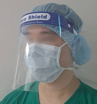 |
Figure 1 Face shield. |
The swabs were placed in a transport medium (AM608-2S, Asan pharm Co.,Seoul, Korea) and delivered to Samkwang Medical Laboratories (Seoul, Korea) in a refrigerated state on the day of the procedure. After mixing the solid medium with a cotton swab, the inspectors streaked the medium onto the blood agar and MacConkey agar plate and incubated it at 37°C for 48 h. The results of bacterial growth were reported as the number of colony-forming units (CFU) in all samples. Any growth of CFU ≥ 1 was classified as a positive colony forming unit (CFU+). Identification of specific bacterial species was not attempted to avoid further clinical intervention of the patients and anesthesiologists.
The primary outcome of this study was whether bacterial exposure (reported as CFU+) occurred during endotracheal extubation; therefore, the rate of CFU+ was compared before and after extubation. The secondary outcome was bacterial exposure during endotracheal intubation, which was presented as the rate of CFU+ before and after intubation. Bacterial exposure was also evaluated in a subgroup of patients who coughed during endotracheal intubation and extubation.
Statistical Analysis
Considering that there was a 17% or greater chance of exposure to droplets or blood to medical personnel during various surgeries and procedures,8,14,15 this study assumed a 17% increase in the CFU+ ratio to be a significant difference (power 0.80, type 1 error rate 5%). The sample size to confirm the 20% difference was 60 patients; therefore, 66 patients were sampled before and after the procedures, considering 10% dropouts.
CFU+ and positive exposure rates before and after the procedure were analyzed using the chi-square test. The correlation between the number of coughing episodes and the CFU count was analyzed using Pearson’s correlation analysis. Statistical analyses were performed using SPSS Statistics 26 (SPSS Inc. USA). Statistical significance was set at P < 0.05.
Results
In the study, there were no excluded cases of unintentional touch to the face shield, and a total of six resident anesthesiologists performed 66 endotracheal intubation and extubation processes in 66 patients. Two of these patients had asthma, and one patient had a history of old pulmonary tuberculosis.
During the process of anesthetic induction, 14 patients exhibited coughing after remifentanil bolus injection, and there was no bacterial growth in either pre- or post-intubation bacterial cultures. In all cases, endotracheal intubation was successful on the first attempt.
During the process of extubation, although there was no bacterial growth in the pre-extubation samples, 15.2% of the post-extubation samples were CFU+ (0/66 [0%] vs 10/66 [15.2%], p=0.001). CFU+ in the post-extubation positive control sampling was 98.5%. A total of 47 patients exhibited coughing during the process of extubation, and bacterial growth was observed only in these patients. In this subgroup where patient coughed, CFU+ was different after extubation compared to before extubation (0/47 [0%] vs 10/47 [21.3%], p=0.001). Among these 47 patients, two patients exhibited coughing during endotracheal suction, 36 patients after the extubation, and the remaining nine patients both during endotracheal suction and after extubation. After the removal of the endotracheal tube, oral suction was performed in 61 patients at the discretion of the anesthesiologists. Median (interquartile range) number of coughing episodes in the subgroup with coughing was 3.0 (1.0–4.0), and there was a correlation between the number of coughing episodes and the CFU count (P < 0.01, correlation coefficient= 0.403). The number of CFU in pre- and post- extubation samples is presented in the Table 1. Hypoxia, with oxygen saturation lower than 90%, did not occur during the extubation process in this study, and none of the anesthesiologists who participated in this study experienced symptoms of respiratory tract or facial infection during the study period.
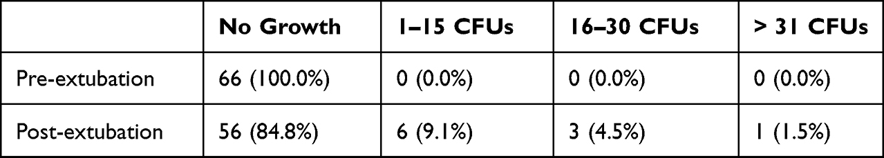 |
Table 1 Number of Bacterial Colony-Forming Unit (CFU) Count in Pre- and Post-Extubation Samples Taken from the Anesthesiologist’s Face Shield |
Discussion
This study demonstrates the bacterial exposure to the face of anesthesiologists during the patient awakening process after general anesthesia, including endotracheal extubation and oral suction procedures. This result is meaningful in that it directly quantified bacterial exposure on the anesthesiologist’s face and showed that the extubation procedure carries a potential risk of infection. In particular, the CFU count confirmed by bacterial culture was correlated with the number of coughing episodes during the process of extubation, implying the importance of coughing for bacterial exposure during this procedure. In contrast, during the general anesthetic induction process, including positive-pressure mask ventilation and endotracheal intubation, we could not confirm bacterial exposure to the anesthesiologist’s face.
In our study design, “awake” endotracheal extubation was performed when the patients were fully alert and able to respond appropriately to anesthesiologists’ commands, which increased the potential risk of post-extubation coughing. Coughing produced the greatest number of aerosol particles among six respiratory activities, including quiet breathing, talking, exercise, shouting, and forced expirations, particularly increasing the particle count 370.8-fold compared with quiet breathing.16 Recent studies that quantified the amount of aerosol generation during general anesthesia have shown that patient coughing during extubation increased the amount of aerosol generation,10–12 which is consistent with our study result that all positive bacterial exposure belonged to extubation cases with patients coughing, and the CFU count was correlated with the number of coughing episodes. According to previous studies,9,11,12 endotracheal extubation produces aerosols, but the amount was comparable to or less than that of coughing. Therefore, based on our study, coughing during the process of extubation could be considered an important factor in bacterial exposure during this procedure. Tight mask sealing using an anesthetic facial mask after the extubation could potentially reduce dispersion of droplets. However, during the process of extubation, the face of the anesthesiologist can be exposed to patient coughing induced either by endotracheal tube suction or oral suction after the removal of the endotracheal tube.
Regarding endotracheal intubation, our study did not find any evidence of bacterial exposure to the anesthesiologist’s face during the procedure. The results of previous studies on whether this procedure is aerosol-generating are inconsistent. While Dhillon et al found that the aerosol concentration was 12 times greater than the baseline during the entire anesthesia induction procedure (passive oxygenation, bag mask ventilation, tube insertion),10 intubation generated only slightly more aerosol than the baseline in another study.9 In our study, bacterial growth was not observed during the anesthetic induction process, even in cases with coughing. During the induction, we used a facial mask for preoxygenation and positive pressure ventilation, and the mask placed between the anesthesiologist’s face and the patient’s mouth could be one of the possible reasons for preventing bacterial exposure during this procedure. Considering that coughing can produce aerosols even under continuous positive airway pressure and high-flow nasal oxygenation ventilation,17 tight mask sealing and minimizing air leaks are still important during anesthetic induction.
During the COVID-19 pandemic era, as airway maneuvers are recognized as a source of infection, careful airway management techniques to reduce the transmission of infection sources have become more important.18 Medical personnel are usually unaware of patients’ body fluid exposure during the procedures, but various medical procedures have the potential to expose health care workers to body fluids and pathogens. Blood spatter and body fluid splashes on protective glasses and masks during various surgeries have been reported previously.14,15 Anesthesiologists also can be easily exposed to blood or secretion from the patient’s upper respiratory tract and stomach, which can be a carrier of pathogens.19,20 The incidence of post-extubation cough was reported to be 10–100%,21,22 and considering the importance of coughing during extubation, the use of appropriate PPE is essential for anesthesiologists in this procedure. Strategies to reduce the incidence of coughing such as “deep” extubation technique,23 or maintenance of low dose opioid24,25 during extubation have been attempted. However, these methods may not be effective for preventing coughing and could even be dangerous for some patient subpopulations such as those with a difficult airway. The anesthesiologists must prioritize the patient’s safety during the process of awakening and extubation. Therefore, the use of standard PPE including high particulate filtering respirator is strongly recommended for this procedure.26,27
This study had some limitations. First, we only investigated bacterial exposure and did not identify specific bacterial species. Exposure to viruses or other microorganisms was not investigated either. The relationship between the transmission of normal commensals and actual infection is not clear, and whether simple facial exposure leads to the transmission of disease or not may depend on additional processes such as droplet inhalation or mucosal penetration by the pathogens. Therefore, it is difficult to generalize the results of this study to the general mechanism of pathogen transmission. Second, we considered the entire process, from endotracheal tube suction to confirmation of the patient’s stability after the endotracheal tube removal, as a single process of extubation. As bacterial swabbing was not performed immediately after endotracheal tube suction, it is not clear which specific procedure caused bacterial exposure. Furthermore, after the removal of the endotracheal tube, it is very difficult to clearly differentiate between spontaneous coughing and that induced by oral suction.
Conclusions
In conclusion, this study demonstrated the possibility of bacterial exposure during patient awakening after general anesthesia. This exposure can pose a potential risk of infection for anesthesiologists. In addition, coughing during extubation appears to play an important role in bacterial exposure during the procedure. Based on the results of our study, appropriate use of PPE is highly recommended for healthcare workers during this procedure.
Abbreviations
CFU, colony forming unit; PPE, personal protective equipment.
Data Sharing Statement
The full data presented in this study are available on request from the corresponding author.
Ethics Approval and Informed Consent
The study protocol was approved by the Institutional Review Board of the Yonsei University Health System, Seoul, South Korea (#4-2020-0529) on 30 June, 2020; and registered at http://clinicaltrials.gov (NCT04673006). This study was conducted in accordance with the ethical guidelines of the Helsinki Declaration. Written informed consent was obtained from all the anesthesiologists and patients enrolled in this study.
Author Contributions
All authors made a significant contribution to the work reported, whether that is in the conception, study design, execution, acquisition of data, analysis and interpretation, or in all these areas; took part in drafting, revising or critically reviewing the article; gave final approval of the version to be published; have agreed on the journal to which the article has been submitted; and agree to be accountable for all aspects of the work.
Funding
This study was supported by a faculty research grant of Yonsei University College of Medicine (6-2020-0162).
Disclosure
The authors declare that they have no competing interests.
References
1. Vijendren A, Yung M, Sanchez J. Occupational health issues amongst UK doctors: a literature review. Occup Med. 2015;65(7):519–528. doi:10.1093/occmed/kqv088
2. Roberge RJ. Face shields for infection control: a review. J Occup Environ Hyg. 2016;13(4):235–242. doi:10.1080/15459624.2015.1095302
3. Siegel JD, Rhinehart E, Jackson M, Chiarello L. 2007 Guideline for isolation precautions: preventing transmission of infectious agents in health care settings. Am J Infect Control. 2007;35(10 Suppl 2):S65–164. doi:10.1016/j.ajic.2007.10.007
4. Lindsley WG, Noti JD, Blachere FM, Szalajda JV, Beezhold DH. Efficacy of face shields against cough aerosol droplets from a cough simulator. J Occup Environ Hyg. 2014;11(8):509–518. doi:10.1080/15459624.2013.877591
5. Greenland KB, Tsui D, Goodyear P, Irwin MG. Personal protection equipment for biological hazards: does it affect tracheal intubation performance? Resuscitation. 2007;74(1):119–126. doi:10.1016/j.resuscitation.2006.11.011
6. Kim DD, Kimura A
7. Tait AR, Tuttle DB. Preventing perioperative transmission of infection: a survey of anesthesiology practice. Anesth Analg. 1995;80(4):764–769. doi:10.1097/00000539-199504000-00020
8. Johnston ER, Habib-Bein N, Dueker JM, et al. Risk of bacterial exposure to the endoscopist’s face during endoscopy. Gastrointest Endosc. 2019;89(4):818–824. doi:10.1016/j.gie.2018.10.034
9. Brown J, Gregson FKA, Shrimpton A, et al. A quantitative evaluation of aerosol generation during tracheal intubation and extubation. Anaesthesia. 2021;76(2):174–181. doi:10.1111/anae.15292
10. Dhillon RS, Rowin WA, Humphries RS, et al. Aerosolisation during tracheal intubation and extubation in an operating theatre setting. Anaesthesia. 2021;76(2):182–188. doi:10.1111/anae.15301
11. Oksanen LM, Sanmark E, Sofieva S, et al. Aerosol generation during general anesthesia is comparable to coughing: an observational clinical study. Acta Anaesthesiol Scand. 2022;66(4):463–472. doi:10.1111/aas.14022
12. Sanmark E, Oksanen LAH, Rantanen N, et al. Aerosol generation during coughing - an observational study. J Laryngol Otol. 2022;137:442–447. doi:10.1017/s0022215122001165.1-15
13. Jansson L, Akel Y, Eriksson R, Lavander M, Hedman J. Impact of swab material on microbial surface sampling. J Microbiol Methods. 2020;176:106006. doi:10.1016/j.mimet.2020.106006
14. Davies CG, Khan MN, Ghauri AS, Ranaboldo CJ. Blood and body fluid splashes during surgery--The need for eye protection and masks. Ann R Coll Surg Engl. 2007;89(8):770–772. doi:10.1308/003588407X209301
15. Endo S, Kanemitsu K, Ishii H, et al. Risk of facial splashes in four major surgical specialties in a multicentre study. J Hosp Infect. 2007;67(1):56–61. doi:10.1016/j.jhin.2007.05.020
16. Wilson NM, Marks GB, Eckhardt A, et al. The effect of respiratory activity, non-invasive respiratory support and facemasks on aerosol generation and its relevance to COVID-19. Anaesthesia. 2021;76(11):1465–1474. doi:10.1111/anae.15475
17. Hamilton FW, Gregson FKA, Arnold DT, et al. Aerosol emission from the respiratory tract: an analysis of aerosol generation from oxygen delivery systems. Thorax. 2022;77(3):276–282. doi:10.1136/thoraxjnl-2021-217577
18. Nestor CC, Wang S, Irwin MG. Are tracheal intubation and extubation aerosol-generating procedures? Anaesthesia. 2021;76(2):151–155. doi:10.1111/anae.15328
19. Shelley KH, Haddadin AS. Is Helicobacter pylori infection an occupational hazard for anesthesiologists? Anesth Analg. 1998;87(4):973–974. doi:10.1213/00000539-199810000-00042
20. Volquind D, Bagatini A, Carneiro Monteiro GM, Londero JR, Benvenutti GD. Occupational hazards and diseases related to the practice of anesthesiology. Braz J Anesthesiol. 2013;63(2):227–232. doi:10.1016/S0034-7094(13)70221-6
21. Park SY, Kim SH, Lee SJ, et al. Application of triamcinolone acetonide paste to the endotracheal tube reduces postoperative sore throat: a randomized controlled trial. Can J Anaesth. 2011;58(5):436–442. doi:10.1007/s12630-011-9478-6
22. Yang SS, Wang NN, Postonogova T, et al. Intravenous lidocaine to prevent postoperative airway complications in adults: a systematic review and meta-analysis. Br J Anaesth. 2020;124(3):314–323. doi:10.1016/j.bja.2019.11.033
23. Neelakanta G, Miller J. Minimum alveolar concentration of isoflurane for tracheal extubation in deeply anesthetized children. Anesthesiology. 1994;80(4):811–813. doi:10.1097/00000542-199404000-00013
24. Lee B, Lee JR, Na S. Targeting smooth emergence: the effect site concentration of remifentanil for preventing cough during emergence during propofol-remifentanil anaesthesia for thyroid surgery. Br J Anaesth. 2009;102(6):775–778. doi:10.1093/bja/aep090
25. Kim HY, Kim JY, Ahn SH, Lee SY, Park HY, Kwak HJ. Predicting effective remifentanil concentration in 95% of patients to prevent emergence cough after laryngomicroscopic surgery. Medicine. 2018;97(26):e11258. doi:10.1097/MD.0000000000011258
26. Siegel JD, Rhinehart E, Jackson M, Chiarello L. The healthcare infection control practices advisory committee, 2007 guideline for isolation precautions: preventing transmission of infectious agents in healthcare settings; 2022. Available from: https://www.cdc.gov/infectioncontrol/guidelines/isolation/index.html.
27. World Health Organization. Standard precautions for the prevention and control of infections: aide-memoire. Available from: who.int/publications/i/item/WHO-UHL-IHS-IPC-2022.1.
 © 2023 The Author(s). This work is published and licensed by Dove Medical Press Limited. The full terms of this license are available at https://www.dovepress.com/terms.php and incorporate the Creative Commons Attribution - Non Commercial (unported, v3.0) License.
By accessing the work you hereby accept the Terms. Non-commercial uses of the work are permitted without any further permission from Dove Medical Press Limited, provided the work is properly attributed. For permission for commercial use of this work, please see paragraphs 4.2 and 5 of our Terms.
© 2023 The Author(s). This work is published and licensed by Dove Medical Press Limited. The full terms of this license are available at https://www.dovepress.com/terms.php and incorporate the Creative Commons Attribution - Non Commercial (unported, v3.0) License.
By accessing the work you hereby accept the Terms. Non-commercial uses of the work are permitted without any further permission from Dove Medical Press Limited, provided the work is properly attributed. For permission for commercial use of this work, please see paragraphs 4.2 and 5 of our Terms.