
Back to Journals » Clinical, Cosmetic and Investigational Dermatology » Volume 19
Remodeling Effect of HA Hybrid Cooperative Complexes on Dermal White Adipose Tissue and Its Structure
Authors Polvere I  , Scrima M , Vito N, Signorino G, Iorio A, Giori AM , Ferravante A
Received 23 April 2026
Accepted for publication 28 June 2026
Published 16 July 2026 Volume 2026:19 617588
DOI https://doi.org/10.2147/CCID.S617588
Checked for plagiarism Yes
Review by Single anonymous peer review
Peer reviewer comments 2
Editor who approved publication: Dr Jeffrey Weinberg
Immacolata Polvere,1 Mario Scrima,1 Nicoletta Vito,1 Giacomo Signorino,1 Antonio Iorio,1 Andrea Maria Giori,2 Angela Ferravante1
1Preclinical & Clinical Research and Safety Department, IBSA Corporate, Ariano Irpino, Italy; 2Preclinical & Clinical Research and Safety Department, IBSA Corporate, Lodi, Italy
Correspondence: Immacolata Polvere, Preclinical & Clinical Research and Safety Department, IBSA Corporate, C/da Camporeale, Ariano Irpino, AV, 83031, Italy, Tel +390371617590, Email [email protected]
Purpose: Skin aging affects white adipose tissue, resulting in volume loss and appearance of deep wrinkles. This study aimed to investigate the effects of Profhilo® Structura, a hybrid cooperative complex of high and low molecular weight hyaluronic acid at the concentration of 45mg/mL, on the stability and size distribution of dWAT adipocyte cells that could potentially mitigate the signs of aging.
Animals and Methods: 15 SHK-1 female mice were used in this study. Six mice were treated with the medical device and nine were used as control group. Each mouse received two consecutive subcutaneous injections, 15 days apart. Two months after the first injection, all animals were sacrificed, and two sites/mouse were sampled. Through Western blot or immunofluorescence analysis, the protein expression levels of PPARγ, Caveolin1, Perilpin1 and Collagen VI were monitored. Using image analysis, the number, diameter, and area of dWAT adipocytes and also the tissue compactness were compared between the two experimental groups.
Results: Image analysis revealed that the distribution of adipocytes based on diameter was different between the two groups, with a greater number of “very large” adipocytes in the treated group compared to the control; consequently, the measure of the total dWAT area was higher in the group treated with the medical device. In parallel, the expression levels of Collagen VI and PPARγ were also higher in the treated group. In addition, the group treated with the medical device showed a higher degree of tissue compactness than the control group.
Conclusion: Profhilo® Structura demonstrated the ability to restore adipose tissue volume loss induced by aging by modulating the size of adipocytes, that phenotypically translates into an increase in the volume of the adipose compartment, while preserving tissue stability through the expression of Collagen VI, one of the main components of the pericellular network of adipocytes.
Keywords: adipocytes, collagen VI, skin aging, hyaluronic acid
Introduction
The hypodermis plays a crucial role in the support and structure of the skin, thanks to the presence of adipocytes, which accumulate lipids and contribute to the skin volume and firmness. However, with age, the hypodermis undergoes a series of changes that can lead to a loss of volume and a reduction in its support function, thus contributing to the formation of wrinkles and furrows.1 Specifically, these changes include a reduction in the thickness of the hypodermis, changes in the intercellular matrix, a reduction in the number of preadipocytes and their differentiation and proliferation abilities, and changes in the size of adipocytes.2
In the hypodermis layer are two distinct types of adipose tissue: dermal white adipose tissue (dWAT) and subcutaneous white adipose tissue (sWAT).3 In this work we focus on the dWAT, the adipose tissue found in the deepest part of the dermis and, in particular, on the size of adipocyte cells that constitute the dWAT. The dWAT performs multiple functions: regulation of the response to skin lesions,4,5 thermoregulation,6 defense against skin infections7 and, moreover, participates in hair regeneration.8 Furthermore, dWAT has recently been recognized as an important organ for both non-metabolic and metabolic health in skin regeneration and rejuvenation.9
During adipogenesis, the process by which mesenchymal stem cells (MSCs) convert into mature adipocytes through an intermediate stage as pre-adipocytes, the cells begin to accumulate triglycerides in their cytoplasm, assuming a unilocular spherical shape in their mature form, characteristic of mature white adipocytes.10 These cellular transformations are the result of regulation involving more than one hundred differentially expressed genes, including the main transcription factors of peroxisome proliferator-activated receptor gamma (PPARγ) and the CCAAT enhancer-binding protein alpha (C/EBPα).1 As already mentioned, during aging we observe a loss of volume of the hypodermis, precisely because the proliferation and differentiation abilities of the skin preadipocytes are reduced with age.9 This reduction was correlated with a lowered expression of PPARγ and C/EBPα in preadipocytes from old subjects.11,12 Mature adipocytes derive from preadipocytes, which are mesenchymal fibroblasts committed to the adipocyte lineage and can be identified by specific cell surface markers,13 such as Caveolin-1 and Perilipin-1, expressed on the plasma membrane of both human and murine mature adipocytes.14 Perilipin-1 localizes to the periphery of lipid droplets and acts as a protective coating against lipases;15 conversely, Caveolin-1 is a component of caveolae, which are the sites of triacylglycerol synthesis.14 Differentiating preadipocytes develop cellular machinery for lipid synthesis and accumulate lipid droplets. The final mature adipocyte contains unilocular or large lipid droplets and produces specialized, hormonally active biopeptides known as adipokines.13
Adipocyte maturation then leads to an increase in cell diameter due to lipid accumulation. Mature adipocytes can vary in size, with diameters ranging from 40 to 200 μm in humans and 20 to 150 μm in mice,16 the latter being, therefore, about 30% smaller than human ones. There is no universally accepted adipocyte size range to define large versus small fat cells, with size limits and cell measurement methods varying between studies. In cross-study comparisons, it was proposed that, in human, adipocyte cells are considered small if the diameter measures less than 70 µm, large for values ranging from 70 to 120 µm, and very large if it is greater than 120 µm.17 Subcutaneous white adipose tissue is composed of a heterogeneous variety of fat cells in terms of size, with a coefficient of variation in cell volume distribution typically exceeding 40%. In addition to the increase in the expression of proteins directly related to adipocyte maturation, changes also occur in the types and levels of extracellular matrix (ECM) components secreted during adipocyte differentiation.18 Because adipocytes lack cytoskeletal structures capable of supporting and stabilizing their shape, larger adipocytes, which also have thinner membranes, are more unstable and can undergo rupture under mechanical stress. In this context, the extracellular matrix, with its intercellular and pericellular collagen structures arranged around mature adipocytes, which have different spatial distributions and mechanical properties, is particularly important.19 Noteworthy in adipocytes is collagen VI (Col VI), a non-fibrillar collagen that is arranged pericellularly, that has been shown to play an important role in adipogenic differentiation.18,20
Although skin aging affects the hypodermis, resulting in volume loss, it is often associated with changes in the epidermis and, even more so, in the dermis. The latter undergoes a series of changes that can include reduced collagen and elastin production, degradation of collagen fibers, reduction in the amount of glycosaminoglycans (GAGs), and increased production of proteolytic enzymes. These changes can lead to the loss of skin elasticity and firmness, the formation of wrinkles and furrows, and the reduction in the barrier function of the skin.21–26 However, despite the importance of the hypodermis in the structure and function of the skin,9 many studies on skin aging and/or corrective actions to counteract the effects of this process predominantly focus on the effects on the dermis. For example, studies monitoring the effects of treatments with hyaluronic acid-based products to counteract skin aging often highlight the effects the treatment can have on the dermis, such as inducing the synthesis of collagen and elastin fibers or even hydration of the tissue and proliferation of dermal fibroblasts.21,22,27–29
In this study, we aimed to explore the effects of treatment with a 45mg/mL hyaluronic acid (HA) based product on dWAT, specifically on the adipocyte cells that constitute this tissue compartment. The product consists of a hybrid cooperative complex (HCC) of high molecular weight (45 mg) and low molecular weight (45 mg) HA in a 2 mL syringe and is the first HA-based medical device certified by European Authorities for restoring adipose tissues (EPT 0477.MDR.22/4992.3 and EPT 0477.MDR.22/4991.3). In clinical practice, it is used to supplement and restore facial adipose tissue. In fact, Forte et al reported optimal injection techniques for the treatment of the superficial, medial, and lateral-temporal fat compartments of the cheeks with the HA hybrid complex 45mg/mL. These techniques were developed on patients with initial hypotrophy of the fat compartments in the preauricular area or sagging cheeks due to hypotrophy in the preauricular and hypertrophy of the middle cheek compartment zygomatic areas.30 HA, the functional ingredient of the medical device tested in this study, is a glycosaminoglycan naturally present in the skin, is known to play an important role in regulating skin hydration and viscoelasticity,27,31 but its effect on the adipose tissue is still unknown. A recent clinical study demonstrated that injections of the 45mg/mL HA hybrid cooperative complexes can significantly improve deep wrinkles, skin thickness leading to a more compacted area while preserving a natural appearance. Specifically, ultrasound analysis demonstrated an increase in adipose tissue volume following treatment with the medical device.32 However, the mechanisms of action through which the HA hybrid complex 45mg/mL exerts these effects remain unclear. To better understand these effects at the cellular level, we conducted an in vivo animal study using a mouse model. The aim of this study was to verify whether the effects observed macroscopically in the clinical study could be attributed to changes at the cellular level, specifically on dermal adipocytes and their extracellular matrix. This is one of the first in vivo animal study evaluating the effect of a non-cross-linked hyaluronic acid-based medical device on the hypodermis, with a particular focus on its impact on the maturation process of dWAT adipocytes. Specifically, to monitor the modulation of the size of adipocytes, we analyzed the cellular dimensions (diameter and area) of the dWAT, and in parallel measured the expression levels of some of the known markers of mature adipocytes, such as PPARγ, Caveolin1, and Perilipin1. We also monitored the expression levels of type VI collagen fibers, which, as mentioned, surround mature adipocytes, giving them greater stability. We also intended to evaluate how the treatment-mediated effect on adipocyte cells could subsequently influence tissue firmness. So, we perform histological analysis to get information about the quality, in terms of compactness, of the treated and untreated sites. To do this, we used a blinded assessment similar to those typically performed in clinical trials, which use tools such as the Wrinkle Severity Rating Scale (WSRS)33 to measure a treatment-induced effect through observation, respecting objective parameters.
Material and Methods
Hyaluronic Acid Product
The formulation tested in this study, HA hybrid complex 45mg/mL, is composed of highly purified HA. Specifically, the medical device supplied by IBSA Farmaceutici Italia S.r.l. (Lodi, Italy), in pre-filled sterile syringes, is an HCC-based compound containing low (L-HA) and high (H-HA) molecular weight HA in a ratio 1 to 1 (4.5% w/v, 45mg/mL), obtained by the patented NaHyCo® Technology,34 which stabilizes HA chain through a thermal treatment and uses high-molecular-weight (1400–2100 kDa) and low-molecular-weight (65–110 kDa) HA molecules as starting material without introducing other chemical compounds or modification.
Preparation of Experimental Animals
The animal protocol (code: Phase A: 718/2023-PR del 08-04-2023) was approved by the Italian “Ministero della Salute”; all the experimental procedures were performed according to Directive 2010/63/UE regarding the protection of animals used for the experimental or other scientific purpose, enforced by the Italian decree n° 26 of March 4, 2014.
15 SHK-1 female laboratory hairless mice, aged 5 to 7 months were used for this experimental study. The animals were used according to Mice were housed under a light-dark cycle, keeping temperature and humidity constant. These parameters were defined as follows: temperature 22 ± 2°C, relative humidity 55 ± 10%, about 15–20 filtered air changes/hour and 12 hours of circadian cycle of artificial light (7 a.m., 7 p.m). Environmental enrichments suitable for the species were made available to the animals. The animals were sacrificed by CO2 (6L/min) euthanasia; in details, the flow rate of CO2 system was ~40% of the cage volume/min, in according to AVMA guidelines.35
Experimental Groups and Treatments
The 15 animals were divided into two experimental groups: one group of 6 animals received treatment with HA hybrid complex 45mg/mL, while the other group consisted of 9 animals, of which 3 were left untreated and 6 were treated with saline solution (0.9% NaCl) to mimic the mechanical stress induced by the injection. All 9 animals constituted the Control group. One subcutaneous injection of 50 µL of HA hybrid complex 45mg/mL or saline solution was given paraspinally along the dorsum of each mouse on the right side of the vertebral column. The injection site was marked immediately after the treatment and this marking was maintained for the entire duration of the study, in order to be able to identify the injection site even after the product has possibly disappeared macroscopically. 15 days after the first treatment, in order to mimic the clinical use of the medical device,32 animals received a second cycle of treatment with the same modality and in the same site of the first one. All enrolled mice were sacrificed two months after the first injection. Given the diffusion of the product, due to low viscosity, and a tan δ >1 (for greater fluidity versus elasticity),35 two skin samples were taken: the injection site, on the right site of the dorsum, and an additional corresponding skin sample from the left side of the dorsum. All analyses were conducted on both skin samples and results are presented as the average of the two sites.
Haematoxylin and Eosin (H&E) Staining
At the end of the treatment, skin samples from both side of the column were collected and fixed in formalin solution. The samples were then included in paraffin blocks and sliced to obtain sections of 4 μm. Slides were stained with haematoxylin and eosin (H&E) according to the kit datasheet (Haematoxilyn and Eosin Stain kit, ScyTek laboratories, #HAE-1). The histological samples were observed under light microscopy using Nikon Eclipse Ti-S, digitalized with a NanoZoomer 2.0rs digital scanner (Hamamatsu) and analysed using NDP view2 software (40X magnification).
Protein Extraction and Western Blot (WB) Analysis
Total proteins from tissue biopsies were extracted by mechanical lysis with tissue lyser (Qiagen) and chemical lysis using RIPA buffer (Merck, #R0278) containing protease inhibitors (Merck, #S8820). The total concentration of extracted proteins was determined by the Bradford assay. For each sample, 30 µg of proteins were separated by SDS-PAGE on acrylamide gel. The separated proteins were transferred onto nitrocellulose/pvdf membrane using Trans-Blot transfer system (Bio-Rad). After transfer, the membrane was blocked for 15 minutes with Blocking solution (Bio-Rad, #12010020) to prevent no-specific binding. After blocking, the membrane was incubated overnight at 4° C with the following primary antibodies: anti-ColVI (Cell Signaling Technology, #52395), anti Perilipin1 (Cell Signaling Technology, #9349), anti-Caveolin1 (Cell Signaling Technology, #3267), anti PPARγ (Cell Signaling Technology, #2435) and anti-β Actin (Cell Signaling Technology, #4970) as loading control. After incubation with the primary antibody, the membrane was washed 3 times for 15 minutes with wash buffer containing 50 mM Tris-HCl (pH 7.6), 150 mM NaCl (Bio-Rad) and 0.1% Tween 20 (Bio-Rad). The membrane was then incubated for 1h at room temperature with the corresponding biotinylated secondary antibody (anti rabbit, Cell Signaling Technology, #7074). Chemiluminescence reaction was carried out with Clarity Western ECL Substrate (Bio-Rad) and signal acquisition was performed through the ChemiDoc Xrs+ and finally, the densitometric analyses were performed using the Image Lab software version 6.1 (Bio-Rad).
Adipocytes Count: Sample Preparation and Imaging
For adipocyte count, two separate 4 μm sections per animal (one per sampling site) resulting from H&E staining were digitized with a NanoZoomer 2.0rs digital scanner (Hamamatsu). A total of 8 images per animal (or rather 4 images per site of each animal) were acquired at 20X magnification for analysis. In detail, the 4 images per site were obtained by selecting two images on the right and two on the left starting from the centre of the section. All images were individually analysed by using the Adiposoft Fiji plugin. Briefly, the software extracted the light-colored fat areas, membranes, and cell nuclei of adipocytes as “white oval or spherical objects.” All “objects” were labelled according to their size, and the output data was stored in an Excel file containing the following information: the number of “objects”/adipocytes counted, the equivalent diameter (in μm), and the area (in μm2) of each counted adipocyte. Considering that murine adipocytes are approximately 30% smaller than the human ones,16 in this study, adipocytes were classified as follows:
- <55 µm were considered small;
- 55−100 µm were considered large;
- >100 µm were considered very large.
Immunofluorescence (IF)
Immunofluorescence (IF) for Col VI in the dWAT was performed on tissue sections fixed in paraformaldehyde and embedded in paraffin. The IF protocol applied was according to Kaindl A. M. et al36 with some modifications. Briefly, the 4 μm tissue sections were subjected to a dewaxing and hydration step, followed by a heat antigen retrieval (unmasking) step using citrate buffer pH 6 at 95–98°C for 15 minutes and then 30 minutes of cooling at room temperature. At this point, a step was added to remove the tissue autofluorescence signal; specifically, the sections were incubated in a 3% H2O2 solution for 30 minutes at room temperature. Subsequently, the sections were rinsed in water and permeabilized in a buffer containing 50 mM Tris-HCl (pH 7.6), 150 mM NaCl, and 0.1% Tween 20 (TBST). Following the permeabilization, a blocking step was performed to eliminate nonspecific binding using bovine serum albumin 5% (BSA 5%) in TBST for 1 hour at room temperature. After blocking, sections were incubated overnight at 4°C with the primary antibody anti-Col VI (Cell Signaling Technology, #52395). The next day, sections were washed in TBST and incubated with secondary antibody Alexa Fluor 568 goat anti-rabbit IgG (H+L) (Invitrogen, # A-11011) for 2 hours in the dark at room temperature. After incubation with the secondary antibody, sections were washed again in TBST to remove excess unbound antibody and counterstained with Hoechst 33342 (Merck, #14533) to stain cell nuclei. The immunofluorescence protocol concluded with the rinsing and mounting steps, with water and aqueous mountant, respectively. Immunofluorescence images were acquired with Nikon Eclipse Ti-S microscope at 60X magnification. The images were processed using Nis-Elements F ink 3.22 imaging software.
Evaluation of Tissue Compactness
To determine the tissue compactness, through observation of the dermis firmness in particular, an internal standardized evaluation scale was used (Table 1). The scale was developed with 3 scoring levels, and a half score system (0.5) was allowed to be used between adjacent scores. Two histological sections, stained with H&E, per animal (one per sample site) were analysed. The images were blind evaluated at 40X magnification considering the entire length of each section. Once the scores were assigned according to the internal scale, the averages of the scores of two sites per animal and the total average of the scores per experimental group were calculated.
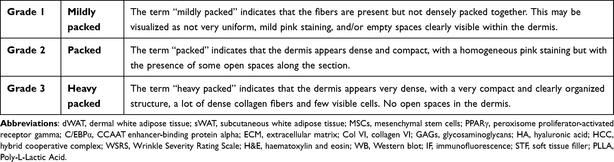 |
Table 1 Dermis Firmness Scoring Internal Scale |
Statistical Analysis
T-test, multiple unpaired t-test and Mann–Whitney test were performed using GraphPad Prism version 10.6.1 for Windows, (GraphPad Software, Boston, Massachusetts USA, www.graphpad.com). Results were expressed as mean ± standard deviation of the mean. All the results were considered to have a statistically significant difference when the p-value was <0.05. Values, such as means and standard deviations, were calculated using Excel.
Results
Evaluation of the Expression of the Transcription Factor PPARγ and of the Mature Adipocyte Markers by WB and IF
To explore the potential modulation of adipocyte differentiation in dWAT between the HA hybrid complex 45mg/mL treated group and the Control group, the expressions of PPAR-γ, Caveolin1, and Perilipin1 were analyzed by WB (Figure 1). In detail, both experimental groups showed similar levels of Caveolin1 (Figure 1B) and Perilipin1 (Figure 1C) expression, but the HA hybrid complex 45mg/mL treated group showed an upregulation of PPAR-γ expression compared to the Control group (Figure 1A).
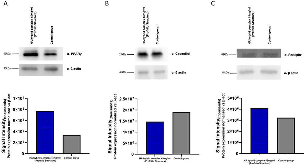 |
Figure 1 Evaluation of expression levels by WB and densiometric analyses of adipogenic differentiation-related proteins and markers of mature adipocytes; PPARγ (A), Caveolin1 (B), Perilipin1 (C). Protein expression is normalized with respect to actin. In detail, the normalization to actin in Figure 1B is the same as in Figure 2B (Data from the same Western blot membrane). |
In addition, the protein expression level of Col VI was monitored. Through WB analysis (Figure 2B) an increase in Col VI expression was observed in the HA hybrid complex 45mg/mL treated group. Col VI, as previously mentioned, is a non-fibrillar component that constitutes the pericellular network that reinforces the adipocyte basement membrane. These results were confirmed by immunofluorescence assays (Figure 2A).
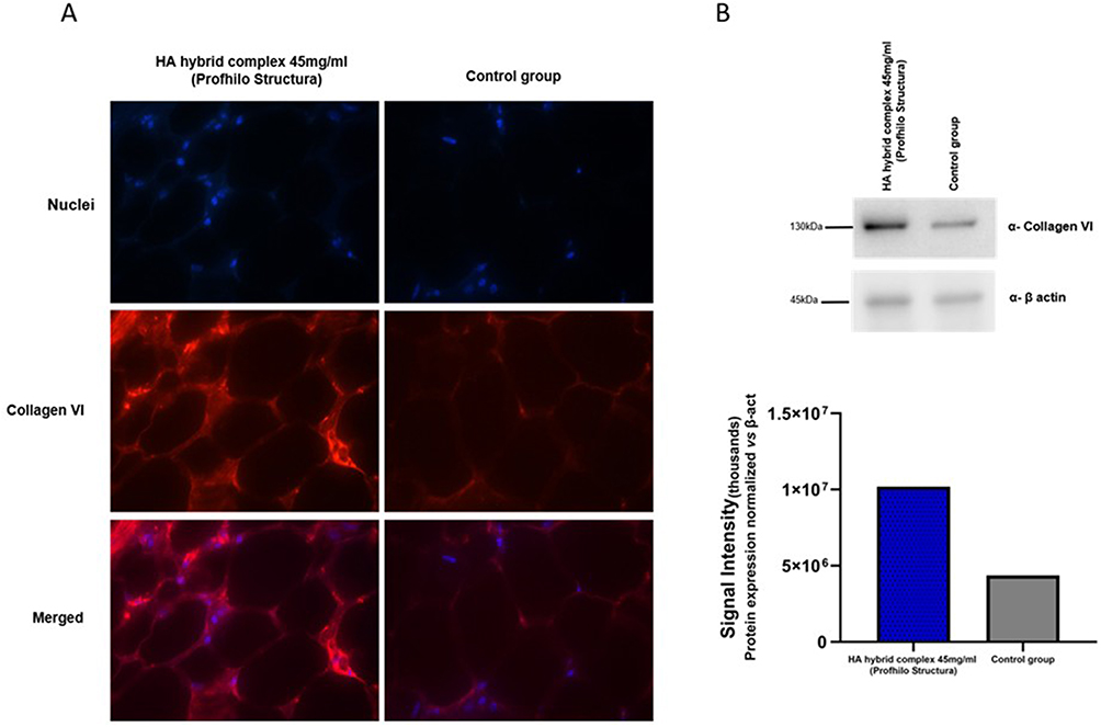 |
Figure 2 Analysis of Col VI in the two experimental groups: HA hybrid complex 45mg/mL (Profhilo Structura) and Control group. Detection of Col VI (red) by immunofluorescence (A). Evaluation of the expression levels of Col VI by WB and densiometric analysis (B). Protein expression levels were normalized respect to actin. In detail, the normalization to actin in Figure 2B is the same as in Figure 1B (Data from the same Western blot membrane). |
These data suggest that HA hybrid complex 45mg/mL treatment supported a mo (fatty acid accumulation), through the increase of PPARγ expression, but at the same time ensured stability of the adipose tissue thanks to the presence of Col VI which has been shown to be particularly important for limiting adipocyte hypertrophy.19
Evaluation of the Effect of HA Hybrid Complex 45mg/mL on the Number and Size of Adipocytes
As mentioned, the maturation state of adipocytes is also defined by the size of cell diameter: an increase of this parameter indicates an enlargement of the cell dimensions due to the accumulation of fat within the adipocyte cell. As reported in the literature,16 in humans, adipocytes have an average size ranging from 40 to 200 µm, while in mice, they are approximately 30% smaller, ranging from 20 to 150 µm. Adipocytes counted by Adiposoft were divided into three main classes, based on the size of their diameter and defined as follows: small (< 55 µm), large (55–100 µm) and very large (>100 µm). The two experimental groups were compared considering: the total number of adipocytes counted, the distribution of adipocytes in the three classes defined above (small, large, very large), the total area of the dWAT and the area per class. Comparative statistical analyses revealed that there was no statistically significant difference in the number of cells between the two experimental groups (Figure 3A), but their distribution varied (Figure 3B). Indeed, treatment with HA hybrid complex 45mg/mL modulated the distribution of adipocytes among the three categories. In particular, the number of very large adipocytes increased compared to the Control group, and a similar trend was also observed for large adipocytes, although it did not reach the statistical significance (Figure 3B).
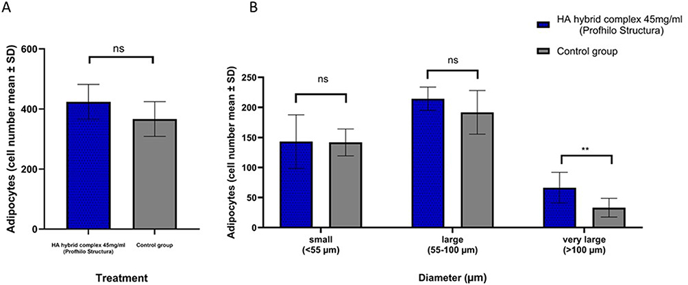 |
Figure 3 Comparison of the number of adipocytes between the HA hybrid complex 45mg/mL (Profhilo Structura)-treated group and the Control group (A). Unpaired t-test (ns: not significant). Comparison of the number of adipocytes in the three classes: small (< 55 um), large (55–100 um) and very large (> 100 um) between the HA hybrid complex 45mg/mL (Profhilo Structura)-treated group and the Control group (B). Cell number is represented as mean ±standard deviation. Multiple Unpaired t-test (ns: not significant; **p< 0.01). |
Consequently, as it can also be seen in the representative images in Figure 4A and B and by analyzing the dWAT area, for this latter parameter, a statistically significant increase was observed in the group treated with HA hybrid complex 45mg/mL compared to the Control group (Figure 4C). Specifically, the area of adipocytes classified as large was greater than in the Control group (Figure 4D).
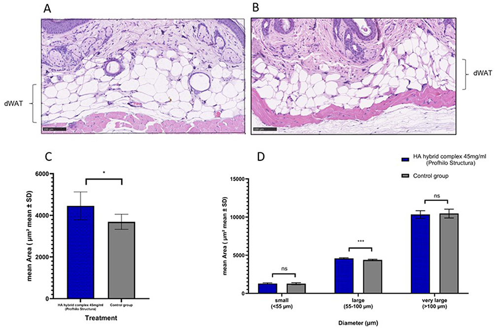 |
Figure 4 Representative images of dWAT from HA hybrid complex 45mg/mL (Profhilo Structura) (A) and Control (B) groups; scale bar= 100 µm. Graphs comparing average adipocyte area between HA hybrid complex 45mg/mL (Profhilo Structura) and control group (C). Unpeaired t-test (*p< 0.05). Graph comparing average adipocyte area in the three classes: small (< 55 um), large (55–100 um) and very large (> 100 um) between the HA hybrid complex 45mg/mL (Profhilo Structura)-treated group and the Control group (D). Multiple Unpaired t-test (ns: not significant; ***p < 0.001). |
Evaluation of the Effect of HA Hybrid Complex 45mg/mL on the Tissue Compactness
The tissue histological analysis, particularly the dermal layer, and the assignment of a score using a three-level internal scale allowed us to determine the tissue compactness in the two experimental groups. HA hybrid complex 45mg/mL further demonstrated its effectiveness with a significant improvement in dermal firmness (Figure 5C). In fact, an improvement by at least 1 grade of the scale was observed in the group treated with HA hybrid complex 45mg/mL, which was assigned an overall score of 2.7 ± 0.5 compared to the control group which achieved a score of 1.6 ± 0.4. As can be observed in the representative images, the dermis of the HA hybrid complex 45mg/mL group (Figure 5B) appears denser than that of the Control group (Figure 5A), with a visible reduction of the number of open spaces in the dermal layer, which is suggestive of higher firmness.
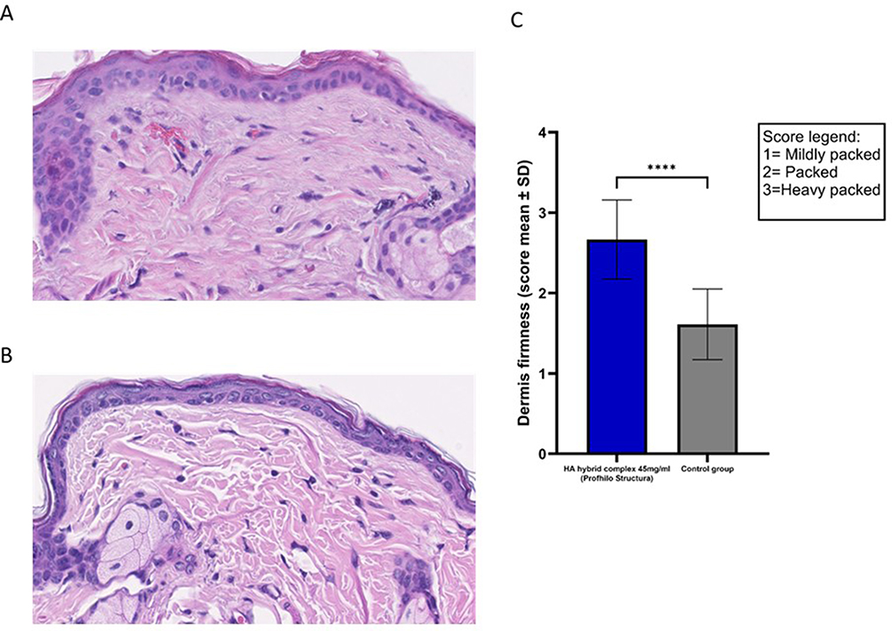 |
Figure 5 Representative images of tissue compactness from HA hybrid complex 45mg/mL (A) and Control groups (B), 60X magnification. Graphs comparing tissue compactness score between HA hybrid complex 45mg/mL and Control groups (C). Mann–Whitney unpaired t-test revealed a statistically significant difference (****p< 0.0001). |
Discussion
In recent years, the scientific and clinical community has increasingly focused on the role of dermal white adipose tissue in health and disease. dWAT, a major component of skin structure, has been recognized as a key factor in regulating skin function and modulating skin aging.9,37 Loss of dWAT volume has been associated with a number of undesirable aesthetic changes, including the formation of wrinkles and furrows, loss of definition of facial contours and consequently a loss of self-confidence.38 Therefore, restoring the volume of dermal adipose tissue has become an important goal in clinical practice, and autologous fat grafting or lipofilling is certainly one of the therapeutic options. Lipofilling is indeed an easily accessible, biocompatible, and economical method that provides both volume enhancement and improved skin quality. However, as with any medical treatment, there are also potential side effects and limitations that must be considered. Since it is a real surgical procedure, there is no shortage of complications ranging from mild ones, such as the appearance of prolonged oedema or erythema, to moderate and severe side effects that require a second corrective operation or even neurosurgical management if an intravascular injection occurs.39 Among the less invasive alternatives, soft tissue filler (STF) injections, such as cross-linked hyaluronic acid-based fillers,1 especially those considered semi-permanent and, in particular, device made up of Poly-L-Lactic Acid (PLLA) have gained importance. In fact, there is several evidence in the literature that shows the volumizing effect and the modulation of the adipogenic process induced by PLLA compounds.40–42
An effect on the hypodermis was also observed in clinical studies involving injections of HA hybrid complex 45mg/mL, the medical device being studied.32,43 Indeed, through self-assessment of the enrolled subjects and ultrasound support, restoration of adipose tissue volume following treatment was demonstrated. These data are particularly important because they are among the first to demonstrate an effect on adipose tissue mediated by a product consisting of cooperative hybrid complexes of high and low molecular weight HA, obtained through NAHYCO technology, a thermally induced process that does not add chemical modifications as occurs in cross-linked products commonly used due to their resistance to degradation and prolonged residence time. Using a non-chemically modified hyaluronic acid product may be safer than both cross-linked HA products and semi-permanent fillers based on calcium hydroxyapatite or PLLA, which have been shown to have adverse effects including edema, moderate swelling, and even more severe lesions such as nodules or foreign body granulomas.44 It is precisely on the basis of the macroscopic observations, id est increase in adipose tissue thickness, from clinical studies that the need arose to perform this in vivo animal study, in order to see the effect of the medical device on adipose tissue “more closely” and better understand its mechanism of action. In this work, we evaluated the maturation process and the morphological changes of the “cellular actors”, such as adipocytes, which constitute the dWAT, following treatment with HA hybrid complex 45mg/mL. Taken together, the data demonstrated the efficacy of the studied medical device in increasing the thickness/area of the dWAT. Specifically, treatment with the HA hybrid complex 45mg/mL showed a modulation in the distribution of cell populations, in terms of size but not number, toward a “more mature” state compared to the Control group. The increased expression of PPARγ observed in the treated-group could suggest the induction of lipogenic effect mediated by medical device. This modulation is likely facilitated by the increased expression of Col VI, which is known to be one of the components of the extracellular matrix distributed pericellularly around mature adipocytes, promoting cell maturation and providing them with greater stability.45,46 These data confirmed the results obtained in previous studies conducted in vitro, in which the treatment with the hybrid compound of high and low molecular weight hyaluronic acid supported the viability of adipocyte e stem cells and activated the adipogenic pathway and therefore cellular differentiation.35 In this in vivo animal study, in addition to analyzing the size (diameter and area) of the adipocyte cells in dWAT, it was possible to observe the appearance of the dermal layer after treatment. Image analysis revealed, in fact, that the treated tissue appeared more compact and that the volume/area occupied by the adipocyte cells in mice treated with the HA hybrid complex 45mg/mL was greater than in the control group. This translates phenotypically as an increase in the thickness of dWAT.
Conclusion
The aforementioned considerations allow us to assert that the present study provides new insight into the potential role of the HA hybrid complex 45mg/mL within adipose tissue and, consequently, its impact on regenerative aesthetic solutions. When administered at the adipocyte level, the HA hybrid complex 45 mg/mL appears to promote a structural and morphological modulation of the adipocyte cells that constitute the white adipose tissue (dWAT), while simultaneously ensuring tissue stability thanks to the presence of collagen VI surrounding the cells. Like any experimental study, this one also has limitations: observing PPARγ expression levels alone can only suggest a treatment-mediated adipogenic effect, and further investigations and studies are needed to adequately demonstrate this, perhaps through functional assays and by expanding the number of key markers regulating adipogenesis. Tissue remodelling and improved structural stability are effects that may indirectly translate into improved tissue compactness and a reduction in both superficial and deep wrinkles. The tissue firmness assessment used in this study also has limitations: as a semi-quantitative analysis using a non-validated rating scale, it provides only a starting point, requiring further investigation. Subsequent analyses will be necessary to confirm the device-mediated effect on tissue firmness in more detail, perhaps using human tissue models.
Disclosure
Immacolata Polvere, Mario Scrima, Nicoletta Vito, Giacomo Signorino, Antonio Iorio, Andrea Maria Giori and Angela Ferravante are employees of IBSA Farmaceutici Italia, IBSA Group. The authors report no other conflicts of interest in this work.
References
1. Nadra K, André M, Marchaud E, et al. A hyaluronic acid-based filler reduces lipolysis in human mature adipocytes and maintains adherence and lipid accumulation of long-term differentiated human preadipocytes. J Cosmet Dermatol. 2021;20(5):1474–12. doi:10.1111/jocd.13794
2. Wollina U, Wetzker R, Abdel-Naser MB, Kruglikov IL. Role of adipose tissue in facial aging. Clin Interv Aging. 2017;12:2069–2076. doi:10.2147/CIA.S151599
3. Boschi F, Negri A, Conti A, Bernardi P, Chirumbolo S, Sbarbati A. The human dermal white adipose tissue (dWAT) morphology: a multimodal imaging approach. Annals Anatomy. 2024;255:152289. doi:10.1016/j.aanat.2024.152289
4. Li Y, Long J, Zhang Z, Yin W. Insights into the unique roles of dermal white adipose tissue (dWAT) in wound healing. Front Physiol. 2024;15:1346612. doi:10.3389/fphys.2024.1346612
5. Wu Z, Wang Z, Chen T, et al. Dermal white adipose tissue: a new modulator in wound healing and regeneration. Regener Ther. 2025;28:115–125. doi:10.1016/j.reth.2024.11.015
6. Alexander CM, Kasza I, Yen CLE, et al. Dermal white adipose tissue: a new component of the thermogenic response. J Lipid Res. 2015;56(11):2061–2069. doi:10.1194/jlr.R062893
7. Juan ZL, Guerrero-Juarez CF, Hata T, et al. Innate immunity. Dermal adipocytes protect against invasive Staphylococcus aureus skin infection. Science. 2015;347(6217):67–71. doi:10.1126/science.1260972
8. Festa E, Fretz J, Berry R, et al. Adipocyte lineage cells contribute to the skin stem cell niche to drive hair cycling. Cell. 2011;146(5):761–771. doi:10.1016/j.cell.2011.07.019
9. Liu M, Lu F, Feng J. Aging and homeostasis of the hypodermis in the age-related deterioration of skin function. Cell Death Dis. 2024;15(6):443. doi:10.1038/s41419-024-06818-z
10. Richard AJ, White U, Elks CM, Stephens JM. Adipose Tissue: physiology to Metabolic Dysfunction In: Feingold KR, Ahmed SF, Anawalt B, et al. editors. Endotext. MDText.com, Inc.; 2000. Available from: http://www.ncbi.nlm.nih.gov/books/NBK555602/.
11. Chon SH, Pappas A. Differentiation and characterization of human facial subcutaneous adipocytes. Adipocyte. 2015;4(1):13–21. doi:10.4161/21623945.2014.955402
12. Tchkonia T, Morbeck DE, von Zglinicki T, et al. Fat tissue, aging, and cellular senescence. Aging Cell. 2010;9(5):667–684. doi:10.1111/j.1474-9726.2010.00608.x
13. Chen SX, Zhang LJ, Gallo RL. Dermal White Adipose Tissue: a Newly Recognized Layer of Skin Innate Defense. J Invest Dermatol. 2019;139(5):1002–1009. doi:10.1016/j.jid.2018.12.031
14. Nicu C, Pople J, Bonsell L, Bhogal R, Ansell DM, Paus R. A guide to studying human dermal adipocytes in situ. Exp Dermatol. 2018;27(6):589–602. doi:10.1111/exd.13549
15. Greenberg AS, Egan JJ, Wek SA, Garty NB, Blanchette-Mackie EJ, Londos C. Perilipin, a major hormonally regulated adipocyte-specific phosphoprotein associated with the periphery of lipid storage droplets. J Biol Chem. 1991;266(17):11341–11346.
16. Hagberg CE, Li Q, Kutschke M, et al. Flow Cytometry of Mouse and Human Adipocytes for the Analysis of Browning and Cellular Heterogeneity. Cell Rep. 2018;24(10):2746–2756.e5. doi:10.1016/j.celrep.2018.08.006
17. Li Q, Spalding KL. The regulation of adipocyte growth in white adipose tissue. Front Cell Dev Biol. 2022;10:1003219. doi:10.3389/fcell.2022.1003219
18. Nakajima I, Muroya S, Tanabe RI, Chikuni K. Positive effect of collagen V and VI on triglyceride accumulation during differentiation in cultures of bovine intramuscular adipocytes. Differentiation. 2002;70(2–3):84–91. doi:10.1046/j.1432-0436.2002.700203.x
19. Kruglikov IL. General Theory of Body Contouring: 2. Modulation of Mechanical Properties of Subcutaneous Fat Tissue. J Cosmet Dermatological Sci Appl. 2014;4(2):117–127. doi:10.4236/jcdsa.2014.42017
20. Liu C, Huang K, Li G, et al. Ascorbic acid promotes 3T3-L1 cells adipogenesis by attenuating ERK signaling to upregulate the collagen VI. Nutr Metab (Lond). 2017;14:79. doi:10.1186/s12986-017-0234-y
21. Shin JW, Kwon SH, Choi JY, et al. Molecular Mechanisms of Dermal Aging and Antiaging Approaches. Int J Mol Sci. 2019;20(9):2126. doi:10.3390/ijms20092126
22. Shin SH, Lee YH, Rho NK, Park KY. Skin aging from mechanisms to interventions: focusing on dermal aging. Front Physiol. 2023;14:1195272. doi:10.3389/fphys.2023.1195272
23. Zorina A, Zorin V, Isaev A, Kudlay D, Vasileva M, Kopnin P. Dermal Fibroblasts as the Main Target for Skin Anti-Age Correction Using a Combination of Regenerative Medicine Methods. Curr Issues Mol Biol. 2023;45(5):3829–3847. doi:10.3390/cimb45050247
24. He T, Fisher GJ, Kim AJ, Quan T. Age-related changes in dermal collagen physical properties in human skin. PLoS One. 2023;18(12):e0292791. doi:10.1371/journal.pone.0292791
25. Tzaphlidou M. The role of collagen and elastin in aged skin: an image processing approach. Micron. 2004;35(3):173–177. doi:10.1016/j.micron.2003.11.003
26. Zouboulis CC, Ganceviciene R, Liakou AI, Theodoridis A, Elewa R, Makrantonaki E. Aesthetic aspects of skin aging, prevention, and local treatment. Clin Dermatol. 2019;37(4):365–372. doi:10.1016/j.clindermatol.2019.04.002
27. Papakonstantinou E, Roth M, Karakiulakis G. Hyaluronic acid: a key molecule in skin aging. Dermatoendocrinol. 2012;4(3):253–258. doi:10.4161/derm.21923
28. Albouy M, Santos MD, Laho K, Couchourel D, Tranchepain F. In Vitro Evaluation of the Effects of Multipoint Hyaluronic Acid-Based Intradermal Fillers on Skin Quality Using a Novel 3D Reconstructed Skin Aging Model. J Cosmet Dermatol. 2025;24(9):e70362. doi:10.1111/jocd.70362
29. Zhou R, Yu M. The Effect of Local Hyaluronic Acid Injection on Skin Aging: a Systematic Review and Meta-Analysis. J Cosmet Dermatol. 2025;24(1):e16760. doi:10.1111/jocd.16760
30. Forte R, Salti G, Tateo A. Profhilo® Structura, Current Status and Future Perspectives: a Practical Review. Plastic Aesthetic Nurs. 2024;44(3):213. doi:10.1097/PSN.0000000000000571
31. Baumann L. Skin ageing and its treatment. J Pathol. 2007;211(2):241–251. doi:10.1002/path.2098
32. Cassuto D, Cigni C, Bellia G, Schiraldi C. Restoring Adipose Tissue Homeostasis in Response to Aging: initial Clinical Experience with Profhilo Structura®. Gels. 2023;9(8):614. doi:10.3390/gels9080614
33. Day DJ, Littler CM, Swift RW, Gottlieb S. The wrinkle severity rating scale: a validation study. Am J Clin Dermatol. 2004;5(1):49–52. doi:10.2165/00128071-200405010-00007
34. De Rosa M, D’agostino A, La Gatta A, Schiraldi C. Hybrid Cooperative Complexes of Hyaluronic Acid. 2012. Available from: https://patentscope.wipo.int/search/en/WO2012032151.
35. American Veterinary Medical Association. Guidelines for the euthanasia of animals. Available from: https://www.avma.org/resources-tools/avma-policies/avma-guidelines-euthanasia-animals.
36. Vassallo V, Di Meo C, Alessio N, et al. Highly Concentrated Stabilized Hybrid Complexes of Hyaluronic Acid: rheological and Biological Assessment of Compatibility with Adipose Tissue and Derived Stromal Cells towards Regenerative Medicine. Int J Mol Sci. 2024;25(4):2019. doi:10.3390/ijms25042019
37. Zaqout S, Becker LL, Kaindl AM. Immunofluorescence Staining of Paraffin Sections Step by Step. Front Neuroanat. 2020;14. doi:10.3389/fnana.2020.582218
38. Kruglikov IL, Scherer PE. Skin aging: are adipocytes the next target? Aging. 2016;8(7):1457–1469. doi:10.18632/aging.100999
39. Gupta MA, Gilchrest BA. Psychosocial aspects of aging skin. Dermatol Clin. 2005;23(4):643–648. doi:10.1016/j.det.2005.05.012
40. Schiraldi L, Sapino G, Meuli J, et al. Facial Fat Grafting (FFG): worth the Risk? A Systematic Review of Complications and Critical Appraisal. J Clin Med. 2022;11(16):4708. doi:10.3390/jcm11164708
41. Kim HW, Jung YA, Yun JM, et al. Effects of Poly-L-Lactic Acid on Adipogenesis and Collagen Gene Expression in Cultured Adipocytes Irradiated with Ultraviolet B Rays. Ann Dermatol. 2023;35(6):424–431. doi:10.5021/ad.22.177
42. Byun KA, Seo SB, Oh S, et al. Poly-D,L-Lactic Acid Fillers Increase Subcutaneous Adipose Tissue Volume by Promoting Adipogenesis in Aged Animal Skin. Int J Mol Sci. 2024;25(23). doi:10.3390/ijms252312739
43. Avelar LE, Nabhani S, Wüst S. Unveiling the Mechanism: injectable Poly-L-Lactic Acid’s Evolving Role—Insights From Recent Studies. J Cosmet Dermatol. 2025;24(1):e16635. doi:10.1111/jocd.16635
44. Sparavigna A, Grimolizzi F, Cigni C, Lualdi R, Bellia G. Efficacy and tolerability of Profhilo® Structura intended to restore lateral cheek fat compartment: an observational pilot study. Health Sci Rep. 2024;7(1):e1743. doi:10.1002/hsr2.1743
45. Guo J, Fang W, Wang F. Injectable fillers: current status, physicochemical properties, function mechanism, and perspectives. RSC Adv. 2023;13(34):23841–23858. doi:10.1039/D3RA04321E
46. Mor-Yossef Moldovan L, Lustig M, Naftaly A, et al. Cell shape alteration during adipogenesis is associated with coordinated matrix cues. J Cell Physiol. 2019;234(4):3850–3863. doi:10.1002/jcp.27157
 © 2026 The Author(s). This work is published and licensed by Dove Medical Press Limited. The
full terms of this license are available at https://www.dovepress.com/terms
and incorporate the Creative Commons Attribution
- Non Commercial (unported, 4.0) License.
By accessing the work you hereby accept the Terms. Non-commercial uses of the work are permitted
without any further permission from Dove Medical Press Limited, provided the work is properly
attributed. For permission for commercial use of this work, please see paragraphs 4.2 and 5 of our Terms.
© 2026 The Author(s). This work is published and licensed by Dove Medical Press Limited. The
full terms of this license are available at https://www.dovepress.com/terms
and incorporate the Creative Commons Attribution
- Non Commercial (unported, 4.0) License.
By accessing the work you hereby accept the Terms. Non-commercial uses of the work are permitted
without any further permission from Dove Medical Press Limited, provided the work is properly
attributed. For permission for commercial use of this work, please see paragraphs 4.2 and 5 of our Terms.
Recommended articles

Efficacy and Tolerability of Cosmetic Serums Enriched with Five Forms of Hyaluronic Acid as Part of Biweekly Diamond Tip Microdermabrasion Treatments for Facial Skin Dryness and Age-Associated Features
Makino ET, Huang PC, Emmerich T, Jiang LI, Mehta RC
Clinical, Cosmetic and Investigational Dermatology 2023, 16:1123-1134
Published Date: 27 April 2023
Applying the MD Codes™ to Treat Emotional and Social Attributes with HA Fillers: A Retrospective Serial Case Study
de Maio M, Brenninkmeijer EEA, Nurlin I, Colucci L, Sanchez T
Clinical, Cosmetic and Investigational Dermatology 2023, 16:3441-3453
Published Date: 29 November 2023
Mitigating Glycation and Oxidative Stress in Aesthetic Medicine: Hyaluronic Acid and Trehalose Synergy for Anti-AGEs Action in Skin Aging Treatment
Chmielewski R, Lesiak A
Clinical, Cosmetic and Investigational Dermatology 2024, 17:2701-2712
Published Date: 28 November 2024