
Back to Journals » International Journal of General Medicine » Volume 17
Receptor-Interacting Protein Kinase 3 as a Serological Biomarker in Relation to Disease Severity and Delirium After Acute Pancreatitis: A Prospective Cohort Study
Authors Ye X, Li B, Xu F, Pan D, Wu J
Received 30 August 2024
Accepted for publication 9 November 2024
Published 15 November 2024 Volume 2024:17 Pages 5309—5323
DOI https://doi.org/10.2147/IJGM.S488540
Checked for plagiarism Yes
Review by Single anonymous peer review
Peer reviewer comments 2
Editor who approved publication: Dr Vinay Kumar
Xiaorong Ye,1 Bingzhen Li,2 Fang Xu,3 Debiao Pan,4 Jing Wu1
1Department of Anorectal Surgery, Lishui Hospital of Wenzhou Medical University, Lishui People’s Hospital, Lishui, Zhejiang Province, People’s Republic of China; 2Department of Gastrointestinal Surgery, Lishui Hospital of Wenzhou Medical University, Lishui People’s Hospital, Lishui, Zhejiang Province, People’s Republic of China; 3Department of Gastroenterology, Lishui Hospital of Wenzhou Medical University, Lishui People’s Hospital, Lishui, Zhejiang Province, People’s Republic of China; 4Department of Hepatobiliary Surgery, Lishui Hospital of Wenzhou Medical University, Lishui People’s Hospital, Lishui, Zhejiang Province, People’s Republic of China
Correspondence: Bingzhen Li, Department of Gastrointestinal Surgery, Lishui Hospital of Wenzhou Medical University, Lishui People’s Hospital, Lishui, Zhejiang Province, People’s Republic of China, Email [email protected]
Objective: Delirium is a common complication of acute pancreatitis. Receptor-interacting protein kinase 3 (RIP3) is an activator of programmed cell necrosis. This study aimed to determine its ability to predict delirium after acute pancreatitis.
Methods: In total, 297 patients with acute pancreatitis were prospectively enrolled in this study. Patients were divided into two subgroups (study and validation groups: 197 and 100 cases, respectively). Serum RIP3 levels were measured in all patients and in 100 healthy controls. Acute Physiology and Chronic Health Evaluation (APACHE) II, Ranson, and sequential organ failure assessment (SOFA) scores were used for the severity assessment. In-hospital delirium was observed as an outcome variable. Multifactorial analyses were performed to discern severity correlations and outcome associations.
Results: Serum RIP3 levels were significantly higher in the patients than in the controls. Serum RIP3 levels had linear relationships under the restricted cubic spline and were independently correlated with APACHE II, Ranson, and SOFA scores. Serum RIP3 levels were linearly correlated with the likelihood of developing in-hospital delirium and exhibited a strong discrimination efficiency under the receiver operating characteristic curve. Serum RIP3 levels, coupled with APACHE II scores, Ranson scores, and SOFA scores, were the four independent predictors of in-hospital delirium. No interactions were revealed regarding its relevance to sex, age, or body mass index in subgroup analysis. These were integrated to form a model graphically represented by a nomogram that showed effective stability, clinical fit, and predictive ability for in-hospital delirium. The model was verified in the validation group.
Conclusion: An incremental trend in serum RIP3 levels was notable after acute pancreatitis. Serum RIP3 levels are independently related to illness severity and occurrence of in-hospital delirium, indicating that serum RIP3 may be a potential biomarker of acute pancreatitis.
Keywords: acute pancreatitis, delirium, receptor-interacting protein kinase 3, severity
Introduction
Acute pancreatitis (AP) is a common, unpredictable, and variable gastrointestinal disease spectrum marked by a local or systemic inflammatory response.1,2 Following AP, dysfunction of the exocrine pancreas and the subsequent inappropriate release and activation of pancreatic enzymes induce an imbalance between protective enzymes and stress signals, thereby triggering an inflammatory response and microcirculatory disturbances.3,4 Acute Physiology and Chronic Health Evaluation (APACHE) II, Ranson, and Sequential Organ Failure Assessment (SOFA) scores are often used to evaluate the severity of AP in clinical practice.5–7 Delirium is an acute episode of blurred consciousness accompanied by inattention, disorganized thinking, incoherent thinking, and perceptual abnormalities. Etiologic factors include infection, acute brain injury disease, or drug intoxication.8,9 Previous studies have shown that delirium is an important cause of long-term cognitive impairment, which increases the risk of poor outcomes and prolongs hospitalization.10 Inflammatory responses, microcirculatory disturbances, and oxidative free-radical responses following acute severe illnesses are prone to damage brain tissues and subsequently cause delirium.11,12 Similarly, a range of pathophysiological processes following AP can provoke delirium.13,14 Early identification of delirium emergence may enable clinicians to appropriately manage sicknesses. Therefore, some biomarkers have been noted in recent decades with respect to their discriminatory capability for the appearance of delirium in a series of severe disorders.15–18
Necroptosis has recently been recognized as a form of cell death.19 Receptor-interacting protein kinase 3 (RIP3), a serine/threonine protein kinase, is identified as one of the most important factors in necroptosis occurrence and progression.20,21 RIP3 is highly expressed in the pancreas of rats with AP and upregulating RIP3 obviously exacerbated AP in in vivo animal models and in vitro cell models.22 Alternatively, there is a dramatically elevated expression of RIP3 after experimental brain injury, such as acute ischemic stroke,23 traumatic brain injury,24 subarachnoid hemorrhage,25 and intracerebral hemorrhage.26 Moreover, compelling evidence has shown that RIP3 may be a detrimental factor in acute brain injury.23–26 Interestingly, RIP3 expression is highly upregulated in hippocampal tissues of rats subjected to AP.27 Notably, blood RIP3 levels were markedly elevated in humans with acute ischemic stroke28 and intracerebral hemorrhage.29 Overall, serum RIP3 level may be a potential biomarker for reflecting local and systemic injuries. In this study, serum RIP3 levels were measured to ascertain their correlation with AP severity and predictive ability for delirium after AP.
Materials and Methods
Study Design and Participant Selection
This study consisted of two parts. One was a cross-sectional study in which patients with AP and controls were recruited to investigate the changes in serum RIP3 levels following AP. The other was a prospective cohort study of patients with AP, with the objective of determining the prognostic role of serum RIP3 in AP, in which all patients were divided into two subgroups (study and validation groups) in accordance with a ratio of 2:1. This study included a consecutively enrolled group of patients with first-episode AP who were admitted to the Lishui People’s Hospital between January 2020 and May 2023. The exclusion criteria were as follows: (1) age <18 years; (2) presence of previous severe systemic diseases or other specific conditions, such as uremia, cardiac failure, cirrhosis, pregnancy, infection in the last month, and recent surgery; and (3) history of neuropsychiatric diseases, such as stroke, moderate-to-severe traumatic brain injury, depression, Parkinson’s disease, Alzheimer’s disease, multiple sclerosis, and craniocerebral neoplasms. This study also included healthy controls who were consecutively recruited at the Physical Examination Center of the Lishui People’s Hospital during the same period. This study was conducted in accordance with the principles set forth in the Declaration of Helsinki, and the research protocol was approved by the Ethics Committee of Lishui People’s Hospital (Opinion Number, LLW-FO-403). Controls and legal representatives of patients provided written informed consent for their willingness to participate in this study.
Data Acquisitions
Patients or relatives were asked to complete a comprehensive and detailed collection of data including age, sex, and body mass index. AP Etiologies were of four types: biliary, alcoholic, hypertriglyceremic, and others. Additionally, two time- parameters were registered: the interval from pain ictus to admission and the duration from pain onset to blood collection. The severity of the disease was assessed using APACHE II scores, Ranson scores, and SOFA scores. Using the Confusion Assessment Method, the patients were assessed twice daily until discharge to identify delirium.30 Key diagnostic features of Confusion Assessment Method include acute onset and fluctuating course of symptoms, inattention, impaired level of consciousness, and cognitive deficits (eg, disorientation, memory deficits, or language changes). Other features that support a diagnosis of delirium include altered sleep-wake cycles, perceptual deficits, delusions, emotional instability, and inappropriate or unsafe behaviors.31,32
Immune Analysis
Peripheral venous blood samples were collected at the time of admission from patients with AP and during routine physical examinations from healthy controls. Conventional laboratory markers such as C-reactive protein levels, blood leukocyte counts, and blood glucose levels were measured using routine methods. The extraction location of RIP3 was from cytoplasm. For quantification of serum RIP3 levels, blood samples were deposited in 5 mL gel-containing biochemistry tubes (Ningbo Siny Medical Technology Co., Ltd., China). After centrifugation at 3000×g for 15 min, the serum samples were transferred to Eppendorf Tubes (Eppendorf Tubes® BioBased, China) and stored at −80°C until further testing. Serum RIP3 levels were measured using an enzyme-linked immunosorbent assay (ELISA) kit (Catalogue number: CSB-EL019737HU; Cusabio Biotech Co., Ltd., China). The detection range was 0.156–10 ng/mL and the detection limit was 0.039 ng/mL, with both the intra-assay coefficient of variance and inter-assay coefficient of variance below 10%. To ensure accuracy and reliability, the procedure was repeated twice for all samples. The technician who conducted the tests was blinded to the patients’ clinical information to ensure unbiased testing and analysis.
Statistical Analysis
Graph plottings and statistical analyses were made by applying SPSS 25.0 (IBM Corp., Armonk, NY, USA), GraphPad Prism 9.0 (GraphPad Software Inc., La Jolla, CA, USA), R 3.5.1 (https://www.r-project.org) and MedCalc 17.4 (MedCalc Software, Mariakerke, Belgium). As deemed suitable, the Kolmogorov–Smirnov test or the Shapiro–Wilk test was in use for assessing the normality of quantitative data. Normally distributed data were expressed as mean ± standard deviation, while non-normally distributed data, as median (25th-75th). Qualitative data were presented in form of counts (proportions). For between-group comparisons, the χ2 test or Fisher’s exact test was used for qualitative data, and the Mann–Whitney U-test or t-test was used for quantitative data. Bivariate correlations were analyzed using the Spearman correlation test. Multiple linear regression model was developed to identify variables independently associated with serum RIP3 levels. To compare the differences between delirium and non-delirium patients, a univariate logistic regression model was established to analyze the relationship between each variable and delirium in AP patients. Binary Logistic regression models were used to identify variables independently associated with delirium after AP. Odds ratio (OR) and corresponding 95% confidence intervals (CI) were calculated to show associations. Subsequently, receiver operating characteristic (ROC) curves were constructed to explore the predictive value of serum RIP3 levels for the development of delirium in patients with AP. The area under the curve (AUC) was estimated, and the Z-test was used to compare the AUC. Also, linear correlations between serum RIP3 levels and other severity indicators, as well as between serum RIP3 levels and delirium risk, were discerned using restricted cubic spline. And, subgroup analysis was made for analyzing whether association of serum RIP3 levels with delirium development possibility was affected by other conventional variables, such as age, gender and body mass index. A nomogram model was developed to predict the risk of delirium. The calibration curve was drawn to verify model stability. The decision curve was plotted to clinical fit of the model. Alternatively, the validation group was employed to confirm prognostic significance of serum RIP3 levels using a series of statistical metrics. Two-sided P-value of <0.05 was considered statistically significant.
Results
Characteristics of the Study Population
Consecutive enrollment of 363 patients with AP was initially completed. Subsequently, 66 patients were excluded from this study on account of the predetermined criteria, and a cumulative total of 297 patients underwent a final analysis, of which 197 were assigned to the study group and 100 to the validation group (Figure 1). Additionally, 100 healthy volunteers were recruited as controls. The differences in age, sex, and body mass index were not statistically significant between the patients and controls (Supplemental Table 1, all P>0.05). The basic features of all the patients, the study group, and the validation group are shown in Supplemental Table 2. The baseline characteristics were not significantly different between the study and validation groups (all P>0.05; Supplemental Table 2).
 |
Figure 1 Flowing-chart for selection of eligible patients with acute pancreatitis. Notes: A total of 363 patients with acute pancreatitis underwent an initial enrollment, sixty-six patients were removed from this study pursuant to the prespecified exclusion criteria, and 297 patients were retained for the final clinical assessment, of which 197 constituted the study group and 100 were assigned to the validation group. |
Correlation Analysis Between Serum RIP3 Levels and Disease Severity
There were no notable differences in terms of serum RIP3 levels among the total patients, those in the study group and in the validation group (all P>0.05; Figure 2); and serum RIP3 levels were significantly higher either in AP patients, those of the study group or the validation group than in controls (all P<0.001; Figure 2). Patients in the study group were divided into three groups in accordance with revised Atlanta classification (RAC), namely, mild pancreatitis (n=102), moderately severe pancreatitis (n=62), and severe pancreatitis (n=33). Using Kruskal–Wallis H-test, patients with severe AP had substantially highest serum RIP3 levels, followed by moderately severe AP and then mild AP (P < 0.001). In the restricted cubic spline analysis framework, serum RIP3 levels were linearly correlated with the APACHE II scores (P for nonlinear > 0.05; Supplemental Figure 1), Ranson scores (P for nonlinear > 0.05; Supplemental Figure 2), and SOFA scores (P for nonlinear > 0.05; Supplemental Figure 3). Spearman’s test showed that serum RIP3 levels were strongly and positively correlated with APACHE II scores (P< 0.001; Supplemental Figure 4), Ranson scores (P< 0.001; Supplemental Figure 5), and SOFA scores (P< 0.001; Supplemental Figure 6). As shown in Supplemental Table 3, in addition to the APACHE II, Ranson, and SOFA scores, other variables, such as blood leukocyte count, serum C-reactive protein levels, blood procalcitonin levels, blood calcium levels, blood glucose levels, and blood creatinine levels, were significantly correlated with serum RIP3 levels (all P<0.05). Similar results were obtained using the univariate linear regression analysis (all P<0.05; Supplemental Table 3). Using a multivariate linear correlation model incorporating the above significantly correlated variables, APACHE II, Ranson and SOFA scores were independently related to serum RIP3 levels (all P<0.05; Table 1).
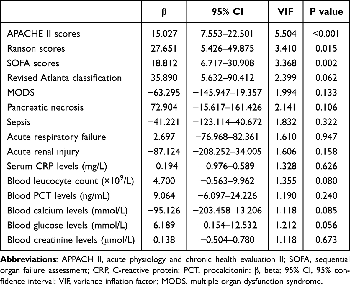 |
Table 1 Correlative Analysis of Serum Receptor-Interacting Protein Kinase 3 Levels Using Multivariate Linear Regression Analysis in Acute Pancreatitis |
 |
Figure 2 Boxplot illustrating serum receptor-interacting protein kinase 3 levels between patients with acute pancreatitis and controls. Abbreviations: RIP3, receptor-interacting protein kinase 3; ns, non-significant. Notes: No significant distinctions were found in terms of serum receptor-interacting protein kinase 3 levels among the three groups, namely, the whole group, study group and validation group (all P>0.05). The levels were substantially lower in the controls than in any of the three groups (all P<0.001). ***P<0.001. |
Relationship Between Serum RIP3 Levels and Delirium After AP
There were a collective of 57 (28.9%) patients with presentation of in-hospital delirium. Patients with severe AP had substantially highest incidence of delirium (45.5%) followed by moderately severe AP (33.9%) and then mild AP (20.6%) (P<0.001). Serum RIP3 levels were significantly higher in patients with delirium than in those without (P<0.001; Supplemental Figure 7). Serum RIP3 levels displayed a marked discrimination efficiency for the probability of delirium occurrence after AP, and an appropriate value was chosen using the Youden method (Figure 3). Under a restricted cubic spline, serum RIP3 levels were linearly related to the possibility of delirium development during hospitalization for AP (P nonlinear >0.05; Figure 4). Table 2 shows that variables such as the APACHE II scores, Ranson scores, SOFA scores, blood leukocyte count, serum C-reactive protein level, blood procalcitonin level, blood calcium level, blood glucose level, blood creatinine level, revised Atlanta classification, pancreatic necrosis, septicaemia, multiple organ dysfunction syndrome, acute respiratory failure, acute kidney injury and serum RIP3 levels were significantly higher in patients with delirium than in the remaining patients (all P<0.05). Consistent results were mirrored by the univariate logistic regression analysis (P<0.05; Table 2). The variables that were significant in the univariate analysis were integrated into the binary logistic regression model, and it was demonstrated that APACHE II scores, Ranson scores, SOFA scores and serum RIP3 levels independently predicted delirium episodes (P<0.005; Table 3). Subgroup analysis showed that the association between serum RIP3 levels and delirium occurrence was not influenced by age, sex, or body mass index (all P > 0.05; Figure 5).
 |
Table 2 Factors Associated with Delirium After Acute Pancreatitis |
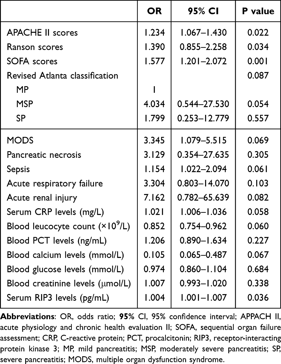 |
Table 3 Multivariate Logistic Regression Analysis of Predictors of Delirium After Acute Pancreatitis |
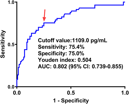 |
Figure 3 Receiver operating characteristic curve showing predictive value of serum receptor-interacting protein kinase 3 levels for in-hospital delirium after acute pancreatitis. In-hospital delirium after acute pancreatitis was efficiently predicted using serum receptor-interacting protein kinase 3 levels, and an appropriate criterion was identified using the Youden method. Red arrow means cutoff value of serum receptor-interacting protein kinase 3 levels. Abbreviation: AUC, area under curve. |
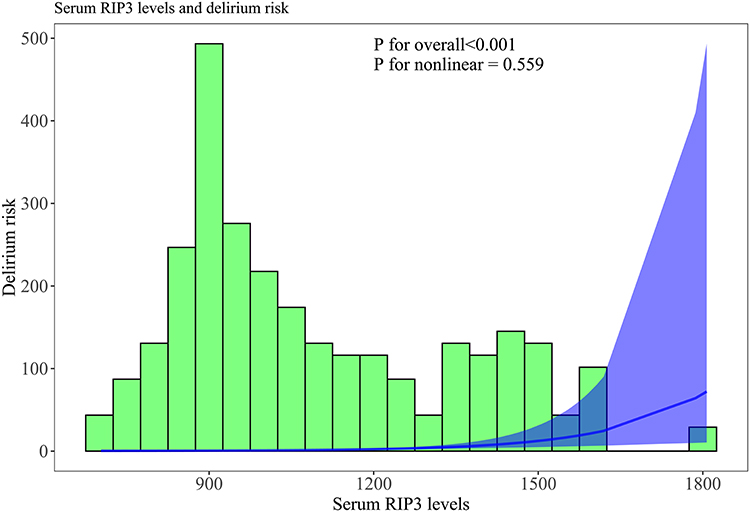 |
Figure 4 Restricted cubic spline depicting linear correlation of serum receptor-interacting protein kinase 3 levels with likelihood of development of in-hospital delirium after acute pancreatitis. Abbreviation: RIP3, receptor-interacting protein kinase 3. Notes: There was a linear correlation between serum receptor-interacting protein kinase 3 levels and the probability of in-hospital delirium following acute pancreatitis (P for nonlinear >0.05). |
 |
Figure 5 Subgroup analysis demonstrating interaction between serum receptor-interacting protein kinase 3 levels and other conventional variables in association with in-hospital delirium following acute pancreatitis. Abbreviations: OR, odds ratio; 95% CI, 95% confidence interval. Notes: The relationship between serum receptor-interacting protein kinase 3 levels and in-hospital delirium following acute pancreatitis was not affected by age, sex, or body mass index (all P > 0.05). |
To determine the additive effect of serum RIP3 levels on common severity metrics, that is, APACHE II, Ranson, and SOFA scores, these four independent parameters of delirium appearance were merged to form a prediction model. A graphical representation of the model was generated using a nomogram (Figure 6). Additionally, the calibration curve confirmed that the model was clinically valid under the decision curve (Figure 7) and comparatively stable (Figure 8). Moreover, the clinical model only contained the APACHE II, Ranson, and SOFA scores, and clinical and biochemical models encompassed all four variables. As shown in Figure 9, serum RIP3 levels showed similar predictive ability as the three severity indicators (all P>0.05); however, the clinical and biochemical models displayed significantly higher predictive capability than any one of the indices (all P<0.05).
 |
Figure 6 Nomogram for visualizing prediction status of the combination model for in-hospital delirium in patients with acute pancreatitis. Abbreviations: RIP3, receptor-interacting protein kinase 3; APACHE II, Acute Physiology and Chronic Health Evaluation II; SOFA, Sequential Organ Failure Assessment. Notes: The nomogram graphically represented the model integrating acute physiology and chronic health evaluation II scores, Ranson scores, sequential organ failure assessment scores, and serum receptor-interacting protein kinase 3 levels. |
 |
Figure 7 Calibration curve showing stability of the model for predicting in-hospital delirium following acute pancreatitis. Notes: The model, in which acute physiology and chronic health evaluation II scores, Ranson scores, sequential organ failure assessment scores, and serum receptor-interacting protein kinase 3 levels were integrated, was comparatively stable for the prediction of in-hospital delirium after acute pancreatitis. |
 |
Figure 8 Decision curve displaying clinical fit of the model for predicting in-hospital delirium following acute pancreatitis. Abbreviations: RIP3, receptor-interacting protein kinase 3; APACHE II, Acute Physiology and Chronic Health Evaluation II; SOFA, Sequential Organ Failure Assessment. Notes: The clinical model included the acute physiology and chronic health evaluation II, Ranson, and sequential organ failure assessment scores. Clinical and biochemical models encompassed the preceding scaling systems and serum receptor-interacting protein kinase 3 levels. Clinical and biochemical models demonstrated efficacious clinical value in predicting in-hospital delirium following acute pancreatitis. |
 |
Figure 9 Receiver operating characteristic curves delineating prediction ability of the model for in-hospital delirium following acute pancreatitis. Abbreviations: RIP3, receptor-interacting protein kinase 3; APACHE II, Acute Physiology and Chronic Health Evaluation II; SOFA, sequential organ failure assessment; ns, non-significant. *P<0.05. Notes: The clinical model included the acute physiology and chronic health evaluation II, Ranson, and sequential organ failure assessment scores. Clinical and biochemical models encompassed the preceding scaling systems and serum receptor-interacting protein kinase 3 levels. When serum receptor-interacting protein kinase 3 levels were compared with those of the other three variables, the predictive ability was not significantly different (all P>0.05). The clinical and biochemical models displayed an effective clinical capability in predicting in-hospital delirium following acute pancreatitis (all P<0.05). |
In the validation group, the four independent predictors of delirium occurrence were repeatedly merged to form a prediction model. The nomogram in this group resembled that in the study group (Supplemental Figure 8). The model exhibited strong stability (Supplemental Figure 9) and high clinical effectiveness (Supplemental Figure 10). In addition, the predictive abilities of the serum RIP3 levels were not significantly different between the study and validation groups (P>0.05; Figure 10), and those of the prediction models were similar between the two groups (P>0.05; Figure 11).
 |
Figure 10 Receiver operating characteristic curve for comparing prediction ability of serum receptor-interacting protein kinase 3 levels between study group and validation group for in-hospital delirium following acute pancreatitis. Abbreviations: RIP3, receptor-interacting protein kinase 3; ns, non-significant. Notes: Serum receptor-interacting protein kinase 3 levels had similar predictive abilities for in-hospital delirium following AP between the study and validation groups (P>0.05). |
 |
Figure 11 Receiver operating characteristic curve for comparison of prediction ability of the models between study group and validation group for in-hospital delirium following acute pancreatitis. Abbreviation: ns, non-significant. Notes: The model included the acute physiology and chronic health evaluation II scores, Ranson scores, sequential organ failure assessment scores, and serum receptor-interacting protein kinase 3 levels. As for predicting in-hospital delirium following acute pancreatitis, the discrimination efficiency of the model was similar between the study and validation groups (P>0.05). |
Discussion
AP is a severe systemic inflammatory disease often associated with organ dysfunction. Early stages of AP can lead to systemic inflammatory response syndrome due to a range of inflammatory factors, while hemodynamic instability often leads to organ failure beyond the pancreas. In previous studies, APACHE II scores, Ranson scores, and SOFA scores have been used to assess and predict the occurrence of organ failure, local pancreatic complications, and systemic complications after AP.6,33,34 Previous studies have found that the APACHE II, Ranson, and SOFA scores are strongly correlated with disease severity in patients with AP and are effective in predicting the occurrence and prognosis of delirium in patients with AP.35,36 In recent decades, biomarkers have gradually attracted the attention of scholars because of their relative objectivity and have been used to predict the occurrence of delirium after AP.
To the best of our knowledge, there is a paucity of data regarding serum RIP3 levels after AP. The main findings of this study were as follows: (1) serum RIP3 levels were markedly higher in patients than in controls; (2) serum RIP3 levels were independently correlated with APACHE II, Ranson, and SOFA scores; (3) serum RIP3 levels were independently associated with the likelihood of in-hospital delirium development; (4) serum RIP3 levels had effective discriminatory ability for patients at risk of in-hospital delirium; and (5) the model with integration of serum RIP3 levels along with the other three severity metrics performed well in both the study and validation groups. Hypothetically, serum RIP3 level, which is closely correlated with AP severity, may be a potential biomarker for predicting the occurrence of in-hospital delirium after AP.
RIP3 is a pivotal protein involved in necroptosis and is highly expressed in the pancreas.22 RIP3 expression is significantly elevated in the blood and pancreatic tissues of rats.37 Accumulating experimental data has demonstrated that RIP3 may be detrimental. Specifically, upregulation of mouse RIP3 expression increased necrosis of alveolar cells in mice with SAP.38 RIP3 knockout significantly reduced pancreatic cell necrosis and facilitated recovery from AP in mice.38 Moreover, RIP3 inhibitors significantly reduce pyroptotic vesicular cell death, pancreatic necrosis, and systemic inflammation.39 In the present study, the serum RIP3 levels were markedly elevated after AP. It was deduced that a portion of RIP3 in peripheral blood may have been derived from pancreatic tissues. RIP3 expression positively correlated with the degree of pancreatic alveolar cell necrosis in an animal model of AP.39 In this study, we found that elevated serum RIP3 levels were significantly correlated with traditional severity scores of AP, including APACHE II, Ranson, and SOFA scores. In conclusion, serum RIP3 levels may reflect AP severity.
Brain injury is a major cause of delirium after severe illness.35 An experimental study has shown that brain tissue is injured in rats with AP, thereby leading to cognitive impairment.27 Interestingly, high RIP3 expression was found in the brain tissues of rats with AP.27 Serum RIP3 levels are strongly correlated with the severity and prognosis of acute brain injury, including acute ischemic stroke28 and intracerebral hemorrhage.29 These data indicate that serum RIP3 may be a biomarker of acute brain injury. Consistently, serum RIP3 levels were independently associated with the emergence of in-hospital delirium after AP, independent of other conventional severity metrics, that is, APACHE II, Ranson, and SOFA scores. Subsequent ROC curve analysis demonstrated that serum RIP3 levels had similar predictive ability for the onset of delirium, as compared to those of the APACHE II scores, Ranson scores and SOFA scores. Overall, the preceding data strongly support the notion that serum RIP3 level may be an encouraging predictor of in-hospital delirium after AP.
To verify the predictive value of serum RIP3 as a biomarker of in-hospital delirium following AP, an integrated model that included serum RIP3 and other independent predictors of in-hospital delirium, namely the APACHE II, Ranson, and SOFA scores, was developed. Its reliability, clinical fit, and discrimination efficiency were validated in the study validation group using a series of statistical methods such as calibration, decision, and ROC curves. Overall, the above data may offer sufficient evidence to conclude that serum RIP3 may have an efficacious ability to discriminate in-hospital delirium following AP.
This study had several advantages and limitations. The advantages are that (1) to the best of our knowledge, serum RIP3 may be determined in AP patients for the first time, and a notable finding is that serum RIP3 may be a potential biomarker for predicting in-hospital delirium in AP patients; (2) all correlations or associations were confirmed using multivariate analysis in this study; and (3) before all tests were performed, linear correlations were validated using a restricted cubic spline; thus, the results were more robust. Several limitations warrant to be considered. First, the serum RIP3 levels were measured in a medium sample size; therefore, the conclusions warrant validation in a larger cohort study. Second, serum RIP3 levels were measured only upon admission in AP patients. It may be of benefit if serum RIP3 levels could be assessed at multiple time-points post-admission This would facilitate the observation of the dynamic changes in serum RIP3 levels throughout the course of AP. Third, our study found that serum RIP3 levels were closely associated with the development of delirium in AP patients. In follow-up studies, we will further explore the mechanistic studies of RIP3 in relation to delirium onset to help to explore the clinical application of RIP3 as a biomarker and its therapeutic potential. Finally, the complications such as pancreatic necrosis, septicaemia, multiple organ dysfunction syndrome, acute respiratory failure and acute kidney injury were not the independent risk factors for delirium after acute pancreatitis in this study, and however, in consideration of a medium sample-size in the current study, this finding needs to be verified by subsequent larger cohort studies.
Conclusion
Patients with AP showed a marked elevation in serum RIP3 levels relative to controls. Moreover, serum RIP3 levels were independently correlated with the APCHCE II, Ranson, and SOFA scores and were independently predictive of in-hospital delirium following AP. Notably, serum RIP3 can take possession of effectively discriminating in-hospital delirium after AP, whether in the study group or validation group, using numerous statistical metrics, such as the ROC curve, calibration curve, and decision curve. Taken together, serum RIP3 may serve as a promising biochemical marker for the evaluation of AP severity and the prediction of delirium during hospitalization after AP.
Abbreviations
AP, acute pancreatitis; RIP3, receptor-interacting protein kinase 3; ROC, receiver operating characteristic; APACHE II, Acute Physiology and Chronic Health Evaluation II; SOFA, Sequential Organ Failure Assessment; AUC, area under the curve.
Data Sharing Statement
Data supporting the findings of this study are available upon request from the corresponding author. The data were not publicly available due to privacy or ethical restrictions.
Author Contributions
All authors made a significant contribution to the work reported, whether that is in the conception, study design, execution, acquisition of data, analysis and interpretation, or in all these areas; took part in drafting, revising or critically reviewing the article; gave final approval of the version to be published; have agreed on the journal to which the article has been submitted; and agree to be accountable for all aspects of the work.
Funding
This study was financially supported by Zhejiang Province Medicine and Health Science and Technology Program (2024KY1873).
Disclosure
The authors declare that they have no competing interests.
References
1. Mederos MA, Reber HA, Girgis MD. Acute pancreatitis: a review. JAMA. 2021;325(4):382–390. doi:10.1001/jama.2020.20317
2. Garg PK, Singh VP. Organ failure due to systemic injury in acute pancreatitis. Gastroenterology. 2019;156(7):2008–2023. doi:10.1053/j.gastro.2018.12.041
3. Zhao Q, Wei Y, Pandol SJ, Li L, Habtezion A. STING signaling promotes inflammation in experimental acute pancreatitis. Gastroenterology. 2018;154(6):1822–1835.e2. doi:10.1053/j.gastro.2018.01.065
4. Wiley MB, Mehrotra K, Bauer J, Yazici C, Bialkowska AB, Jung B. Acute pancreatitis: current clinical approaches, molecular pathophysiology, and potential therapeutics. Pancreas. 2023;52(6):e335–e343. doi:10.1097/MPA.0000000000002259
5. Chauhan R, Saxena N, Kapur N, Kardam D. Comparison of modified Glasgow-Imrie, Ranson, and Apache II scoring systems in predicting the severity of acute pancreatitis. Pol Przegl Chir. 2022;95(1):6–12. doi:10.5604/01.3001.0015.8384
6. Wu S, Zhou Q, Cai Y, Duan X. Development and validation of a prediction model for the early occurrence of acute kidney injury in patients with acute pancreatitis. Ren Fail. 2023;45(1):2194436. doi:10.1080/0886022X.2023.2194436
7. Cazacu SM, Parscoveanu M, Cartu D, et al. NLR48 is better than CRP, and mCTSI, and similar to BISAP and SOFA scores for mortality prediction in acute pancreatitis: a comparison of 6 scores. J Inflamm Res. 2023;16:4793–4804. doi:10.2147/JIR.S432408
8. Maldonado JR. Acute brain failure: pathophysiology, diagnosis, management, and sequelae of delirium. Crit Care Clin. 2017;33(3):461–519. doi:10.1016/j.ccc.2017.03.013
9. Hshieh TT, Inouye SK, Oh ES. Delirium in the elderly. Psychiatr Clin North Am. 2018;41(1):1–17. doi:10.1016/j.psc.2017.10.001
10. Ornago AM, Pinardi E, Okoye C, et al. Delirium prevalence and delirium literacy across Italian hospital wards: a secondary analysis of data from the World Delirium Awareness Day 2023. Eur Geriatr Med. 2024. doi:10.1007/s41999-024-01019-5
11. Vasunilashorn SM, Dillon ST, Inouye SK, et al. High C-reactive protein predicts delirium incidence, duration, and feature severity after major noncardiac surgery. J Am Geriatr Soc. 2017;65(8):e109–e116. doi:10.1111/jgs.14913
12. Neerland BE, Hall RJ, Seljeflot I, et al. Associations between delirium and preoperative cerebrospinal fluid C-reactive protein, interleukin-6, and interleukin-6 receptor in individuals with acute hip fracture. J Am Geriatr Soc. 2016;64(7):1456–1463. doi:10.1111/jgs.14238
13. Dong Z, Song J, Ge M, et al. Effectiveness of a multidisciplinary comprehensive intervention model based on the hospital elderly life program to prevent delirium in patients with severe acute pancreatitis. Ann Palliat Med. 2020;9(4):2221–2228. doi:10.21037/apm-20-913
14. Potolidis E, Mandros C, Karakitsos D, Kountra PM. Quetiapine-associated pancreatitis in a geriatric critical care patient with delirium. Case Rep Psychiatry. 2012;2012:625954. doi:10.1155/2012/625954
15. Pang H, Kumar S, Ely EW, Gezalian MM, Lahiri S. Acute kidney injury-associated delirium: a review of clinical and pathophysiological mechanisms. Crit Care. 2022;26(1):258. doi:10.1186/s13054-022-04131-9
16. Wittwer ED, Cerhan JH, Schroeder DR, Schaff HV, Mauermann WJ. Impact of ketamine versus propofol for anesthetic induction on cognitive dysfunction, delirium, and acute kidney injury following cardiac surgery in elderly, high-risk patients. Ann Card Anaesth. 2023;26(3):274–280. doi:10.4103/aca.aca_106_22
17. Larsen LK, Møller K, Petersen M, Egerod I. Delirium prevalence and prevention in patients with acute brain injury: a prospective before-and-after intervention study. Intensive Crit Care Nurs. 2020;59:102816. doi:10.1016/j.iccn.2020.102816
18. Hager DN, Dinglas VD, Subhas S, et al. Reducing deep sedation and delirium in acute lung injury patients: a quality improvement project. Crit Care Med. 2013;41(6):1435–1442. doi:10.1097/CCM.0b013e31827ca949
19. Wen P, Zhao Y, Yang M, et al. Identification of necroptosis-related genes in ankylosing spondylitis by bioinformatics and experimental validation. J Cell Mol Med. 2024;28(14):e18557. doi:10.1111/jcmm.18557
20. Sun L, Wang H, Wang Z, et al. Mixed lineage kinase domain-like protein mediates necrosis signaling downstream of RIP3 kinase. Cell. 2012;148(1–2):213–227. doi:10.1016/j.cell.2011.11.031
21. Cho YS, Challa S, Moquin D, et al. Phosphorylation-driven assembly of the RIP1-RIP3 complex regulates programmed necrosis and virus-induced inflammation. Cell. 2009;137(6):1112–1123. doi:10.1016/j.cell.2009.05.037
22. Wang Y, Song M, Zhou P, Wang J, Zheng J, Xu H. TNFAIP3-upregulated RIP3 exacerbates acute pancreatitis via activating NLRP3 inflammasome. Int Immunopharmacol. 2021;100:108067. doi:10.1016/j.intimp.2021.108067
23. Wang Q, Yang F, Duo K, et al. The role of necroptosis in cerebral ischemic stroke. Mol Neurobiol. 2024;61(7):3882–3898. doi:10.1007/s12035-023-03728-7
24. Wang L, Zhang Y, Huang M, Yuan Y, Liu X. RIP3 in necroptosis: underlying contributions to traumatic brain injury. Neurochem Res. 2024;49(2):245–257. doi:10.1007/s11064-023-04038-z
25. Chen T, Pan H, Li J, et al. Inhibiting of RIPK3 attenuates early brain injury following subarachnoid hemorrhage: possibly through alleviating necroptosis. Biomed Pharmacother. 2018;107:563–570. doi:10.1016/j.biopha.2018.08.056
26. Huang S, Hu W, Rao D, et al. RIPK3-dependent necroptosis activates MCP-1-mediated inflammation in mice after intracerebral hemorrhage. J Stroke Cerebrovasc Dis. 2022;31(1):106213. doi:10.1016/j.jstrokecerebrovasdis.2021.106213
27. Ou X, Hua Y, Liao X, Gong C, Kang Y. Cognitive impairments induced by severe acute pancreatitis are attenuated by berberine treatment in rats. Mol Med Rep. 2018;18(3):3437–3444. doi:10.3892/mmr.2018.9313
28. Chen B, Hang J, Zhao Y, et al. Correlation between plasma levels of RIP3 and acute ischemic stroke with large-artery atherosclerosis. Curr Neurovasc Res. 2022;19(1):30–37. doi:10.2174/1567202619666220214105208
29. Li B, Wu D, Song W, Jiang W, Li W. A prospective cohort study regarding usability of serum RIPK3 as a biomarker in relation to early hematoma growth and neurological outcomes after acute intracerebral hemorrhage. Clin Chim Acta. 2024;562:119850. doi:10.1016/j.cca.2024.119850
30. Zhou C, Wang H, Wang L, Zhou Y, Wu Q. Diagnostic accuracy of the family confusion assessment method for delirium detection: a systematic review and meta-analysis. J Am Geriatr Soc. 2024;72(3):892–902. doi:10.1111/jgs.18692
31. Ely EW, Inouye SK, Bernard GR, et al. Delirium in mechanically ventilated patients: validity and reliability of the confusion assessment method for the intensive care unit (CAM-ICU). JAMA. 2001;286(21):2703–2710. doi:10.1001/jama.286.21.2703
32. Ely EW, Margolin R, Francis J, et al. Evaluation of delirium in critically ill patients: validation of the Confusion Assessment Method for the Intensive Care Unit (CAM-ICU). Crit Care Med. 2001;29(7):1370–1379. doi:10.1097/00003246-200107000-00012
33. Thandassery RB, Yadav TD, Dutta U, Appasani S, Singh K, Kochhar R. Hypotension in the first week of acute pancreatitis and APACHE II score predict development of infected pancreatic necrosis. Dig Dis Sci. 2015;60(2):537–542. doi:10.1007/s10620-014-3081-y
34. Adam F, Bor C, Uyar M, Demırağ K, Çankayalı İ. Severe acute pancreatitis admitted to intensive care unit: SOFA is superior to Ranson’s criteria and APACHE II in determining prognosis. Turk J Gastroenterol. 2013;24(5):430–435. doi:10.4318/tjg.2013.0761
35. Gong JD, Qi XF, Zhang Y, Li HL. Increased admission serum cold-inducible RNA-binding protein concentration is associated with prognosis of severe acute pancreatitis. Clin Chim Acta. 2017;471:135–142. doi:10.1016/j.cca.2017.06.002
36. Xu WB, Hu QH, Wu CN, Fan ZK, Song ZF. Serum soluble fibrinogen-like protein 2 concentration predicts delirium after acute pancreatitis. Brain Behav. 2019;9(4):e01261. doi:10.1002/brb3.1261
37. Su YR, Hong YP, Mei FC, et al. High-fat diet aggravates the intestinal barrier injury via TLR4-RIP3 pathway in a rat model of severe acute pancreatitis. Mediators Inflamm. 2019;2019:2512687. doi:10.1155/2019/2512687
38. Zhou X, Xie L, Xia L, et al. RIP3 attenuates the pancreatic damage induced by deletion of ATG7. Cell Death Dis. 2017;8(7):e2918. doi:10.1038/cddis.2017.313
39. Wu J, Mulatibieke T, Ni J, et al. Dichotomy between receptor-interacting protein 1- and receptor-interacting protein 3-mediated necroptosis in experimental pancreatitis. Am J Pathol. 2017;187(5):1035–1048. doi:10.1016/j.ajpath.2016.12.021
 © 2024 The Author(s). This work is published and licensed by Dove Medical Press Limited. The
full terms of this license are available at https://www.dovepress.com/terms
and incorporate the Creative Commons Attribution
- Non Commercial (unported, 3.0) License.
By accessing the work you hereby accept the Terms. Non-commercial uses of the work are permitted
without any further permission from Dove Medical Press Limited, provided the work is properly
attributed. For permission for commercial use of this work, please see paragraphs 4.2 and 5 of our Terms.
© 2024 The Author(s). This work is published and licensed by Dove Medical Press Limited. The
full terms of this license are available at https://www.dovepress.com/terms
and incorporate the Creative Commons Attribution
- Non Commercial (unported, 3.0) License.
By accessing the work you hereby accept the Terms. Non-commercial uses of the work are permitted
without any further permission from Dove Medical Press Limited, provided the work is properly
attributed. For permission for commercial use of this work, please see paragraphs 4.2 and 5 of our Terms.