Back to Journals » International Journal of General Medicine » Volume 16
Recent Advances in Ultrasound of Soft Tissue Lesions
Authors Pluetrattanabha N, Direksunthorn T
Received 13 January 2023
Accepted for publication 24 March 2023
Published 30 March 2023 Volume 2023:16 Pages 1163—1170
DOI https://doi.org/10.2147/IJGM.S404682
Checked for plagiarism Yes
Review by Single anonymous peer review
Peer reviewer comments 2
Editor who approved publication: Dr Woon-Man Kung
Nakaraj Pluetrattanabha,1 Thanyaporn Direksunthorn2
1Surat Skin-Hair-Nails Clinic, Surat Thani, Thailand; 2School of Medicine, Walailak University, Nakhon Si Thammarat, Thailand
Correspondence: Thanyaporn Direksunthorn, School of Medicine, Walailak University, 222 Thaiburi, Thasala, Nakhon Si Thammarat, Thailand, Email [email protected]
Abstract: The superficial soft tissue lesions are commonly discovered during routine clinical practice. However, their diagnosis can be challenging. Non-invasive imaging can differentiate the features of various superficial soft tissue lesions. Moreover, imaging-based evaluations can help guide treatment and surgical planning, evaluate tumor extension, and perform staging and follow-up. Novel imaging modalities and techniques have been developed to improve diagnostic performance and differentiate between benign and malignant lesions in vivo. The authors reviewed the literature to determine how ultrasound has been utilized to diagnose and treat superficial soft tissue lesions.
Keywords: soft tissue lesion, ultrasound, Doppler ultrasound
Introduction
The superficial soft tissue lesions are commonly discovered during clinical practice and routine imaging as known cases or incidental findings. However, interpreting clinical and imaging features is challenging for physicians and radiologists. The clinical presentation and imaging characteristics can give a definite diagnosis for some lesions, such as benign subcutaneous lipoma. However, the imaging characteristics of many other soft tissue lesions are equivocal and non-specific, requiring biopsy to exclude malignancy, as histopathology remains the gold standard. The non-invasive imaging can help differentiate the features of various soft tissue lesions to help narrow the differential diagnosis.
Regarding soft tissue tumors, imaging-based assessments can guide treatment and surgical planning. Identifying the tumor’s characteristics, location, and blood supply can guide treatment, thus enabling less extensive procedures and avoiding unnecessary biopsies of benign lesions. After an initial differential diagnosis is made based on the case presentation, a more specific diagnosis can be made by combining that information with the imaging findings.
The basic concepts behind ultrasound that can be utilized for diagnosing superficial soft tissue conditions and some interesting cases are presented in this pictorial review.
Ultrasound
Ultrasound is non-invasive, safe, and does not use radiation. Ultrasound is based on the sound wave reflection through the tissue. Each tissue reflects these sound waves differently depending on the intrinsic structure and density of the tissue. Ultrasound can reveal the anatomical location, lesion type (solid, cystic, or mixed components), three-dimensional size, relation to the surrounding structures, vascularity, and identification of the most suitable site for surgery. The major advantage of ultrasound is enabling dynamic evaluation of soft tissue lesions such as assessment of compressibility of vascular channels in a vascular malformation, and assessment of the mobility of lesions in relation to adjacent tissue planes. Ultrasound also plays a role in real-time guidance for the treatment of soft tissue lesions such as sclerotherapy.
High-Frequency Ultrasound
The higher the frequency of the ultrasound waves, the better the resolution of images closer to the transducer. Low-frequency and high-frequency ultrasound are both commercially available and used for different targets. Low-frequency ultrasound with convex transducers can visualize deeper structures, such as the visceral organs (eg, liver, spleen, or kidney). The linear and hockey-stick transducers are designed for more superficial lesions. High-frequency ultrasound gives high-resolution images of the superficial tissues, but the visual depth is decreased. There is a trade-off between the quality of superficial pictures and observational depth. The skin ultrasound is operated in the range of 13.5 to 100 MHz and is most frequently used at 20 MHz.1 The ultrasound of subcutaneous lesions and lymph nodes is operated at 5 to 12 MHz.2
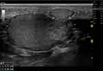
Ultrasound has several clinical applications in the assessment and diagnosis of superficial soft tissue conditions, including benign cysts (Figure 1), lymphadenopathy (Figure 2), inflammatory diseases, tissue edema, wound healing, soft tissue foreign bodies, and abscess (Figure 3). Ultrasound can be used for parasitic tissue infection. When live cysticerci are involved, ultrasound may show a well-defined, round, or elliptical cystic lesion with an eccentric echogenic mural nodule representing the scolex, a characteristic sonographic feature of cysticercosis (Figure 4).3 Ultrasound is also helpful for assessing malignant skin cancers and measuring tumor thickness, particularly in patients with malignant melanoma.
Doppler Ultrasound
Doppler ultrasound is a technique for examining tissues during motion or blood flow. It is based on the principle of relative motion between the transducer and the ultrasound wave reflector (tissues or blood) leading to an apparent change in the frequency of the ultrasound wave. Doppler ultrasound can be used to evaluate the vascular supply to the soft tissue tumors, helping to differentiate between malignant and benign tumors and vascular and non-vascular tumors (Figure 5). The deep and superficial venous systems of the extremities can be assessed by Doppler ultrasound for deep vein thrombosis, thrombophlebitis (Figure 6), and chronic venous insufficiency.
Vector Flow Imaging
High-frame rate vector flow imaging (VFI) is a novel quantitative technique for superficial vessels, initially developed for use on the carotid arteries.4 VFI depicts the speed and direction of blood cells flowing through the region of interest where the low-speed blood cells, high-speed blood cells, and reverse cells flow through a point of interest in a short time. The VFI measures the average speed of all blood cells and shows spatiotemporal characteristics of blood flow that can evaluate the specific flow pattern visually and quantitatively. The VFI displays vectors of velocity, streamline, and vorticity distribution. The direction of the arrows indicates the flow direction for streamlined distribution. The arrows’ color represents the flow’s velocity: green for low speeds, yellow and orange for medium rates, and red for higher velocities. The length of the arrows represents the magnitude of the flow; the longer the arrows, the faster the blood flow. The potential clinical applications for patients with vascular disease of the lower extremities and venous ulcers are an evaluation of chronic venous insufficiency and deep vein thrombosis (Figure 7).
Ultrasound Elastography
Ultrasound elastography is a technique for assessing the degree of tissue elasticity. In recent years, elastography-based imaging has gained attention for non-invasive assessment of tissue’s mechanical properties. The qualitative and quantitative changes in soft tissue elasticity due to various diseases and pathologies are measured using the ultrasound transducer. The mechanical force, compression, or shear wave, is applied to the lesion for tissue stiffness measurement. The potential clinical applications of ultrasound elastography for soft tissue lesions include assessing skin tumors, vascular disease of the lower extremities, pressure ulcers, lymphedema, and age-related skin changes.5–7 Ultrasound elastography is also an adjunctive technique to evaluate breast masses (Figure 8) and lymph nodes.5 Dasgeb et al reviewed the results of ultrasound elastography in benign and malignant skin tumors and suggested that malignant lesions had significantly lower elasticity than benign lesions.8
Limitations of the Ultrasound
The potential limitations of the ultrasound are the resolution of the images and soft tissue characterization compared to magnetic resonance imaging (MRI) (Figure 9a and b). Although high-frequency ultrasound actually has a higher spatial resolution for superficial lesions than MRI, the resolution for deeper soft tissue lesions is higher on MRI. MRI imparts excellent structural information and soft tissue characterization, which leads to a differential diagnosis of soft tissue lesions. MRI exhibits local tumor extension and the relationship of a tumor to adjacent fascia, bones, muscles, nerves, and vessels. Sedaghat et al reported that the MRI configuration of soft-tissue sarcoma (STS) correlates with the grade of malignancy.9 Higher-grade STS are multilobulated, whereas low-grade STS are ovoid or streaky. Infiltrative lesions suggest a higher grade.9 Additionally, MRI is a valuable tool for postsurgical follow-up of various soft tissue tumors10–13 such as recurrent dermatofibrosarcoma protuberans (DFSP) which appear as nodular, homogeneous, and well-defined enhancing lesions.10 Furthermore, MRI can assess the occurrence of post-treatment changes such as subcutaneous and muscle edema.11 Further studies on an ultrasound could focus on correlating MRI configurations to US configurations.
Conclusion
Non-invasive imaging techniques can be utilized to differentiate the features of various superficial soft tissue lesions. Comprehensive imaging knowledge can help physicians and radiologists identify and distinguish lesions with characteristic imaging patterns that require further investigation. Imaging-based assessments have potential roles in guiding treatment and surgical planning, evaluating tumor extension, staging, and performing follow-up. Novel imaging modalities and techniques have been developed to achieve better diagnostic performance while differentiating between benign and malignant lesions in vivo.
Acknowledgments
The authors would like to thank Assoc. Prof. Dr. Jitlada Meephansan for her support.
Funding
The authors received no financial support for the research, authorship, and/or publication of this article.
Disclosure
The authors report no conflicts of interest in this work.
References
1. Rallan D, Harland CC. Skin imaging: is it clinically useful? Clin Exp Dermatol. 2004;29(5):453–459. doi:10.1111/j.1365-2230.2004.01602.x
2. Jemec GB, Gniadecka M, Ulrich J. Ultrasound in dermatology. Part I. High frequency ultrasound. Eur J Dermatol. 2000;10(6):492–497.
3. Sharma P, Neupane S, Shrestha M, Dwivedi R, Paudel K. An ultrasonographic evaluation of solitary muscular and soft tissue cysticercosis. Kathmandu Univ Med J. 2010;8(30):257–260. doi:10.3126/kumj.v8i2.3571
4. Yiu BY, Lai SS, Yu AC. Vector projectile imaging: time-resolved dynamic visualization of complex flow patterns. Ultrasound Med Biol. 2014;40(9):2295–2309. doi:10.1016/j.ultrasmedbio.2014.03.014
5. Garra BS. Imaging and estimation of tissue elasticity by ultrasound. Ultrasound Q. 2007;23(4):255–268. doi:10.1097/ruq.0b013e31815b7ed6
6. Deprez JF, Cloutier G, Schmitt C, et al. 3D ultrasound elastography for early detection of lesions. evaluation on a pressure ulcer mimicking phantom. Annu Int Conf. 2007;2007:79–82. doi:10.1109/IEMBS.2007.4352227
7. Fujimura T, Osanai O, Moriwaki S, Akazaki S, Takema Y. Development of a novel method to measure the elastic properties of skin including subcutaneous tissue: new age-related parameters and scope of application. Skin Res Technol. 2008;14(4):504–511. doi:10.1111/j.1600-0846.2008.00325.x
8. Dasgeb B, Siegel E. Elastographic quantitative analysis combined with high frequency imaging for characterization of benign and malignant skin lesions.
9. Sedaghat S, Ravesh M, Sedaghat M, Both M, Jansen O. Configuration of soft-tissue sarcoma on MRI correlates with grade of malignancy. Radiol Oncol. 2021;55(2):158–163. doi:10.2478/raon-2021-0007
10. Sedaghat S, Schmitz F, Sedaghat M, Nicolas V. Appearance of recurrent dermatofibrosarcoma protuberans in postoperative MRI follow-up. J Plast Reconstr Aesthet Surg. 2020;73(11):1960–1965. doi:10.1016/j.bjps.2020.08.089
11. Sedaghat S, Schmitz F, Grözinger M, Sedaghat M. Malignant peripheral nerve sheath tumours in magnetic resonance imaging: primary and recurrent tumour appearance, post-treatment changes, and metastases. Polish J Radiol. 2020;85(1):196–201. doi:10.5114/pjr.2020.94687
12. Sedaghat S, Surov A, Krohn S, Sedaghat M, Reichardt B, Nicolas V. Configuration of primary and recurrent aggressive fibromatosis on contrast-enhanced MRI with an evaluation of potential risk factors for recurrences in MRI follow-up. Konfiguration primärer und rezidivierender aggressiver Fibromatosen in der kontrastmittelgestützten MRT mit einer Evaluation potenzieller Risikofaktoren für Rezidive in MRT-Verlaufskontrollen. Rofo. 2020;192(5):448–457. doi:10.1055/a-1022-4546
13. Sedaghat S, Salehi Ravesh M, Sedaghat M, Meschede J, Jansen O, Both M. Does the primary soft-tissue sarcoma configuration predict configuration of recurrent tumors on magnetic resonance imaging? Acta Radiologica. 2022;63(5):642–651. doi:10.1177/02841851211008381
 © 2023 The Author(s). This work is published and licensed by Dove Medical Press Limited. The full terms of this license are available at https://www.dovepress.com/terms.php and incorporate the Creative Commons Attribution - Non Commercial (unported, v3.0) License.
By accessing the work you hereby accept the Terms. Non-commercial uses of the work are permitted without any further permission from Dove Medical Press Limited, provided the work is properly attributed. For permission for commercial use of this work, please see paragraphs 4.2 and 5 of our Terms.


