Back to Journals » Drug Design, Development and Therapy » Volume 17
Recent Advances in the Nanoshells Approach for Encapsulation of Single Probiotics
Received 4 May 2023
Accepted for publication 16 August 2023
Published 8 September 2023 Volume 2023:17 Pages 2763—2774
DOI https://doi.org/10.2147/DDDT.S419897
Checked for plagiarism Yes
Review by Single anonymous peer review
Peer reviewer comments 2
Editor who approved publication: Professor Jianbo Sun
Cheng Chen,1 Ziyu Zhu2
1The People’s Hospital of Danyang, Affiliated Danyang Hospital of Nantong University, Danyang, Jiangsu Province, 212300, People’s Republic of China; 2The Affiliated Huai’an Hospital of Xuzhou Medical University and the Second People’s Hospital of Huai’an, Huai’an, 223002, People’s Republic of China
Correspondence: Ziyu Zhu, Email [email protected]
Abstract: The intestine, often referred to as the “second brain” of the human body, houses a vast microbial community that plays a crucial role in maintaining the host’s balance and directly impacting overall health. Probiotics, a type of beneficial microorganism, offer various health benefits when consumed. However, probiotics face challenges such as acidic conditions in the stomach, bile acids, enzymes, and other adverse factors before they can colonize the intestinal tissues. At present, pills, dry powder, encapsulation, chemically modified bacteria, and genetically engineered bacteria have emerged as the preferred method for the stable and targeted delivery of probiotics. In particular, the use of nanoshells on the surface of single probiotics has shown promise in regulating their growth and differentiation. These nanoshells can detach from the probiotics’ surface upon reaching the intestine, facilitating direct contact between the probiotics and intestinal mucosa. In this perspective, we provide an overview of the current developments in the formation of nanoshells mediated by single probiotics. We also discuss the advantages and disadvantages of different nanocoating strategies and explore future trends in probiotic protection.
Keywords: microbial community, intestinal mucosa, probiotics, nanoshells
Introduction
The intestine tract is a vital organ in the human body inhabited by a huge microbial community. This community plays an indispensable role in maintaining the body’s homeostasis and regulating the immune system, directly impacting the overall health of the host.1,2 Imbalances in the intestinal flora, disruptions in the metabolism of intestinal bacteria, and the invasion of pathogens are closely associated with various diseases, including inflammatory bowel disease, obesity, diabetes, and certain tumors.3–5 To improve overall health, consuming suitable probiotics can enhance immune responses, restore balance to the intestinal flora, and inhibit the colonization of pathogenic bacteria.6,7 The probiotic properties of these beneficial microorganisms are closely linked to their viability and quantity.8 Unfortunately, the complex environment of the digestive tract, including enzyme invasions, gastric acid attacks, and continuous intestinal peristalsis, can significantly impact the survival rate and intestinal colonization of probiotics, thereby compromising their beneficial effects.9,10
To address the aforementioned problems, several strategies have been developed to improve the survival rate and intestinal colonization ability of probiotics. These strategies include pills, dry powder, encapsulation, chemically modified bacteria, and genetically engineered bacteria.11–13 However, each protection strategy has its own advantages and disadvantages. For instance, pills and dry powder can enhance the bioavailability of probiotics to some extent but fail to achieve effective colonization in the intestinal tract.14 Genetically engineered probiotics exhibit excellent tolerance and resistance, making them suitable as drug carriers to inhibit pathogenic microorganisms or directly eliminate them. However, their tedious preparation process hinders large-scale manufacturing.15,16 On the other hand, surface modification, unlike genetic engineering, is an effective method that can introduce various functional motifs onto the surface of probiotics to protect them from enzyme and gastric acid attacks in the gastrointestinal tract. However, it does not provide control over the metabolism and activities of probiotics.17,18 Despite the promising nature of these protection methods for oral probiotic delivery, few have been proven effective in disease prevention or treatment. This is due to challenges such as low permeability, low stability, short sustenance period, and the complex gastrointestinal environment, which result in significant probiotic death, limited colonization, and proliferation in intestinal tissues.19,20 Therefore, innovative strategies are needed to develop oral probiotic bioagents that can overcome these limitations.
Recent studies have demonstrated that the formation of nanoshells on the surface of single probiotics can effectively protect them from physical and biological threats, enabling them to maintain their biological function in harsh environments. Additionally, these nanoshells enhance the adhesion of probiotics to the intestinal mucosal epithelial tissue, preventing their removal by the flowing liquid (Figure 1a-c).21–25 The nanoshells on the surface of a single probiotic serve as a physical barrier, a chemically selective channel between the external and internal probiotics, and a mechanically robust exoskeleton. In comparison to previous encapsulation methods that only provided temporary protection until probiotics split, the generation of nanoshells mediated by a single probiotic offers long-term probiotic protection and mucosal adhesion. The selective permeability of the nanoshells allows for the transfer of nutrients, oxygen, and probiotic metabolites, thereby maintaining the function and viability of probiotics both in vitro and in vivo. This paper aims to discuss and summarize some typical natural derivative materials that can wrap a single probiotic by coating it with a nano-thickness layer. This approach enables probiotics to continuously withstand the harsh environmental conditions during transportation, thereby improving their bioavailability and intestinal colonization. This comprehensive review serves as a valuable resource for selecting effective and efficient nanoshells materials for targeted delivery of probiotics.
 |
Figure 1 Schematic of single probiotics nanoencapsulation and against harsh environment in human digestive system. (a) Representation of probiotics without nanocoating. (b) Nanoencapsulation of probiotics. (c) Gut bacteria pathway via the gastrointestinal tract and the importance of probiotics encapsulation. Data from these studies.22–25 |
Layer-by-Layer Assembly Mediated Nanoshells
The coating of artificial polymer nanoshells onto the surface of probiotics through layer-by-layer (LBL) assembly has proven to be a successful method for isolating probiotics from harsh environmental attacks, such as stomach acid and rapid intestinal transit time. This coating technique enhances the viability and intestinal adhesion performance of probiotics during oral delivery.26,27 The mechanism behind LBL nanoshells construction relies on electrostatic interactions between oppositely charged polyelectrolytes. The process begins with the deposition of polycation onto the negatively charged surface of probiotics, forming the first layer. Subsequently, alternating deposition of polyanion and polycation compounds takes place until the nanoshell achieves the desired robustness, thickness, and functionality.28 As shown in Figure 2a, the LBL assembly of multilayered nanoshells on the probiotics’ surface requires centrifugation and washing at each step to remove unbound polyelectrolytes. This process can be laborious and time-consuming.
 |
Figure 2 (a) Layer-by-layer self-assembly as the most widely utilized approach to the formation of polymeric nanocoating. After deposition of each layer, cells are collected by centrifugation, washed with buffer, and then collected again by centrifugation. (b) Schematic layer-by-layer templating of chitosan and alginate on bacteria surface. |
However, the merit of this LBL nanocoating mediated nanoshell on the surface of individual probiotics lies in its ability to effectively protect the probiotics during oral delivery. By encapsulating the probiotics with an optimal amount of polymers, the nanoshell shields them from the harsh gastric acid and enzymatic attacks, while preserving their original morphology and activity. Furthermore, it enhances the probiotics’ adhesion in the intestinal tract, thereby exerting precise control over their proliferation and division.29 Therefore, I believe that the materials formed by LBL assembly mediated nanoshell for probiotics have a wide range of applicability, including positively charged proteins, polycationic electrolytes, and polycationic polysaccharides, etc. Besides, the polycation layer used first is in direct contact with the surface of probiotics, and the biocompatibility of the synthesized polycation should be the core issues of electrostatic LBL assembly, because direct exposure of probiotics to polycation can lead to membrane perforation. Importantly, this nanofilm formation approach heavily relied on the multi-step LBL deposition process, and the templates removed process would deteriorate nanoshells integrity and needs harsh removal condition that may induce incompatible with proteins, and probiotics.
Therefore, the biocompatibility of polycation is the core premise that hinders the development of LBL assembly. It has been reported that the cytotoxicity of polycation mainly depends on charge density and molecular weight.30 These problems can be solved by using natural cationic polyelectrolyte with low cytotoxicity and high cell compatibility, so natural polyelectrolyte (hyaluronic acid, alginate, collagen, chitosan, etc.) have been widely used in cell nanoencapsulation.31,32 Based on this, Aaron C. Anselmo and co-workers utilized cationic polysaccharide chitosan (CHI) and the anionic polysaccharide alginate alternated deposited into the Bacillus coagulans surface via electrostatic interactions to form three bilayers (CHI/ALG)3 that can address the chemical, physical, and probiotics oral delivery challenges while enhancing the survival of Bacillus coagulans against acidic and bile salt insults, and enhance the mucoadhesion ability on intestinal tissues to improve the bloating with irritable bowel syndrome and colitis treatment effects (Figure 2b).11 Chitosan-alginate multilayers nanoshells formed on Bacillus coagulans surface indicated that polycationic compounds provided the material–material and cell–material interactions suitable to construction of LBL assembly multilayer nanoshells on the probiotics surface without disturbing probiotics-membrane integrity.
Generally speaking, natural biopolymers are generally considered to be more suitable bio-friendly nanocoating materials than synthetic polyelectrolytes, which may overcome the cytotoxicity of the first polycation layer on probiotics films. This makes all kinds of naturally derived polycation compounds widely used in LBL assembly of the first layer of multilayer nanoshells. In particular, harnessing cytocompatible and edible components derived from nature to form nanoshells on the surface of single probiotics using a one-step method is a suitable approach for protecting and maintaining the viability of living probiotics.
Polyphenolic Compounds Mediated Nanoshells
For the nanocapsulation of single probiotics, the formation of a nanofilm primarily relies on layer-by-layer (LBL) deposition. However, this process is laborious and involves multiple steps, making it unsuitable for large-scale production. Therefore, there is an urgent need for a convenient and effective method to protect probiotics from harmful and deadly external environment. Inspired by the fact that prokaryotes can produce a series of extracellular matrix polymers, researchers have developed different interfering reaction pathways to produce new synthetic polymers according to the different cell targeting mechanisms and unique intracellular environmental condition (the redox enzyme cascade reaction).33,34 In addition to the main purpose of cell protection, recent studies have focused on inducing various oxidative polymerization reactions in the environment of probiotics to form nanoshells on the surface of a single probiotic. Therefore, probiotics-mediated formation of synthetic polymers would be the sustainable nanoplatform to investigate their hierarchical assembly to generate multifunctional bionic systems.
Polyphenolic compounds that contain pyrogallol or catechol groups in their structure, in which catechol groups can be easily covalent cross-linking via probiotics-mediated catalytic process to form self-encapsulating protective nanoshells on the probiotics surface.35,36 Among various polyphenolic compounds, dopamine, caffeine acid and pyrocatechol, are the typical and widely applied probiotics coating materials (Figure 3a), which can be oxidized into quinones under the alkaline condition (pH = 8.5), and a series of cross-coupling reaction and polymerization assembly occur, and then forming a phenolic nanofilm on single-probiotics surface (Figure 3b).37,38 However, some probiotics cannot bear this alkaline condition, resulting in most probiotics dying during the polymerization process.39
 |
Figure 3 Schematic of probiotics-mediated oxidation of polyphenolic compounds bearing catechol groups by probiotic cells. (a) Structures of the phenolic precursors used in this study. (b) Process overview of probiotics-mediated oxidation of catechol compounds and in situ nanoencapsulation. (c) Possible mechanism of the probiotics-mediated phenolic oxidation using probiotics. Data from these studies.37,38 |
As literatures report that Lactobacillus species have rich transition metals in their biological systems that not only can maintain the stability of proteins structure, but also playing a catalytic role in the biological process, respond to oxidative stress, and activating phenol compounds oxidation as well as inducing the cross-linking reaction to conduct self-assembly of surface polymers.40,41 Inspired by this redox-active transition metals system, Franco-Centurion and his colleagues used the essential nutrient manganese contained in Lactobacillus to regulate the oxidation of phenolic compounds, and then inducing Lactobacillus-mediated polymerization and nanofilm formation on the single Lactobacillus surface that can enhance the viability and functionality of encapsulating Lactobacillus.42 During the process of oxidative polymerization of phenolic compounds, Lactobacillus playing the role of both templates and catalysts. The mechanism is that the interfacial manganese ion released by Lactobacillus can further promote the oxidative polymerization of phenolic compounds and form nanoscale shells on single Lactobacillus surface.43,44
Therefore, manganese ions in Lactobacillus playing an important role in the oxidative polymerization of dopamine. The released manganese ions from Lactobacillus can participate in a redox reaction cycle to induce dopamine oxidation and then formation a reactive complex between manganese ions and the dopamine (semi-quinone free radical) as a ligand, thus rapidly forming a polydopamine nanoshells wrapped in single Lactobacillus surface (Figure 3c).39 This nanocoating method utilizes the combination of dopamine and Lactobacillus, and utilizes manganese ions released by Lactobacillus to mediate the oxidation of phenolic compounds to form nanoshells to encapsulate a single Lactobacillus in situ, which can improve the survival rate of Lactobacillus in acidic stomach environment, enhance the adhesion behavior to intestinal epithelial cells, and endow Lactobacillus with excellent antioxidant properties by the presence of hydroquinone moieties in the polydopamine nanoshells without further modification.45 More importantly, the formed polydopamine nanoshells acted as the physical barrier of Lactobacillus can significantly control the nutrient access and effectively delay Lactobacillus growth. Overall, Lactobacillus-mediated phenolic compounds oxidative polymerization and formation nanoshells on single-Lactobacillus surface have provided a valuable solution to address the bottleneck in Lactobacillus delivery and also served as Lactobacillus-mediated nanoplatform to provide cytoprotection, adherence ability and other advanced functions to Lactobacillus beneficial for human health. However, harnassing the biointerfaces of Lactobacillus’ redox enzyme cascade reaction to induce oxidative polymerization of phenolic compounds is only suitable for Lactobacillus species, such as Lactobacillus helveticus and Lactobacillus plantarum, and not suitable for all probiotics nanocoating.
Biphasic Interfacial Mediated Nanoshells
Polyphenolic compounds are adopted for nanoshells formation on single-probiotics surface, which can endow their different functions to adapt for harsh environment changes, but operation processes are very strict, not biocompatible and lots of time are wasted to obtain nanometer-thick film.46,47 Thanks to the development of interfacial supramolecular self-assembly at the heterogeneous system, that can quickly, simply and effectively in situ formation a high degree of uniformity nanoshells on single-probiotics surface with the help of microfluidic technology.48 The mechanism of this biphasic interfacial reaction system is that each of these two immiscible phases contains one of the self-assembling components and rapid formed a nanolayer with one-step in the flow microfluidic device.49
Different from the traditional reports that have all utilized synthetic molecules for biphasic interfacial reactions, which needs cumbersome synthesis process, and incompatible with living cells.50 Kim and his co-workers used this biphasic interfacial reaction to form a uniform Feш-TA metal-organic coordination complex (Feш-TA-MOC) nanoshells via using the cell-compatible and even edible complex Feш and tannic acid (TA).47 Inspired by the catecholato iron (III) complexes found in biological system, this Feш-TA-MOC nanoshells formation do not require any synthetic challenges, and the mechanism of Feш-TA-MOC nanoshells formation process was ascribed to the pyrogallol moiety in TA, which acted as the bidentate ligand for Feш to form bis or tris complexes.51 More importantly, the interfacial Feш-TA-MOC nanoshells formed in this biphasic interfacial system are highly versatile and widely applied to various kinds of interfaces, including water–oil interface, hydrogel–water interface, and cellular interfaces (Figure 4a). In order to further illustrate the application of this two-phase interface in the process of nanocapsulation, Kim and his colleagues used the water–oil interface as the sample to form two-phase supramolecular Feш-TA-MOC nanoshells by flow focusing microfluidic device (Figure 4b). The obtained Feш-TA-MOC nanoshells were homogeneous and monodisperse, which is especially advantageous useful to research the probiotics activity, and the uptake behavior of nutrients by the encapsulated probiotics in a specific confinement and intercellular distance.52 Mimicking like germination process of dormant spores, the other advantage of Feш-TA-MOC nanoshells is that the probiotics were temporal cytoprotection by the Feш-TA-MOC nanoshells, which can be degraded to the external environment stimuli (ascorbic acid, hydrochloric acid, and ethylenediaminetetraacetic acid), and the extra Feш-TA-MOC nanoshells can be further modified with different biomolecules, giving extra functions to probiotics.53
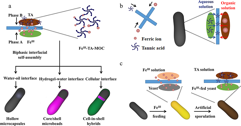 |
Figure 4 (a) Schematic representation of interfacial supramolecular self-assembly of FeIII and TA at various interfaces. FeIII ions are preloaded into one phase (Phase A), and the supramolecular self-assembly is induced at the interface of two immiscible phases by TA in the other phase (Phase B). (b) Schematic representation for formation of FeIII–TA-MOC hollow nanoshell. (c) Schematic representation for self-responsive FeIII-TA-MOC nanoshell formation on baker’s yeast. |
Although this method can successfully form a nanoshell on the surface of probiotics, which has no obvious effect on the viability of probiotics viability, they all involved passive modification processes that were manipulated by the external chemical reaction. In contrast, the natural process of sporulation is a stimulus-responsive and autonomous process.54 Therefore, to develop a stimulus-responsive, active cell system, in which probiotics respond to the environment changes and formed a hard shell by theirself, that would be a huge leap in the single-probiotics nanoencapsulation. Inspired by this concept, Kim and his co-workers feed S. cerevisiae (baker’s yeast) with Feш for 12h, and then Feш fed baker’s yeast was immersed into the TA solution (Figure 4c). During the interfacial reaction, Feш ions flowed out of the baker’s yeast which was responsible for formation of the Feш-TA-MOC nanoshells. These artificial nanoshells can not only protect and preserve cells from external pressure in vitro, but also delay the growth of cells and choose the right time for cell therapy. Therefore, this efflux Feш ions-driven Feш-TA-MOC nanoshells formed opened a new window for the biphasic interfacial reaction of probiotics in a heterogeneous system.
The demonstration of these two works has the similarities and differences in the working mechanism and detailed chemical scheme, they all depend on the formation of biphase interface of the Feш-TA-MOC nanoshells complex. From the proof-of-concept, we can speculate that Feш-TA nanoshells not only acted as the protective film to protect probiotics from external pressure damage but also as the cargo carrier for drugs delivery. Overall, the interface reaction strategy of forming edible nanoshells on the surface of single-probiotics is a general strategy for developing durable, functional and oral nanocapsules.
Liposomes Mediated Nanoshells
Liposomes can also be considered as promising materials for encapsulating microbial cells, providing protection for probiotics during oral delivery, particularly in gastrointestinal transit.55 What is more, liposomes-mediated nanoshells can be designed with diverse structures by altering liposome size, composition, membrane fluidity, and surface properties.56 Currently, the liposomes-mediated nanocoating approach can be categorized into two types: natural cell membrane formation and lipid compound-mediated cell membrane formation. Vesicles derived from natural cell membrane can endow coating material with longer blood circulation time, avoid being eliminated by the immune system of the body, and reduce the nonspecific absorption of protein during transportation.57,58 Besides, there are many kinds of natural cell membrane, including cancer cells, platelet, neutrophil, macrophage, and erythrocyte membranes. Due to the CD47 protein present on the surface of erythrocyte membranes (EM) that can avoid EM coated materials eliminated by body immune system.59 Thus, Cao Zhengping and his colleagues coated Escherichia coli Nissle 1917 (EcN) with EM (CMCB) via mechanical extrusion through the porous polycarbonate membrane (Figure 5a), and the obtained CMCB shown the typically core-shell structure and the shell with the thickness for about 100 nm (Figure 5b).60 In CMCB system, EcN skillfully utilized the low immunogenicity of EM to endow EcN with anti-phagocytosis ability and low inflammation in transportation. At the same time, the biological activity of EcN did not change significantly, because the coated EM was finally split and removed by EcN division, the removed coating membranes were visualized by transmission electron microscope (Figure 5c) as well as the schematic illustration for coating membranes removed process is shown in Figure 5d. Comparing with untreated EcN, the obtained CMCB has higher blood reservation time, reduce inflammatory response and higher tumor tissues accumulated ability. More importantly, through switching the culture media can tune the coating membranes removal process from hours to days, which indicated that EcN can realize long-term application and on-demand release behavior. Besides, EM mediated nanocoating is a versatile modality to fabricate biologically functional EcN to improve therapy effect and solve the safety problems of some probiotics. Therefore, we can anticipate that probiotics camouflaged with different cell membranes can meet different disease needs and provide an unparalleled tool for probiotics in biomedical field. However, the application of cell membrane-mediated nanocoating encountered some problems and challenges before clinical application, such as the source of the cell membrane, incomplete or uneven coverage of probiotics may cause immune response. Besides, the membrane-mediated coating process is still at the early stage, and maintaining the integrity of the membrane in the process of extraction and fusion is an urgent core problem. Generally speaking, cell membrane-mediated nanocoating has great application prospect, which can meet the needs of probiotics to resist harsh physiological environment and external attacks, and meet the need of probiotics to play a beneficial role in the specific place at a specific time. However, researchers need repeated experiments to prove its biological safety before it can be applied in clinic.
 |
Figure 5 (a) Schematic illustration for the preparation of EM-coated-EcN by extruding bacteria with cell membranes. (b) Representative transmission electron microscope images of uncoated EcN and EM-coated-EcN. (c) A representative image of membrane shedding captured by TEM. (d) Schematic illustration for the removal of coating membranes. Adapted from Cao ZP, Cheng SS, Wang XY et al. Camouflaging bacteria by wrapping with cell membranes. Nat Commun. 2019. 10 (1): 3452. Creative Commons.60 |
Different from cell membrane mediated nanoshells formed on single-probiotics surface, lipid membrane via a biointerfacial supramolecular self-assembly deposited into the probiotics surface by simply vortexing probiotics with biocompatible lipids without tedious separation procedures and multiple steps, which can endow wrapped probiotics a substantial chemical biostability against various extreme environment and friendly for manufacturing scale-up.61 Moreover, the coating lipid membrane does not affect the growth of probiotics and keep intact before probiotics division, which can significantly improve probiotics’ bioavailability, colonization and retain their innate bioactivity. Once it reaches the targeting sites, the lipid membrane formed on the surface of probiotics can be degraded by lipolysis, and the viability, the adhesion, settlement and proliferation of probiotics are not affected.62 Inspired by the merits of lipid membrane, Cao Zhengping and his co-workers used Food and Drug Administration (FDA) approved materials cholesterol and dioleoylphosphatydic acid to coat EcN in calcium phosphate buffer by vortexing less than 15 min (Figure 6a).63 In the process of lipid film formation, calcium ions contained in solution can assist the self-assembly of diacylphosphoric acid to the surface of the negative surface of EcN, and the existence of cholesterol can further stabilize the self-assembled lipid membrane to isolate EcN more consistently.64 Thus, the lipid membrane nanoshells can intact and stable protecting EcN from the attack of gastric acid, alkalis, antibiotics, intestinal juice and oxidative stress conditions before EcN split and fat decomposed (Figure 6b). Because of the dynamic behavior of self-assembly and disassembly of coated lipid membrane, it not only provides a simple, universal and efficient way to improve the colonization and bioavailability of EcN, but also shown a wide range of adaptability to different strains.
 |
Figure 6 (a) Schematic illustration of the preparation of lipid membrane coated bacteria by biointerfacial supramolecular self-assembly. (b) The presence of coating membranes endows probiotic bacteria with exceptional resistance to various harsh environmental conditions. Adapted from Cao ZP, Wang XY, Pang Y et al. Biointerfacial self-assembly generates lipid membrane coated bacteria for enhanced oral delivery and treatment. Nat Commun. 2019. 10 (1): 5783. Creative Commons.63 |
Thus, lipid membranes mediated nanoshells approach provided a simple but a highly efficient approach to improve probiotics viability and bioactivity and exhibit incomparable advantages during probiotics delivery process. Thus, we can anticipate that utilizing FDA approved materials to generate lipid membranes on the surface of probiotics with simple and convenient approach is the first step. The core elements of lipid membranes nanoencapsulation are not just limited to protect the bioavailable of probiotics but should be used to further functionalize the lipid membranes to make it suitable for the needs of different diseases.
Challenge and Prospects
Numerous cell-compatible materials and methods have been explored for surface coating of probiotics to enhance their activity. However, the fragility and susceptibility of probiotic cell membranes to damage during the encapsulation process remain a challenge.65 To address these limitations, it is crucial to develop cell-compatible materials, such as polymers, liposomes, and coacervates. Additionally, combining synthetic materials with natural materials can offer potential solutions to the existing issues.
The advancement of the single-probiotic nanocoating technique has allowed for the encapsulation of individual probiotics within a confined three-dimensional space. This technique enables studies at the individual probiotic level, while selectively allowing the passage of small molecules and nutrients, and facilitating probiotics-to-gut communication. According to different nanocoating mechanisms, the coating process can be divided into two categories: “growth from the surface of probiotics” and “deposition on the surface of probiotics”. In the “growing from” method, synthetic polymers were grafted onto the surface of probiotics by the cytocompatible approach, which can successfully form the probiotics-in-shell structure and keep the viability of probiotics but they needing passive processes that were determined by the chemical reactions through external manipulation. For the “deposition to” strategy, some prefabricated materials are deposited on the surface of single probiotics, showing typical core-shell structure characteristics.66
The great advantage of this probiotics-in-shell engineering is that they can generate exogenous functions that are neither naturally nor innately achievable, such as controlling the biological activity and metabolism of single living probiotics.67,68 Therefore, in this review, we only summarized the advantages and disadvantages of some typical natural cell-compatible materials on the surface modification of living single probiotics, in order to understand and adjust the behavior of probiotics, and improve their tolerance to unfriendly environments (acidic environment, temperature fluctuation and intestinal peristalsis) to enhance their therapeutic effects (Table 1). To sum up, the process of nanoshells formation mediated by single probiotic gives probiotics unparalleled biological function, which goes beyond the traditional probiotics surface engineering without complicated operation processes. Despite the significant progress that has been achieved with the single probiotics nanocoating approach, the research progress of probiotics nanocoating is still in its infancy which needs repeatability experimentation before clinical application.
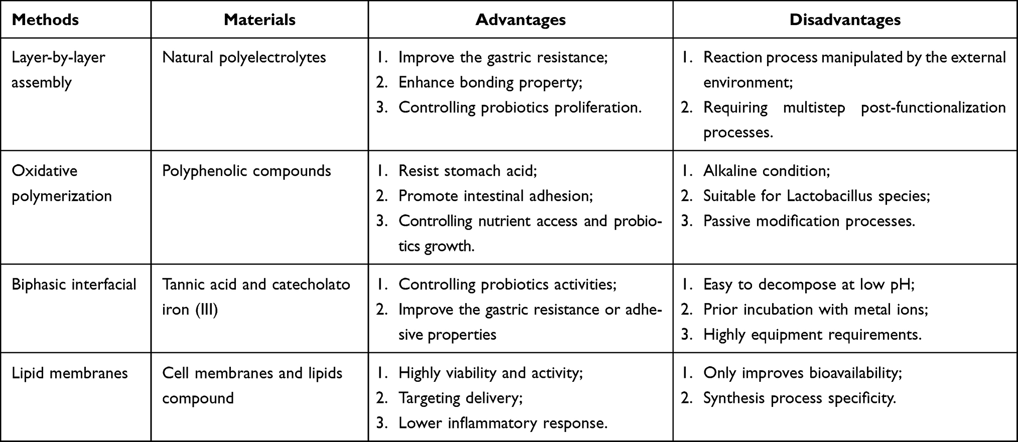 |
Table 1 The Advantages and Disadvantages of Different Nanoshell to Protect Probiotics |
In the future, the progress of single probiotics nanocoating should focus on selecting appropriate biomimetic materials to form multifunctional bioactive nanoshells according to the special characteristics of disease, so as to change the surrounding environment and act as the supplement of drugs to enhance the disease therapeutic effects, rather than just avoiding probiotics attacked by the complex and harsh physiological environment or just making probiotics more suitable for colonization. Or the composition of the intestinal flora of patients should be tested before disease treatment, then artificially supplementing the major intestinal lost probiotics as well as utilizing the required therapeutic substances needed for the disease to encapsulate these probiotics that can effectively achieve the cascade effect of disease. More important, the nanoshells that coat the probiotics can be further modified with some functional molecule that promote the colonization of the probiotics at the desirable sites in the gut and achieve drugs targeting delivery and release. Thus, the technologies and methods of surface protection and modification of probiotics still have a lot of room for improvement, which requires a lot of resources and persistence. Furthermore, researchers need to address fundamental issues regarding the interactions between coating materials and probiotic surfaces. This will facilitate the use of more Food and Drug Administration (FDA) approved materials for protecting probiotics, without compromising their bioactivity. Additionally, it will aid in the scale-up of manufacturing processes and subsequent clinical translation of probiotic-based products.
Conclusion
Although probiotic embedding technology has been developed for several decades, there is still ample room for exploring suitable encapsulating materials and methods. In this study, we have reviewed and summarized some simple yet valuable and versatile approaches for integrating environmentally friendly materials with probiotics. These approaches aim to establish a protective barrier on the surface of individual probiotics, isolating them from harsh external conditions when delivered into the gastrointestinal tract. Additionally, the biostructure of the probiotics-in-shell can significantly enhance the functionality of the encapsulated probiotics at the individual level, while also providing novel or even abiotic functions. This enables the probiotics to exhibit flexible biochemical properties, adapting to changes in both in vivo and in vitro environments. The insights gained from this research can provide valuable experiences for the development of next-generation cellular biological systems in the field of probiotic surface engineering and other related biological disciplines. However, when designing novel and multifunctional single probiotic nanoshells, it is important to consider the types of probiotics, the interaction between the coating process, and the choice of encapsulating materials.
Author Contributions
All authors have made substantial contributions to the work, including conception, study design, data acquisition, analysis and interpretation. They have also been involved in drafting, revising, and critically reviewing the article. Furthermore, all authors have given their final approval for the version to be published and have agreed on the journal to which the article has been submitted. They also accept accountability for all aspects of the work.
Funding
There is no funding to report.
Disclosure
The authors declare that they have no competing interests in this work.
References
1. Fredrik B, Ruth EL, Justin LS, et al. Host-bacterial mutualism in the human intestine. Science. 2005;307(5717):1915–1920. doi:10.1126/science.1104816
2. Wang XQ, Zhang AH, Miao JH, et al. Gut microbiota as important modulator of metabolism in health and disease. Rsc Adv. 2018;8(74):42380–42389. doi:10.1039/c8ra08094a
3. James LA, Ian DW, Julian T, et al. Gut microbiota modulation of chemotherapy efficacy and toxicity. Nat Rev Gastroenterol Hepatol. 2017;14(6):356–365. doi:10.1038/nrgastro.2017.20
4. Taekil E, Yong SK, Chang HC, et al. Current understanding of microbiota- and dietary-therapies for treating inflammatory bowel disease. J Microbiol. 2018;56(3):189–198. doi:10.1007/s12275-018-8049-8
5. Louis HSL, Sunny HW. Microbiota, Obesity and NAFLD. Adv Exp Med Biol. 2018;1061:111–125. doi:10.1007/978-981-10-8684-7_9
6. Willem MDV, Herbert T, Matthias VH, et al. Gut microbiome and health: mechanistic insights. Gut. 2022;71(5):1020–1032. doi:10.1136/gutjnl-2021-326789
7. Abbas MS, Afzaal M, Saeed F, et al. Probiotic viability as affected by encapsulation materials: recent updates and perspectives. Int J Food Properties. 2023;26(1):1324–1350. doi:10.1080/10942912.2023.2213408
8. Montoya-soto JG, González-Laredo RF, Medina-Torres L, et al. Recent developments on wall materials for the microencapsulation of probiotics: a review. Tecnociencia Chihuahua. 2023;1(4):2683–3360. doi:10.54167/tch.v17i1.1140
9. Kourosh KZ, Kyle JB, Rebecca EB, et al. Intestinal gases: influence on gut disorders and the role of dietary manipulations. Nat Rev Gastroenterol Hepatol. 2019;16(12):733–747. doi:10.1038/s41575-019-0193-z
10. Kourosh KZ, Stephanie AW, Kamyar KZ, et al. Considering the Effects of Microbiome and Diet on SARS-CoV-2 Infection: nanotechnology Roles. ACS Nano. 2020;14(5):5179–5182. doi:10.1021/acsnano.0c03402
11. Aaron CA, Kevin JM, Jamie W, et al. Layer-by-Layer Encapsulation of Probiotics for Delivery to the Microbiome. Adv Mater. 2016;28(43):9486–9490. doi:10.1002/adma.201603270
12. Li ZH, Adam MB, Nitzan G, et al. Biofilm-Inspired Encapsulation of Probiotics for the Treatment of Complex Infections. Adv Mater. 2018;30(51):e1803925. doi:10.1002/adma.201803925
13. Wang YT, Tang Y, Du Y, et al. Genetically engineered bacteria-mediated multi-functional nanoparticles for synergistic tumor-targeting therapy. Acta Biomater. 2022;150:337–352. doi:10.1016/j.actbio.2022.07.056
14. Lee H, Kim N, Hyeong BR, et al. A Decade of Advances in Single-Cell Nanocoating for Mammalian Cells. Adv Healthc Mater. 2021;10(13):e2100347. doi:10.1002/adhm.202100347
15. Jacob DP, Emma P, Beth AM, et al. Engineered Probiotic for the Inhibition of Salmonella via Tetrathionate-Induced Production of Microcin H47. ACS Infect Dis. 2018;4(1):39–45. doi:10.1021/acsinfecdis.7b00114
16. Caroline BK, Yves AM, Marja KP, et al. An engineered E. coli Nissle improves hyperammonemia and survival in mice and shows dose-dependent exposure in healthy humans. Sci Transl Med. 2019;11(475):eaau7975. doi:10.1126/scitranslmed.aau7975
17. Himanshu KS, Dipak DP, Dushyant AS, et al. Development of microencapsulation delivery system for long-term preservation of probiotics as biotherapeutics agent. Biomed Res Int. 2013;2013:620719. doi:10.1155/2013/620719
18. Kenneth T, James KT, Harald B, et al. Synthetic consortia of nanobody-coupled and formatted bacteria for prophylaxis and therapy interventions targeting microbiome dysbiosis-associated diseases and co-morbidities. Microb Biotechnol. 2019;12(1):58–65. doi:10.1111/1751-7915.13355
19. Cornelius CD, Wang J, Abdul WB, et al. Targeted delivery of probiotics to enhance gastrointestinal stability and intestinal colonisation. Int J Pharm. 2017;530(1–2):224–229. doi:10.1016/j.ijpharm.2017.07.068
20. Kong SS, Zhang YHH, Zhang WQ. Regulation of Intestinal Epithelial Cells Properties and Functions by Amino Acids. Biomed Res Int. 2018;2018:2819154. doi:10.1155/2018/2819154
21. Artis D. Epithelial-cell recognition of commensal bacteria and maintenance of immune homeostasis in the gut. Nat Rev Immunol. 2008;8(6):411–420. doi:10.1038/nri2316
22. Wang MM, Yang J, Li M, et al. Enhanced viability of layer-by-layer encapsulated Lactobacillus pentosus using chitosan and sodium phytate. Food Chem. 2019;285:260–265. doi:10.1016/j.foodchem.2019.01.162
23. Flemming HC, Wingender J, Szewzyk U, et al. Biofilms: an emergent form of bacterial life. Nat Rev Microbiol. 2016;14(9):563–575. doi:10.1038/nrmicro.2016.94
24. Yan J, Bassler BL. Surviving as a Community: antibiotic Tolerance and Persistence in Bacterial Biofilms. Cell Host Microbe. 2019;26(1):15–21. doi:10.1016/j.chom.2019.06.002
25. Franco C, Abdul WB, Liu JY, et al. Nanoencapsulation for Probiotic Delivery. ACS Nano. 2021;15(12):18653–18660. doi:10.1021/acsnano.1c09951
26. Choi YH, Phan B. Methods and Applications of Biomolecular Surface Coatings on Individual Cells. ACS Appl Bio Mater. 2020;3(10):6556–6570. doi:10.1021/acsabm.0c00867
27. Fischer D, Li YX, Barbara A, et al. In vitro cytotoxicity testing of polycations: influence of polymer structure on cell viability and hemolysis. Biomaterials. 2003;24(7):1121–1131. doi:10.1016/s0142-9612(02)00445-3
28. Germain M, Balaguer P, Nicolas JC, et al. Protection of mammalian cell used in biosensors by coating with a polyelectrolyte shell. Biosens Bioelectron. 2006;21(8):1566–1573. doi:10.1016/j.bios.2005.07.011
29. Michael TC, George T, Vitaliy VK, et al. Layer-by-layer coating of alginate matrices with chitosan-alginate for the improved survival and targeted delivery of probiotic bacteria after oral administration. J Mater Chem B. 2013;1(1):52–60. doi:10.1039/c2tb00126h
30. Jeong HJ, Hwang JS, Lee H, et al. In vitro blood cell viability profiling of polymers used in molecular assembly. Sci Rep. 2017;7(1):9481. doi:10.1038/s41598-017-10169-5
31. Zhou JY, James WY, Sung WK, et al. Intracellular kinetics of non-viral gene delivery using polyethylenimine carriers. Pharm Res. 2007;24(6):1079–1087. doi:10.1007/s11095-006-9229-5
32. Haesslein A, Ueda H, Hacker MC, et al. Long-term release of fluocinolone acetonide using biodegradable fumarate-based polymers. J Controlled Release. 2006;114(2):251–260. doi:10.1016/j.jconrel.2006.05.024
33. Lee H, Dellatore SM, Miller WM, et al. Mussel-inspired surface chemistry for multifunctional coatings. Science. 2007;318(5849):426–430. doi:10.1126/science.1147241
34. Ejima H, Richardson JJ, Laing K, et al. One-step assembly of coordination complexes for versatile film and particle engineering. Science. 2013;341(6412):154–157. doi:10.1126/science.1237265
35. Yang J, Martien ACS, Marleen K. Jack of all trades: versatile catechol crosslinking mechanisms. Chem Soc Rev. 2014;43(24):8271–8298. doi:10.1039/c4cs00185k
36. Leonhard M. The Emerging Role of the Mammalian Glycocalyx in Functional Membrane Organization and Immune System Regulation. Front Cell Dev Biol. 2020;8:253.
37. Zhou JJ, Lin ZX, Ju Y, et al. Polyphenol-Mediated Assembly for Particle Engineering. Acc Chem Res. 2020;53(7):1269–1278. doi:10.1021/acs.accounts.0c00150
38. Sileika TS, Barrett DG, Zhang R, et al. Colorless multifunctional coatings inspired by polyphenols found in tea, chocolate, and wine. Angew Chem Int Ed. 2013;52(41):10766–10770. doi:10.1002/anie.201304922
39. Mikko S, Lauri M, Henri K, et al. Effects of pH and Oxidants on the First Steps of Polydopamine Formation: a Thermodynamic Approach. J Phys Chem B. 2018;122(24):6314–6327. doi:10.1021/acs.jpcb.8b02304
40. Lauren DP, Eric PS. Transition Metals and Virulence in Bacteria. Annu Rev Genet. 2016;50:67–91. doi:10.1146/annurev-genet-120215-035146
41. Mikko S, Matti T, Timo P, et al. Preparation of Thin Melanin-Type Films by Surface-Controlled Oxidation. Langmuir. 2016;32(16):4103–4112. doi:10.1021/acs.langmuir.6b00402
42. Franco C, Salma M, Mahroo B, et al. Cell-Mediated Biointerfacial Phenolic Assembly for Probiotic Nano Encapsulation. Adv Funct Mater. 2022;32:2200775. doi:10.1002/adfm.202200775
43. Geng J, Li WS, Zhang YC, et al. Radical polymerization inside living cells. Nat Chem. 2019;11(6):578–586. doi:10.1038/s41557-019-0240-y
44. Liu YY, Yan GQ, Gao MX, et al. Magnetic capture of polydopamine-encapsulated Hela cells for the analysis of cell surface proteins. J Proteomics. 2018;172:76–81. doi:10.1016/j.jprot.2017.10.009
45. Ju KY, Lee YW, Lee SH, et al. Bioinspired polymerization of dopamine to generate melanin-like nanoparticles having an excellent free-radical-scavenging property. Biomacromolecules. 2011;12:625–632. doi:10.1021/bm101281b
46. Kim JY, Lee HJ, Park TY, et al. Artificial Spores: cytocompatible Coating of Living Cells with Plant-Derived Pyrogallol. Chem Asian J. 2016;11(22):3183–3187. doi:10.1002/asia.201601237
47. Kim BJ, Han S, Lee KB, et al. Biphasic Supramolecular Self-Assembly of Ferric Ions and Tannic Acid across Interfaces for Nanofilm Formation. Adv Mater. 2017;29(28):1700784. doi:10.1002/adma.201700784
48. Kim MJ, Yeo JS, Highley CB, et al. One-Step Generation of Multifunctional Polyelectrolyte Microcapsules via Nanoscale Interfacial Complexation in Emulsion (NICE). ACS Nano. 2015;9:8269–8278. doi:10.1021/acsnano.5b02702
49. Guo JL, Ping Y, Ejima H, et al. Engineering Multifunctional Capsules through the Assembly of Metal-Phenolic Networks. Angew Chem Int Edit. 2014;53(22):5546–5551. doi:10.1002/anie.201311136
50. Yang YF, Wang FW, Yang Q, et al. Hollow metal-organic framework nanospheres via emulsion-based interfacial synthesis and their application in size-selective catalysis. ACS Appl Mater Interfaces. 2014;6(20):18163–18171. doi:10.1021/am505145d
51. Wilker JJ. The iron-fortified adhesive system of marine mussels. Angew Chem Int Ed. 2010;49(44):8076–8078. doi:10.1002/anie.201003171
52. Park JH, Kim KH, Lee J, et al. A cytoprotective and degradable metal-polyphenol nanoshells for single-cell encapsulation. Angew Chem Int Ed. 2014;53(46):12420–12425. doi:10.1002/anie.201405905
53. Lee HJ, Park J, Han SY, et al. Ascorbic acid-mediated reductive disassembly of Fe3+-tannic acid shells in degradable single-cell nanoencapsulation. Chem Commun (Camb). 2020;56(89):13748–13751. doi:10.1039/d0cc05856d
54. McKenney PT, Driks A, Eichenberger P. The Bacillus subtilis endospore: assembly and functions of the multilayered coat. Nat Rev Microbiol. 2013;11(1):33–44. doi:10.1038/nrmicro2921
55. Chen JH, Gao P, Yuan SJ, et al. Oncolytic Adenovirus Complexes Coated with Lipids and Calcium Phosphate for Cancer Gene Therapy. ACS Nano. 2016;10(12):11548–11560. doi:10.1021/acsnano.6b06182
56. Pinlla CMB, Lopes NA, Brandelli A. Liposome-mediated encapsulation of antimicrobials and probiotics. Liposomal Encapsulation Food Sci Technol. 2023;10(6):966–976. doi:10.1016/B978-0-12-823935-3.00011-4
57. Fang ZZ, Yang EL, Du Y, et al. Biomimetic smart nanoplatform for dual imaging-guided synergistic cancer therapy. J Mater Chem B. 2022;10(6):966–976. doi:10.1039/d1tb02306c
58. Li SY, Cheng H, Xie BR, et al. Cancer Cell Membrane Camouflaged Cascade Bioreactor for Cancer Targeted Starvation and Photodynamic Therapy. ACS Nano. 2017;11(7):7006–7018. doi:10.1021/acsnano.7b02533
59. Fang ZZ, Zhu ZY, Zhuang ZJ, et al. Cascade biomimetic intelligent nanotheranostic agents for imaging-guided tumor synergistic therapy. Nanomedicine. 2023;18(1):35–52. doi:10.2217/nnm-2022-0266
60. Cao ZP, Cheng SS, Wang XY, et al. Camouflaging bacteria by wrapping with cell membranes. Nat Commun. 2019;10(1):3452. doi:10.1038/s41467-019-11390-8
61. Biou V. Lipid-membrane protein interaction visualised by cryo-EM: a review. Biochim Biophys Acta Biomembr. 2022;13(1):184068. doi:10.1016/j.bbamem.2022.184068
62. Sardar A, Dewangan N, Panda B, et al. Lipid and Lipidation in Membrane Fusion. J Membr Biol. 2022;255(6):691–703. doi:10.1007/s00232-022-00267-5
63. Cao ZP, Wang XY, Pang Y, et al. Biointerfacial self-assembly generates lipid membrane coated bacteria for enhanced oral delivery and treatment. Nat Commun. 2019;10(1):5783. doi:10.1038/s41467-019-13727-9
64. Martina SC, Nuccio SP, Liu H, et al. Microcins mediate competition among Enterobacteriaceae in the inflamed gut. Nature. 2016;540(7632):280–283. doi:10.1038/nature20557
65. Guo JL, Miguel S, Kelsey KS, et al. Light-driven fine chemical production in yeast biohybrids. Science. 2018;362(6416):813–816. doi:10.1126/science.aat9777
66. Kieran RC, Christopher Y. Uncovering pseudotemporal trajectories with covariates from single cell and bulk expression data. Nat Commun. 2018;9:2442.
67. Okamoto Y, Kojima R, Schwizer F, et al. A cell-penetrating artificial metalloenzyme regulates a gene switch in a designer mammalian cell. Nat Commun. 2018;9(1):1943. doi:10.1038/s41467-018-04440-0
68. Su DY, Qi JR, Liu XM, et al. Enzyme-Modulated Anaerobic Encapsulation of Chlorella Cells Allows Switching from O2 to H2 Production. Angew Chem Int Ed. 2019;58(12):3992–3995. doi:10.1002/anie.201900255
 © 2023 The Author(s). This work is published and licensed by Dove Medical Press Limited. The
full terms of this license are available at https://www.dovepress.com/terms
and incorporate the Creative Commons Attribution
- Non Commercial (unported, 3.0) License.
By accessing the work you hereby accept the Terms. Non-commercial uses of the work are permitted
without any further permission from Dove Medical Press Limited, provided the work is properly
attributed. For permission for commercial use of this work, please see paragraphs 4.2 and 5 of our Terms.
© 2023 The Author(s). This work is published and licensed by Dove Medical Press Limited. The
full terms of this license are available at https://www.dovepress.com/terms
and incorporate the Creative Commons Attribution
- Non Commercial (unported, 3.0) License.
By accessing the work you hereby accept the Terms. Non-commercial uses of the work are permitted
without any further permission from Dove Medical Press Limited, provided the work is properly
attributed. For permission for commercial use of this work, please see paragraphs 4.2 and 5 of our Terms.