")
Back to Journals » International Journal of General Medicine » Volume 16
Radiographic Analysis of Morphological Variations of Sella Turcica in Different Skeletal Patterns Among Saudi Subpopulations
Authors Issrani R, Alanazi SH, Alrashed FF, Alrasheed SS, Bader AK, Prabhu N, Alam MK , Khan ZA, Khan TU
Received 4 April 2023
Accepted for publication 16 May 2023
Published 15 June 2023 Volume 2023:16 Pages 2481—2491
DOI https://doi.org/10.2147/IJGM.S413903
Checked for plagiarism Yes
Review by Single anonymous peer review
Peer reviewer comments 2
Editor who approved publication: Dr Woon-Man Kung
Rakhi Issrani,1 Sarah Hatab Alanazi,2 Fouz Fawaz Alrashed,2 Shouq Saud Alrasheed,2 Alzarea K Bader,3 Namdeo Prabhu,4 Mohammad Khursheed Alam,1,5,6 Zafar Ali Khan,4 Tahir Ullah Khan7
1Department of Preventive Dentistry, College of Dentistry, Jouf University, Sakaka, Kingdom of Saudi Arabia; 2College of Dentistry, Jouf University, Sakaka, Kingdom of Saudi Arabia; 3Department of Prosthetic Dental Sciences, College of Dentistry, Jouf University, Sakaka, Kingdom of Saudi Arabia; 4Department of Oral & Maxillofacial Surgery and Diagnostic Sciences, College of Dentistry, Jouf University, Sakaka, Kingdom of Saudi Arabia; 5Center of Transdisciplinary Research (CFTR), Saveetha Dental College, Saveetha Institute of Medical and Technical Sciences, Saveetha University, Chennai, India; 6Department of Public Health, Faculty of Allied Health Sciences, Daffodil International University, Dhaka, Bangladesh; 7Department of Oral & Maxillofacial Surgery, Lady Reading Hospital Medical Teaching Institute, Peshawar, Pakistan
Correspondence: Rakhi Issrani, Department of Preventive Dentistry, College of Dentistry, Jouf University, Sakaka, Kingdom of Saudi Arabia, Email [email protected]; [email protected]
Background: Size and shape of the sella turcica is considered vital for many radiographic analyses.
Objectives: To assess and compare the linear dimensions and shape of sella turcica on digital lateral cephalograms in Saudi subpopulation with different skeletal patterns, age groups and genders.
Methodology: A total of 300 digital lateral cephalograms were retrieved from the hospital archive. The selected cephalograms were grouped based on the age, gender, and skeletal types. The linear dimensions and shape of sella turcica were measured on each radiograph. Data were analyzed using an independent t-test and a one-way ANOVA. To test the inter-relationship of age, gender, and skeletal type with the dimensions of sella turcica, regression analyses were used. Statistical significance was set at P ≤ 0.01.
Results: Significant differences in linear dimensions between the age groups (P < 0.001) and genders (P < 0.001) were noted. On comparing sella size with different skeletal types, a significant difference was found for all sella dimensions (P < 0.001). The mean length, depth and diameter among skeletal class III were significantly higher than that among classes I and II. On comparing age, gender, and skeletal type with size of sella, age and skeletal type were significantly related to the change of length, depth and diameter (P < 0.001), whereas gender was found to be significantly related only to a change in length of the sella (P < 0.01). For the sella shape, normal morphology was noted in 44.3% of patients.
Conclusion: According to the findings of this study, the measurements of sella can be used as reference standards for future studies in Saudi subpopulation.
Keywords: cephalograms, morphology, sella turcica, skeletal, Saudi Arabia
Introduction
Numerous cranial landmarks are used to trace cephalograms. The sella turcica (sella point S) has been frequently used in many cephalometric investigations as a significant landmark.1 The sella turcica is a saddle-shaped structure that lies in the middle cranial fossa and houses the pituitary gland.2 The structure consists of two anterior and posterior clinoid processes, tuberculum sellae, and the pituitary or hypophyseal fossa covered by the diaphragma sellae.3 Because it is a fixed landmark in cephalometric analysis, the anterior wall of the sella turcica is utilized to analyze craniofacial growth and jaw inter-relationships in orthodontics.4 Information can be used to identify the underlying reason in addition to evaluating pituitary gland pathologies or syndromes related to the craniofacial region, such as acromegaly or gigantism, Cushing’s disease, galactorrhea, hyperthyroidism, and menstruation problems.1,5,6 It is therefore essential to know the normal anatomy and morphological variations of the sella turcica in order to describe the craniofacial patterns, to detect any underlying diseases before their clinical manifestation occurs and to plan and predict treatments and their outcomes.1,4
Several craniofacial anomalies and syndromes can be diagnosed by identifying normal patterns of the sella.7 However, these patterns vary from individual to individual. As such, race-specific evaluations must be conducted.1,6,8,9
Thus, this study aimed to determine the linear dimensions and shape of the sella turcica on digital lateral cephalograms of Saudi subpopulations with different skeletal patterns, age groups, and genders. The null hypothesis was that there is no association between the linear dimension and shape of sella turcica and age, gender, or skeletal patterns.
Materials and Methods
In a hospital-based setting, a retrospective observational study was conducted. Jouf University’s Local Committee for Bioethics provided the ethical approval for the project (approval number 1–03-43) and all the procedures in this study were in compliance with the Helsinki Declaration. At the time of treatment, patient’s informed consent was obtained after elaborately explaining the treatment needs and goals along with the possible complications that could be expected. For patients under 18 years of age, their parent/legal guardian provided informed consent.
Sample Size
G-Power 3.0 software was used to estimate sample size. ANOVA and F-test sample sizes were calculated using fixed effects, omnibus, one-way, for 3 groups. Alkofide estimated that a sample size of 297 would be sufficient for an alpha of 0.05, a power of 95%, and an effect size of 0.23 (based on the difference in the dimensions of the sella turcica among skeletal classes).6 A final sample size of 300 was used.
Inclusion and Exclusion Criteria
The inclusion criteria were: i) clearest representation of the sella turcica in the lateral cephalogram; ii) aged between 9 and 26 years; and iii) only Saudi nationals. Those excluded were: i) history of trauma/surgery to the craniofacial region; ii) presence of craniofacial congenital deformities; and iii) incomplete medical histories and clinical records.
Evaluation of Cephalograms
A digital cephalogram taken between 2018 and 2021 was retrieved from the hospital records. Soredex Cranex D Digital Dental X-Ray units were used to obtain the cephalograms at an exposure time of 13.9 seconds with 81 kVp/10 mA. The images retrieved were those taken for the purpose of diagnosing and planning orthodontic treatment. There were 616 radiographs evaluated, of which 300 cephalograms were selected, as shown in Figure 1.
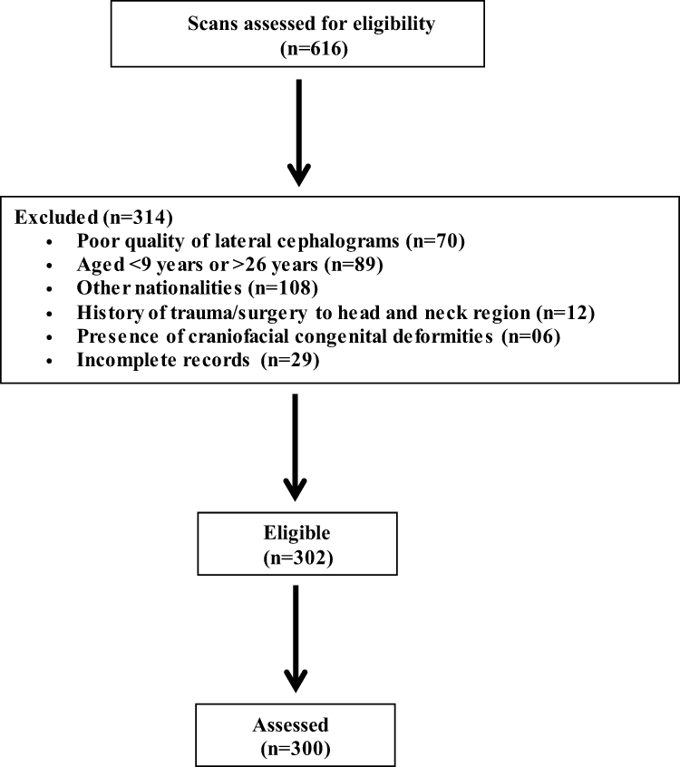 |
Figure 1 Flow chart of study participants. |
The following parameters were studied in the selected cephalograms-
Distribution into Skeletal Classes
Cephalograms are classified into the following groups based on the ANB angle (angle between Nasion, skeletal A-point, and skeletal B-point):
Class I- ANB angle between 1° and 3°;
Class II- ANB angle >4°
Class III- ANB angle <0°.
Furthermore, Wits analysis was utilized in order to overcome the shortcomings of ANB angle.
Size of Sella Turcica
Silverman10 and Kisling11 determined the linear dimensions (length, depth, and antero-posterior diameter) of the sella. A midsagittal line was used to evaluate the reference lines (Figure 2).
- The distance between the tuberculum and the tip of the dorsum sella was used to calculate the length of the sella.
- The depth was determined by drawing a line perpendicular from the line joining tuberculum sella and dorsum to the deepest point on the floor.
- The antero-posterior diameter of sella turcica was measured from the farthest point on postero-inferior aspect of the hypophyseal fossa to the superior most point on tuberculum sella.
 |
Figure 2 Normal sella turcica morphology and reference lines used for measuring sella size. |
Shape of Sella Turcica
We also studied five morphological variations of the sella (oblique anterior wall, double contour of the floor, sella turcica bridging, irregularity (notching) in the posterior part of the dorsum sella and pyramidal shape of dorsal sellae) as described by Axelsson et al.7
Additionally, the cephalograms were divided into two groups based on an individual’s age: prepubertal (9–14 years), as well as during or postpubertal (15–26 years). We based our classification on the findings of previous studies which showed that the shape of the sella does not change significantly after 12 years of age; females at about 15 years of age have completed their pubertal growth; the size of the sella in young adult females and males has been reported to be nearly identical, with the exception of when pregnant.6 To ensure consistency and reproducibility, the measurements were performed through automated Lateral Cephalometric analysis using a specialized Artificial Intelligence algorithm technique [Webceph (Korea)]. Two oral radiologists examined the selected radiographs independently using this software.
Measurement of Error
We selected 20 lateral cephalograms at random and reevaluated them after 14 days in order to reduce errors due to intra-operator variability. In this study, we utilized the intra-class correlation coefficient, which measured the reliability of the error.12 The average intra-class correlation coefficient was 0.931, which showed excellent reliability.
Statistical Analysis
The mean differences in linear dimensions of sella between different age groups and between genders were calculated using an independent t-test. The relationship between the skeletal type and the size of the sella turcica was studied using one-way ANOVA and the linear dimensions were assessed using a multiple comparison test. A regression analysis was used to evaluate the inter-relationships between age, gender, and skeletal type. SPSS software version 20.0 (IBM Corp., Armonk, NY, USA) was used for statistical analysis, with a significance level of P < 0.01.
Results
Based on age, gender, and skeletal type, the distribution of patients is shown in Table 1. There were 151 male patients (50.3%) and 149 female patients (49.7%) in the study. The mean age of the subjects was 19.8 years.
 |
Table 1 Distribution of Patients According to Age, Gender, and Skeletal Classes |
Size of the Sella Turcica
The mean values for length, diameter, and depth of sella were 11.57±4.73, 11.54±6.13, and 9.54±5.23 mm, respectively.
The linear dimensions of the sella turcica according to the age group are presented in Table 2. Age-wise comparisons revealed that all the linear measurements, ie, length, depth and antero-posterior diameter were significantly larger in patients belonging to 15–26 years age group as compared to that among 9–14 years age group (P < 0.001).
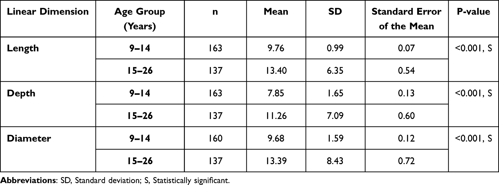 |
Table 2 Linear Dimensions of Sella (in Millimetres) According to Age Group |
The linear dimensions of sella turcica for males and females are shown in Table 3. When comparing linear dimensions of sella turcica between genders, it was found that females exhibited significantly larger linear dimensions as compared to the males (P < 0.001).
 |
Table 3 Linear Dimensions of Sella Turcica (in Millimetres) for Females and Males |
In order to determine if subjects with different skeletal patterns presented with different linear dimensions, irrespective of age or gender, a one-way ANOVA test was performed. Mean length, depth and antero-posterior diameter were found to be significantly different among different skeletal classes (P < 0.001). All the linear dimensions were found to be larger among patients with class III malocclusion as shown in Table 4.
 |
Table 4 One-Way ANOVA Testing the Effects of Skeletal Class on Sella Linear Dimensions (in Millimetres) |
Post hoc pairwise comparison of linear dimensions among different skeletal classes showed that mean length, depth and antero-posterior diameter among skeletal class III were significantly higher than that among skeletal classes I and II (P < 0.001; Table 5).
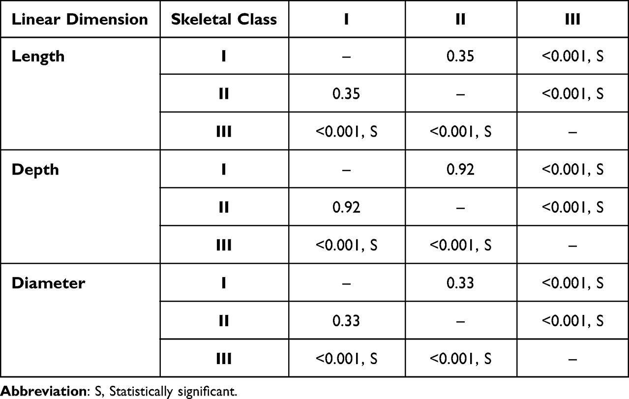 |
Table 5 Post Hoc Pairwise Comparison (Scheffe Test) Between Classes for Length, Depth & Diameter (in Millimetres) as the Dependent Variables |
Table 6 demonstrates the inter-relationship between the variables (age, gender, and skeletal type) and size of the sella turcica. The results revealed that age and skeletal type were significantly related to all the linear dimensions, ie, length, depth, and diameter (P < 0.001) whereas gender was only related to a change in length of the sella turcica (P<0.01).
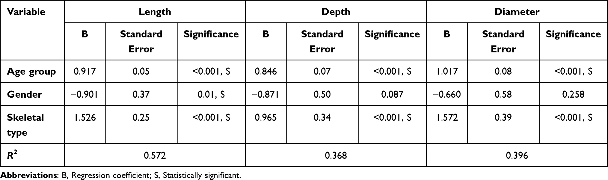 |
Table 6 Regression Analysis for Variables (Age Group, Gender, and Skeletal Type) and Linear Dimensions |
Shape of the Sella Turcica
The morphology of the sella turcica appeared to be normal in shape in the majority of patients (44.3%). Variation in morphological appearance was present in 55.7% of the individuals; oblique anterior wall was found in 22.0% of cases followed by double contour (17.3%), pyramidal shape (11.7%), sella bridging (3.7%) whereas the least frequent was irregular dorsum sella (1.0%).
On comparing the morphological variations between genders, sella turcica appeared normal in 43.0% of female patients and 45.6% of male patients. Furthermore, comparison between the two age groups showed that sella turcica appeared normal in 51.5% subjects belonging to 9–14 years age group and in 35.7% subjects belonging to 15–26 years age group. Variations in morphological of sella turcica appeared more common in female patients and in older age group. Table 7 demonstrates the frequency distribution of different shapes of sella turcica among genders and in two age groups.
 |
Table 7 Frequency Distribution of Different Shapes of Sella Turcica Among Both Genders and in Two Age Groups |
When the morphology was assessed for different skeletal types, sella turcica appeared normal in 50.5% of class I patients, 43.6% of class II patients and 35.6% of class III patients. Variations in morphological of sella turcica appeared more common in class III patients followed by class II patients and the least variation was noted in class I patients. Table 8 demonstrates the frequency distribution of morphological variations of sella among different skeletal types.
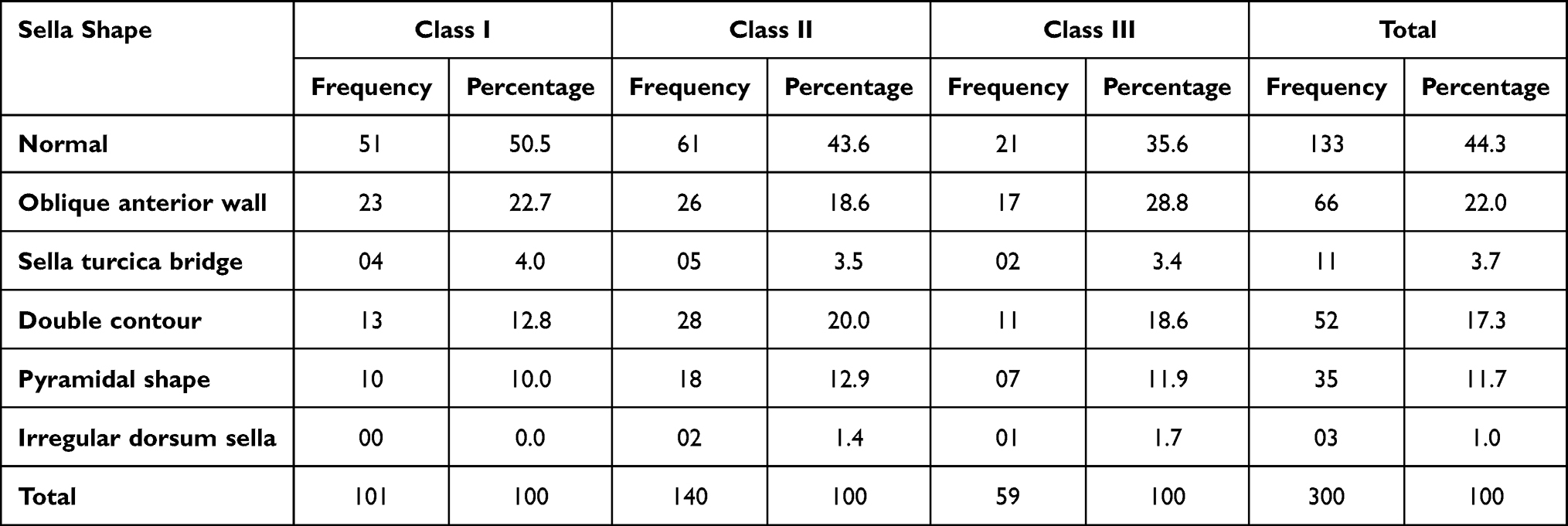 |
Table 8 Frequency Distribution of Different Shapes of Sella Turcica in Skeletal Classes |
Discussion
The evaluation of the sella turcica aids in the diagnosis and treatment of orthodontic problems. In addition, by superimposing the scans longitudinally, it is helpful for determining craniofacial growth.12 In this retrospective investigation, Saudi subpopulations with various skeletal types were examined for the linear dimensions and morphological appearance of the sella turcica.
As the person ages, the size of the sella correlates with the size of the pituitary gland.13,14 In this study, postpubertal age group individuals had significantly larger linear dimensions of the sella than their prepubertal counterparts. Similar findings were noted by Sathyanarayana et al,1 Yasa et al,3 Alkofide,6 Hasan et al,15 Hasan et al,16 Mustafa et al.17 However, the studies done by Al-Mohana et al4 and Valizadeh et al18 noticed significant differences only in antero-posterior diameter of sella among age groups. Muhammed et al8 also found that older patients had a larger diameter and longer length of their sella than younger patients. According to Nagaraj et al,19 the depth and antero-posterior diameter of the sella turcica increased as the age of the patient increased. According to Shrestha et al9 and Chaitanya et al,20 there was no significant difference in linear dimensions with age, although there was a gradual increase in linear dimensions of sella with age. Interestingly, Choi et al21 found that the linear dimensions of sella turcica showed a positive linear trend until 25 years of age, but after 26 years of age, the size of the sella was not significantly increased.
In order to determine if gender affected sella size, Chou et al22 utilized cone beam computed tomography to measure sella sizes. They found significant differences between sexes in the dimensions of sella, with males having larger dimensions. In contrast, Sathyanarayana et al,1 Al-Mohana et al4 and Axelsson et al7 observed a significant difference only in the length of the sella between the genders, whereas Ghaida et al23 observed only a significant difference in the height of the sella. According to Usman et al,24 computerized tomography was used to analyze the dimensions of the sella and concluded that males possess significantly larger sellae than females, especially regarding diameters and lengths. From approximately 1 to 13 years of age, Silverman10 found that males had a larger pituitary fossa than females. Due to the pubertal development spurt, which begins 2 years earlier in women than in males, the pituitary fossa in women significantly changes in size from 11 to 15 years of age. Later in life, male growth acceleration happen a couple of years later than female growth acceleration, giving both sexes nearly the same area of sella. Magat et al,25 Franscis26 and Haas,27 however, have reported that the sella turcica of females is larger than that of males. Unlike these studies, many researchers have found no significant difference in all three dimensions of the sella.3,6,9,15,18–20,28–34 In the current study, significantly larger dimensions of the sella were noted in female patients compared to the male patients. The differences between studies could be explained by the fact that study samples are from different age groups.
Correlation of skeletal patterns with sella size is still unclear. A study by Filipovie et al34 found that the size of the sella was larger in class III patients and smaller in class II patients. According to Akofide,6 individuals with skeletal class II patterns have a smaller sella diameter than individuals with skeletal class III patterns. It was found by Shrestha et al9 that patients with class III malocclusions had longer and wider sellas than those with class I and class II malocclusions. Individuals with the class III pattern had significantly larger diameters than patients with the class II pattern, according to Sinha et al.33 In the study done by Al-Mohana et al,4 individuals with class II had a larger sella depth compared to individuals with skeletal class III pattern. Valizadeh et al18 found that sella turcica was significantly higher in length in patients with skeletal class III malocclusion than in those with class I and class II skeletal types. Contrarily, Boddeti et al35 concluded that class I skeletal pattern shows larger dimensions of sella whereas Chaitanya et al,20 Magat et al,25 Shah et al,30 Meyer Marcotty et al,36 and Tepedino et al37 stated that there was insignificant difference between the skeletal patterns and linear dimension of sella turcica. We found that all the sella dimensions were larger in individuals with skeletal class III when compared to individuals with skeletal class I or II patterns. There might be differences in the results of previous studies as a result of different landmarks, radiographic techniques, or differences in the sample composition (age, gender, ethnicity, etc.).
The sella turcica has been described in several research studies as having different shapes.38–40 The variations in the sella turcica had been classified in a variety of ways, but in many cases the results were very difficult to interpret.38,41–43 In a recent study by Using Axelsson et al,7 six main types of seal shapes were distinguished; normal sella turcica, oblique anterior wall, double-contoured sella, sella turcica bridges, pyramidal shapes of dorsum sellae, and irregularities (notches) in the posterior part of the sella. 44.3% of participants in the present study had normal morphology, whereas 55.7% had variations in morphological appearance. This is similar to the findings of Al-Mohana et al4 who concluded that 56% of the patients had morphological variations in the sella shape and only 44% demonstrated normal morphology. This is also in accordance with the findings of Nagaraj et al19 and Magat et al.25 Researchers such as Sathyanarayana et al,1 Alkofide,6 Axelsson et al,7 Shrestha et al,9 Sinha et al,21 Usman et al,24 Shah et al,30 and Islam et al32 found normal sella morphology more common than individuals with other variations of morphology.
According to this study, oblique anterior walls made up 22.0% of the common morphological variant types. This is in agreement with studies by Chaitanya et al19 and Usman et al,24 where the authors found the oblique anterior wall as the most common variant (18.5% and 9.1% respectively). Additionally, Axelsson et al7 reported that 26% of patients had an oblique anterior wall, Magat et al25 reported that 14.4% of patients had an oblique anterior wall, Sinha et al33 found in 11.33% of patients, Alkofide6 found in 9.4% of subjects, and Islam et al32 noted in 4.8% of patients.
The occurrence of a sella bridge in normal individuals is not uncommon and has been estimated at between 0.0% and 22% of patients7,9,24,25,32,33,39,40,44 with an increase in occurrence in patients with craniofacial deviations.5 In the present study, sella turcica bridging was found in only 3.7% of the subjects which is within the range as previously reported.
The least frequent morphological type observed in this study was the irregular (notching) shape of sellae on the dorsum (1.0%), in agreement with Al-Mohana et al.4 On the contrary, it was the most frequent type in the studies done by Sathyanarayana et al,1 Alkofide6 and Shrestha et al.9 Additionally, Islam et al32 noted irregular dorsum sella in 16.2% of patients, Sinha et al33 noted in 10% of the subjects, Magat et al25 in irregular dorsum sella in 8.6% of patients, and Usman et al24 in 5.1% of patients. This study found normal morphology to be more common in males than females, along with class I and II skeletal patterns that are similar to Boddeti et al.35
The strength of this paper is equal distribution of patients with different malocclusions that provides better understanding which could not be found in previous studies. Also, the regression analysis for variables like age, gender and skeletal type authenticates the results that may be used as reference standards for Saudi patients when studying sella turcica morphology. Furthermore, the use of Artificial Intelligence based research tool ensures consistency and reproducibility.
Limitations
Several limitations were identified in the study. Firstly, the research was conducted at a single institution, it cannot be said that the study reflects the overall characteristics of the Saudi population. Second, only patients undergoing lateral cephalometric imaging were included in the study; as a result, caution should be exercised when interpreting the results. Finally, the cephalometric digital image was two-dimensional, which does not accurately depict the three-dimensional structure of the sella turcica. In spite of this, the dimensions reported in this study can still be used as a guideline.
Recommendations
Further studies on inter-relation between the dimensions of the sella turcica and pathological conditions should be conducted with the use of advanced imaging methods.
Conclusions
For all linear dimensions of the sella, there was a significant difference between pre-pubertal (9–14 years) and post-pubertal (15–26 years) age groups, with the size of the sella being greater in the post-pubertal age group. The size of the sella was significantly larger in females than males. There was a significant difference between different skeletal types when the sella size was compared. Sella size was found to be larger in class III patients. As age, gender, and skeletal type were compared to sella dimensions, age and skeletal type were significantly correlated with each dimension, whereas gender was significantly correlated only with a change in length of the sella. 44.3% of patients had normal sella turcica shape. This study provided reference data for assessing racial, gender and age-specific variations in sella turcica in Saudi Arabian patients based on the linear dimensions and morphological types of sella turcica.
Data Sharing Statement
The data set used in the current study will be made available on request from Dr. Rakhi Issrani; [email protected].
Disclosure
The authors report no conflicts of interest in this work.
References
1. Sathyanarayana HP, Kailasam V, Chitharanjan AB. The size and morphology of sella turcica in different skeletal patterns among South Indian population: a lateral cephalometric study. J Ind Orthod Soc. 2013;47(4):266–271. doi:10.1177/0974909820130507S
2. Kjaer I. Sella turcica morphology and the pituitary gland-a new contribution to craniofacial diagnostics based on histology and neuroradiology. Eur J Orthod. 2015;37(1):28–36. doi:10.1093/ejo/cjs091
3. Yasa Y, Bayrakdar IS, Ocak A, Duman SB, Dedeoglu N. Evaluation of sella turcica shape and dimensions in cleft subjects using cone-beam computed tomography. Med Princ Pract. 2017;26(3):280–285. doi:10.1159/000453526
4. Al-Mohana RA, Muhammed FK, Li X, Lubamba GP. The bridging and normal dimensions of sella turcica in Yemeni individuals. Oral Radiol. 2022;38(1):162–170. doi:10.1007/s11282-021-00541-7
5. Becktor JP, Einersen S, Kjaer I. A sella turcica bridge in subjects with severe craniofacial deviations. Eur J Orthod. 2000;22(1):69–74. doi:10.1093/ejo/22.1.69
6. Alkofide EA. The shape and size of the sella turcica in skeletal Class I, Class II, and Class III Saudi subjects. Eur J Orthod. 2007;29(5):457–463. doi:10.1093/ejo/cjm049
7. Axelsson S, Storhaug K, Kjaer I. Post-natal size and morphology of the sella turcica. Longitudinal cephalometric standards for Norwegians between 6 and 21 years of age. Eur J Orthod. 2004;26(6):597–604. doi:10.1093/ejo/26.6.597
8. Muhammed FK, Abdullah AO, Liu Y. Morphology, incidence of bridging, dimensions of sella turcica, and cephalometric standards in three different racial groups. J Craniofac Surg. 2019;30(7):2076–2081. doi:10.1097/SCS.0000000000005964
9. Shrestha GK, Pokharel PR, Gyawali R, Bhattarai B, Giri J. The morphology and bridging of the sella turcica in adult orthodontic patients. BMC Oral Health. 2018;18(1):45. doi:10.1186/s12903-018-0499-1
10. Silverman FN. Roentgen standards for size of the pituitary fossa from infancy through adolescence. Am J Roentgenol Radium Ther Nucl Med. 1957;78(3):451–460. PMID: 13458563.
11. Kisling E. Cranial Morphology in Down’s Syndrome. A Comparative Roentgencephalometric Study in Adult Males [Thesis]. Copenhagen; Munksgaard; 1966.
12. Shoukri MM. Measures of Interobserver Agreement. Boca Raton: Chapman & Hall/CRC Press; 2004:5–10.
13. Sari S, Sari E, Akgun V, et al. Measures of pituitary gland and stalk: from neonate to adolescence. J Pediatr Endocrinol Metab. 2014;27(11–12):1071–1076. doi:10.1515/jpem-2014-0054
14. Chauhan P, Kalra S, Mongia SM, Ali S, Anurag A. Morphometric analysis of sella turcica in North Indian population: a radiological study. Int J Res Med Sci. 2014;2(2):521–526. doi:10.5455/2320-6012.ijrms20140529
15. Hasan HA, Alam MK, Abdullah YJ, et al. 3DCT morphometric analysis of sella turcica in Iraqi population. J Hard Tissue Biol. 2016;25:227–232. doi:10.2485/jhtb.25.227
16. Hasan HA, Alam MK, Yusof A, Mizushima H, Kida A, Osuga N. Size and morphology of sella turcica in Malay populations: a 3D CT study. J Hard Tissue Biol. 2016;25:313–320. doi:10.2485/jhtb.25.313
17. Mustafa AG, Abu Ghaida JH, Mistareehi AJ, Allouh MZ, Mistarihi SM. A cephalometric morphometric study of age- and gender-dependent shape patterns of the sella turcica. Italian J Anat Embryol. 2018;123(1):32–45.
18. Valizadeh S, Shahbeig S, Mohseni S, Azimi F, Bakhshandeh H. Correlation of shape and size of sella turcica with the type of facial skeletal class in an Iranian group. Iran J Radiol. 2015;12(3):e16059. doi:10.5812/iranjradiol.12(3)2015.16059
19. Nagaraj T, Shruthi R, James L, Keerthi I, Balraj L, Goswami RD. The size and morphology of sella turcica: a lateral cephalometric study. J Med Radiol Pathol. 2015;1:3–7. doi:10.15713/ins.jmrps.14
20. Chaitanya B, Pai KM, Chhaparwal Y. Evaluation of the effect of age, gender, and skeletal class on the dimensions of sella turcica using lateral cephalogram. Contemp Clin Dent. 2018;9(2):195–199. doi:10.4103/ccd.ccd_805_17
21. Choi WJ, Hwang EH, Lee SR. The study of shape and size of normal sella turcica in cephalometric radiographs. Korean J Oral Maxillofac Radiol. 2001;31:43–49.
22. Chou ST, Chen CM, Chen PH, Chen YK, Chen SC, Tseng YC. Morphology of sella turcica and bridging prevalence correlated with sex and craniofacial skeletal pattern in Eastern Asia population: CBCT study. Biomed Res Int. 2021;6646406. doi:10.1155/2021/6646406
23. Abu Ghaida JH, Mistareehi AJ, Mustafa AG, Mistarihi SM, Ghozlan HH. The normal dimensions of the sella turcica in Jordanians: a study on lateral cephalograms. Folia Morphol. 2017;76(1):1–9. doi:10.5603/FM.a2016.0038
24. Usman Z, Zagga AD, Yunusa GH, et al. Shapes and sizes of sella turcica using Computerized Tomography (CT) from tertiary hospital in Sokoto. Asian J Med Health. 2020;18(1):8–15. doi:10.9734/ajmah/2020/v18i130175
25. Magat G, Ozcan Sener S. Morphometric analysis of the sella turcica in Turkish individuals with different dentofacial skeletal patterns. Folia Morphol. 2018;77(3):543–550. doi:10.5603/FM.a2018.0022
26. Francis CC. Growth of the human pituitary fossa. Hum Biol. 1948;20(1):1–20. PMID: 18912277.
27. Haas LL. The size of the sella turcica by age and sex. Am J Roentgenol. 1954;72:754–761.
28. Konwar SK, Singhla A, Bayan R. Morphological (length, depth, and diameter) study of sella turcica in different mandibular growth patterns in Indians. IJDMS. 2016;3(3):4–9. doi:10.5958/2394-4196.2016.00009.1
29. Otuyemi O, Fadeju A, Adesina B, Otuyemi D. A Cephalometric analysis of the morphology and size of sella turcica in Nigerians with normal and bimaxillary incisor protrusion. J West Afr Coll Surg. 2017;7(2):93–111. PMID: 29951468.
30. Shah A, Bashir U, Ilyas T. The shape and size of the sella turcica in skeletal class I, II and III in patients presenting at Islamic International Dental Hospital, Islamabad. Pak Oral Dent J. 2011;31(1):102–108.
31. Elnour H, Al-Otaibi A, Al-Otaibi M, et al. Evaluation of shape and size of sella turcica using computerized tomography in Saudi populations. Sch J Appl Med Sci. 2020;8:2058–2063. doi:10.36347/sjams.2020.v08i09.016
32. Islam M, Alam MK, Yusof A, et al. 3D CT Study of morphological shape and size of sella turcica in Bangladeshi population. J Hard Tissue Biol. 2017;26(1):1–6. doi:10.2485/jhtb.26.1
33. Sinha S, Shetty A, Nayak K. The morphology of sella turcica in individuals with different skeletal malocclusions – a cephalometric study. Transl Res Anat. 2020;18:100054. doi:10.1016/j.tria.2019.100054
34. Filipovie G, Burie M, Janosevie M, Stosie M. Radiological measuring of sella turcica’s size in different malocclusions. Acta Stomatologica Naissi. 2011;27:1035–1042. doi:10.5937/asn1163035F
35. Boddeti SS, Varma NS, Sasidhar YN, Ujwala T, Navya P. The size and the morphology of sella turcica in different skeletal patterns of South Indians: a lateral cephalogrametric study. Int J Oral Health Med Res. 2016;3(1):13–16.
36. Meyer-Marcotty P, Reuther T, Stellzig-Eisenhauer A. Bridging of the sella turcica in skeletal Class III subjects. Eur J Orthod. 2010;32(2):148–153. doi:10.1093/ejo/cjp081
37. Tepedino M, Laurenziello M, Guida L, et al. Morphometric analysis of sella turcica in growing patients: an observational study on shape and dimensions in different sagittal craniofacial patterns. Sci Rep. 2019;9(1):19309. doi:10.1038/s41598-019-55916-y
38. Gordon MB, Bell AL. A roentgenographic study of the sella turcica in normal children. N Y State J Med. 1922;22:54–59.
39. Kantor ML, Norton LA. Normal radiographic anatomy and common anomalies seen in cephalometric films. Am J Orthod Dentofacial Orthop. 1987;91(5):414–426. doi:10.1016/0889-5406(87)90394-5
40. Tetradis S, Kantor ML. Prevalence of skeletal and dental anomalies and normal variants seen in cephalometric and other radiographs of orthodontic patients. Am J Orthod Dentofacial Orthop. 1999;116(5):572–577. doi:10.1016/s0889-5406(99)70191-5
41. Davidoff LM, Epstein BS. The Abnormal Pneumoencephalogram. Philadelphia: Lea and Febiger; 1950.
42. Fournier AM, Denizet D. La selle turcique en omega. [Omega-shaped sella turcica]. Mars Med. 1965;102(6):503–509. French. PMID: 5294513.
43. Kier EL. “J” and “omega” shape of sella turcica. Anatomic clarification of radiologic misconceptions. Acta Radiol Diagn. 1969;9:91–94. PMID: 5381081.
44. Camp JD. Normal and pathological anatomy of the sella turcica as revealed by roentgenograms. Am J Roentgenol. 1924;12:143–156.
 © 2023 The Author(s). This work is published and licensed by Dove Medical Press Limited. The full terms of this license are available at https://www.dovepress.com/terms.php and incorporate the Creative Commons Attribution - Non Commercial (unported, v3.0) License.
By accessing the work you hereby accept the Terms. Non-commercial uses of the work are permitted without any further permission from Dove Medical Press Limited, provided the work is properly attributed. For permission for commercial use of this work, please see paragraphs 4.2 and 5 of our Terms.
© 2023 The Author(s). This work is published and licensed by Dove Medical Press Limited. The full terms of this license are available at https://www.dovepress.com/terms.php and incorporate the Creative Commons Attribution - Non Commercial (unported, v3.0) License.
By accessing the work you hereby accept the Terms. Non-commercial uses of the work are permitted without any further permission from Dove Medical Press Limited, provided the work is properly attributed. For permission for commercial use of this work, please see paragraphs 4.2 and 5 of our Terms.