
Back to Journals » International Journal of Women's Health » Volume 18

Pulmonary Benign Metastasizing Leiomyoma with Abdominal Wall and Pelvic Cavity Implantation: A Case Report and Literature Review
Authors Liang D, Cao Y  , Shi H
Received 18 April 2026
Accepted for publication 19 June 2026
Published 9 July 2026 Volume 2026:18 618061
DOI https://doi.org/10.2147/IJWH.S618061
Checked for plagiarism Yes
Review by Single anonymous peer review
Peer reviewer comments 2
Editor who approved publication: Dr Vinay Kumar
Video abstract of “Leiomyoma with abdominal wall and pelvic cavity implantation” [618061].
Views: 6
Dandan Liang, Yang Cao, Honghui Shi
National Clinical Research Center for Women’s Health and Obstetric and Gynecologic Diseases, Department of Obstetrics and Gynecology, Peking Union Medical College Hospital, Chinese Academy of Medical Sciences & Peking Union Medical College, Beijing, People’s Republic of China
Correspondence: Honghui Shi, National Clinical Research Center for Women’s Health and Obstetric and Gynecologic Diseases, Department of Obstetrics and Gynecology, Peking Union Medical College Hospital, Chinese Academy of Medical Sciences & Peking Union Medical College, Beijing, People’s Republic of China, Email [email protected]
Abstract: Benign metastasizing leiomyoma (BML) is a rare condition characterized by extrauterine proliferation of benign smooth muscle cells. We present a rare instance of pulmonary BML with concurrent abdominal wall and pelvic implantation. A 47-year-old female presented with palpable abdominal wall nodules and pelvic lesions following a history of myomectomy and hysterectomy. Imaging revealed vascularized nodules in the abdominal wall, rectal mesentery, and bilateral lungs. Surgical excision of abdominal and pelvic nodules confirmed cellular leiomyoma. Post-oophorectomy, lung nodules initially regressed but subsequently progressed. Wedge resection confirmed pulmonary BML. Literature review highlights substantial heterogeneity in BML cases. Although many cases are associated with prior uterine surgery, cases without surgical history suggest that spontaneous dissemination may also occur. In the present case, the coexistence of pulmonary, abdominal wall, and pelvic lesions raises the possibility that hematogenous or lymphatic dissemination and iatrogenic implantation may have coexisted, although this hypothesis requires further molecular validation. Standardization in the definition and classification of BML may help improve diagnosis and management. Careful surgical handling, incision protection, and long-term follow-up are warranted in patients with recurrent or atypically behaving leiomyomas.
Keywords: benign metastasizing leiomyoma, abdominal wall implantation, pulmonary metastases, iatrogenic dissemination, case report
Introduction
Benign metastasizing leiomyoma (BML) was first reported by Steiner in 1939 when it was named fibroleiomyomatous hamartoma. It is an uncommon condition in which solitary or multiple benign aggregates of smooth muscle cells are present at a distant location, most commonly the lungs.1 Clonality studies suggest that BML nodules are derived from uterine leiomyomas, spreading by vascular or lymphatic channels.2,3 Further cytogenetic data identified recurrent 19q and 22q deletions in both uterine and metastasized leiomyomas,4 indicating a subset may be predisposed to disseminate. Here we report a case of pulmonary benign metastasizing leiomyoma (PBML) with abdominal wall and pelvic cavity implantation and discuss the possible pathogenesis of multiple lesions.
Pathologically, BML is characterized by extrauterine proliferation of smooth muscle cells with morphologically benign features, usually lacking significant cytological atypia, coagulative tumor cell necrosis, or high mitotic activity. Immunohistochemistry typically supports smooth muscle differentiation and hormone responsiveness, with expression of smooth muscle markers and estrogen and progesterone receptors. Despite its benign histological appearance, BML can present with distant lesions, most commonly in the lungs, creating diagnostic and conceptual challenges. Proposed mechanisms include hematogenous or lymphatic dissemination from uterine leiomyoma, iatrogenic implantation during prior uterine surgery, and underlying molecular or cytogenetic alterations that may predispose a subset of leiomyomas to disseminate. When pulmonary lesions coexist with abdominal wall or pelvic lesions, a single mechanism may be insufficient to explain the full disease distribution.
This case report has been reported in line with the SCARE 2025 guidelines.5
Case Presentation
Clinical History
A 47‑year‑old G1P1 female presented with palpable nodules on the right lower abdominal wall for 4 years, accompanied by intermittent pain. She underwent two abdominal myomectomies in other hospitals in 2008 and 2016, respectively. In 2019, a transabdominal hysterectomy and bilateral salpingectomy were performed for recurrent uterine fibroids. Four months after the operation, she noticed palpable nodules in the right lower abdomen along the incision, with intermittent stabbing pain increasing in frequency and severity, along with urinary frequency, hesitancy, and nocturia up to 4 times per night.
Diagnostic Assessment
In December 2023, the patient presented to our institution. Ultrasonography revealed multiple irregular hypoechoic lesions with clear borders and rich blood flow in the lower abdominal wall. A well‑defined heterogeneous nodule with rich blood flow was identified in the anterior aspect of the rectum (Figure 1). Contrast‑enhanced magnetic resonance imaging (MRI) showed multiple round lesions with T1 isointense and T2 slightly hyperintense to hypointense signals in the lower abdominal wall from the midline to the right rectus muscle, and in the left anterior aspect of the rectum, demonstrating significant enhancement (Figure 2). CA19‑9 and CA‑125 levels were within the normal range.
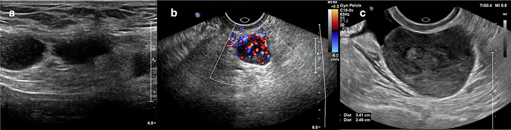 |
Figure 1 Ultrasonography showing multiple irregular hypoechoic lesions with clear borders (a) and rich blood flow (b) in the lower abdominal wall. A well-defined heterogeneous nodule with rich blood flow measuring 3.4×2.8×2.5 cm was identified in the anterior aspect of the rectum (c). |
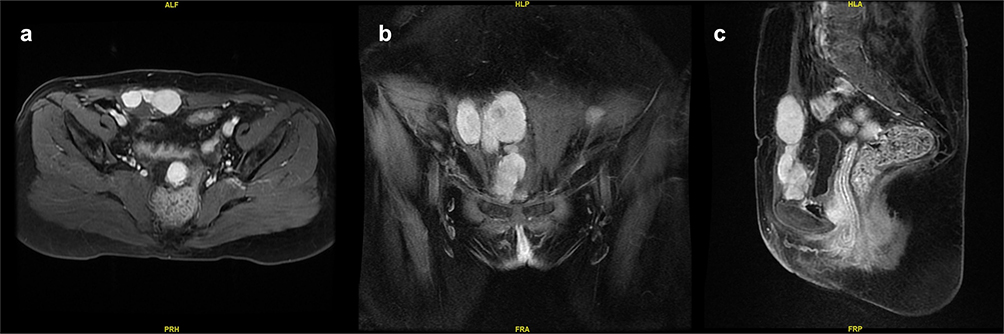 |
Figure 2 Pelvic magnetic resonance imaging findings. (a) Axial image showing multiple nodules in the lower anterior abdominal wall and a pelvic lesion anterior to the rectum. (b) Coronal image showing multiple nodules extending from the midline to the right lower abdominal wall/rectus region. (c) Sagittal image showing the spatial relationship between the abdominal wall lesions and the pelvic lesion anterior to the rectum. The lesions showed marked enhancement on contrast-enhanced MRI, and the largest abdominal wall lesion measured approximately 32 mm × 23 mm × 47 mm. |
Pathological consultation of the 2019 hysterectomy specimen revealed multiple uterine leiomyomas, with one submucosal cellular leiomyoma showing degeneration and occasional mitotic activity, raising concern for uncertain malignant potential. Due to the uncertain malignant potential of the leiomyoma, a chest CT scan was performed, showing multiple nodules in both lungs, with the largest measuring approximately 13.5 mm in the lingula of the superior lobe of the left lung (Figure 3). The trachea and bronchi were patent, and no significant mediastinal lymphadenopathy was observed. BML was suspected.
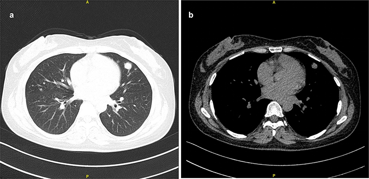 |
Figure 3 Chest computed tomography findings. (a) Lung-window axial CT image showing multiple bilateral pulmonary nodules, including the largest nodule in the lingula of the left upper lobe. (b) Mediastinal-window axial CT image showing the same lesion. The largest lesion measured approximately 13.5 mm × 12.2 mm. |
Management
After consultation with a thoracic surgeon, it was decided to address the symptomatic abdominal and pelvic lesions first, performing bilateral oophorectomy simultaneously, and then observe the postoperative changes of the lung nodules. The patient underwent exploratory laparotomy on December 5, 2023 (Figure 4). A right paramedian incision was employed. The nodules in the abdominal wall were beneath the anterior rectus sheath with relatively clear boundaries to the surrounding muscles; however, there were no distinct boundaries at the base of the nodules with the anterior rectus sheath. A 3‑cm nodule was observed in the anterior aspect of the mesorectum and removed completely. Bilateral oophorectomy was performed on grossly normal‑looking ovaries. The estimated blood loss was 100 mL, and postoperative recovery was uneventful.
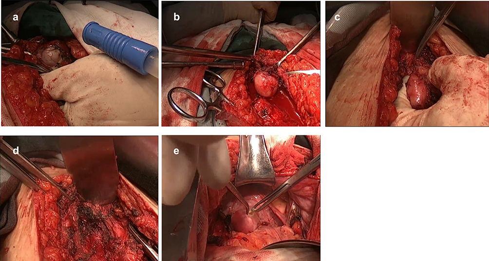 |
Figure 4 Intraoperative findings: Nodules on the left side (a) and right side (b) of the incision. A nodule protruding from the pubic bone (c); note the lack of clear boundary between the nodule base and the pubic bone. All visible lesions at the base were excised and cauterized (d). A 3 cm nodule located in the anterior aspect of the mesorectum (e). |
Pathology
Pathology revealed spindle cell tumors consistent with cellular leiomyoma for both abdominal wall nodules and the mesorectal nodule. Immunohistochemistry (IHC) of the abdominal and mesorectal nodules was slightly different, with a Ki‑67 index of 5% in the abdominal wall lesions and 20% in the mesorectal lesion (Figure 5).
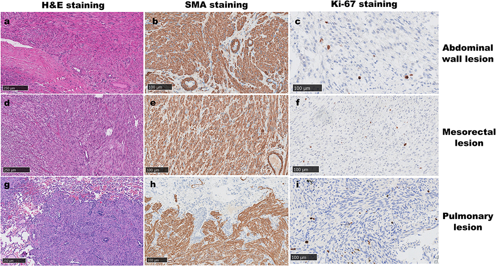 |
Figure 5 Representative histopathological and immunohistochemical findings of the abdominal wall, mesorectal, and pulmonary lesions. (a) H&E staining of the abdominal wall lesion showing spindle cell proliferation with benign smooth muscle morphology. (b) SMA staining of the abdominal wall lesion showing positivity for smooth muscle differentiation. (c) Ki-67 staining of the abdominal wall lesion showing proliferative activity. (d) H&E staining of the mesorectal lesion showing spindle cell proliferation with benign smooth muscle morphology. (e) SMA staining of the mesorectal lesion showing positivity for smooth muscle differentiation. (f) Ki-67 staining of the mesorectal lesion showing proliferative activity. (g) H&E staining of the pulmonary lesion showing spindle cell proliferation in the lung parenchyma, without definite cytological atypia or coagulative tumor cell necrosis, consistent with benign metastasizing leiomyoma. (h) SMA staining of the pulmonary lesion showing positivity for smooth muscle differentiation. (i) Ki-67 staining of the pulmonary lesion showing proliferative activity. Together, these findings support smooth muscle differentiation and suggest heterogeneous proliferative activity among lesions from different anatomical sites. Scale bars: 250 μm in (a, d, and g); 100 μm in (b, c, e, f, h, and i). |
Follow-Up and Definitive Diagnosis
Postoperative chest CT scans in March 2024 revealed partial remission compared to previous imaging, with a decrease in the size of the largest lingular nodule. However, in August 2024, CT showed an increase in the number and size of nodules in both lungs, with the largest lingular nodule increasing to approximately 17 mm in diameter. To establish a definitive diagnosis, the patient underwent wedge resection of the left upper and lower lobes at another hospital in September 2024. Pathology revealed multiple spindle cell tumors in the lung tissue, suggestive of smooth muscle origin tumors. There was no definite cellular atypia or coagulative necrosis, with rare mitotic figures, morphologically consistent with benign metastasizing leiomyoma. No tumors were observed in the lung pleura or staple line. IHC showed positive staining for smooth muscle markers with a Ki‑67 index of 30%. The patient is under regular follow‑up, with pelvic MRI in October 2024 showing no signs of recurrence.
Discussion
In a systematic review including 385 cases of benign metastasizing leiomyoma (BML), the most common metastatic site was the lungs (330/385, 85.7%), followed by the pelvic/abdominal cavity and retroperitoneum (17.9%). Multisite metastases were observed in 16.6% of cases.6 In another systematic review of 161 cases, the mean age at primary uterine surgery and BML diagnosis was 38.5 and 47.3 years, respectively, and lung metastases were documented in 137/161 (85.1%) cases.7 A series of 23 PBML cases from a single facility included patients without a history of leiomyoma or without surgical treatment before PBML diagnosis.8 In a case report and literature review of 12 cases of BML with abdominal wall metastasis, only 5/12 (41.7%) cases had concomitant pulmonary metastasis, and 5/12 (41.7%) had undergone hysterectomy before BML diagnosis, deviating from the previous systematic reviews.9
A thorough analysis of these data suggests several points. First, a proportion of patients had no history of uterine leiomyoma, which cannot be explained by vascular or lymphatic spread of leiomyoma cells and may suggest a different pathogenesis for BML. Second, some patients have a history of leiomyoma but no prior uterine surgery, indicating that metastasis of leiomyoma cells can occur spontaneously without surgical manipulation. However, 78.3% to 84.8% of patients had a history of uterine surgery prior to BML diagnosis, raising concerns that surgical procedures may potentially promote or exacerbate metastasis.6–8
These findings suggest that BML is unlikely to represent a single homogeneous entity. Clonality studies and recurrent cytogenetic alterations support the concept that at least some BML lesions originate from uterine leiomyomas and disseminate through hematogenous or lymphatic routes. In contrast, abdominal wall lesions occurring near prior surgical incisions are more compatible with local iatrogenic implantation, particularly after myomectomy or hysterectomy. Therefore, pulmonary and abdominal wall/pelvic lesions may not necessarily share the same route of spread, even when they coexist in the same patient.
In the two systematic reviews, the incidence of lung metastasis in BML was around 85%, and hysterectomy was the most common surgery before BML.6,7 However, in the 12 cases of BML with concomitant abdominal wall metastasis, the rate of lung metastasis was only 41.7%, and myomectomy accounted for 58.3% of previous surgeries.9 Abdominal wall nodules are more likely to result from direct implantation of leiomyoma cells onto the incision during myomectomy. Sandberg et al reported positive cytology for the presence of leiomyoma cells in post‑myomectomy washings, supporting this viewpoint.10 Furthermore, we hypothesize that mechanical compression of the uterus during hysterectomy may have contributed to the hematogenous spread of leiomyoma cells, while surgical manipulation could be associated with direct implantation from leiomyoma cell spillage onto the incision. However, the exact role of compression-induced vascular spread remains a hypothesis requiring further investigation. Therefore, it is important to adhere to tumor‑free principles and utilize effective incision protection devices during surgery to reduce implantation at the incision site.
This highlights the diagnostic challenges in BML due to unclear pathogenesis. The current definition of BML may encompass diseases with different pathogenic mechanisms but similar clinical and pathological presentations. The term “metastasizing” is more suitable for diseases that spread through vascular or lymphatic routes, but not direct implantation. For patients without a history of uterine leiomyoma, pulmonary lesions should be differentiated from sporadic pulmonary lymphangioleiomyomatosis (LAM). Standardizing the definition and reporting of BML can help clarify its pathogenesis and implement appropriate preventive measures.6–9,11,12
In the present case, the coexistence of pulmonary lesions with abdominal wall and mesorectal nodules raises the possibility that more than one dissemination pathway may have been involved. The pulmonary lesions may reflect hematogenous or lymphatic spread from a uterine smooth muscle tumor, whereas the abdominal wall lesions along the prior incision are more compatible with iatrogenic implantation. However, the precise sequence and mechanism of dissemination cannot be determined from this single case. Alternative explanations should also be considered. Therefore, the proposed relationship among the uterine, abdominal wall, mesorectal, and pulmonary lesions should be interpreted cautiously. For leiomyoma patients experiencing multiple recurrences, reevaluation of pathology and close postoperative follow‑up are crucial. In cases where pathology indicates increased cellularity and/or uncertain malignant potential, leiomyomas may be predisposed to dissemination.11 Treatment should be based on the patient’s age, fertility desire, tumor location, number, growth rate, and resectability,12 rather than whether the tumor originated from metastasis or implantation. In our case, the Ki-67 index varied across anatomical sites, with 5% in the abdominal wall, 20% in the mesorectal lesion, and 30% in the pulmonary lesion, raising the possibility of site-specific proliferative behavior or microenvironmental influences.
Many patients with metastasizing leiomyoma experience an indolent course. Expectant management is an option for asymptomatic disease.6 Such tumors are hormone dependent and express estrogen and progesterone receptors. Use of agents that decrease or modulate estrogen or progesterone, or anti‑angiogenesis agents, has resulted in tumor regression.13 Surgical interventions, including resection of metastatic lesions, uterus, and bilateral ovaries, can be considered; however, the role of oophorectomy in BML remains unclear. In this case, the pulmonary nodule shrank and later enlarged again after castration surgery. Therefore, for young women with BML, especially those with asymptomatic multiple nodules, ovary‑sparing therapy and close monitoring could be more appropriate.
This case has several limitations. First, molecular or clonality analyses were not performed; therefore, we could not definitively confirm that the uterine, abdominal wall, mesorectal, and pulmonary lesions shared a common clonal origin. Second, although the coexistence of pulmonary lesions and abdominal wall/pelvic nodules raises the possibility of both hematogenous dissemination and iatrogenic implantation, the exact mechanism and sequence of spread cannot be proven in a single case. Third, interpretation of proliferative activity across different anatomical sites should be cautious, because Ki-67 assessment may be affected by sampling, hotspot selection, technical differences, interobserver variation, and tumor heterogeneity. Further molecular studies and accumulation of similar cases are needed to clarify the pathogenesis of BML with extrapulmonary involvement.
Conclusion
This case highlights the diagnostic and conceptual challenges of BML, especially when abdominal wall and pelvic cavity implantation coexist with pulmonary involvement. The coexistence of lesions at these sites suggests, but does not prove, that hematogenous or lymphatic dissemination and iatrogenic implantation may coexist in some patients. Molecular or clonality studies are needed to validate this proposed mechanism. Clinically, this case emphasizes the importance of meticulous surgical technique, effective incision protection, differential diagnosis of pulmonary nodules, pathological reassessment, and long-term surveillance in patients with recurrent or atypically behaving uterine leiomyomas. Standardization in the definition, classification, and reporting of BML may help improve diagnosis, management, and prevention strategies.
Ethics Approval and Informed Consent
Ethical approval was waived by the Ethics Committee of Peking Union Medical College Hospital because this study is a single case report and did not involve any additional intervention beyond routine clinical care. All patient information was anonymized before submission.
Consent for Publication
Written informed consent was obtained from the patient for publication of this case report and accompanying images.
Acknowledgments
We are grateful to the patient who provided consent and clinical information for this case report.
Author Contributions
All authors made a significant contribution to the work reported, whether that is in the conception, study design, execution, acquisition of data, analysis and interpretation, or in all these areas; took part in drafting, revising or critically reviewing the article; gave final approval of the version to be published; have agreed on the journal to which the article has been submitted; and agree to be accountable for all aspects of the work.
Funding
This research received no funding.
Disclosure
The authors report no conflicts of interest in this work.
References
1. Clark DH, Weed JC. Metastasizing leiomyoma: a case report. Am J Obstet Gynecol. 1977;127(6):672–7. doi:10.1016/0002-9378(77)90373-8
2. Patton KT, Cheng L, Papavero V, et al. Benign metastasizing leiomyoma: clonality, telomere length and clinicopathologic analysis. Mod Pathol. 2006;19(1):130–140. doi:10.1038/modpathol.3800504
3. Tietze L, Günther K, Hörbe A, et al. Benign metastasizing leiomyoma: a cytogenetically balanced but clonal disease. Hum Pathol. 2000;31(1):126–128. doi:10.1016/S0046-8177(00)80210-5
4. Nucci MR, Drapkin R, Dal Cin P, Fletcher CDM, Fletcher JA. Distinctive cytogenetic profile in benign metastasizing leiomyoma: pathogenetic implications. Am J Surg Pathol. 2007;31(5):737–743. doi:10.1097/01.pas.0000213414.15633.4e
5. Kerwan A, Al-Jabir A, Mathew G, et al. Revised surgical case report (SCARE) guideline: an update for the age of artificial intelligence. Premier J Sci. 2025;10:100079.
6. Liu Y, Li D, Li X, Wang J, Wang J. Benign metastasizing leiomyoma: is “wait and watch” strategy feasible? Reprod Sci. 2023;30(12):3568–3577. doi:10.1007/s43032-023-01314-9
7. Barnaś E, Książek M, Raś R, Skręt A, Skręt-Magierło J, Dmoch-Gajzlerska E. Benign metastasizing leiomyoma: a review of current literature in respect to the time and type of previous gynecological surgery. PLoS One. 2017;12(4):e0175875. doi:10.1371/journal.pone.0175875
8. Fan R, Feng F, Yang H, et al. Pulmonary benign metastasizing leiomyomas: a case series of 23 patients at a single facility. BMC Pulm Med. 2020;20(1):292. doi:10.1186/s12890-020-01330-4
9. Hu J, Tian S, Pan Q, Yu Y. Multiple benign metastasizing leiomyoma in the abdominal wall: a case report and literature review. Front Oncol. 2024;14:1391850. doi:10.3389/fonc.2024.1391850
10. Sandberg EM, van den Haak L, Bosse T, Jansen FW. Disseminated leiomyoma cells can be identified following conventional myomectomy. BJOG. 2016;123(13):2183–2187. doi:10.1111/1471-0528.14265
11. Momeni-Boroujeni A, Nucci MR, Chapel DB. Risk stratification of uterine smooth muscle tumors: the role of morphology, immunohistochemistry, and molecular testing. Adv Anat Pathol. 2025;32(1):44–56. doi:10.1097/PAP.0000000000000478
12. Bucuri CE, Ciortea R, Malutan AM, et al. Disseminated peritoneal leiomyomatosis-a challenging diagnosis-mimicking malignancy scoping review of the last 14 years. Biomedicines. 2024;12(8):1749. doi:10.3390/biomedicines12081749
13. Yang JW, Hua Y, Xu H, He L, Huo HZ, Zhu CF. Treatment of leiomyomatosis peritonealis disseminata with goserelin acetate: a case report and review of the literature. World J Clin Cases. 2021;9(19):5217–5225. doi:10.12998/wjcc.v9.i19.5217
 © 2026 The Author(s). This work is published and licensed by Dove Medical Press Limited. The
full terms of this license are available at https://www.dovepress.com/terms
and incorporate the Creative Commons Attribution
- Non Commercial (unported, 4.0) License.
By accessing the work you hereby accept the Terms. Non-commercial uses of the work are permitted
without any further permission from Dove Medical Press Limited, provided the work is properly
attributed. For permission for commercial use of this work, please see paragraphs 4.2 and 5 of our Terms.
© 2026 The Author(s). This work is published and licensed by Dove Medical Press Limited. The
full terms of this license are available at https://www.dovepress.com/terms
and incorporate the Creative Commons Attribution
- Non Commercial (unported, 4.0) License.
By accessing the work you hereby accept the Terms. Non-commercial uses of the work are permitted
without any further permission from Dove Medical Press Limited, provided the work is properly
attributed. For permission for commercial use of this work, please see paragraphs 4.2 and 5 of our Terms.
Recommended articles
A Case of Microwave Ablation Activating Immune Response in Pulmonary Metastases from Hepatocellular Carcinoma
Wan J, Jin C, Li C, Zhang W, Zhou Y, Yu C, Fan Z, Qu X
Journal of Hepatocellular Carcinoma 2026, 13:532524
Published Date: 17 March 2026