
Back to Journals » Drug Design, Development and Therapy » Volume 19
Prospects and Challenges of Chitosan-Based Drug Carriers for Anticancer Agents’ Delivery Against Lung Cancer: A Review
Authors Liu Y  , Yang B, Chen H, Liao Y, Zhang S, Yang J , Chen W , Liu H
Received 19 July 2025
Accepted for publication 4 October 2025
Published 25 October 2025 Volume 2025:19 Pages 9581—9599
DOI https://doi.org/10.2147/DDDT.S544567
Checked for plagiarism Yes
Review by Single anonymous peer review
Peer reviewer comments 2
Editor who approved publication: Dr Muzammal Hussain
Yan Liu,1,* Baolian Yang,2,* Hao Chen,1 Yumei Liao,3 Shiyu Zhang,3 Jianqiong Yang,4 Weiliang Chen,1 Hai Liu1
1Jiangxi Province Key Laboratory of Pharmacology of Traditional Chinese Medicine, College of Pharmacy, Gannan Medical University, Ganzhou, 341000, People’s Republic of China; 2Department of Manufacturing Laboratory, Ganzhou Hospital of Traditional Chinese Medicine, Ganzhou, 341000, People’s Republic of China; 3Innovation and Translational Medicine Research Institute, Gannan Medical University, Ganzhou, 341000, People’s Republic of China; 4Ganzhou Key Laboratory of Antitumor Effects of Natural Products, The First Affiliated Hospital of Gannan Medical University, Ganzhou, 341000, People’s Republic of China
*These authors contributed equally to this work
Correspondence: Weiliang Chen; Hai Liu, Jiangxi Province Key Laboratory of Pharmacology of Traditional Chinese Medicine, College of Pharmacy, Gannan Medical University, Ganzhou, 341000, People’s Republic of China, Email [email protected]
Abstract: Lung cancer remains one of the leading causes of cancer-related mortality and morbidity worldwide. Despite significant advancements in diagnostic techniques and therapeutic modalities such as surgery, chemotherapy, and radiotherapy, there is an urgent need for innovative therapeutic approaches due to severe adverse reactions, escalating multidrug resistance, an increased risk of tumor recurrence, and the complexity of the tumor microenvironment (TME). Nanotechnology-based drug delivery systems have emerged as a novel strategy for lung cancer treatment by enhancing drug targeting, reducing toxic side effects, and improving bioavailability. Chitosan (CS) has emerged as a promising candidate material for anticancer drug delivery carriers due to its unique physicochemical properties, biocompatibility, and biodegradability. This review summarizes recent research progress on CS nanocarriers for lung cancer treatment and elucidates their challenges and future prospects for clinical transformation.
Keywords: chitosan, lung cancer, nanocarriers, drug delivery systems, phototherapy
Introduction
As a persistent and critical challenge to global public health, lung cancer significantly undermines human health while imposing a substantial economic burden.1,2 According to statistics from the International Agency for Research on Cancer (IARC), approximately 2.5 million new cases and 1.8 million deaths were attributed to lung cancer worldwide in 2022, making it the leading cause of cancer-related mortality.3 Smoking remains the primary risk factor for lung cancer-related deaths, while genetic susceptibility, occupational exposures (such as asbestos and radon gas), and environmental factors also contribute to its development and progression,4,5 as depicted in Figure 1. The World Health Organization (WHO) classifies lung cancer into non-small cell lung cancer (NSCLC) and small cell lung cancer (SCLC) based on histopathology. NSCLC predominates, accounting for approximately 85% of cases, which include adenocarcinoma, squamous cell carcinoma, and large cell carcinoma.6,7 Both NSCLC and SCLC are characterized by high proliferation rates, strong early metastatic potential, and poor prognosis.8 Furthermore, the high heterogeneity of lung cancer allows it to occur in any part of the bronchial tree, resulting in clinical symptoms and signs that vary depending on the tumor’s origin.8
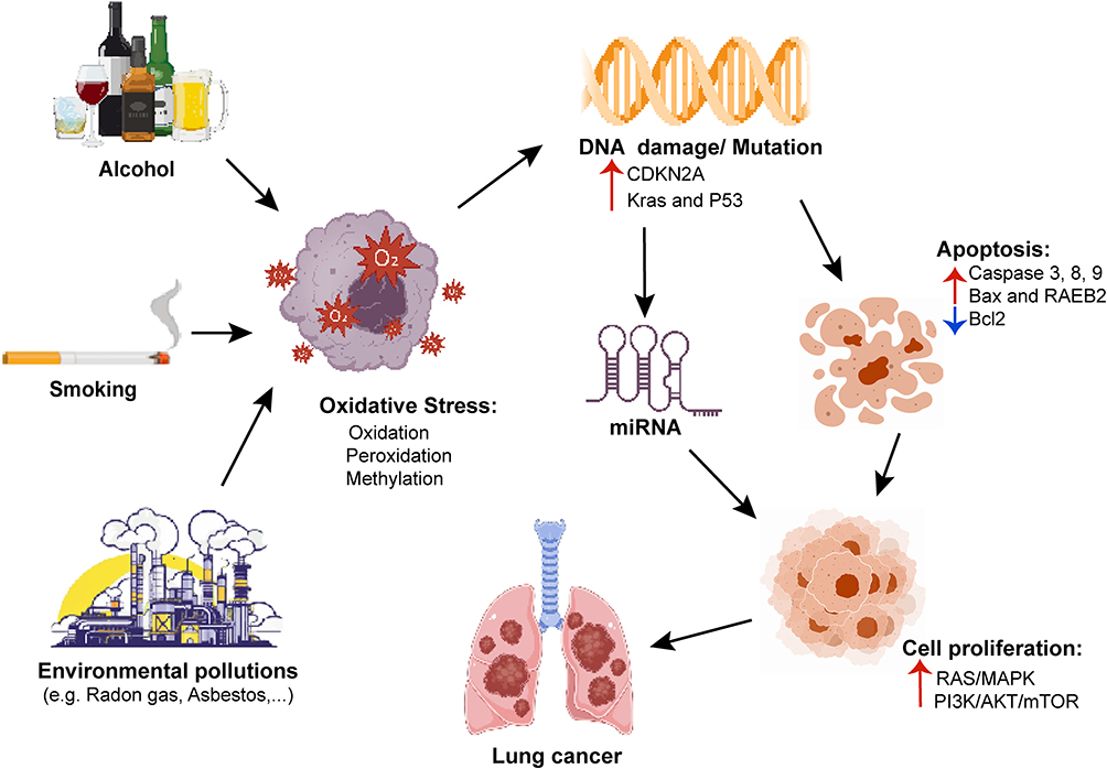 |
Figure 1 Risk factors and related pathogenesis of lung cancer. |
Current treatment modalities for lung cancer include surgery, radiotherapy, chemoradiotherapy, immunotherapy, and targeted therapy. Clinical decision-making is primarily guided by the tumor type and stage, as well as the patients’ physical condition.9,10 Despite significant advancements in lung cancer therapy over recent decades, persistent challenges--such as severe adverse effects, low bioavailability, and drug resistance--continue to hinder the clinical effectiveness of these interventions.11–13 For example, chemotherapeutic agents like cisplatin, paclitaxel, and docetaxel (DTX) can cause side effects, including nausea, vomiting, alopecia, and myelosuppression, some of which may be life-threatening. Additionally, targeted and immunotherapeutic drugs may induce adverse reactions such as rash, cough, diarrhea, and visual impairment.14 Furthermore, long-term treatment can lead to the development of multidrug resistance (MDR), resulting in treatment failure and tumor recurrence.15 Therefore, the development of innovative drug delivery systems designed to enhance therapeutic efficacy and reduce toxicity has emerged as a critical area of research in lung cancer therapy.
In recent years, nanotechnology-based drug delivery systems have garnered extensive attention in the field of tumor therapy, offering new strategies for lung cancer treatment by improving drug targeting, reducing toxic side effects, and enhancing drug stability and solubility.15–17 Currently, FDA-approved nanomedicines such as Abraxane® and Pazenirl® are used in the clinical treatment of lung cancer and have shown advantages in improving drug solubility and prolonging blood circulatory time. However, due to the limitations of systemic administration, they still face challenges in terms of targeting and therapeutic index.17 CS-based nanodelivery systems offer a promising direction to address these shortcomings, leveraging their unique mucoadhesive properties, excellent biocompatibility, and enhanced drug penetration capabilities.18 Derived from natural sources, CS exhibits properties such as hemostatic, bacteriostatic, anti-cholesterol, and anticancer effects.19,20 Furthermore, it can be chemically modified to enhance its solubility, drug-loading capacity, bioavailability, and targeting ability.21,22 Based on its cationic nature, CS can increase adhesion through electrostatic interactions with mucosal surfaces, prolong the contact time between the drug and mucosa, and facilitate cellular internalization.23,24 Moreover, the positive charge on its surface enables CS to interact with anionic drugs and polyanionic biomolecules (such as siRNA or DNA), thereby facilitating drug encapsulation and controlled release.25 This characteristic has been utilized in various therapeutic fields, particularly in oncology, where it enhances the specificity and efficacy of chemotherapeutic agents while minimizing systemic toxicity.26 Therefore, this review aims to summarize the latest research advancements on CS-based nanodelivery platforms, explore their potential in drug and gene delivery, and delineate the applications of CS nanocomplexes in phototherapeutic strategies for lung cancer.
Chitosan and Its Physicochemical Properties
CS is a linear polysaccharide polymer formed by the linkage of glucose molecules through β-1,4-glycosidic bonds.27 CS is obtained by the deacetylation of chitin, which is widely found in marine crustaceans such as shrimp and crabs.28 Depending on the conditions of deacetylation, CS exhibits a molecular weight (MW) ranging from 10 to 1000 kDa and a degree of deacetylation (DD) between 70% and 95%.29 Due to the protonation of its amino groups, CS dissolves in acidic solutions with a pH below 6.5, while exhibiting poor solubility under neutral and alkaline conditions.30 As the second most abundant natural polymer after cellulose, CS is an ideal candidate material in pharmacology and therapy, owing to its excellent biodegradability, biocompatibility, and non-toxicity.31,32 Table 1 illustrates the applications of chitosan-based nanocarriers in lung cancer therapy.
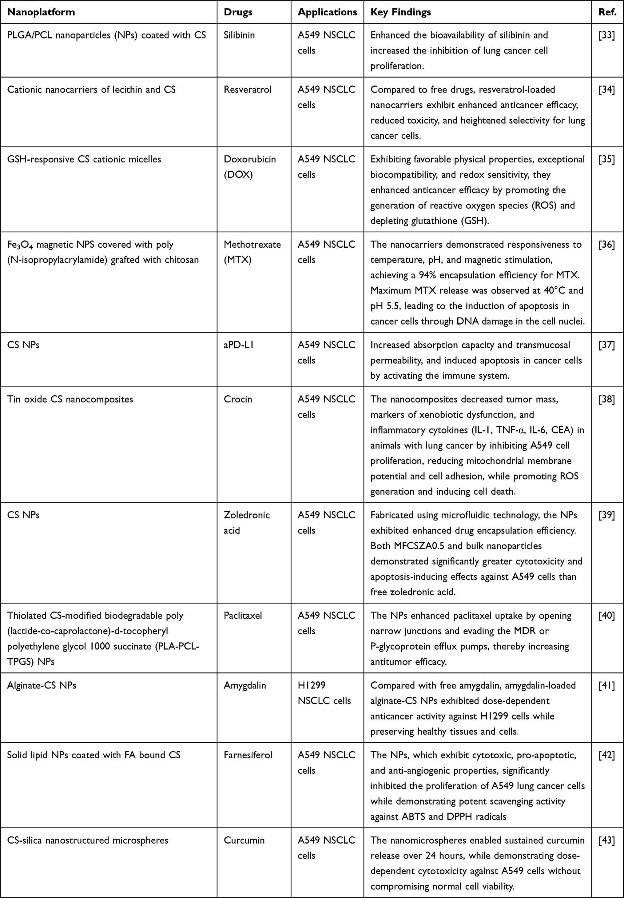 |
Table 1 Applications of Chitosan-Based Nanocarriers in Lung Cancer Therapy |
The MW and DD significantly influence the physicochemical properties and biological activity of CS and its derivatives. CS with a high MW can enhance the stability of complexes and prolong the circulation time of nanoparticles in the bloodstream; however, it reduces their intracellular release rate, thereby increasing tumor targeting selectivity. In contrast, low MW chitosan exhibits the opposite effects.43,44 DD controls the free amino groups in the CS structure, which not only determines its positive charge density and the capacity to affiliate with DNA/siRNA molecules but also facilitates the evasion of nanovehicles from endolysosomal sequestration, thereby improving transfection efficiency.43,45 CS with a high DD increases the surface charge density of nanoparticles, significantly enhancing cellular uptake and antitumor effects.43,46 Additionally, CS with lower MW is often associated with a higher degree of deacetylation, exhibiting enhanced tolerance to amino group protonation and solubility, along with increased toxicity towards malignant cells.47
The presence of hydroxyl, amino, and other functional groups in CS molecules enables chemical modifications, including acylation, esterification, alkylation, and grafting. These modifications yield chitosan derivatives with desired physical, chemical, and biological properties,48 as depicted in Figure 2. Research indicates that CS possesses anticancer activity due to its cationic nature, MW, and DD, while demonstrating minimal toxicity to non-malignant cells.49 CS is more readily protonated and soluble in the acidic tumor microenvironment.49 Through electrostatic repulsion, CS disperses and undergoes a conformational change from a tightly curled state to an extended linear form, achieving a ζ potential as high as +30 mV.50 This property enables CS to effectively interact with negatively charged tumor cell membranes and tumor vascular endothelial cells, which overexpress anionic surface groups such as phospholipids, glycoproteins, and proteoglycans.51,52 Consequently, CS can modify cell membrane permeability and penetrate intracellularly to exert antitumor effects by inhibiting the formation of matrix metalloproteinase 9 (MMP-9) protein, as demonstrated in in vitro studies of malignant cell lines. In a physiological environment, CS may exhibit reduced affinity for normal cells, indicating its selective targeting of malignant cells.53
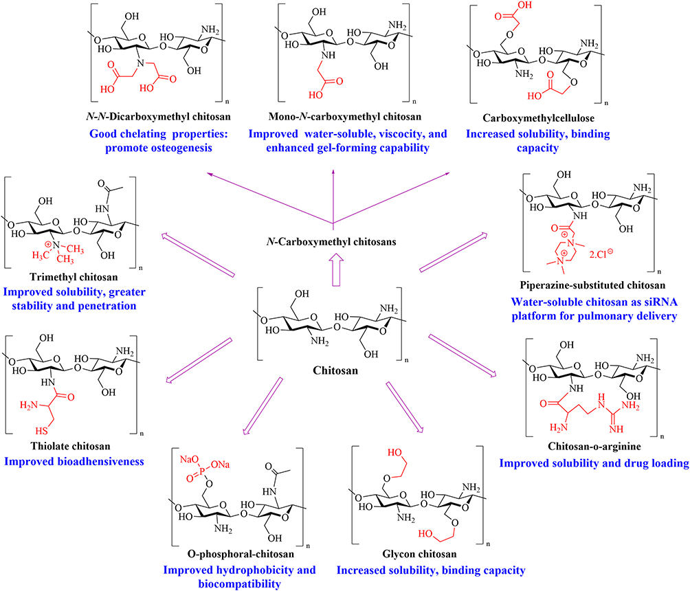 |
Figure 2 Chitosan derivatives and their properties for improved drug delivery. |
The viscosity and mucosal adhesion properties of CS play a pivotal role in regulating the entrapment, absorption, and release of drugs. As a cationic polymer, it engages with the negatively charged membranes of cancer cells, thereby prolonging the residence time at the target site and enhancing absorption capacity.24,54 The positive charge of CS can also prevent therapeutic agents from being degraded by enzymes during lysosomal transport through the proton sponge effect.55 Additionally, the positive charge of CS enables it to disrupt the tight junctions between epithelial cells by activating the protein kinase C pathway, which decreases the transepithelial resistance of the epithelial cell membrane52 and disrupts connection proteins such as zonula occludens-1 (ZO-1) and occludin, thereby facilitating paracellular and transcellular drug transport.56,57 As a result, CS-based drug carriers can be strategically designed to release their therapeutic contents within the tumor microenvironment, achieving precise treatment.
Chitosan-Based Targeted Drug Delivery Systems
Traditional drug delivery systems face critical limitations, including non-specific distribution, low bioavailability, severe adverse reactions, and rapid bodily clearance, which collectively hinder their therapeutic efficacy.58 To address the aforementioned challenges, CS-based nanocarriers offer a novel approach for targeted cancer therapy. These nanocarriers not only accumulate selectively in tumor tissues via the retention (EPR) effect, but also can be functionalized with targeting molecules (eg, antibodies or peptides) to enhance their specificity and therapeutic potency against cancer cells.59 Furthermore, CS nanocarriers can selectively disrupt tumor cell membranes, enhancing the bioavailability of drugs in lung cancer cells while preserving the safety of healthy cells.60 Figure 3 illustrates the mechanisms of CS-NPs for delivering chemotherapeutic drugs through stimuli-responsive, passive, and active targeting strategies.
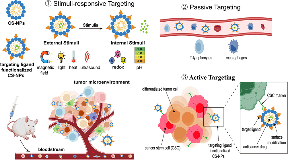 |
Figure 3 Mechanisms of CS-NPs for delivering chemotherapeutic drugs through stimuli-responsive, passive, and active targeting strategies. |
Medication Delivery Utilizing Passive Targeting
Nanocarriers exploit the hyperpermeability of tumor capillaries and the EPR effect for passive targeting to cancerous sites, where they accumulate due to vascular leakage and insufficient lymphatic drainage in the tumor microenvironment.61 Moreover, the impaired lymphatic function also facilitates the escape of nanocarriers from defective tumors, enabling them to target lung cancer. Passive targeting relies on the physicochemical properties of nanocarriers, such as size, shape, and surface characteristics, and tumor biological features like vascular distribution and leakage.62 Nanoparticles with a particle size less than 200 nm effectively evade immune clearance and prolong circulation time in the bloodstream. Moreover, in the acidic tumor microenvironment, CS protonation induces nanoparticle swelling and subsequent drug release into target malignant cells.25,63 Altwaijry et al prepared CS-modified lipid nanoparticles (ERT-CLNPs) encapsulating Erlotinib (ERT) through probe sonication and the ionic gelation technique. ERT-CLNPs, with a particle size below 200 nm, exhibited low polydispersity, a cationic zeta potential, and high encapsulation efficiency, while demonstrating excellent gastrointestinal and storage stability. Compared with pure ERT, ERT-CLNPs demonstrated significantly enhanced mucoadhesion and intestinal permeability, while exhibiting superior cytotoxicity toward lung cancer cells.64 A poly-lactic glycolic acid (PLGA)/CS nanoparticle was synthesized through a single emulsification and evaporation method for targeted delivery of Meclinertant to treat NSCLC. The Meclinertant encapsulated in PLGA/CS NPs exhibited pH-dependent release, with enhanced drug release under acidic conditions, aligning with the characteristics of the tumor microenvironment. Compared to the free drug, PLGA/CS NPs loaded with Meclinertant demonstrated a more significant anti-proliferative effect on A549 cells. Cell uptake and double staining experiments revealed that PLGA NPs coated with CS substantially enhanced cellular uptake capacity and induced apoptosis in A549 cells.65 Sun et al encapsulated Bortezomib (BTZ) and Vincristine (VCR) in polyethylene glycol (PEG)/CS nanoparticles (PEG/CSNPs) to enhance drug solubility. The study revealed that compared to free drugs or single-drug-loaded CSNPs, PEG/CSNPs loaded with BTZ and VCR exhibited significantly enhanced cytotoxicity against NCI-H661, NCI-H460, A549, H157, and H1299 lung cancer cells, demonstrating synergistic effects between BTZ and VCR with substantially reduced half-maximal inhibitory concentrations (IC50). FITC/Annexin-V and PI staining assays indicated that PEG/CSNPs induced apoptosis in NCI-H661 cells by upregulating the expression of pro-apoptotic proteins, such as Bax, Cyt-C, and caspase-3, −8, and −9.66 Copper-citrate complexes were loaded into CS nanoparticles (CuCC NPs) using the conventional ion gelation method. The nanoparticles enhanced accumulation within tumor tissues via the EPR effect. In response to the acidic and GSH-rich tumor microenvironment, CuCC NPs enabled the controlled release of Cu2+ and mediated efficient chemodynamic therapy to effectively kill A549 cells. Additionally, histopathological and hematological analyses confirmed that CuCC NPs induced massive apoptosis in tumor tissues while maintaining an excellent safety profile.67 Nijhawan et al employed a Quality-by-Design approach to encapsulate erlotinib hydrochloride (ERT-HCl) within CS/PLGA NPs. Drug release studies revealed that the nanoparticles sustained drug release for up to 72 h, following zero-order release kinetics, which indicated that the release process was governed by Fickian diffusion.68 In a separate study, Karami et al employed double nanoemulsion techniques to synthesize a hydrogel nanocarrier, which was formulated with CS, alumina (γ-Al2O3), and graphene quantum dots (GQDs) for targeted delivery of quercetin (QC). Cytotoxicity experiments revealed that the interaction between QC and the nanocarrier resulted in a gradual release, effectively inhibiting the growth and proliferation of lung cancer cells, which indicated the potential of the nanocarrier as a novel nanosystem for targeting tumor tissues.69 Ray et al created an inhalable nanocomposite (NIC-CS-PCL-NA) comprising niclosamide (NIC) encapsulated within CS-functionalized poly (ε-caprolactone) (PCL) for NSCLC treatment. In vitro experiments revealed that NIC-CS-PCL-NA exhibited dose-dependent cytotoxic effects, which could effectively enhance autophagic flux, inhibit cellular invasiveness, and induce apoptosis in A549 lung cancer cells. In a mouse model of lung cancer, inhaled NIC-CS-PCL-NA was observed to deposit and retain in the lungs, effectively inhibiting tumor growth while exhibiting minimal toxicity to other major organs. These findings position it as a promising treatment for NSCLC.70 The incorporation of zinc phthalocyanine-perfluorinated resin (ZnPc-PFR) and compound 1 into CS-fructose (CH-Fru) yielded CH-Fru@ZnPc-PFR@1, which was further complexed with gefitinib to form the nanocomposites (CH-Fru@ZnPc-PFR@1@Gefitinib). This nanocomposite exhibited dose-dependent inhibitory effects against A549, NCI-H1734, and NCI-H1299 NSCLC cells, with the high-dose group (200 μM) demonstrating stronger inhibitory effect than the low-dose group (100 μM). These findings indicated synergistic effects between compound 1 and gefitinib in suppressing the proliferation of NSCLC cells.71
Medication Delivery Utilizing Active Targeting
Although passive targeting based on the EPR effect lays the groundwork for nanomedicine delivery, its clinical translation is frequently limited by tumor heterogeneity and variations in vascular leakage, which may lead to heterogeneous drug distribution and off-target toxicity.72,73 To overcome these limitations, researchers can achieve precise drug delivery via ligand-receptor interactions for specific recognition. Nanocarriers functionalized with ligands, such as folic acid (FA), monoclonal antibodies, and aptamers,10 can selectively recognize overexpressed target receptors on cancer cell surfaces (such as EGFR, folate receptors, sigma receptors) or target specific membrane proteins on immunosuppressive macrophages within the tumor microenvironment, thereby effectively differentiating them from healthy cells.74 As an illustration, Feng et al successfully synthesized CS NPs loaded with bacterial vesicles and DOX for targeted drug delivery to lung cancer cells. Through surface modification with FA, the nanoparticles achieved targeted delivery, induced the release of ROS, and activated the STING signaling pathway via mitochondrial DNA damage, thereby promoting dendritic cell (DC) maturation. Consequently, mature DCs recruited CD4+ and CD8+ cytotoxic T cells and enhance the secretion of IFN-β, IFN-γ, and IL-12.75 Patel et al successfully synthesized inhalable polymer nanoparticles encapsulating silibinin, functionalized with FA to confer targeting capability against lung cancer cells. Their findings demonstrated that the nanoparticles significantly reduced the viability of A549 cells compared with free silibinin. Histological analyses showed that, by exploiting the overexpression of FA receptors in lung cancer cells, the nanoparticles effectively accumulated in lung tissue, thus significantly inhibiting tumor volume expansion.76 Monoclonal antibodies are widely used for targeting cell receptors due to their high specificity in vivo and stability in serum.77 Gold nanoparticles conjugated with CS and loaded with lapatinib were utilized for targeted drug delivery against lung cancer cells. The nanoparticles not only demonstrated dose- and time-dependent cytotoxicity towards A549 cells, but also enhanced attachment to the cell surface and internalization into cancer cells due to their positive charge. PCR analysis revealed that the nanoparticles could significantly downregulated the expression levels of LINC01615 and related genes such as IL11 and MMP14, indicating that they effectively destroyed the tumor growth and metastasis pathway.78 Imatinib was loaded into CS polyethylene glycol nanoparticles and modified with Ramucirumab antibody to achieve targeted therapy for A549 cells. The results demonstrated enhanced Imatinib release from the nanoparticles in acidic media compared to physiological conditions. Relative to free Imatinib, the nanoparticles exhibited superior pro-apoptotic effects, and induced G1-phase cell cycle arrest in 8.17% of cells.79 Najmeh et al designed an aerosol-assisted synthesis method for CS-based nanocarriers (GefNC) loaded with gefitinib. GefNC exhibited pH sensitivity, with enhanced drug release in acidic environments compared to physiological conditions, indicating potential for targeted delivery to cancer cells. MTT assays demonstrated that the IC50 value of GefNC against lung cancer cells was lower than that of free drugs, while the empty nanocarrier did not elicit appreciable cytotoxicity.80 Zhang et al fabricated CS iodoacetamide (CsIA)-coated liposome NPs for lung cancer treatment. Trastuzumab (TZ) was conjugated to the nanoparticle surface to target lung cancer cells overexpressing the HER2 receptor, enabling sustained drug release at the tumor sites. Compared with free drug and unconjugated liposome NPs, the TZ-conjugated NPs loaded with SN-38 accumulated extensively within Calu-3 lung adenocarcinoma cells, and significantly potentiated the anticancer effects on Calu-3 cells.81 Furthermore, peptides or protein ligands have been utilized for the functional modification of CS, enhancing the specific recognition of cancer targets through active targeting strategies.82 In another study, a thermally induced aggregation method was employed to load camptothecin (CPT) into a formulation of pH/temperature dual-responsive poly(acrylic acid)-b-poly(N-isopropyl acrylamide) block copolymer, which were then surface-modified with CS and fucoidan (Fu) to form Fu-CPT-NPs for lung cancer treatment. Cell experiments confirmed that Fu-CPT-NPs exhibited excellent biocompatibility and antitumor activity. The NPs effectively delivering CPT to H460 lung cancer cells through the interaction of Fu with P-selectin, enhancing cellular uptake and inducing apoptosis in cancer cells. Animal studies showed that Fu-CPT-NPs inhibited lung tumor growth by promoting tumor cell apoptosis without causing significant tissue damage.83 Kordbacheh et al embedded DOX in a ZIF-8 (Zn, Fe) bimetallic-organic framework (BMOF), whose surface was coated with curcumin-loaded CS-grafted- polycaprolactone (Cs-g-PCL) and subsequently functionalized with transactivator of transcription (TAT) peptide for targeted delivery. Cs-g-PCL regulated the release rate of curcumin through pH-dependent behavior, promoting drug accumulation in tumor tissues, while TAT peptide modification further prolonged drug release duration, establishing a synergistic controlled-release system. In vitro studies indicated that the combined delivery of DOX and Cur significantly reduced the viability of A549 lung cancer cells, with the TAT peptide and DOX-Cur combination exhibiting a synergistic apoptotic induction effect. In vivo studies in tumor-bearing mice showed that the TAT peptide-modified nanosystem significantly inhibited tumor growth compared to the unmodified Cs-g-PCL/Cur/BMOF/DOX, reaching a maximum relative tumor volume inhibition rate of 0.25.84 Hu et al designed a copper ferrite nanocomposite, which was functionalized with vitamin E succinic acid (VES)- ethylene glycol CS polymer (GV). The nanocomposites utilized the protonation of GV polymer and polylysine (PL) in the acidic tumor microenvironment to facilitate drug release, along with the active targeting of human serum albumin to specifically induce tumor cell death. The nanocomposites exhibited cytotoxicity against lung cancer cells by elevating intracellular ROS levels, thus triggering cuproptosis and ferroptosis. Moreover, the nanocomposites not only showed superior antitumor efficacy and biocompatibility compared to carboplatin, but also potentially inhibited tumor cell growth by activating the p53 signaling pathway. In vivo studies showed that the nanocomposite effectively suppressed the growth of subcutaneous A549 tumors in nude mice.85
Medication Delivery Utilizing Magnetic Targeting
Magnetic targeting is a technique that harnesses external magnetic fields to direct magnetic drug carriers to the target site. For instance, magnetic nanoparticles can direct and concentrate drugs in specific areas of the body under the influence of an external magnetic field.86 Controlled by an external magnetic field, composites formed from CS and magnetic nanoparticles are directed to the tumor site, ensuring targeted drug delivery with minimal impact on surrounding healthy tissues.87 Yuly et al developed CS-based microencapsulated systems incorporating magnetite nanoparticles and microzeolites for gemcitabine (GEM) delivery in lung cancer therapy. This system exhibited a drug encapsulation efficiency of over 99% and achieved a slow and controlled release of GEM at pH 7.4 and 5.0 for up to 24 hours. Cell viability assays revealed that the cell viability of A549 and H1299 cells treated with the nanoparticles was significantly lower than that of cells treated with pure GEM.88 Peptidyl arginine deiminase 4 inhibitor was loaded into magnetic CS NPs to achieve dual-target drug delivery via magnetic targeting and pH-responsive release. In vitro experiments demonstrated that an external magnetic field enhanced the uptake of nanoparticles by cancer cells, inducing tumor cell apoptosis. In vivo studies demonstrated that nanoparticles effectively inhibited the growth and metastasis of lung cancer in Lewis mice by suppressing the expression of citrulline histone (H3cit) under the influence of an external magnetic field.89 Additionally, Sheng et al prepared novel core-shell Fe3O4 NPs, which were synthesized by functionalizing gold NPs and CS-agarose composites. In vitro experiments showed that nanocomposites inhibited the proliferation of various cancer cell lines, exhibiting IC50 values of 160, 173, 142, and 212 for NCI-H1563, NCI-H1573, NCI-H1975, and NCI-H661 cells, respectively.90 A composite capsule was prepared by an ionic gelation method, in which GEM was incorporated into CS-magnetite-zeolite composite matrix, and the synergistic regulation of drug loading and magnetic targeting was realized. The composite capsule exhibited an encapsulation efficiency of over 86% for GEM and showed a sustained release profile over 24 hours. Cell viability assays revealed that the composite capsule exhibited similar cytotoxicity to free GEM but at a 45-fold lower concentration, suggesting its potential to reduce the required dosage and potential side effects.91
Chitosan Miscellaneous Nanocomplexes in Lung Cancer Therapy
CS plays multiple roles in the development of metal-based, carbon-based, and silica-based hybrid nanoparticles. Due to its cationic nature, the primary amine groups in the CS structure, along with adjacent hydroxyl groups, can reduce metal cations (such as Ag+, Au3+), thereby promoting the synthesis of metal nanoparticles (MNPs). In addition, the modification of gold NPs, silver NPs, and quantum dots with CS enhances their stability and optical properties while reducing their cytotoxicity.92,93 Aldosari et al doped gold into modified graphene oxide CS NPs (GO@CSP/Au NPs) for the treatment of lung cancer. MTT assays demonstrated that GO@CSP/Au NPs inhibited A549 cell viability in a dose-dependent manner, with an IC50 value of 35.2 μg/mL, indicating potential antineoplastic efficacy against lung cancer.94 Wang et al synthesized silver nanoparticles (AgNPs) mediated by curcumin on CS-modified kaolin. The kaolin minerals promoted the in-situ nucleation of AgNPs on the surface and within the interlayers of the modified kaolin, effectively preventing particle aggregation and achieving a uniform distribution with a particle size range of 20–30 nm. The nanocomposites possessed potent antioxidant activity and exhibited IC50 values of 110 μg/mL, 96 μg/mL and 38 μg/mL against HLC-1, LC-2/ad and PC-14 lung cancer cells, respectively, among which PC-14 showed the strongest anticancer effect.95 In another study, the integration of Cu2O into CS/guar gum (GG) composite hydrogel successfully yielded nanoparticles (CS-GG/Cu2O NPs). This synthesis approach demonstrated CS-GG/Cu2O NPs could inhibit the expression of pivotal proteins (PI3K, AKT, mTOR) in the phosphatidylinositol 3-kinase (PI3K)/protein kinase B (AKT)/mammalian target of rapamycin (mTOR) signaling pathway in NCI-H661 lung cancer cells, thereby increasing ROS levels and promoting apoptosis, and inhibiting the cell cycle in lung cancer cells.96 Abiraman et al utilized acryloyl CS grafted with [2-(methacryloyloxy)ethyl] trimethyl ammonium chloride acryloyl piperazinium chloride-based polymers as a supporting agent to synthesize spherical zinc oxide nanoparticles (ZnO NPs). The results indicated that, in comparison to uncapped ZnO, the capped ZnO NPs exhibited a more potent inhibitory effect on the A549 cancer, with nanoparticles capped with a hybrid polymer containing an acryloyl ethyl substituent in the piperazine ring exhibiting the maximum inhibitory effect on cancer cells.97 Khichar and associates extracted components from Terminalia Arjuna using water and ethanol to functionally modify CS-loaded AgNPs through biological methods. Cytotoxicity assays revealed that the two types of nanoparticles inhibited the viability of A549 lung cancer cells in a dose-and time-dependent manner, with IC50 values of 400 μg/mL and 300 μg/mL after 24 and 72 hours, respectively.98 Venkidasamy and coworkers synthesized vanillic acid nanocomposites incorporating silver and CS NPs. Apoptosis assays revealed that the nanocomposites induced cellular apoptosis by upregulating the expression of pro-apoptotic proteins, such as BAX, Casp3, Casp7, and cytochrome C, while concurrently suppressing the gene expression of Bcl-2. Moreover, the nanocomposites exhibited a dose-dependent increase in the expression of the tumor suppressor gene p53, indicating a multi-pathway synergistic mechanism underlying its pro-apoptotic effects.99 Thenmozhi et al developed a biosynthesized curcumin-loaded cadmium oxide-silicon dioxide nanocomposites utilizing Pithecellobium dulce leaf extract as a reducing and capping agent. Their research indicated that the nanocomposites exhibited a dose-dependent cytotoxicity effect against various human cancer cell lines, with a particularly pronounced impact on A549 lung cancer cells (IC50 value of 31.10 μg/mL). Furthermore, the nanocomposites demonstrated good antibacterial activity and anti-inflammatory effects.100
Nanosystems composed of CS and quantum dots endow nanoparticles with stability, functionality, cell binding and uptake, while also modulating drug release, increasing tumor drug accumulation, inhibiting degradation, and reducing toxicity associated with inorganic metal ions.101–103 Tahamtan et al encapsulated QC within CS/starch/GQDs/titanium dioxide porous nanocomposite using a water-in-oil-in-water (W/O/W) multiple emulsion method. The release behavior of QC from the nanocomposites followed the Fickian diffusion mechanism and exhibited responsiveness to environmental stimuli such as pH and temperature. MTT assays showed that the nanocarrier exhibited good biocompatibility; compared to free QC, the drug-loaded nanocomposites significantly enhanced cytotoxicity against the A549 lung cancer cell line.104 Furthermore, carbon nanomaterials (CNMs) have been widely applied in the biomedical field due to their unique physicochemical properties, including high stability, conductivity, thermal conductivity, and optical properties.105 The application of CNMs coated with CS has been shown to augment their stability, diminish cytotoxicity, and exhibit pH-responsive release characteristics in tumor microenvironments with pH levels below 6.5, thereby effectively facilitating drug delivery to tumor tissues.106,107 Fateme et al created a pH-responsive nanocarrier (CS/HNTs/GQDs) composed of CS, halloysite nanotubes (HNT) and GQDs for targeted QC to lung cancer cells. The incorporation of HNTs enhanced QC drug loading capacity and encapsulation efficiency, while the presence of GQDs accelerated QC release kinetics under different pH conditions. MTT assays revealed that CS/HNTs/GQDs@QC exhibited the strongest cytotoxicity against both L929 and A549 cells compared to other groups, with significantly greater effects on A549 cells. Furthermore, CS/HNTs/GQDs@QC induced the highest apoptotic proportion of apoptosis, accounting for 74.4% early apoptosis and 5.15% late apoptosis.108 AbouAitah et al fabricated multi-walled carbon nanotubes (MWCNTs) loaded with ferulic acid (FUA) and diosgenin (DGN) for lung cancer treatment. Initially, the MWCNTs were functionalized with carboxylic acid or amine functionalities, followed by coating with CS or a CS-stearic acid complex. Cytotoxicity assays showed that, compared with free DNG and FUA, the MWCNTs exhibited a significantly superior inhibitory effect on the viability of A549 cells. The MWCNTs loaded with DGN and FUA exhibited synergistic and additive interactions, but antagonistic effects were observed at elevated concentrations. Consequently, this nanosystem has emerged as a promising drug delivery platform, which has the potential to enhance the therapeutic activity of natural prodrugs.109
Chitosan-Alginate Nanocarrier in Lung Cancer Therapy
CS and alginate are biopolymers with opposite charges. CS, a cationic polysaccharide, contains amino groups on its molecular chains that can be protonated in an acidic environment, thereby conferring cationic properties to CS. Due to its cationic nature, CS enables electrostatic interactions with negatively charged polymers, providing unique advantages in chemotherapeutic drug delivery. Alginate, an anionic polysaccharide, is composed of α-L-guluronic (G) and β-D-mannuronic (M) acids.110 The carboxyl groups in its structure confer good water solubility and pH responsiveness when the pH exceeds the pKa values of guluronic and mannuronic acids (3.38 and 3.65).111 The protonated amino groups of CS electrostatically interact with carboxylate groups of alginate, forming a variety of polyelectrolyte complexes.112 Eldin et al designed a novel core-shell nanocarrier system where aminated mesoporous silica nanoparticles (MSNs) served as the core, encapsulating DTX. An intermediate layer of alginate controlled the release of DTX, while the outermost layer, composed of GMC-loaded PEG-CS, ensured the stealth properties and rapid dissemination of GMC. The core-shell nanocarriers were capable of depositing in the lungs and demonstrated excellent aerosolization behavior. Compared with the individual drugs, the combined use of both drugs within the nanocarriers elicited a stronger cytotoxic effect against A549 cells. Additionally, in vivo biological distribution studies confirmed the accumulation of the nanocarriers in the lungs, and histopathology assessments indicated the biological safety of the core-shell nanocarriers.113 Gefitinib was loaded into self-assembled CS/alginate nanocarriers, which displayed pH sensitivity, releasing a higher concentration of gefitinib in acidic conditions. MTT assays revealed that the nanocarriers had a significantly lower IC50 value than free gefitinib, with no appreciable cytotoxic effects observed.114 Yaşar et al developed a CS-alginate liposome to encapsulate mallow extract. The anti-proliferative efficacy of the liposome loaded with mallow extract was evaluated using the CCK8 assay in A549 cell. Treatment of A549 cells with these liposomes elicited a dose- and time-dependent cytotoxic response. As the treatment time extended to 48 hours, the amount of extract released from the liposomes gradually increased, resulting in increased cell death.115
Chitosan Nanocomposites in Delivery of Bioactive Compounds for Lung Cancer
Bioactive compounds derived from plants, such as polyphenols, flavonoids, and polysaccharides, possess various biological activities including antibacterial, anti-inflammatory, anti-aging, and anticancer effects.116 They can exhibit antitumor activities by interfering with signaling pathways (eg, PI3K/Akt, NF-κB, TGF β) and regulating cellular metabolism.117 Moreover, their beneficial impacts on cancer treatment have been confirmed in both in vitro and in vivo studies.118 Nevertheless, these phytochemicals possess some physicochemical limitations, such as poor solubility and low bioavailability, as well as pharmacokinetic issues such as short half-life, non-specific biodistribution, and narrow therapeutic window, which restrict their clinical translation.119 CS-based nanocomplexes, with their biocompatibility, modifiability, and sustained-release properties, can effectively encapsulate phytochemicals, enhance their stability and bioavailability, prolong circulation time, improve targeting, and reduce toxicity to normal tissues.120 El-Ghannam et al synthesized a beetroot@CS nanocomposite (BR@CS NC) by electrostatic interaction. MTT assays demonstrated that the cytotoxicity of BR@CS NC on A549 cells was dose-dependent, reaching 89% cell mortality at 250 μg/mL, which was significantly higher than that of free BR (5–7%). A549 cells treated with BR@CS NC not only exhibited extensive late-stage apoptosis and numerous necrotic changes but also elevated expression levels of the Caspase 3 (CasP3) and p53 genes.121 Abulaiti et al fabricated an oleanolic acid-conjugated CS nanocomplex (OAC) and explored its anticancer efficacy against lung cancer. In A549 and NCI-H460 lung cancer cells, OAC significantly inhibited cell viability, migration, and invasion, while inducing apoptosis and autophagy in a dose-dependent manner. Western blot and immunofluorescence analyses revealed that OAC attenuated the phosphorylation of signal transducer and activator of transcription 3 (STAT3) in lung cancer cells, leading to the upregulation of LC3-II protein expression and the formation of GFP-labeled LC3-positive autophagic vacuoles. This indicated that OAC induced autophagy by modulating the STAT3/Bcl-2 pathway, thereby exerting its anticancer effects.122 Alali et al developed a spray-dried solid dispersion of apigenin (AGN) based on CS (AC2 SDSD), and evaluated its antiproliferative efficacy against A549 cells. Compared with pure AGN, AC2 SDSD not only exhibited stronger antioxidant activity but also significantly potentiated antiproliferative effects in A549 cells. Its mechanism of action involved the inhibition of the PI3K/Akt signaling pathway, activation of the apoptosis pathway, promotion of cancer cell death, and inhibition of lung tumor growth.123 Gomathi et al created a new drug delivery material by conjugating CS with Spondias pinnata phytocompounds, and evaluated the cytotoxicity of the composites against A549 cells. The results revealed that compared with the leaf extraction of Spondias pinnata, the composites could significantly reduce the cell viability.124 CS NPs loaded with star anise extract (SAE) were synthesized by ionic gelation. Compared with free SAE, CSNPs loaded with SAE inhibited NCl-H460 lung cancer cells in a dose-dependent manner, suggesting enhanced cellular uptake of the nanoparticles. In vivo studies demonstrated that CSNPs loaded with SAE exhibited the most potent antitumor activity, as seen by decreased levels of malondialdehyde, tumor protein 53 (p53), tumor necrosis factor-alpha and fibronectin, compared with other treatment groups.125
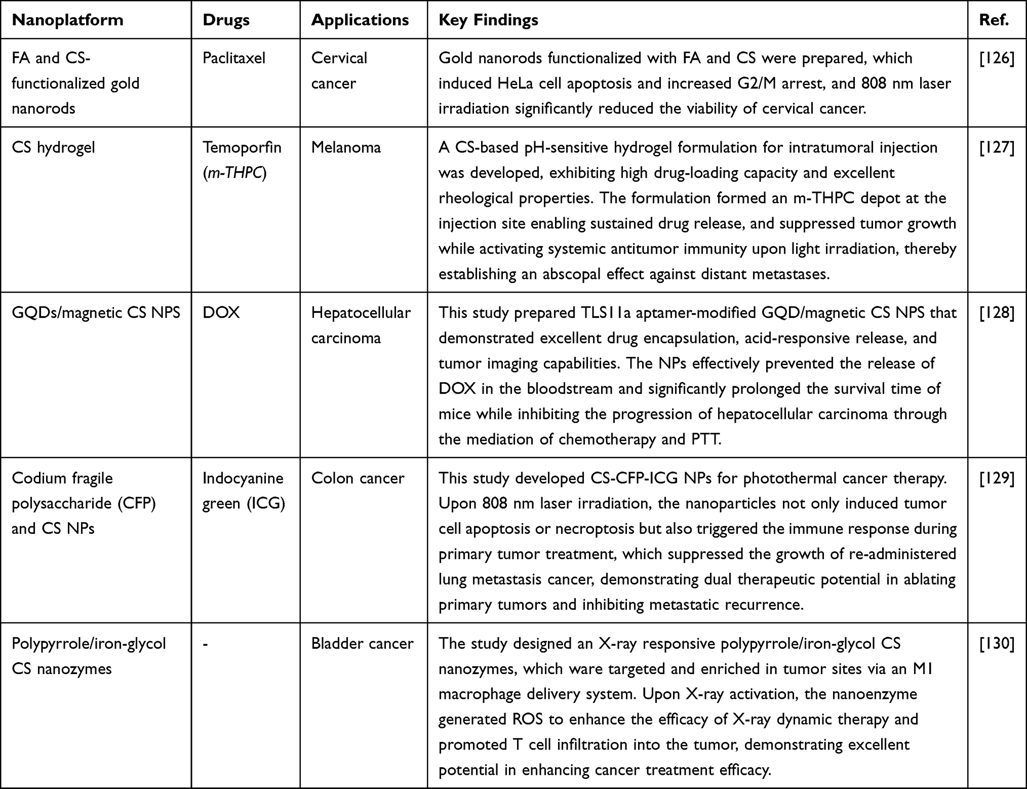 |
Table 2 Chitosan-Based Nanostructures for Cancer Phototherapy |
Chitosan Nanocarriers for Phototherapy and Gene Delivery
Phototherapy includes two modalities: photodynamic therapy (PDT) and photothermal therapy (PTT). Although they differ in mechanisms of action, both ultimately induce tumor cell death to achieve antitumor effects (Table 2 provides an overview of CS-based nanostructures for cancer phototherapy). PDT selectively kills cancer cells through the cytotoxic action of photosensitizers (PS) activated by light.131–133 Its non-invasive nature, minimal side effects, and targeting advantages have garnered significant attention in clinical applications.134–136 However, the uneven distribution and poor penetration of PS within tumors137,138 result in suboptimal efficacy and potential resistance, which hinder the effectiveness of PDT.139,140 Therefore, researchers have employed the strategy of loading PS onto nanomaterials, which promotes ROS generation, disrupts the oxidative-antioxidative balance, inhibits tumor growth, damages DNA, and ultimately leads to cancer cells death, thereby providing a new approach to enhance the efficacy of PDT.31 Guo et al developed a novel probiotic-based PS delivery system (EWC), utilizing Escherichia coli Nissle 1917 (EcN) as the carrier. The system facilitated the electrostatic adsorption of Chlorin e6 (Ce6) to the surface of EcN with the assistance of water-soluble CS. In vitro experiments demonstrated that EWC exhibited low toxicity to normal cells but strong photodynamic cytotoxicity against A549 cells. 3D fluorescence imaging revealed that EWC could penetrate tumor spheroids and effectively inhibit their growth. In mouse models, EWC not only demonstrated good biocompatibility but also enhanced the targeted distribution and accumulation of Ce6 at tumor sites, effectively inhibiting tumor progression upon light stimulation.141 In contrast, PTT utilizes the photothermal conversion capability of near-infrared (NIR) light-responsive nanomaterials to convert light energy into thermal energy, inducing cancer cell death through localized hyperthermia.142 Its deep tissue penetration capability and minimal damage to normal tissues render it an effective therapeutic strategy.143 An et al designed a CS-based hydrogel nanocomposite with NIR light response, which induced apoptosis in lung cancer cells through a triple synergistic mechanism combining photothermal, chemodynamic, and chemotherapeutic effects. The nanocomposite, loaded with carbon nanodots, platinum nanoparticles (PtNPs), and DOX, demonstrated favorable sustained drug release properties. Upon NIR irradiation, carbon nanodots amplified oxidative stress and initiated apoptosis through ROS generation and activation of caspase-3/8/9 pathways. The synergistic effects of PtNPs and DOX involved PtNPs-mediated enhancement of ROS production and DOX-induced amplification of cytotoxic effects, resulting in pronounced cytotoxicity and apoptotic induction in A549 and H1299 cells under NIR stimulation. Consequently, this innovative nanocomposite system offered a promising strategy for targeted lung cancer therapy.144
With the emergence of chemoresistance, gene delivery technology has witnessed substantial advancements in exploring new avenues for cancer treatment and enhancing drug sensitivity.145,146 However, naked gene delivery faces challenges such as easy degradation in the bloodstream and poor cellular internalization, resulting in low transfection efficiency.147 To overcome these challenges, researchers have proposed strategies utilizing nanoscale structures to achieve precise gene targeting.148,149 Furthermore, CSNPs have emerged as ideal vectors in the field of gene delivery because of their excellent biocompatibility, biodegradability, and the ability to protect nucleic acids. The cationic nature of CS enables it to interact electrostatically with negatively charged nucleic acids, forming stable complexes that protect genetic material from enzymatic degradation and facilitate cellular uptake of nucleic acids.150 In cancer treatment, small interfering RNA (siRNA) downregulates the expression of oncogenes by binding to complementary mRNA, inhibiting cancer cell proliferation, inducing cell death, and preventing lymphatic metastasis.142 Zhang et al created a novel gene vector, vitamin E succinate-CS-histidine (VES-CTS-His, VCH), which was combined with STAT3-shRNA recombinant plasmid by electrostatic interactions to form stable particles. The nanoparticles exhibited typical nanoscale dimensions, allowing them to accumulate in tumor tissues via the EPR effect. They could enter tumor cells through endocytosis. VCH exhibited good pH responsiveness, allowing it to dissociate in the acidic microenvironment of tumor tissues and release the recombinant plasmid. By leveraging the RNA interference effect, the expression of STAT3 protein was downregulated, which inhibited the PI3K/mTOR signaling pathway. This action significantly enhanced the effect of inducing apoptosis and inhibiting growth in NSCLC cells, thereby achieving the goal of tumor treatment.151 Furthermore, several studies have combined chemotherapeutic drugs with siRNA in nanoscale drug delivery systems, which can synergistically enhance the anticancer effects and improve treatment efficiency. DOX and Bcl-2 siRNA were incorporated into a pH-sensitive nanofibrous hybrid formulation consisting of liposome, CS, poly (ethylene oxide), and PCL (CS/PEO/PCL) for treating lung cancer. Cellular experiments indicated that the liposome-incorporated CS/PEO/PCL core-shell nanofibers loaded with DOX and Bcl-2 achieved sustained release of DOX and siRNA, and mediated the maximum down-regulation of Bcl-2 siRNA, which promoted apoptosis and reduced the viability of A549 lung cancer cells. In vivo experiments showed that mice treated with CS/PEO/PCL nanofibers incorporated with DOX-Bcl-2 liposomes could inhibit tumor growth to the greatest extent without changing their body weight.152 Sun et al synthesized a novel multifunctional micelle (VCPH), which is composed of VES, CS, histidine and polyethylene glycol monomethyl ether. DOX was incorporated into the self-assembled VCPH micelles, and the recombinant pGPU6/GFP/Neo STAT3-shRNA (pDNA) was loaded by electrostatic action, thereby forming dual-loaded nanoparticles (DOX/VCPH/pDNA). The nanoparticles exhibited pH-responsive drug release behavior, with a higher release rate in acidic environments compared to neutral conditions. The nanoparticles upregulated PTEN protein expression, which inhibited the PI3K/AKT pathway and induced apoptosis of adjacent tumor cells. They also downregulated CD31 protein expression, inhibited angiogenesis, and decreased tumor cell viability. In addition, the nanoparticles significantly downregulated the expression level of STAT3 protein, thus enhancing the efficacy of chemotherapy.153 The construction and development of this dual-load collaborative delivery system provides important enlightenment for the combined treatment of tumors and offers an effective approach for the precise treatment of NSCLC.
Conclusion and Future Perspective
Lung cancer remains a formidable challenge in oncology, necessitating innovative and targeted treatment approaches. Traditional treatments are costly and are associated with issues such as non-specificity, low bioavailability, and rapid clearance. CS-based nanodelivery systems have emerged as promising platforms for lung cancer therapy due to their unique biocompatibility, cationic properties, and functional versatility. Extensive research has been conducted on CS nanocarriers to explore their distinctive properties and multifunctionality, particularly in targeted cancer therapy and drug delivery. These nanocarriers efficiently load chemotherapeutic drugs, increasing the intracellular accumulation of drugs, enhancing cytotoxic effects, reducing chemoresistance, and minimizing side effects. They also deliver biomacromolecules such as nucleic acids, proteins, and peptides, prolonging their circulation time, enhancing intracellular accumulation, and protecting them from degradation. Notably, despite the delivery of natural active substances being largely overlooked, studies have demonstrated the potential of CS nanocarriers to inhibit lung cancer. The combination of phototherapy and chemotherapy mediated by CS nanocarriers can synergistically contribute to lung cancer treatment. Furthermore, CS nanocarriers can be surface-modified to achieve precise release of active drugs in the TEM, reducing toxicity to normal tissues.
However, CS-based nanoformulations face several limitations. The drug-loading capacity of CS-based nanoformulations depends on the physicochemical properties of the drug and the preparation techniques. Additionally, due to the aggregation characteristics of nanocarriers, the stability of the formulation in physiological environments is reduced, leading to issues such as drug crystalline transformation, heterogeneity in particle size distribution, and burst release effects. Although CS-based nanoformulations have been proven effective in animals, their clinical translation faces multiple challenges. On one hand, the current safety profile of CS nanocarriers remains insufficient, and there is a lack of adequate clinical data to confirm their safety and efficacy in humans. On the other hand, species-dependent immune responses and the differences between mouse models and human pathophysiology collectively hinder their progression toward clinical application. Additionally, the industrialization process encounters bottlenecks such as the low water solubility of CS, limited drug-loading capacity, and difficulties in quality control for large-scale production. Future research could focus on improving the solubility and drug-loading capacity of CS through physical and chemical modifications, and constructing multi-component tumor-homing nanosystems. This approach would enhance the scalability of production, tumor-targeting ability, and therapeutic efficacy of CS-based nanoformulations, thereby providing a solid foundation for their clinical translation and industrial application.
Disclosure
The authors report no conflicts of interest in this work.
References
1. Solanki R, Bhatia D. Stimulus-Responsive Hydrogels for Targeted Cancer Therapy. Gels. 2024;10(7):440. doi:10.3390/gels10070440
2. Kumar RR, Kumar A, Chuang CH, Shaikh MO. Recent advances and emerging trends in cancer biomarker detection technologies. Ind Eng Chem Res. 2023;62(14):5691–5713. doi:10.1021/acs.iecr.2c04097
3. Bray F, Laversanne M, Sung H, et al. Global cancer statistics 2022: GLOBOCAN estimates of incidence and mortality worldwide for 36 cancers in 185 countries. CA Cancer J Clin. 2024;74(3):229–263. doi:10.3322/caac.21834
4. Daylan AEC, Miao E, Tang K, Chiu G, Cheng H. Lung cancer in never smokers: delving into epidemiology, genomic and immune landscape, prognosis, treatment, and screening. Lung. 2023;201(6):521–529. doi:10.1007/s00408-023-00661-3
5. Schabath MB, Cote ML. Cancer progress and priorities: lung cancer. Cancer Epidemiol Biomarkers Prev. 2019;28(10):1563–1579. doi:10.1158/1055-9965.EPI-19-0221
6. Ramos-Paradas J, Gómez-Sánchez D, Rosado A, et al. Comprehensive characterization of human lung large cell carcinoma identifies transcriptomic signatures with potential implications in response to immunotherapy. J Clin Med. 2022;11(6):1500. doi:10.3390/jcm11061500
7. Hendriks LEL, Remon J, Faivre-Finn C, et al. Non-small-cell lung cancer. Nat Rev Dis Primer. 2024;10(1):71. doi:10.1038/s41572-024-00562-6
8. Ezhilarasan D, Lakshmi T, Mallineni SK. Nano-Based Targeted Drug Delivery for Lung Cancer: therapeutic Avenues and Challenges. Nanomed. 2022;17(24):1855–1869. doi:10.2217/nnm-2021-0364
9. Vinod SK, Hau E. Radiotherapy treatment for lung cancer: current status and future directions. Respirology. 2020;25(S2):61–71. doi:10.1111/resp.13870
10. Narmani A, Jafari SM. Chitosan-based nanodelivery systems for cancer therapy: recent advances. Carbohydr Polym. 2021;272:118464. doi:10.1016/j.carbpol.2021.118464
11. Ioele G, Chieffallo M, Occhiuzzi MA, et al. Anticancer drugs: recent strategies to improve stability profile, pharmacokinetic and pharmacodynamic properties. Molecules. 2022;27(17):5436. doi:10.3390/molecules27175436
12. Rodríguez F, Caruana P, De La Fuente N, et al. Nano-Based Approved Pharmaceuticals for Cancer Treatment: present and Future Challenges. Biomolecules. 2022;12(6):784. doi:10.3390/biom12060784
13. Li Y, Yan B, He S. Advances and challenges in the treatment of lung cancer. Biomed Pharmacother. 2023;169:115891. doi:10.1016/j.biopha.2023.115891
14. Ettinger DS, Wood DE, Aisner DL, et al. Non–Small Cell Lung Cancer, Version 3.2022, NCCN Clinical Practice Guidelines in Oncology. J Natl Compr Canc Netw. 2022;20(5):497–530. doi:10.6004/jnccn.2022.0025
15. Dubey SK, Bhatt T, Agrawal M, et al. Application of chitosan modified nanocarriers in breast cancer. Int J Biol Macromol. 2022;194:521–538. doi:10.1016/j.ijbiomac.2021.11.095
16. Alrushaid N, Khan FA, Al-Suhaimi EA, Elaissari A. Nanotechnology in cancer diagnosis and treatment. Pharmaceutics. 2023;15(3):1025. doi:10.3390/pharmaceutics15031025
17. Silva AC, Costa MP, Zacaron TM, et al. The role of inhaled chitosan-based nanoparticles in lung cancer therapy. Pharmaceutics. 2024;16(8):969. doi:10.3390/pharmaceutics16080969
18. Almajidi YQ, Ponnusankar S, Chaitanya MVNL, et al. Chitosan-based nanofibrous scaffolds for biomedical and pharmaceutical applications: a comprehensive review. Int J Biol Macromol. 2024;264(Pt2):130683. doi:10.1016/j.ijbiomac.2024.130683
19. Dziadek M, Dziadek K, Salagierski S, et al. Newly crosslinked chitosan- and chitosan-pectin-based hydrogels with high antioxidant and potential anticancer activity. Carbohydr Polym. 2022;290:119486. doi:10.1016/j.carbpol.2022.119486
20. Imran H, Tang Y, Wang S, et al. Optimized DOX drug deliveries via chitosan-mediated nanoparticles and stimuli responses in cancer chemotherapy: a review. Molecules. 2023;29(1):31. doi:10.3390/molecules29010031
21. Magli S, Rossi GB, Risi G, et al. Design and Synthesis of Chitosan—Gelatin Hybrid Hydrogels for 3D Printable in vitro Models. Front Chem. 2020;8:524. doi:10.3389/fchem.2020.00524
22. Wu X, Xin Y, Zhang H, Quan L, Ao Q. Biopolymer-based nanomedicine for cancer therapy: opportunities and challenges. Int J Nanomed. 2024;19:7415–7471. doi:10.2147/IJN.S460047
23. Aibani N, Rai R, Patel P, Cuddihy G, Wasan EK. Chitosan nanoparticles at the biological interface: implications for drug delivery. Pharmaceutics. 2021;13(10):1686. doi:10.3390/pharmaceutics13101686
24. Lim C, Hwang DS, Lee DW. Intermolecular interactions of chitosan: degree of acetylation and molecular weight. Carbohydr Polym. 2021;259:117782. doi:10.1016/j.carbpol.2021.117782
25. Virmani T, Kumar G, Sharma A, et al. Amelioration of cancer employing chitosan, its derivatives, and chitosan-based nanoparticles: recent updates. Polymers. 2023;15(13):2928. doi:10.3390/polym15132928
26. Yadav S, Singh A, Palei NN, Pathak P, Verma A, Yadav JP. Chitosan-based nanoformulations: preclinical investigations, theranostic advancements, and clinical trial prospects for targeting diverse pathologies. AAPS Pharm Sci Tech. 2024;25(8):263. doi:10.1208/s12249-024-02948-x
27. Mohite P, Shah SR, Singh S, et al. Chitosan and chito-oligosaccharide: a versatile biopolymer with endless grafting possibilities for multifarious applications. Front Bioeng Biotechnol. 2023;11:1190879. doi:10.3389/fbioe.2023.1190879
28. Alemu D, Getachew E, Mondal AK. Study on the physicochemical properties of chitosan and their applications in the biomedical sector. Int J Polym Sci. 2023;2023(1):5025341. doi:10.1155/2023/5025341
29. Yadav M, Kaushik B, Rao GK, Srivastava CM, Vaya D. Advances and challenges in the use of chitosan and its derivatives in biomedical fields: a review. Carbohydr Polym Technol Appl. 2023;5:100323. doi:10.1016/j.carpta.2023.100323
30. Ma J, Wang Y, Lu R. Mechanism and application of chitosan and its derivatives in promoting permeation in transdermal drug delivery systems: a review. Pharmaceuticals. 2022;15(4):459. doi:10.3390/ph15040459
31. Sachdeva B, Sachdeva P, Negi A, et al. Chitosan nanoparticles-based cancer drug delivery: application and challenges. Mar Drugs. 2023;21(4):211. doi:10.3390/md21040211
32. Aguanell A, Del Pozo ML, Pérez-Martín C, et al. Chitosan sulfate-lysozyme hybrid hydrogels as platforms with fine-tuned degradability and sustained inherent antibiotic and antioxidant activities. Carbohydr Polym. 2022;291:119611. doi:10.1016/j.carbpol.2022.119611
33. Raval M, Patel P, Airao V, Bhatt V, Sheth N. Novel silibinin loaded chitosan-coated PLGA/PCL nanoparticles based inhalation formulations with improved cytotoxicity and bioavailability for lung cancer. Bionanoscience. 2021;11(1):67–83. doi:10.1007/s12668-020-00797-z
34. Kamel R, EL-Deeb NM, Abbas H. Development of a potential anti-cancer pulmonary nanosystem consisted of chitosan-doped LeciPlex loaded with resveratrol using a machine learning method. J Drug Deliv Sci Technol. 2022;70:103259. doi:10.1016/j.jddst.2022.103259
35. Yuan Y, Chen Q, Wang Z, et al. Low molecular weight chitosan based GSH-responsive self-assembled cationic micelle with enhanced anti-tumor effect by combining oxidative damage and chemotherapy. Int J Biol Macromol. 2024;268:131736. doi:10.1016/j.ijbiomac.2024.131736
36. Moradi S, Najjar R, Hamishehkar H, Lotfi A. Triple-responsive drug nanocarrier: magnetic core-shell nanoparticles of Fe3O4@poly(N-isopropylacrylamide)-grafted-chitosan, synthesis and in vitro cytotoxicity evaluation against human lung and breast cancer cells. J Drug Deliv Sci Technol. 2022;72:103426. doi:10.1016/j.jddst.2022.103426
37. Jin Q, Zhu W, Zhu J, et al. Nanoparticle-mediated delivery of inhaled immunotherapeutics for treating lung metastasis. Adv Mater. 2021;33(7):2007557. doi:10.1002/adma.202007557
38. Seshadri VD, Oyouni AAA, Hawsawi YM, et al. Chemopreventive role of tin oxide-chitosan-polyethylene glycol-crocin nanocomposites against lung cancer: an in vitro and in vivo approach. Process Biochem. 2022;120:186–194. doi:10.1016/j.procbio.2022.05.028
39. Khayati M, Safari H, Bagheri F, Kheiri H. Evaluation of the cytotoxic effect of chitosan-zoledronic acid nanoparticles and free zoledronic acid against a549 lung cancer cells using microfluidic technology. Microfluid Nanofluidics. 2025;29(4):20. doi:10.1007/s10404-025-02794-8
40. Jiang L, Li X, Liu L, Zhang Q. Thiolated chitosan-modified PLA-PCL-TPGS nanoparticles for oral chemotherapy of lung cancer. Nanoscale Res Lett. 2013;8(1):66. doi:10.1186/1556-276X-8-66
41. Sohail R, Abbas SR. Evaluation of amygdalin-loaded alginate-chitosan nanoparticles as biocompatible drug delivery carriers for anticancerous efficacy. Int J Biol Macromol. 2020;153:36–45. doi:10.1016/j.ijbiomac.2020.02.191
42. Homayouni Tabrizi M, Soltani M, Es-haghi A. Preparation and characterization of the farnesiferol C-loaded solid lipid nanoparticles decorated with folic acid-bound chitosan and evaluation of its in vitro anti-cancer and anti-angiogenic activities. J Mol Liq. 2023;382:121908. doi:10.1016/j.molliq.2023.121908
43. Ding J, Guo Y. Recent advances in chitosan and its derivatives in cancer treatment. Front Pharmacol. 2022;13:888740. doi:10.3389/fphar.2022.888740
44. Mushtaq A, Li L, A A, Grøndahl L. Chitosan nanomedicine in cancer therapy: targeted delivery and cellular uptake. Macromol Biosci. 2021;21(5):2100005. doi:10.1002/mabi.202100005
45. Shakil MS, Mahmud KM, Sayem M, et al. Using chitosan or chitosan derivatives in cancer therapy. Polysaccharides. 2021;2(4):795–816. doi:10.3390/polysaccharides2040048
46. Jhaveri J, Raichura Z, Khan T, Momin M, Omri A. Chitosan nanoparticles-insight into properties, functionalization and applications in drug delivery and theranostics. Molecules. 2021;26(2):272. doi:10.3390/molecules26020272
47. Zaiki Y, Iskandar A, Wong TW. Functionalized chitosan for cancer nano drug delivery. Biotechnol Adv. 2023;67:108200. doi:10.1016/j.biotechadv.2023.108200
48. Chen Q, Qi Y, Jiang Y, et al. Progress in research of chitosan chemical modification technologies and their applications. Mar Drugs. 2022;20(8):536. doi:10.3390/md20080536
49. Adhikari HS, Yadav PN. Anticancer Activity of Chitosan, Chitosan Derivatives, and Their Mechanism of Action. Int J Biomater. 2018;2018:2952085. doi:10.1155/2018/2952085
50. Mourya VK, Inamdar NN. Trimethyl chitosan and its applications in drug delivery. J Mater Sci Mater Med. 2009;20(5):1057–1079. doi:10.1007/s10856-008-3659-z
51. Quiñones ED, Lu TY, Liu KT, Fan YJ, Chuang EY, Yu J. Glycol chitosan/iron oxide/polypyrrole nanoclusters for precise chemodynamic/photothermal synergistic therapy. Int J Biol Macromol. 2022;203:268–279. doi:10.1016/j.ijbiomac.2022.01.085
52. Rasul RM, Tamilarasi Muniandy M, Zakaria Z, et al. A review on chitosan and its development as pulmonary particulate anti-infective and anti-cancer drug carriers. Carbohydr Polym. 2020;250:116800. doi:10.1016/j.carbpol.2020.116800
53. Farshbaf M, Davaran S, Zarebkohan A, Annabi N, Akbarzadeh A, Salehi R. Significant role of cationic polymers in drug delivery systems. Cells Nanomed Biotechnol. 2017;8(46):1872–1891. doi:10.1080/21691401.2017.1395344
54. Yu S, Xu X, Feng J, Liu M, Hu K. Chitosan and chitosan coating nanoparticles for the treatment of brain disease. Int J Pharm. 2019;560:282–293. doi:10.1016/j.ijpharm.2019.02.012
55. Li W, Suarato G, Cathcart JM, Sargunas PR, Meng Y. Design, characterization, and intracellular trafficking of biofunctionalized chitosan nanomicelles. Biointerphases. 2020;15(6):61003. doi:10.1116/6.0000380
56. Rohtagi P, Garg U, Triveni, et al. Chitosan and hyaluronic acid-based nanocarriers for advanced cancer therapy and intervention. Biomater Adv. 2024;157:213733. doi:10.1016/j.bioadv.2023.213733
57. Zaiki Y, Lim LY, Wong TW. Critical material designs for mucus- and mucosa-penetrating oral insulin nanoparticle development. Int Mater Rev. 2023;68(2):121–139. doi:10.1080/09506608.2022.2040293
58. Rana A, Adhikary M, Singh PK, Das BC, Bhatnagar S. “Smart” drug delivery: a window to future of translational medicine. Front Chem. 2023;10:1095598. doi:10.3389/fchem.2022.1095598
59. Dixit T, Dave N, Basu K, Sonawane P, Gawas T, Ravindran S. Nano-radiopharmaceuticals as therapeutic agents. Front Med. 2024;11:1355058. doi:10.3389/fmed.2024.1355058
60. Fernandes Q, Therachiyil L, Khan AQ, et al. Shrinking the battlefield in cancer therapy: nanotechnology against cancer stem cells. Eur J Pharm Sci. 2023;191:106586. doi:10.1016/j.ejps.2023.106586
61. Zhang H, Zhou F, Yang Q, Huang M. Targeting the oral tumor microenvironment by nanoparticles: a review of progresses. J Drug Deliv Sci Technol. 2024;91:105248. doi:10.1016/j.jddst.2023.105248
62. Awad NS, Salkho NM, Abuwatfa WH, Paul V, AlSawaftah NM, Husseini GA. Tumor vasculature vs tumor cell targeting: understanding the latest trends in using functional nanoparticles for cancer treatment. Opennano. 2023;11:100136. doi:10.1016/j.onano.2023.100136
63. Bae YH, Park K. Advanced drug delivery 2020 and beyond: perspectives on the future. Adv Drug Deliv Rev. 2020;158:4–16. doi:10.1016/j.addr.2020.06.018
64. Altwaijry N, Gilani SJ, Sultan AM, Fatima F. Erlotinib encapsulated oral chitosan modified lipid nanoparticles for lung cancer. J Drug Deliv Sci Technol. 2025;105:106612. doi:10.1016/j.jddst.2025.106612
65. Patel P, Shoyele NO, Shoyele SA. Preparation and evaluation of meclinertant-loaded PLGA/chitosan nanoparticles as a potential treatment for non-small cell lung cancer using A549 cells. J Drug Deliv Sci Technol. 2025;107:106831. doi:10.1016/j.jddst.2025.106831
66. Sun W, Li S. Fabrication of Co-delivery of bortezomib and vincristine loaded chitosan cross-linked polymeric nanoparticles: evaluation of anticancer activity and apoptosis induction in lung cancer cells. J Polym Environ. 2025;33(2):867–884. doi:10.1007/s10924-024-03451-x
67. Sun H, Zhang L, Zhao N, Xin H. Cu2+ –citrate–chitosan complex nanoparticles for the chemodynamic therapy of lung cancer. ACS Omega. 2024;9(7):8425–8433. doi:10.1021/acsomega.3c09619
68. Nijhawan HP, Prabhakar B, Yadav KS. Central composite design augmented quality-by-design-based systematic formulation of erlotinib hydrochloride-loaded chitosan-poly (lactic-co-glycolic acid) nanoparticles. Ther Deliv. 2024;15(6):427–447. doi:10.1080/20415990.2024.2342771
69. Karami M, Pourmadadi M, Abdouss M, Kalaee M. Synthesis, characterization, and In vitro analysis of a chitosan/gamma alumina/graphene quantum dots/bio-hydrogel for quercetin delivery to lung cancer cells. Bionanoscience. 2025;15(1):62. doi:10.1007/s12668-024-01751-z
70. Ray E, Jadhav K, Kadian M, et al. Inhalable chitosan-coated nano-assemblies potentiate niclosamide for targeted abrogation of non-small-cell lung cancer through dual modulation of autophagy and apoptosis. Int J Biol Macromol. 2024;279(Pt4):135411. doi:10.1016/j.ijbiomac.2024.135411
71. Li H, Yang J, Sun R, Zhang Y. Development of polymer-based fluorescent nanocomposites for synergistic inhibition of non-small cell lung cancer with gefitinib. J Fluoresc. 2025;2025:1–12. doi:10.1007/s10895-025-04143-y
72. Zhu X, Yu Z, Feng L, et al. Chitosan-based nanoparticle co-delivery of docetaxel and curcumin ameliorates anti-tumor chemoimmunotherapy in lung cancer. Carbohydr Polym. 2021;268:118237. doi:10.1016/j.carbpol.2021.118237
73. Kim J, Cho H, Lim DK, Joo MK, Kim K. Perspectives for improving the tumor targeting of nanomedicine via the EPR effect in clinical tumors. Int J Mol Sci. 2023;24(12):10082. doi:10.3390/ijms241210082
74. Guan B, Zhang X. Aptamers as versatile ligands for biomedical and pharmaceutical applications. Int J Nanomed. 2020;15:1059–1071. doi:10.2147/IJN.S237544
75. Feng J, Liu Y, Zheng X, et al. Protein-assisted synthesis of chitosan-coated minicells enhance dendritic cell recruitment for therapeutic immunomodulation within pulmonary tumors. Carbohydr Polym. 2024;334:122031. doi:10.1016/j.carbpol.2024.122031
76. Patel P, Raval M, Airao V, et al. Formulation of folate receptor-targeted silibinin-loaded inhalable chitosan nanoparticles by the QbD approach for lung cancer targeted delivery. ACS Omega. 2024;9(9):10353–10370. doi:10.1021/acsomega.3c07954
77. Sabra R, Billa N, Roberts CJ. Cetuximab-conjugated chitosan-pectinate (modified) composite nanoparticles for targeting colon cancer. Int J Pharm. 2019;572:118775. doi:10.1016/j.ijpharm.2019.118775
78. Rezaei Aghdam H, Peymani M, Salehzadeh A, Rouhi L, Zarepour A, Zarrabi A. Precision Nanomedicine: lapatinib-Loaded Chitosan-Gold Nanoparticles Targeting LINC01615 for Lung Cancer Therapy. AAPS J. 2024;27(1):4. doi:10.1208/s12248-024-00990-y
79. Kamali M, Jafari H, Taati F, Mohammadnejad J, Daemi A. Synthesis of chitosan polyethylene glycol antibody complex for delivery of imatinib in lung cancer cell lines. J Biochem Mol Toxicol. 2024;38(8):e23787. doi:10.1002/jbt.23787
80. Khademzadeh N, Madrakian T, Ahmadi M, et al. Microfluidic aerosol-assisted synthesis of gefitinib anticancer drug nanocarrier based on chitosan natural polymer. J Drug Deliv Sci Technol. 2024;99:105992. doi:10.1016/j.jddst.2024.105992
81. Zhang X, Sun W, Liu C. Fabrication of bioconjugates clacked chitosan-coated SN-38 liposomal nanoparticles: improved precise chemotherapy efficacy in lung cancer cells and its apoptosis. Biotechnol Appl Biochem. 2025;e2740. doi:10.1002/bab.2740
82. Wang G, Li R, Parseh B, Du G. Prospects and challenges of anticancer agents’ delivery via chitosan-based drug carriers to combat breast cancer: a review. Carbohydr Polym. 2021;268:118192. doi:10.1016/j.carbpol.2021.118192
83. Don TM, Hong YT, Jheng PR, Chuang EY, Huang YC. Improved camptothecin encapsulation and efficacy by environmentally sensitive block copolymer/chitosan/fucoidan nanoparticles for targeting lung cancer cells. Int J Biol Macromol. 2024;277(Pt 2):133901. doi:10.1016/j.ijbiomac.2024.133901
84. Kordbacheh H, Eslami S, Rezaee A, et al. Cell-penetrating peptide functionalized ZIF-8 (Zn, Fe)/doxorubicin/chitosan-grafted-polycaprolactone/curcumin against A549 lung cancer cells. J Polym Environ. 2025;33(1):581–598. doi:10.1007/s10924-024-03382-7
85. Hu G, Cheng W, Gu M, et al. Vitamin e succinate-glycol chitosan modified copper ferrite nanocomposites for lung cancer: targeted oxidative stress regulation induces cuproptosis and ferroptosis. Chem Eng J. 2024;493:152408. doi:10.1016/j.cej.2024.152408
86. Xie A, Hanif S, Ouyang J, et al. Stimuli-responsive prodrug-based cancer nanomedicine. Ebiomedicine. 2020;56:2352–3964. doi:10.1016/j.ebiom.2020.102821
87. Alia K, Atia D, Tedjani ML, et al. Characterization optimization of synthesis chitosanclay/benzoin/Fe3O4 composite for adsorption of thionine dye by design expert study. Sci Rep. 2024;14(1):23373. doi:10.1038/s41598-024-75016-w
88. Guarín-González YA, Cabello-Guzmán G, Plessing CV, et al. Multifunctional nano-in-microparticles for targeted lung cancer cells: synthesis, characterization and efficacy assessment. Mater Today Chem. 2024;38:102072. doi:10.1016/j.mtchem.2024.102072
89. Lu Y, Wang X, Jia Y, et al. PAD4 inhibitor-loaded magnetic Fe3O4 nanoparticles for magnetic targeted chemotherapy and magnetic resonance imaging of lung cancer. Int J Nanomed. 2025;20:3031–3044. doi:10.2147/IJN.S502814
90. Sheng Y, Wu B, Zhang C. Gold nanoparticles decorated chitosan-agarose modified magnetic nanocomposite: catalytic application for the preparation of tetrazoles and biological evaluation for the treatment of lung carcinoma. J Polym Environ. 2024;32(10):4945–4958. doi:10.1007/s10924-024-03252-2
91. Guarín-González YA, Cabello-Guzmán G, Reyes-Gasga J, et al. Dual-action gemcitabine delivery: chitosan–magnetite–zeolite capsules for targeted cancer therapy and antibacterial defense. Gels. 2024;10(10):672. doi:10.3390/gels10100672
92. Chouhan D, Mandal P. Applications of chitosan and chitosan based metallic nanoparticles in agrosciences-a review. Int J Biol Macromol. 2021;166:1554–1569. doi:10.1016/j.ijbiomac.2020.11.035
93. Sharifiaghdam M, Shaabani E, Asghari F, Faridi-Majidi R. Chitosan coated metallic nanoparticles with stability, antioxidant, and antibacterial properties: potential for wound healing application. J Appl Polym Sci. 2022;139(10):51766. doi:10.1002/app.51766
94. Aldosari FM, El-kott AF, Alshehri AS, et al. Chitosan-functionalization graphene oxide for immobilizing gold nanoparticles: application of its catalytic performance in the reduction of methylene blue and methyl Orange and investigation of its anti-lung cancer effects. J Inorg Organomet Polym Mater. 2025. doi:10.1007/s10904-025-03708-5
95. Wang Y, Liu J, El-kott AF, AlSheri AS, Ghamry HI. Curcumin-mediated synthesis of silver nanoparticles immobilized on chitosan-modified kaolin: investigation of its catalytic activity, antioxidant and anti-lung cancer effects. Int J Biol Macromol. 2025;307:141540. doi:10.1016/j.ijbiomac.2025.141540
96. Cheng X, Hemmati S, Pirhayati M, Zangeneh MM, Veisi H. Decoration of copper nanoparticles (Cu2O NPs) over chitosan-guar gum: its application in the sonogashira cross-coupling reactions and treatment of human lung adenocarcinoma. Int J Biol Macromol. 2025;305:141122. doi:10.1016/j.ijbiomac.2025.141122
97. Abiraman T, Muthumanickam D, Murugesan AK, Malathi G, Liu B, Balasubramanian S. Water-soluble as well as bio-compatible acryloyl chitosan-grafted piperazinium polymers stabilized zinc oxide nanoparticles and their anticancer study. Surf Interfaces. 2025;59:105906. doi:10.1016/j.surfin.2025.105906
98. Khichar S, Dashora S, Patel DK, et al. Bio-functionalization of chitosan@Ag nanoparticles using terminalia arjuna and their antioxidant, antibacterial and cytotoxic activity. Int J Biol Macromol. 2025;301:140507. doi:10.1016/j.ijbiomac.2025.140507
99. Venkidasamy B, Subramanian U, Almoallim HS, Alharbi SA, Lakshmikumar RRC, Thiruvengadam M. Vanillic Acid Nanocomposite: synthesis, Characterization Analysis, Antimicrobial, and Anticancer Potentials. Molecules. 2024;29(13):3098. doi:10.3390/molecules29133098
100. Ramasamy T, Venkatachalam P, Mayilsamy M, Ellappan V, Cinnaraj R, Ramasamy S. Biosynthesis and characterization of curcumin loaded CdO-SiO2@chitosan nanocomposite: multifunctional agent for enhanced drug delivery vehicles for cancer therapy. Nano-Struct Nano-Objects. 2024;38:101169. doi:10.1016/j.nanoso.2024.101169
101. Abdelhamid HN, El-Bery HM, Metwally AA, Elshazly M, Hathout RM. Synthesis of CdS-modified chitosan quantum dots for the drug delivery of sesamol. Carbohydr Polym. 2019;214:90–99. doi:10.1016/j.carbpol.2019.03.024
102. Nair R, Paul P, Maji I, et al. Exploring the current landscape of chitosan-based hybrid nanoplatforms as cancer theragnostic. Carbohydr Polym. 2024;326:121644. doi:10.1016/j.carbpol.2023.121644
103. Manan FAA, Yusof NA, Abdullah J, et al. Drug release profiles of mitomycin C encapsulated quantum dots–chitosan nanocarrier system for the possible treatment of non-muscle invasive bladder cancer. Pharmaceutics. 2021;13(9):1379. doi:10.3390/pharmaceutics13091379
104. Tahamtan S, Shamsabadipour A, Pourmadadi M, et al. Biocompatible chitosan/starch/graphene quantum dots/titanium dioxide nanocomposite: a stimuli-responsive, porous nanocarrier for prolonged quercetin delivery in lung cancer treatment. Bionanoscience. 2024;14(3):2491–2508. doi:10.1007/s12668-024-01461-6
105. Khorsandi Z, Borjian-Boroujeni M, Yekani R, Varma RS. Carbon nanomaterials with chitosan: a winning combination for drug delivery systems. J Drug Deliv Sci Technol. 2021;66:102847. doi:10.1016/j.jddst.2021.102847
106. Cirillo G, Vittorio O, Kunhardt D, et al. Combining carbon nanotubes and chitosan for the vectorization of methotrexate to lung cancer cells. Materials. 2019;12(18):2889. doi:10.3390/ma12182889
107. Zavareh HS, Pourmadadi M, Moradi A, Yazdian F, Omidi M. Chitosan/carbon quantum dot/aptamer complex as a potential anticancer drug delivery system towards the release of 5-fluorouracil. Int J Biol Macromol. 2020;165(Pt A):1422–1430. doi:10.1016/j.ijbiomac.2020.09.166
108. Rezaei Abbas Abad F, Pourmadadi M, Abdouss M. Development and characterization of a novel pH-responsive nanocarrier for enhanced quercetin delivery and cytotoxicity in lung cancer. Inorg Chem Commun. 2024;170:113175. doi:10.1016/j.inoche.2024.113175
109. AbouAitah K, Abdelaziz AM, Higazy IM, et al. Functionalized Carbon Nanotubes for Delivery of Ferulic Acid and Diosgenin Anticancer Natural Agents. ACS Appl Bio Mater. 2024;7(2):791–811. doi:10.1021/acsabm.3c00700
110. Chen X, Zhao X, Wang G. Review on marine carbohydrate-based gold nanoparticles represented by alginate and chitosan for biomedical application. Carbohydr Polym. 2020;244:116311. doi:10.1016/j.carbpol.2020.116311
111. Ibrahim HK, Sorour RMH, Salah Ad-din I. Application of mathematical modelling to alginate chitosan polyelectrolyte complexes for the prediction of system behavior with Venlafaxine HCl as a model charged drug. Saudi Pharm J. 2022;30(10):1507–1520. doi:10.1016/j.jsps.2022.07.013
112. Brahmi M, Essifi K, Bakirhan NK, El Bachiri A, Ouldriane SD, Tahani A. New insights into physicochemical aspects involved in the formation of chitosan@alginate biobased polyelectrolyte complexes on natural montmorillonite clay surface. J Mol Liq. 2023;387:122635. doi:10.1016/j.molliq.2023.122635
113. Eldin MH, Arafa KK, Gamal O, Keshk S, El-Sherbiny IM. Inhalable pH-responsive core-shell nanocarriers with PEGylated chitosan/alginate layer-by-layer coating for sequential drug release in lung cancer therapy. Int J Biol Macromol. 2025;307:141851. doi:10.1016/j.ijbiomac.2025.141851
114. Alizadeh H, Ahmadi M, Heydari Shayesteh O. On chip synthesis of a pH sensitive gefitinib anticancer drug nanocarrier based on chitosan/alginate natural polymers. Sci Rep. 2024;14(1):772. doi:10.1038/s41598-024-51483-z
115. Yaşar Ü, Gül Yaşar Z. Investigation of mallow oil-entrapped alginate/chitosan liposomes in human lung carcinoma cells. J Radiat Res Appl Sci. 2024;17(2):100926. doi:10.1016/j.jrras.2024.100926
116. Nhn D, Truong QT, Le PK, Ha AC. Recent developments in chitosan hydrogels carrying natural bioactive compounds. Carbohydr Polym. 2022;294:119726. doi:10.1016/j.carbpol.2022.119726
117. Pathak N, Singh P, Singh PK, et al. Biopolymeric nanoparticles based effective delivery of bioactive compounds toward the sustainable development of anticancerous therapeutics. Front Nutr. 2022;9:963413. doi:10.3389/fnut.2022.963413
118. Chavda VP, Patel AB, Mistry KJ, et al. Nano-drug delivery systems entrapping natural bioactive compounds for cancer: recent progress and future challenges. Front Oncol. 2022;12:867655. doi:10.3389/fonc.2022.867655
119. Rizwanullah M, Amin S, Mir SR, Fakhri KU, Rizvi Mohd MA. Phytochemical based nanomedicines against cancer: current status and future prospects. J Drug Target. 2018;26(9):731–752. doi:10.1080/1061186X.2017.1408115
120. Calzoni E, Cesaretti A, Polchi A, Di Michele A, Tancini B, Emiliani C. Biocompatible polymer nanoparticles for drug delivery applications in cancer and neurodegenerative disorder therapies. J Funct Biomater. 2019;10(1):4. doi:10.3390/jfb10010004
121. El-ghannam G, Moawad M, Abo-Elfadl MT, Elfeky SA. Beetroot extract@chitosan nanocomposite as a promising approach towards cancer therapy. Int J Biol Macromol. 2024;261:129700. doi:10.1016/j.ijbiomac.2024.129700
122. Abulaiti A, Sun X, Yibulayin W, He D, KeMing X. Oleanolic acid conjugated chitosan nanocomplex exerts anti-tumor effects by inhibiting autophagy in lung cancer cells through the signal transducers and activators of transcription 3/B cell lymphoma-2 signaling pathway. J Physiol Pharmacol. 2024;75(3):315–326. doi:10.26402/jpp.2024.3.08
123. Alali AS, Muqtader Ahmed M, Fatima F, Anwer M, Ibnauf M, Aboudzadeh MA. Chitosan-based spray-dried solid dispersions of apigenin in a
124. Gomathi M, Deepa N, Muraleedharan A, et al. Novel drug delivery materials: chitosan polymers conjugated with spondias pinnata phytocompounds for enhanced anti-microbial and anti-cancer properties. Polym Adv Technol. 2024;35(9):e6561. doi:10.1002/pat.6561
125. Abdelaziz MA, Alalawy AI, Sobhi M, et al. Elaboration of chitosan nanoparticles loaded with star anise extract as a therapeutic system for lung cancer: physicochemical and biological evaluation. Int J Biol Macromol. 2024;279:135099. doi:10.1016/j.ijbiomac.2024.135099
126. Lee YJ, Kim YJ, Park Y. Folic acid and chitosan-functionalized gold nanorods and triangular silver nanoplates for the delivery of anticancer agents. Int J Nanomed. 2022;17:1881–1902. doi:10.2147/IJN.S354866
127. Gierlich P, Donohoe C, Behan K, Kelly DJ, Senge MO, Gomes-da-silva LC. Antitumor Immunity Mediated by Photodynamic Therapy Using Injectable Chitosan Hydrogels for Intratumoral and Sustained Drug Delivery. Biomacromolecules. 2024;25(1):24–42. doi:10.1021/acs.biomac.3c00591
128. Chen L, Hong W, Duan S, Li Y, Wang J, Zhu J. Graphene quantum dots mediated magnetic chitosan drug delivery nanosystems for targeting synergistic photothermal-chemotherapy of hepatocellular carcinoma. Cancer Biol Ther. 2022;23(1):281–293. doi:10.1080/15384047.2022.2054249
129. Ryu D, Park HB, An EK, et al. Photoimmunotherapy using indocyanine green-loaded Codium fragile polysaccharide and chitosan nanoparticles suppresses tumor growth and metastasis. J Nanobiotechnol. 2024;22(1):650. doi:10.1186/s12951-024-02944-0
130. Chuang AEY, Tao YK, Dong SW, Nguyen HT, Liu CH. Polypyrrole/iron-glycol chitosan nanozymes mediate M1 macrophages to enhance the X-ray-triggered photodynamic therapy for bladder cancer by promoting antitumor immunity. Int J Biol Macromol. 2024;280:135608. doi:10.1016/j.ijbiomac.2024.135608
131. Liu Z, Xie Z, Li W, et al. Photodynamic immunotherapy of cancers based on nanotechnology: recent advances and future challenges. J Nanobiotechnol. 2021;19(1):160. doi:10.1186/s12951-021-00903-7
132. Deng X, Shao Z, Zhao Y. Solutions to the Drawbacks of Photothermal and Photodynamic Cancer Therapy. Adv Sci. 2021;8(3):2002504. doi:10.1002/advs.202002504
133. Han HS, Choi KY. Advances in nanomaterial-mediated photothermal cancer therapies: toward clinical applications. Biomedicines. 2021;9(3):305. doi:10.3390/biomedicines9030305
134. Kwiatkowski S, Knap B, Przystupski D, et al. Photodynamic therapy – mechanisms, photosensitizers and combinations. Biomed Pharmacother. 2018;106:1098–1107. doi:10.1016/j.biopha.2018.07.049
135. Huo M, Wang L, Zhang L, Wei C, Chen Y, Shi J. Photosynthetic Tumor Oxygenation by Photosensitizer-Containing Cyanobacteria for Enhanced Photodynamic Therapy. Angew Chem Int Ed. 2020;59(5):1906–1913. doi:10.1002/anie.201912824
136. Sobhani N, Samadani AA. Implications of photodynamic cancer therapy: an overview of PDT mechanisms basically and practically. J Egypt Natl Cancer Inst. 2021;33(1):34. doi:10.1186/s43046-021-00093-1
137. Lee D, Kwon S, Jang S, Park E, Lee Y, Koo H. Overcoming the obstacles of current photodynamic therapy in tumors using nanoparticles. Bioact Mater. 2022;8:20–34. doi:10.1016/j.bioactmat.2021.06.019
138. Wang Z, Xu FJ, Yu B. Smart Polymeric Delivery System for Antitumor and Antimicrobial Photodynamic Therapy. Front Bioeng Biotechnol. 2021;9:783354. doi:10.3389/fbioe.2021.783354
139. Liao S, Cai M, Zhu R, et al. Antitumor effect of photodynamic therapy/sonodynamic therapy/sono-photodynamic therapy of chlorin e6 and other applications. Mol Pharm. 2023;20(2):875–885. doi:10.1021/acs.molpharmaceut.2c00824
140. Yu XT, Sui SY, He YX, Yu CH, Peng Q. Nanomaterials-based photosensitizers and delivery systems for photodynamic cancer therapy. Biomater Adv. 2022;135:212725. doi:10.1016/j.bioadv.2022.212725
141. Guo Y, Wan P, Xu Y, et al. Facultative probiotics enable improved tumor distribution and deep penetration of photosensitizer for enhanced photodynamic therapy. Colloids Surf B Biointerfaces. 2025;246:114372. doi:10.1016/j.colsurfb.2024.114372
142. Li T, Ashrafizadeh M, Shang Y, Nuri Ertas Y, Orive G. Chitosan-functionalized bioplatforms and hydrogels in breast cancer: immunotherapy, phototherapy and clinical perspectives. Drug Discov Today. 2024;29(1):103851. doi:10.1016/j.drudis.2023.103851
143. Agwa MM, Elmotasem H, Moustafa RI, Abdelsattar AS, Mohy-Eldin MS, Fouda MMG. Advent in proteins, nucleic acids, and biological cell membranes functionalized nanocarriers to accomplish active or homologous tumor targeting for smart amalgamated chemotherapy/photo-therapy: a review. Int J Biol Macromol. 2023;253:127460. doi:10.1016/j.ijbiomac.2023.127460
144. An X, Bai J, Li Y. Fabrication of carbon nanodots/platinum functionalized nanocomposite hydrogel formulation for near-infrared responsive delivery of doxorubicin to promote necroptosis for mitigating lung cancer cells: in vitro photodynamic therapy. Macromol Mater Eng. 2025;2025:2400442. doi:10.1002/mame.202400442
145. Huang CZ, Zhou Y, Tong QS, et al. Precision medicine-guided co-delivery of ASPN siRNA and oxaliplatin by nanoparticles to overcome chemoresistance of colorectal cancer. Biomaterials. 2022;290:121827. doi:10.1016/j.biomaterials.2022.121827
146. Zhong C, Jiang WJ, Yao Y, et al. CRISPR screens reveal convergent targeting strategies against evolutionarily distinct chemoresistance in cancer. Nat Commun. 2024;15(1):5502. doi:10.1038/s41467-024-49673-4
147. Santana-Armas ML, Tros De Ilarduya C. Strategies for cancer gene-delivery improvement by non-viral vectors. Int J Pharm. 2021;596:120291. doi:10.1016/j.ijpharm.2021.120291
148. Li J, Zeng H, Li L, Song M, Dong M. Biomembrane-wrapped gene delivery nanoparticles for cancer therapy. Front Bioeng Biotechnol. 2023;11:1211753. doi:10.3389/fbioe.2023.1211753
149. Zhang J, Zhang T, Gao J. Biocompatible iron oxide nanoparticles for targeted cancer gene therapy: a review. Nanomaterials. 2022;12(19):3323. doi:10.3390/nano12193323
150. Antoniou V, Mourelatou EA, Galatou E, Avgoustakis K, Hatziantoniou S. Gene Therapy with Chitosan Nanoparticles: modern Formulation Strategies for Enhancing Cancer Cell Transfection. Pharmaceutics. 2024;16(7):868. doi:10.3390/pharmaceutics16070868
151. Zhang W, Song L, Zhou Y, et al. Study on the inhibition of non-small cell lung cancer mediated by chitosan-based gene carrier delivering STAT3-shRNA. Int J Biol Macromol. 2025;284:138211. doi:10.1016/j.ijbiomac.2024.138211
152. Kordbacheh H, Bahmani E, Bybordi S, et al. Co-delivery of bcl-2 siRNA and doxorubicin using liposome-incorporated poly(ε-caprolactone) /chitosan nanofibers for the treatment of lung cancer. J Drug Deliv Sci Technol. 2024;99:105994. doi:10.1016/j.jddst.2024.105994
153. Sun L, Sun J, Li C, et al. STAT3-specific nanocarrier for shRNA/drug dual delivery and tumor synergistic therapy. Bioact Mater. 2024;41:137–157. doi:10.1016/j.bioactmat.2024.07.010
 © 2025 The Author(s). This work is published and licensed by Dove Medical Press Limited. The
full terms of this license are available at https://www.dovepress.com/terms
and incorporate the Creative Commons Attribution
- Non Commercial (unported, 4.0) License.
By accessing the work you hereby accept the Terms. Non-commercial uses of the work are permitted
without any further permission from Dove Medical Press Limited, provided the work is properly
attributed. For permission for commercial use of this work, please see paragraphs 4.2 and 5 of our Terms.
© 2025 The Author(s). This work is published and licensed by Dove Medical Press Limited. The
full terms of this license are available at https://www.dovepress.com/terms
and incorporate the Creative Commons Attribution
- Non Commercial (unported, 4.0) License.
By accessing the work you hereby accept the Terms. Non-commercial uses of the work are permitted
without any further permission from Dove Medical Press Limited, provided the work is properly
attributed. For permission for commercial use of this work, please see paragraphs 4.2 and 5 of our Terms.
Recommended articles
Intracellular Uptake of Magnetic Nanocapsules with Ionic Chitosan Shells and Magnetically Triggered Cargo Release
Gumieniczek-Chłopek E, Odrobińska-Baliś J, Gilarska A, Opiła G, Ibarra MR, Kapusta C, Zapotoczny S
Nanotechnology, Science and Applications 2025, 18:263-275
Published Date: 7 June 2025
Biomimetic Aggregation-Induced Emission Luminogens Mediated Effective Phototherapy and Immune Checkpoint Blockade for the Synergistic Treatment of Lung Cancer

Yang L, Wei M, Deng X, He L, Lin Y, Lin X, Zhou D, Li M, Qin A, Zhang L, Ouyang Z
International Journal of Nanomedicine 2025, 20:10177-10194
Published Date: 24 August 2025
Precision Nanomedicine for Cancer: Innovations, Strategies, and Translational Challenges
Wen F, Wang L, Li X, Zhao J, Xu T, Zhu J, Ma L, Wang X
OncoTargets and Therapy 2025, 18:1125-1148
Published Date: 10 October 2025