Back to Journals » OncoTargets and Therapy » Volume 11
Prognostic value of CD44v6 expression in breast cancer: a meta-analysis
Authors Qiao GL, Song LN, Deng ZF, Chen Y, Ma LJ
Received 4 November 2017
Accepted for publication 23 June 2018
Published 4 September 2018 Volume 2018:11 Pages 5451—5457
DOI https://doi.org/10.2147/OTT.S156101
Checked for plagiarism Yes
Review by Single anonymous peer review
Peer reviewer comments 3
Editor who approved publication: Dr Samir Farghaly
Guang-Lei Qiao, Li-Na Song, Zhou-feng Deng, Ying Chen, Li-Jun Ma
Department of Oncology, Tongren Hospital, Shanghai Jiao Tong University School of Medicine, Shanghai, China
Purpose: The prognostic value and clinical significance of CD44 variant isoform v6 (CD44v6) in breast cancer remains controversial. Our study aimed to generalize the correlation between CD44v6 expression and clinicopathological features and prognosis in breast cancer by using a meta-analysis.
Methods: We performed a comprehensive search of relevant literature from PubMed, Cochrane Database, and EMBASE database that were published before January 2018. The pooled ORs and HRs with 95% CIs were used to estimate the effects.
Results: Thirteen articles comprising 1,458 patients were included for analysis. The results revealed that CD44v6 expression was associated with histological grade (overall: OR=1.56, 95% CI [1.06, 2.29], P=0.023; Asian: OR=1.78, 95% CI [1.12, 2.85], P=0.016) and lymph node metastasis (overall: OR=1.96, 95% CI [1.01, 3.78], P=0.046; Asian: OR=2.11, 95% CI [1.00, 4.44], P=0.049). CD44v6 expression was significantly associated with poor prognosis in patients with breast cancer (overall survival: overall: HR=1.55, 95% CI [1.09, 2.22], P=0.015; Asian: HR=2.22, 95% CI [1.34, 3.68], P=0.002).
Conclusion: Our meta-analysis demonstrates that CD44v6 is significantly associated with poor prognosis, histological grade, and lymph node metastasis in breast cancer patients, especially among Asian patients.
Keywords: CD44v6, breast cancer, prognosis, metastasis, meta-analysis
Introduction
Breast cancer is the most common malignancy and the leading cause of cancer death in women worldwide.1 Despite recent progresses in its treatment, the prognosis of breast cancer remains unsatisfactory.2 This is mainly due to the lack of specific and effective prognostic factors. It is necessary to explore more reliable biomarkers that are strongly associated with the progression and prognosis of breast cancer. Recent research has suggested that the expression of CD44 variant isoform v6 (CD44v6) may be one of the potential prognostic biomarkers for breast cancer.3,4
CD44 is a cell surface glycoprotein and plays critical roles in cell motility, proliferation, and survival.5,6 CD44v6 is the chief variant isoform of CD44 and regulates tumor invasion and metastasis.7,8 In fact, CD44v6 can regulate extracellular matrix and suppress tumor apoptosis by PI2K/Akt and MAPK activation.9,10 The prognostic value of CD44v6 has been reported in various types of tumors, including gastric cancer, hepatocellular carcinoma, esophageal cancer, lung cancer, head and neck squamous cell carcinoma, and osteosarcoma.11–16 With respect to breast cancer, the relationship between CD44v6 and prognosis was still controversial.17–19 To address this issue, we conducted a meta-analysis of all the eligible studies to evaluate the predictive value of CD44v6 in clinicopathological features and prognosis of breast cancer.
Materials and methods
Search strategy
We searched literature from PubMed, Cochrane Database, and EMBASE database up to January 2018. The following search terms were used: breast cancer and CD44v6 and prognosis or survival. The language was limited to English.
Inclusion criteria
The studies selected in this meta-analysis were randomized-controlled studies or observational studies (case–control or cohort) that evaluated the relationship between CD44v6 expression and the clinicopathological features or prognosis of breast cancer. Eligible studies met the following criteria: 1) patients were pathologically confirmed to have breast cancer; 2) CD44v6 expression was evaluated by immunohistochemistry (IHC); and 3) the association of CD44v6 expression with clinicopathological features and prognosis was analyzed.
Exclusion criteria
Studies were excluded on the basis of the following criteria: 1) review articles or letters; 2) the study of CD44v6 mRNA expression by RT-PCR; 3) insufficient data to determine the prognostic value of CD44v6; and 4) studies with fewer than 20 analyzed patients.
Data extraction and quality assessment
The following data of the eligible studies were independently extracted by two reviewers (Guang-Lei Qiao and Li-Na Song): first author, country, publication year, number of patients, numbers of different clinicopathological parameters, detection method, cutoff, follow-up period, and prognostic outcomes (overall survival [OS], disease-free survival [DFS]).
The quality of the included studies was assessed by the Newcastle–Ottawa Scale criteria. High-quality studies refer to those scored 5 or above 5.
Statistical analysis
The estimated OR was used to summarize the relationship between CD44v6 expression and the clinicopathological features of breast cancer. The HR and 95% CI were used to summarize the effect measures for the OS and DFS. If the HR and 95% CI were not reported in the original study, these values were estimated from available data using software designed by Tierney et al.20 The subgroup analyses were performed by ethnicity. All statistical values were reported with the two-sided P-value threshold for statistical significance set at 0.05. Heterogeneity was evaluated with the Q test and I2 statistic. When heterogeneity was absent (I2<50%), a fixed-effects model was used. Otherwise, a random-effects model was used. Publication bias was analyzed using Egger’s test and Begg’s test. One-way sensitivity analyses were performed to evaluate the stability of the meta-analysis results. Analysis was performed using STATA 12.0 (StataCorp LP, College Station, TX, USA).
Results
Selection and characteristics of the studies
After the systematic literature search, 98 potentially relevant papers were retrieved. By screening the titles and abstracts, 60 potential studies were retrieved. Then, 47 studies were excluded because they were not written in English (20 studies), used RT-PCR for studying CD44v6 mRNA expression (two studies), had insufficient data to conduct meta-analysis (23 studies), included fewer than 20 patients (one study), or had IHC cutoff >50% (one study). Finally, 13 articles were included in the final meta-analysis, comprising 1,458 patients (Figure 1).3,4,21–31 The population was from Asia and Europe (China, Japan, Germany, Ireland, Austria, Bulgaria, France, and the Netherlands). The detailed characteristics of the studies are summarized in Table 1.
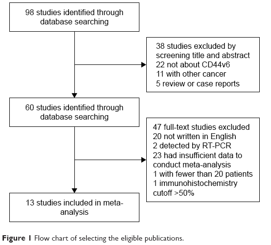 | Figure 1 Flow chart of selecting the eligible publications. |
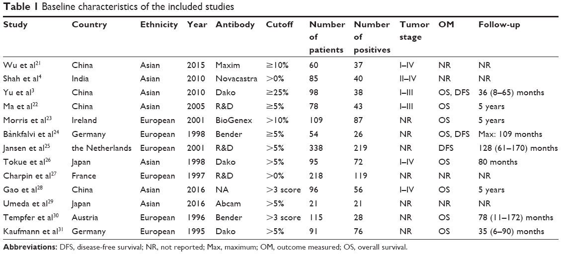 | Table 1 Baseline characteristics of the included studies |
Meta-analysis results
Correlation of CD44v6 with clinicopathological features
The forest plot of OR was evaluated for the relationship between CD44v6 expression and clinicopathological features including age, tumor diameter, histological type and grade, pTNM stage, lymph node metastasis, and menopausal status. In pooled analysis, CD44v6 expression was significantly associated with histological grade (overall: OR=1.56, 95% CI [1.06, 2.29], P=0.023; Asian: OR=1.78, 95% CI [1.12, 2.85], P=0.016, fixed-effect) (Figure 2A) and lymph node metastasis (overall: OR=1.96, 95% CI [1.01, 3.78], P=0.046; Asian: OR=2.11, 95% CI [1.00, 4.44], P=0.049, random-effect) (Figure 2B) in breast cancer. However, we did not find that CD44v6 expression was associated with age, tumor diameter, histological type, pTNM stage, and menopausal status. These results are summarized in Table 2.
 | Figure 2 Associations of CD44v6 expression with clinicopathological features. (A) Histological grade. (B) Lymph node metastasis. |
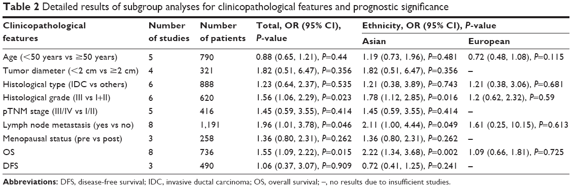 | Table 2 Detailed results of subgroup analyses for clinicopathological features and prognostic significance |
The prognostic effect of CD44v6 on survival in breast cancer
Survival analysis was performed on HR for OS and DFS in eight (730 patients) and three (247 patients) studies, respectively. The pooled analysis revealed that CD44v6 expression was highly correlated with poor OS (HR=1.55, 95% CI [1.09, 2.22], P=0.015, fixed-effect) (Figure 3), but not with poor DFS (HR=1.06, 95% CI [0.37, 3.07], P=0.909, random-effect).
 | Figure 3 Meta-analysis of the correlation of CD44v6 expression with overall survival. |
In the subgroup analysis, we found the significant prognostic effect of CD44v6 expression in Asian patients (OS: HR=2.22, 95% CI [1.34, 3.68], P=0.002, fixed-effect) (Table 2).
Publication bias and sensitivity analyses
Begg’s test and Egger’s test were performed to evaluate the publication bias. There was no evidence of publication bias for the pooled analysis of OS (PBegg=0.536, PEgger=0.733) (Figure 4A) and DFS (PBegg=1, PEgger=0.781) (Figure 4B). Sensitivity analyses were performed by excluding one study in turn to check whether individual study affected the final results. All the results were not materially altered.
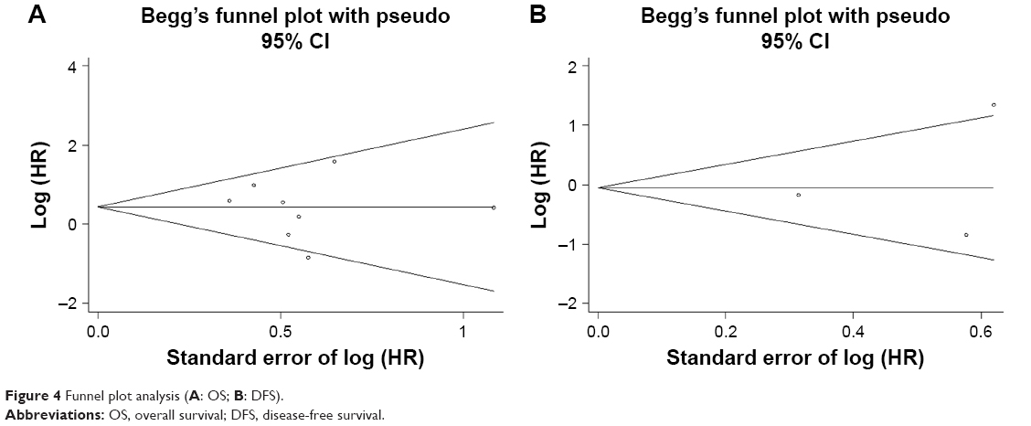 | Figure 4 Funnel plot analysis (A: OS; B: DFS). |
Discussion
Breast cancer remains the most common cancer in woman. Despite remarkable progresses in its treatment, the prognosis is still not optimistic.2 Prognostic factors are correlated with some clinical outcomes, such as OS or DFS, independent of any treatment. The accumulating evidence showed that CD44v6 might be a potential prognostic marker for solid tumors.13–16 There are many reports about the prognostic significance of CD44v6 in breast cancer. However, the results were inconsistent. Therefore, the quantitative meta-analysis about the association of CD44v6 with prognostic factor in breast cancer is required. This is the first meta-analysis to evaluate the clinicopathological features and prognostic significance of CD44v6 in breast cancer by summarizing all relevant studies.
We performed a comprehensive meta-analysis to assess the association between CD44v6 expression and clinicopathological features of breast cancer. The results showed that CD44v6 expression was significantly associated with histological grade and lymph node metastasis. However, no correlation was observed between CD44v6 expression and age, tumor diameter, histological type, pTNM stage, or menopausal status. Our results suggested that the expression of CD44v6 in tumor cells might enhance their potential for metastasis in the regional lymph nodes. Günthert et al7 found that transfection of tumor cells with CD44v6 could enhance metastasis to lymph nodes. Kaufmann et al31 demonstrated that CD44v6 could help tumor cells escape the immune system to promote lymph node metastasis. Thus, our results are in line with those of basic studies. CD44v6 expression may be considered as a marker of breast cancer that indicates lymph node metastasis. However, the correlation of CD44v6 expression with tumor diameter, pTNM stage, or menopausal status was not observed. These might be due to the different sample cohorts studied, as well as the smaller number of studies included.
The pooled data showed promising prognostic effect of CD44v6 expression in breast cancer samples for OS. The patients with CD44v6 expression had a 1.55 times higher risk of poor prognosis than those without CD44v6 expression. The subgroup analysis based on ethnicity was conducted to further evaluate prognostic value of CD44v6. The results showed that patients with CD44v6 expression had poor OS in the Asian subgroup. This may be attributed to the differences in gene and environment among the ethnicity. These results were consistent with prior reports of meta-analysis in gastric cancer,13 pharyngolaryngeal cancer,32 colorectal cancer,33 hepatocellular carcinoma,14 esophageal cancer,15 and osteosarcoma.16
The glycoprotein CD44 is a receptor for extracellular matrix components and is the most common cancer stem cell (CSC) marker in multiple types of cancers.34 CD44 is a complex family of molecules, including standard isoform of CD44 (CD44s) and CD44v1-v10 isoform. To date, only the particular variant CD44v6 was found to be related to aggressive tumor behavior and prognosis in breast cancer.31,35 This meta-analysis revealed that CD44v6 expression was associated with histological grade, lymph node metastasis, and poor prognosis in breast cancer. CD44v6 also has great implications for targeted therapy and prognostic imaging. Qian et al found an antihuman CD44v6 functionalized nanoparticle for targeted drug delivery to pancreatic cancer, resulting in tumor cell apoptosis.36 The CD44v6 monoclonal antibody-conjugated nanoprobes showed excellent stability, targeting ability against CD44-expressing gastric CSC, high photothermal conversion, and ablation ability. The CD44v6 nanoprobes exhibited potential for applications of gastric cancer targeted imaging and photothermal therapy.37 In head and neck squamous cell carcinoma, bivatuzumab could direct mertansine activity to CD44v6-expressing tumor cells. Although effective, the greatly toxic payload resulted in skin toxicity and termination of the program.38
There are limitations to this meta-analysis. First, the population data were extracted from the included studies and individual data were unavailable. Second, the heterogeneity could not be eliminated, and we used the random-effects model to obtain more conservative estimates. Third, due to lack of available data, we were unable to perform subgroup analyses based on breast cancer subtypes (ER/PR/HER2 negative or positive). Despite these limitations, this meta-analysis is the first study to analyze the prognostic value of CD44v6 expression in breast cancer.
Conclusion
This meta-analysis showed that CD44v6 expression is significantly associated with a poor survival, histological grade, and lymph node metastasis in breast cancer patients, especially among Asian patients. These results should be confirmed by adequate, high-quality, well-designed multicenter studies.
Acknowledgments
This work was supported by Shanghai Tongren Hospital (number TRYJ201514), the Project of Shanghai Municipal Commission of Health and Family Planning (number 20174Y0231), the National Natural Science Foundation of China (number 81672335), and the Shanghai Jiaotong University “medical professionals cross fund” (number YG2016ZD10).
Disclosure
The authors report no conflicts of interest in this work.
References
Torre LA, Bray F, Siegel RL, Ferlay J, Lortet-Tieulent J, Jemal A. Global cancer statistics, 2012. CA Cancer J Clin. 2015;65(2):87–108. | ||
Siegel RL, Miller KD, Jemal A. Cancer statistics, 2018. CA Cancer J Clin. 2018;68(1):7–30. | ||
Yu P, Zhou L, Ke W, Li K. Clinical significance of pAKT and CD44v6 overexpression with breast cancer. J Cancer Res Clin Oncol. 2010;136(8):1283–1292. | ||
Shah NG, Trivedi TI, Vora HH, et al. CD44v6 expression in primary breast carcinoma in western India: a pilot clinicopathologic study. Tumori. 2010;96(6):971–977. | ||
Naor D, Sionov RV, Ish-Shalom D. CD44: structure, function, and association with the malignant process. Adv Cancer Res. 1997;71:241–319. | ||
Nagano O, Saya H. Mechanism and biological significance of CD44 cleavage. Cancer Sci. 2004;95(12):930–935. | ||
Günthert U, Hofmann M, Rudy W, et al. A new variant of glycoprotein CD44 confers metastatic potential to rat carcinoma cells. Cell. 1991;65(1):13–24. | ||
Matuschek C, Lehnhardt M, Gerber PA, et al. Increased CD44s and decreased CD44v6 RNA expression are associated with better survival in myxofibrosarcoma patients: a pilot study. Eur J Med Res. 2014;19:6. | ||
Marhaba R, Bourouba M, Zöller M. CD44v6 promotes proliferation by persisting activation of MAP kinases. Cell Signal. 2005;17(8):961–973. | ||
Jung T, Gross W, Zöller M. CD44v6 coordinates tumor matrix-triggered motility and apoptosis resistance. J Biol Chem. 2011;286(18):15862–15874. | ||
Ruibal Á, Aguiar P, del Río MC, Nuñez MI, Pubul V, Herranz M. Cell membrane CD44v6 levels in squamous cell carcinoma of the lung: association with high cellular proliferation and high concentrations of EGFR and CD44v5. Int J Mol Sci. 2015;16(3):4372–4378. | ||
Athanassiou-Papaefthymiou M, Shkeir O, Kim D, et al. Evaluation of CD44 variant expression in oral, head and neck squamous cell carcinomas using a triple approach and its clinical significance. Int J Immunopathol Pharmacol. 2014;27(3):337–349. | ||
Fang M, Wu J, Lai X, et al. CD44 and CD44v6 are correlated with gastric cancer progression and poor patient prognosis: evidence from 42 studies. Cell Physiol Biochem. 2016;40(3–4):567–578. | ||
Fu Y, Geng Y, Yang N, et al. CD44v6 expression is associated with a poor prognosis in Chinese hepatocellular carcinoma patients: A meta-analysis. Clin Res Hepatol Gastroenterol. 2015;39(6):736–739. | ||
Hu B, Luo W, Hu RT, Zhou Y, Qin SY, Jiang HX. Meta-analysis of prognostic and clinical significance of CD44v6 in esophageal cancer. Medicine. 2015;94(31):e1238. | ||
Zhang Y, Ding C, Wang J, et al. Prognostic significance of CD44V6 expression in osteosarcoma: a meta-analysis. J Orthop Surg Res. 2015;10:187. | ||
Guriec N, Gairard B, Marcellin L, et al. CD44 isoforms with exon v6 and metastasis of primary N0M0 breast carcinomas. Breast Cancer Res Treat. 1997;44(3):261–268. | ||
Hefler L, Tempfer C, Haeusler G, et al. Cytosol concentrations of CD44 isoforms in breast cancer tissue. Int J Cancer. 1998;79(5):541–545. | ||
Afify A, Mcniel MA, Braggin J, Bailey H, Paulino AF. Expression of CD44s, CD44v6, and hyaluronan across the spectrum of normal-hyperplasia-carcinoma in breast. Appl Immunohistochem Mol Morphol. 2008;16(2):121–127. | ||
Tierney JF, Stewart LA, Ghersi D, Burdett S, Sydes MR. Practical methods for incorporating summary time-to-event data into meta-analysis. Trials. 2007;8:16. | ||
Wu XJ, Li XD, Zhang H, et al. Clinical significance of CD44s, CD44v3 and CD44v6 in breast cancer. J Int Med Res. 2015;43(2):173–179. | ||
Ma W, Deng Y, Zhou L. The prognostic value of adhesion molecule CD44v6 in women with primary breast carcinoma: a clinicopathologic study. Clin Oncol. 2005;17(4):258–263. | ||
Morris SF, O’Hanlon DM, Mclaughlin R, Mchale T, Connolly GE, Given HF. The prognostic significance of CD44s and CD44v6 expression in stage two breast carcinoma: an immunohistochemical study. Eur J Surg Oncol. 2001;27(6):527–531. | ||
Bànkfalvi A, Terpe HJ, Breukelmann D, et al. Gains and losses of CD44 expression during breast carcinogenesis and tumour progression. Histopathology. 1998;33(2):107–116. | ||
Jansen RH, Joosten-Achjanie SR, Arends JW, et al. CD44v6 is not a prognostic factor in primary breast cancer. Ann Oncol. 1998;9(1):109–111. | ||
Tokue Y, Matsumura Y, Katsumata N, Watanabe T, Tarin D, Kakizoe T. CD44 variant isoform expression and breast cancer prognosis. Jpn J Cancer Res. 1998;89(3):283–290. | ||
Charpin C, Garcia S, Bouvier C, et al. Automated and quantitative immunocytochemical assays of CD44v6 in breast carcinomas. Hum Pathol. 1997;28(3):289–296. | ||
Gao YL, Xing LQ, Ren TJ, et al. The expression of osteopontin in breast cancer tissue and its relationship with p21ras and CD44V6 expression. Eur J Gynaecol Oncol. 2016;37(1):41–47. | ||
Umeda T, Ishida M, Murata S, et al. Immunohistochemical analyses of CD44 variant isoforms in invasive micropapillary carcinoma of the breast: comparison with a concurrent conventional invasive carcinoma of no special type component. Breast Cancer. 2016;23(6):869–875. | ||
Tempfer C, Lösch A, Heinzl H, et al. Prognostic value of immunohistochemically detected CD44 isoforms CD44v5, CD44v6 and CD44v7-8 in human breast cancer. Eur J Cancer. 1996;32A(11):2023–2025. | ||
Kaufmann M, Heider KH, Sinn HP, von Minckwitz G, Ponta H, Herrlich P. CD44 variant exon epitopes in primary breast cancer and length of survival. Lancet. 1995;345(8950):615–619. | ||
Chai L, Liu H, Zhang Z, et al. CD44 expression is predictive of poor prognosis in pharyngolaryngeal cancer: systematic review and meta-analysis. Tohoku J Exp Med. 2014;232(1):9–19. | ||
Fan CW, Wen L, Qiang ZD, et al. Prognostic significance of relevant markers of cancer stem cells in colorectal cancer – a meta analysis. Hepatogastroenterology. 2012;59(117):1421–1427. | ||
Ryoo IG, Choi BH, Ku SK, Kwak MK. High CD44 expression mediates p62-associated NFE2L2/NRF2 activation in breast cancer stem cell-like cells: Implications for cancer stem cell resistance. Redox Biol. 2018;17:246–258. | ||
Wang Z, Wang Q, Wang Q, Wang Y, Chen J. Prognostic significance of CD24 and CD44 in breast cancer: a meta-analysis. Int J Biol Markers. 2017;32(1):75–82. | ||
Qian C, Wang Y, Chen Y, et al. Suppression of pancreatic tumor growth by targeted arsenic delivery with anti-CD44v6 single chain antibody conjugated nanoparticles. Biomaterials. 2013;34(26):6175–6184. | ||
Liang S, Li C, Zhang C, et al. CD44v6 monoclonal antibody-conjugated gold nanostars for targeted photoacoustic imaging and plasmonic photothermal therapy of gastric cancer stem-like cells. Theranostics. 2015;5(9):970–984. | ||
Spiegelberg D, Nilvebrant J. CD44v6-targeted imaging of head and neck squamous cell carcinoma: antibody-based approaches. Contrast Media Mol Imaging. 2017;2017:2709547. |
 © 2018 The Author(s). This work is published and licensed by Dove Medical Press Limited. The
full terms of this license are available at https://www.dovepress.com/terms
and incorporate the Creative Commons Attribution
- Non Commercial (unported, 3.0) License.
By accessing the work you hereby accept the Terms. Non-commercial uses of the work are permitted
without any further permission from Dove Medical Press Limited, provided the work is properly
attributed. For permission for commercial use of this work, please see paragraphs 4.2 and 5 of our Terms.
© 2018 The Author(s). This work is published and licensed by Dove Medical Press Limited. The
full terms of this license are available at https://www.dovepress.com/terms
and incorporate the Creative Commons Attribution
- Non Commercial (unported, 3.0) License.
By accessing the work you hereby accept the Terms. Non-commercial uses of the work are permitted
without any further permission from Dove Medical Press Limited, provided the work is properly
attributed. For permission for commercial use of this work, please see paragraphs 4.2 and 5 of our Terms.