
Back to Journals » Drug Design, Development and Therapy » Volume 20
Pregnanolone-Based Prodrugs as a Strategy for Neuroprotective Drug-Like Compounds: Pregnanolone Pyroglutamate and Its Age‑dependent Anticonvulsant Effects in the 6-hz Seizure Model in Immature Rats
Authors Mareš P, Chodounská H, Szczurowska E  , Kubová H, Kudova E
Received 12 November 2025
Accepted for publication 16 February 2026
Published 17 March 2026 Volume 2026:20 566239
DOI https://doi.org/10.2147/DDDT.S566239
Checked for plagiarism Yes
Review by Single anonymous peer review
Peer reviewer comments 2
Editor who approved publication: Professor Tamer Ibrahim
Pavel Mareš,1 Hana Chodounská,2 Ewa Szczurowska,2 Hana Kubová,1 Eva Kudova2
1Institute of Physiology, Academy of Sciences of the Czech Republic, Prague, Czech Republic; 2Institute of Organic Chemistry and Biochemistry, Academy of Sciences of the Czech Republic, Prague, Czech Republic
Correspondence: Eva Kudova, Email [email protected] Pavel Mareš, Email [email protected]
Purpose: Pediatric epilepsy remains a major therapeutic challenge due to the high prevalence of pharmacoresistant seizures, and the lack of drugs tailored to immature brain physiology. This study tested a pregnanolone‑based prodrug strategy in which the metabolically labile C‑3 hydroxyl group is transiently masked with a polar moiety designed to regenerate pregnanolone in vivo and thereby preserve or enhance its anticonvulsant efficacy.
Methods: Four C‑3‑conjugated pregnanolone analogues (compounds 1– 4) were synthesized and characterized as positive modulators of γ‑aminobutyric acid type A receptors (GABAARs) in vitro. Their in vivo activity was evaluated in the 6-Hz transcorneal stimulation model in 15‑ and 25‑day‑old Wistar rats, with detailed quantification of seizure duration and severity, and direct comparison to the endogenous neurosteroid allopregnanolone and the approved neuroactive steroid zuranolone.
Results: All four prodrugs showed nanomolar GABAA receptor modulation, consistent with the expected main neurosteroid mechanism of action. Compound 1 (pregnanolone pyroglutamate), designed to be enzymatically cleaved to release pregnanolone, displayed the most consistent and robust in vivo efficacy, producing marked, age‑dependent reductions in seizure duration and severity across multiple parameters in both age groups. Additionally, compounds 2 and 4 displayed enhanced efficacy in younger animals. Plasma stability experiments demonstrated rapid biotransformation of compound 1 to pregnanolone, supporting the hypothesis that its anticonvulsant effect is mediated by in vivo regeneration of the parent neurosteroid.
Conclusion: These findings validate a pregnanolone‑prodrug approach in which C‑3 masking groups are used primarily to deliver and release endogenous pregnanolone in vivo, and they identify compound 1 as a lead candidate for further optimization as an anticonvulsant for pediatric epilepsy. The work provides a translationally relevant framework for developing neurosteroid‑based prodrugs with improved drug‑like properties for pharmacoresistant seizures in the developing brain.
Keywords: neuroactive steroids, seizures, 6-Hz model, anticonvulsant action, immature rats, allopregnanolone, zuranolone
Introduction
Neurosteroids are endogenous steroids that are either synthesized within the brain or derived from peripheral glands.1 Their synthetic analogues are called neuroactive steroids (NAS). Both neurosteroids and neuroactive steroids rapidly modulate neuronal excitability by interacting with ligand-gated ion channels,2–6 voltage-gated ion channels,6–8 or G-protein coupled receptors.9–12 Consequently, they have been studied as modulators of seizure susceptibility,13–16 anxiety and stress,17–20 pain,21–23 neurodegenerative disorders,24–27 and many other nervous system disorders.28 The neurotherapeutic potential of neurosteroids was first demonstrated by the endogenous compound allopregnanolone (ALLO, 3α-hydroxy-5α-pregnan-20-one, Figure 1), a potent allosteric modulator of the γ‐aminobutyric acid type A receptors (GABAARs) and the first FDA-approved neurosteroid (brexanolone, ZULRESSO®) to treat moderate-to-severe postpartum depression.
 |
Figure 1 Structures of allopregnanolone, ganaxolone, and zuranolone. |
Numerous studies have described the anticonvulsant effects of ALLO in experimental models of seizures, including pentylenetetrazol-, bicuculline-, pilocarpine-, 6-Hz-induced convulsions or status epilepticus in rodents.29–33 Moreover, ALLO has also been tested in translational human clinical trials to treat super-refractory status epilepticus.34 The abovementioned published data underscores the antiepileptogenic properties of neurosteroids and NAS, positioning them as promising candidates for epilepsy management, including in catamenial epilepsy, stress-sensitive seizure disorders, or rare forms of epilepsy.35
Ganaxalone (ZTALMY®, 3α-hydroxy-3β-methyl-5α-pregnan-20-one, Figure 1) is the first-in-class neurosteroid approved in 2022 for the treatment of seizures associated with CDKL5 deficiency disorder (CDD) in patients ≥ 2 years of age.36,37 As a positive allosteric modulator of GABAARs, ganaxolone shows robust anticonvulsant efficacy in preclinical and clinical studies, including partial, catamenial, and refractory status epilepticus seizures,38–40 with surprisingly very mild adverse effects such as somnolence.41,42 The 3β-methyl group in ganaxolone enhances metabolic stability by resisting cytochrome P450 degradation. Subsequent modifications at C-19 (demethylation) and C-21 (heterocyclic substitution) defined a novel class of NAS.43
Zuranolone (SAGE-217, ZURZUVAE®, Figure 1) was approved in 2023 as the first oral treatment of postpartum depression in adults.44–48 The potent modulator of synaptic and extrasynaptic GABAARs,43 zuranolone, is not being developed for epilepsy but has been evaluated in clinical trials for major depressive disorder, anxiety, and insomnia.45,49–54
Our research focuses on developing novel NAS molecules to treat seizure disorders and epilepsy in immature rats.55,56 Our novel compounds are structural analogs of pregnanolone (PA, 3α‑hydroxy‑5β‑pregnan‑20‑one; Figure 2), an endogenous isomer of ALLO and progesterone metabolite, crucial for menstrual cycle regulation, pregnancy, and neuroprotection.57–60 Notably, both PA and zuranolone share a 5β‑pregnane skeleton. Like ALLO, PA belongs to a family of GABA-potentiating NAS.61,62 We previously reported63 a strong neuroprotective effect of PA in the pentylenetetrazol seizure model in animals aged 7, 12, 18, and 25 days with the most pronounced effect in the 7-, 12-, and 18-day-old groups.
 |
Figure 2 Biosynthesis of reduced metabolites of progesterone. |
Seizure disorders are among the most common and clinically significant neurological conditions in children. Childhood epilepsy encompasses a heterogeneous group of syndromes with distinct diagnostic criteria, management, and outcomes.64–66 Antiseizure drugs (ASDs) achieve seizure freedom in only about 70% of pediatric patients,67 while adverse effects are frequent, affecting up to 90% of children.65,66,68 Many ASDs may also disrupt normal brain development and, together with seizures and underlying pathology, contribute to long-term neuropsychiatric comorbidities.69 Despite this major unmet need, preclinical ASD screening still relies predominantly on adult animal models. The systematic use of developmental seizure and epilepsy models, followed by dedicated pediatric trials, is therefore crucial for designing safer, age-specific therapies.70
The 6‑Hz model is a widely used model of limbic seizures, applied primarily in mice and more recently adapted to rats71 and immature rodents.56 Its main advantages are throughput capability, cost‑effectiveness, and a proven ability to detect compounds with novel mechanisms of action. As a result, the 6-Hz test is employed by the NIH Epilepsy Therapy Screening Program for evaluating novel anticonvulsant candidates.72 Despite extensive research in adult rodents, the utility of this model in developing animals remains largely unexplored. It should be noted that this acute model does not reproduce the complex pathophysiology of chronic epilepsy, and convulsions induced by higher current intensities - initially described as relatively drug‑resistant - have yielded inconsistent results in epileptic animals, where disease‑related network changes can markedly alter seizure sensitivity to antiseizure drugs.73 Consequently, data derived from 6‑Hz screening must be interpreted cautiously and not assumed to predict efficacy in chronic epilepsy. To our knowledge, no studies have assessed novel antiseizure compounds in immature animals using the 6-Hz model. Currently, only one published study from our laboratory provides a methodological description of seizures induced by transcorneal 6-Hz stimulation in developing rats.56
Therefore, our research, which evaluates new compounds showing promising efficacy in the pentylenetetrazol-induced seizure model, aims to extend this assessment to the 6-Hz model. This may facilitate the identification of novel, drug-like molecules with improved efficacy and tolerability, potentially addressing unmet therapeutic needs in pediatric epilepsy.
Herein, we report the structure-activity relationship of NAS compounds 1–4 (Figure 3) in a 6-Hz seizure model in immature rats and compare their effects to those of ALLO and zuranolone. The synthetic strategy for ganaxolone and zuranolone involves introducing a 3β-methyl group. The purpose of the 3β-methyl substitution is to block oxidation, glucuronidation, and sulfation at position C-3. However, this modification, in the case of ganaxolone, reduces aqueous solubility, which can be overcome by enhancing solubility with cyclodextrins (eg, Captisol)74 or by using a suspension formulation.75 In our approach, we chemically introduced a prodrug moiety at the C-3 position as a widely used solution in medicinal chemistry.76–78 Our prodrug design uses a polar moiety to transiently mask the metabolically labile C‑3 hydroxyl group of pregnanolone, with the primary aim of testing the hypothesis that this protected form will be enzymatically converted back to pregnanolone in vivo and thereby reproduce its pharmacological effects.
 |
Figure 3 Concept of the study and structures of compounds 1–4. (A) Major steroidal metabolites identified in the hippocampus of male Wistar rats (P12 and P25 days old) after the administration of pregnanolone glutamate (i.p., 1 mg/kg in CDX).55 The values below the name of the steroid compound (blue color) represent the fold increase in the concentration of that particular compound in the rat hippocampus above the basal levels 20 min after pregnanolone glutamate application. (B) Structures of compounds 1–4. The texts below the structures summarize the concept of the structure-activity relationship and the rationale for the design of these compounds. |
Compounds 1–4 were designed as C-3 conjugates of the endogenous neurosteroid pregnanolone, building directly on prior in vivo work79 where a larger series of 3α,5β-neuroactive steroids was synthesized and evaluated in a pentylenetetrazole (PTZ) induced seizure model. The most potent anticonvulsant analogues were identified as compounds 1–3 and were included as exemplary structures in the corresponding patent.79 Compound 4 was synthesized as a structural analogue of compound 3, mimicking the 5-membered ring of pyroglutamic acid, to further elucidate the key structural features relevant to biological activity. The fact that pregnanolone-based C-3 prodrugs are pharmacologically active is further supported by the characterization of pregnanolone glutamate,55 a linear analogue of pregnanolone pyroglutamate 1 with a potent anticonvulsant effect, which was shown to act as a CNS-active prodrug metabolized in vivo, with pregnanolone demonstrated as the major primary metabolite and 17-hydroxy-pregnanolone identified as the major secondary metabolite. These data provided a mechanistic rationale to prioritize pregnanolone prodrugs in the present study, while reserving 17-hydroxy-pregnanolone prodrugs for a separate project.
On this basis, the central hypothesis of the current work is that masking the metabolically labile C‑3 hydroxyl group of pregnanolone with a pyroglutamate (and related) promoiety will regenerate pregnanolone in vivo, preserve GABAergic anticonvulsant activity, and thus retain or enhance efficacy when challenged in the 6-Hz seizure model in immature rats.
Materials and Methods
Animals
Experiments were performed in 15 and 25-day-old male Wistar albino rats (n=333). The day of birth was counted as zero (P0). The number of animals used in each experimental group is listed in Tables S1 and S2 (Supporting Information file). Rats were housed in a controlled environment (temperature 22 ± 1 °C, humidity 50–60%, lights on 0600–1800 h) with free access to food and water. The sample size was determined in advance according to previous experience with the given tests and followed the principles of the three R’s (Replacement, Reduction, and Refinement; https://www.nc3rs.org.uk/the-3rs). Outcome measures were prospectively selected. All procedures involving animals and their care were conducted according to the ARRIVE guidelines in compliance with national (Act No 246/1992 Coll.) and international laws and policies (EEC Council Directive 86/609, OJ L 358, 1, December 12, 1987; Guide for the Care and Use of Laboratory Animals, US National Research Council, 1996). The Ethical Committee of the Czech Academy of Sciences approved the experimental protocol (Approval No. 35–2022P). The administration of the tested drugs was not associated with intense pain or adverse events. Seizures induced by transcorneal stimulation are brief and mild. These seizures are not accompanied by pain or significant stress reactions, such as vocalization, stooped posture, or piloerection. After a seizure, the animals behave normally, and this model is not associated with any mortality or morbidity later in life.
For plasma stability assessment, four adult male CD-1 mice (AnLab, Czech Republic) were quickly decapitated under deep general anesthesia (2% isoflurane). Their blood was collected in Li-heparin tubes and then centrifuged at 1,400 RPM for 12 minutes at 4°C. The plasma was then separated and used for stability measurement, as described below.
Synthesis of Compounds 1–4
Compounds were synthesized according to the patent application.79 In brief, compounds 1–4 were prepared as follows.
(3R,5R,8R,9S,10S,13S,14S,17S)-17-Acetyl-10,13-dimethylhexadecahydro-1H-cyclopenta[a]phenanthren-3-yl (S)-5-oxopyrrolidine-2-carboxylate (pregnanolone pyroglumate, 1). A mixture of pregnanolone (318 mg, 1.0 mmol), 4-dimethylaminopyridine (30 mg, 0.25 mmol), 1-ethyl-3-(3-dimethylaminopropyl)carbodiimide hydrochloride (419 mg, 2.7 mmol), and hydroxybenzotriazole (297 mg, 2.2 mmol) was dried under vacuum at room temperature for 1 hour. Then, dry dimethylformamide (10 mL) was added under an inert atmosphere, followed by slow dropwise addition of L-pyroglutamic acid (194 mg, 1.5 mmol) in dry DMF (5 mL). The reaction mixture was allowed to stir overnight at room temperature. After solvent evaporation, the residue was purified by column chromatography (5% acetone/chloroform). Compound 1 (370 mg, 89%) was obtained as a solid material: m.p. 167−168 °C (chloroform, diethyl ether), [α]D20 +105.6 (c 0.3, CHCl3). 1H NMR (400 MHz, CDCl3): δ 0.60 (3H, s, H-18), 0.94 (3H, s, H-19), 2.11 (3H, s, H-21), 4.16–4.24 (1H, m, H-C2´), 4.80 (1H tt, J = 11.4, 4.8 Hz, H-3), 5.86 (1H, s, N-H). 13C NMR (101 MHz, CDCl3): δ 209.7, 177.6, 171.5, 75.9, 64.0, 56.8, 55.6, 44.5, 41.9, 40.6, 39.3, 35.9, 35.1, 34.7, 32.2, 31.7, 29.3, 26.9, 26.7, 26.4, 25.1, 24.6, 23.4, 23.1, 21.0, 13.6. IR spectrum (CHCl3): 1734, 1702 (C = O), 1230 (C-O). MS: ESI m/z 452.3 (100%, M+Na). HR-MS (ESI) m/z: for C26H39NO4Na [M+Na] calcd, 452.27713; found, 452.26742. For C26H39NO4 (429.6) calcd: 72.69%, C; 9,15%, H; 3.26%, N. Found: 72.29%, C; 9,15%, H; 3.11%, N.
(3R,5R,8R,9S,10S,13S,14S,17S)-17-Acetyl-10,13-dimethylhexadecahydro-1H-cyclopenta[a]phenanthren-3-yl (S)-6-oxopiperidine-2-carboxylate (compound 2). A mixture of pregnanolone (318 mg, 1.0 mmol), 4-dimethylaminopyridine (30 mg, 0.25 mmol), and (S)-6-oxo-2-piperidinecarboxylic acid (213 mg, 1.5 mmol) was dried under vacuum at room temperature for 1 hour. Then, dry dichloromethane (2 mL) was added under an inert atmosphere, followed by slow dropwise addition of 1-ethyl-3-(3-dimethylaminopropyl)carbodiimide hydrochloride (419 mg, 2.7 mmol) in dry dichloromethane (10 mL). The reaction mixture was allowed to stir overnight at room temperature. After 18 h, the reaction mixture was poured into water. The combined extracts were washed with an aqueous solution of hydrochloric acid (5%), aqueous solution of sodium bicarbonate, brine, and dried over anhydrous sodium sulfate. After solvent evaporation, the residue was purified by column chromatography on silica gel (3% acetone/chloroform). Compound 2 (142 mg, 32%) was obtained as a solid material: m.p. 139−141 °C (acetone/n-heptane), [α]D20 +103.2 (c 0.3, CHCl3). 1H NMR (400 MHz, CDCl3): δ 0.60 (3H, s, H-18), 0.94 (3H, s, H-19), 4.05 (1H, m, H-C2´), 4.81 (1H, m, H-3), 6.14 (1H, s, N-H). 13C NMR (101 MHz, CDCl3): δ 209.7, 171.4, 170.5, 76.1, 64.0, 56.8, 55.1, 44.5, 42.0, 41.0, 39.3, 35.9, 35.0, 34.7, 32.2, 31.7, 31.2, 27.0, 26.7, 26.4, 25.6, 24.6, 23.4, 23.1, 21.0, 19.7, 13.6. IR spectrum (CHCl3): 3402 (NH); 1734, 1698, 1663 (C = O). MS: ESI m/z 466.3 (100%, M+Na). HR-MS (ESI) m/z: for C27H41NO4Na [M+Na] calcd, 466.29278; found, 466.29283.
(3R,5R,8R,9S,10S,13S,14S,17S)-17-Cyano-10,13-dimethylhexadecahydro-1H-cyclopenta[a]phenanthren-3-yl (S)-5-oxopyrrolidine-2-carboxylate (compound 3). A mixture of 3α-hydroxy-5β-androstan-17β-carbonitrile (250 mg, 0.83 mmol), 4-dimethylaminopyridine (25 mg, 0.21 mmol), and L-pyroglutamic acid (139 mg, 1.07 mmol) was dried under vacuum at room temperature for 1 hour. Then, dry dichloromethane (2 mL) was added under an inert atmosphere, followed by slow dropwise addition of 1-ethyl-3-(3-dimethylaminopropyl)carbodiimide hydrochloride (347 mg, 2.2 mmol) in dry dichloromethane (10 mL). The reaction mixture was allowed to stir overnight at room temperature. After 18 h, the reaction mixture was poured into water. The combined extracts were washed with an aqueous solution of hydrochloric acid (5%), aqueous solution of sodium bicarbonate, brine, and dried over anhydrous sodium sulfate. After solvent evaporation, the residue was purified by column chromatography on silica gel (20% acetone/chloroform). Compound 3 (180 mg, 52%) was obtained as a solid material: m.p. 188−189 °C (chloroform/diethyl ether), [α]D20 +75.5 (c 0.2, CHCl3). 1H NMR (400 MHz, CDCl3): δ 0.91 (3H, s, H-18), 0.96 (3H, s, H-19), 4.25 (1H, ddd, J = 8.8, 5.1, 0.7 Hz, H-C2´), 4.80 (1H tt, J = 11.3, 4.8 Hz, H-3), 5.92 (1H, s, N-H). 13C NMR (101 MHz, CDCl3): δ 177.7, 171.5, 121.4, 75.8, 55.6, 54.5, 44.7, 41.8, 40.5, 40.5, 37.4, 36.3, 35.1, 34.8, 32.2, 29.3, 26.8, 26.8, 26.6, 26.4, 25.0, 24.7, 23.3, 20.6, 14.8. IR spectrum (CHCl3): 2237 (CN), 1735, 1705 (C = O), 1229 (C-O). MS: ESI m/z 435.3 (100%, M+Na). HR-MS (ESI) m/z: for C25H36N2O3Na [M+Na] calcd, 435.26181; found, 345.26135. For C25H36N2O3 (412.6) calcd: 72.78%, C; 8.80%, H; 6.79%, N. Found: 72.39%, C; 8.64%, H; 6.33%, N.
(3R,5R,8R,9S,10S,13S,14S,17S)-17-cyano-10,13-dimethylhexadecahydro-1H-cyclopenta[a]phenanthren-3-yl acetylglycinate (compound 4). A mixture of 3α-hydroxy-5β-androstan-17β-carbonitrile (300 mg, 1.0 mmol), 4-dimethylaminopyridine (30 mg, 0.25 mmol), 1-ethyl-3-(3-dimethylaminopropyl)carbodiimide hydrochloride (419 mg, 2.7 mmol), and hydroxybenzotriazole (297 mg, 2.2 mmol) was dried under vacuum at room temperature for 1 hour. Then, dry dimethylformamide (10 mL) was added under an inert atmosphere, followed by slow dropwise addition of L-acetylglycine (176 mg, 1.5 mmol) in dry DMF (5 mL). The reaction mixture was allowed to stir overnight at room temperature. After solvent evaporation, the residue was purified by column chromatography (5% acetone/chloroform). Compound 4 (310 mg, 78%) was obtained as a solid material: m.p. 158−159 °C (chloroform/diethyl ether), [α]D20 +74.5 (c 0.2, CHCl3). 1H NMR (400 MHz, CDCl3): δ 0.90 (3H, s, H-18), 0.95 (3H, s, H-19), 2.04 (3H, s, H-C5´), 2.28 (1H, t, J = 8.8 Hz, H-17), 4.00 (2H, dd, J = 1.8, 5.0 Hz, H-C2´), 4.80 (1H, tt, J = 4.8, 11.4 Hz, H-3), 5.96 (1H, s, N-H). 13C NMR (101 MHz, CHCl3): δ 170.2, 169.7, 121.4, 75.8, 54.5, 44.7, 41.8, 40.5, 40.5, 37.4, 36.4, 35.1, 34.8, 32.2, 26.9, 26.8, 26.7, 26.5, 24.7, 23.4, 23.2, 20.7, 14.5. IR spectrum (CHCl3): 3436, 1516 (NH); 1735, 1675 (C = O); 1192 (C−O). MS: ESI m/z 423.3 (100%, M+Na). HR-MS (ESI) m/z: for C24H36N2O3Na [M+Na] calcd, 423.26181; found, 423.26162. For C24H36N2O3 (400.6) calcd: 71.96% C; 9.06% H; 6.99% N. Found: 71.87% C; 9.04% H; 6.75% N.
Drugs
Allopregnanolone and zuranolone are commercially available; allopregnanolone (CAS 516–54-1, Carbosynth Ltd., United Kingdom, Catalogue No. FA158661), zuranolone (CAS 1632051–40-1, ChemShuttle, Hayward, CA, USA, Catalogue No. 186443). Compounds were dissolved in a solution of 3 g of (2-hydroxypropyl)-β-cyclodextrin (CDX, Sigma-Aldrich, St. Louis, MO, USA) and 157 mg of citric acid (Sigma-Aldrich, St. Louis, MO, USA) in 30 mL of distilled water. The pH was adjusted to 7.36 with NaOH (Sigma-Aldrich, St. Louis, MO, USA). Compounds 1–4 were dissolved in a concentration of 1 mg/mL, ALLO in a concentration of 2 mg/mL, and zuranolone in a concentration of 1 mg/mL. All compounds were administered intraperitoneally. Compounds 1–4 were injected in doses of 5 and 10 mg/kg. Based on our published data with pentylenetetrazol-induced seizures, allopregnanolone was used in doses of 10 and 20 mg/kg80 and zuranolone was used in doses of 0.5, 1, 5, and 10 mg/kg.81 In general, the 10-mg/kg dose of various NAS exhibits marked anticonvulsant action against PTZ.63,80,82 Control rats received cyclodextrin solution (10 mL/kg). Each age and dose group comprised ten animals. The first stimulation started 20 min after the injection of the drugs.
Stability in Plasma
For the evaluation of the stability of compound 1 in plasma, compound 2 was used as the internal standard (IS). For identification of pregnanolone, pregnanolone-D5 was used (IsoSciences, Ambler, PA, USA). Methanol (HPLC Super Gradient grade, Macron, Gliwice, Poland), formic acid, and ammonium acetate (puriss. p.a., Fluka, Buchs, Switzerland) were used for HPLC/MS-MS analysis. The HPLC/MS-MS system consisted of the P4000 pump, SCM 1000 degasser, AS3000 autosampler, TSQ Quantum Discovery Max triple quadrupole mass spectrometer with electro-spray ion source and data station with Xcalibur software, version 2.0.7 (Thermo Electron Corporation, Waltham, MA, USA). The separation was performed on a pentafluorophenyl-propyl column (Discovery HS F5, 50×2.1 mm, particle size 3 µm, Supelco, Bellefonte, PA, USA) protected with a Phenomenex (Torrance, CA, USA) C18 4×3 mm precolumn.
Stock solutions of compounds 1 and 2 (IS), pregnanolone, and pregnanolone-D5 were prepared by dissolving the respective substances in methanol. Working solutions were obtained by further dilution with the same solvent. All solutions were stored in the dark at –18 °C. The IS solution contained 113.9 pg/µL of compound 2 and 880.4 pg/µL of pregnanolone-D5 in methanol. Plasma samples (25 µL) were transferred to polypropylene tubes, and 100 µL of the IS solution was added. The samples were vortex-mixed for 15s at 2000 rpm and centrifuged for 2 min at 4000 rpm. The resulting supernatant was transferred to a 0.3 mL polypropylene autosampler vial, and 10 µL was injected into the chromatographic system. Chromatographic and mass spectrometric conditions are described in the Supporting Information file (Table S3).
Stimulation
Ten to five minutes before each stimulation, the eyes of the tested animals were locally anaesthetized with 0.5% mesocain (Mesocain 1%, Zentiva, Prague). A three-second series of biphasic pulses with a frequency of 6-Hz was applied bicorneally. Three intensities of stimulation current were used – 40, 60, and 80 mA in 15-day-old rats and 20, 40, and 60 mA in the 25-day-old animals. These intensities were selected according to our developmental study of 6-Hz seizures in developing rats.56 The stimulation series elicited clonic movements of the forelimbs with a 6-Hz frequency. The intensity of these movements increased with higher intensities of stimulation current. Three stimulation series were applied at 20-minute intervals. Clonic seizures, if they occurred, began immediately after the end of stimulation. The semiology of these seizures was identical to classical minimal pentylenetetrazol seizures and seizures appearing in the kindling model (clonic seizures with preserved righting ability). The severity of seizures classified with Racine’s scale83 might increase in the course of a fit. The highest grade of seizures was used in statistics. The first appearance of seizures served for the calculation of threshold current intensities; in addition, the duration of seizures was measured. After the experiment was completed, animals were deeply anesthetized using 2% isoflurane, and the depth of anesthesia was monitored by pinching the tip of the tail. When there was no response to pinching, the animal was quickly decapitated.
Statistics
At the beginning of the experiment, individual animals from each litter were randomly allocated to a particular treatment group. All efforts were made to minimize the number of animals used and their suffering. Data analysis was done blindly to the treatment. Each treatment and stimulation intensity group consisted of nine to ten animals. Data were analyzed using GraphPad Prism 7.0 (GraphPad Software, United States) software.
Using the D’Agostino-Pearson normality test, all data sets were first analyzed to determine whether the values were derived from a Gaussian distribution. Threshold intensities were analyzed with the Kruskal–Wallis test corrected for multiple comparisons by controlling the False Discovery Rate (FDR) of Benjamini, Krieger, and Yekutieli (Q = 0.05 taken as discovery). Seizure durations were analyzed only in groups composed of at least three animals that developed convulsive scores 3–5 with analysis of variance (ANOVA) using One-way ANOVA followed by FDR-corrected multiple comparison tests. Seizure severity (score 1–5) was analyzed with analysis of variance (ANOVA) using One-way ANOVA followed by FDR-corrected multiple comparison tests. The incidence of convulsive seizure score 3–5 was analyzed using Fisher’s exact test. Probability values <0.05 were considered statistically significant.
Competition Binding Assays
Radioligand binding experiments were performed in Eurofins Cerep, Celle l’Evescault, France, based on previously published protocols.84 Briefly, rat cerebral cortex tissue was used to prepare a membrane fraction (120 µg protein/sample). Samples were further incubated (120 min, 22°C) with [35S]-t-butylbicyclophosphorothionate ([35S]TBPS) at a concentration of 3 nM (Kd 14.6 nM). Incubation was conducted in a buffer (50 mM Na2HPO4/KH2PO4, pH 7.4) and 500 mM NaCl without (control) and with test compounds at their increasing concentrations (0.3 nM–10 µM). Nonspecific binding was determined using 20 µM picrotoxinin (in the absence of GABA, Kd 3.2 µM).85 Further, using the 96-well harvester (UNIFILTER), the samples were rapidly filtered under vacuum through glass fiber filters (Whatman GF/B) presoaked with 0.3% PEI and rinsed with cold 50 mM Tris-HCl. Dried filters were soaked in the scintillation cocktail (Microscint), and radioactivity was counted using the scintillation counter (Topcount). Picrotoxinin, as the standard reference compound, was tested in each experiment at several concentrations to obtain a competition curve and calculate its IC50 (210–280 nM). The results are summarized in the Supporting Information file.
Results
Compounds 1–4 (Figure 3) were designed as analogues of the endogenous neurosteroid pregnanolone, based on our previous study describing the in vivo metabolites of pregnanolone glutamate,55 in which pregnanolone was identified as the major primary metabolite. Pregnanolone glutamate carries an L-glutamic acid moiety at position C-3. The selection of an amino acid–based substituent was supported by our earlier studies employing metabolically stable glutamate- and aspartate-bearing steroids as negative allosteric modulators of N-methyl-D-aspartate receptors. Despite their polar character, the corresponding amide analogues exhibited high permeability together with robust CNS behavioral effects in vivo after systemic administration, indicating that the amino acid moiety represents a validated structural motif for CNS-active steroidal ligands. Building on this, the series progresses from pregnanolone glutamate to its cyclic pyroglutamate analogue 1 (Figure 3), in which the side chain is rendered neutral to favor CNS permeation, then to the six-membered ring analogue 2, and finally to the open-chain amino acid derivative 4 obtained by disruption of the cyclic pyroglutamic scaffold. Carbonitrile groups are well‑established and useful in CNS drug discovery.86 Therefore, bioisosteric substitution of C-20 ketone (strong H‑bond acceptor) with a C-17 nitrile (weaker acceptor, no H‑bond donor) was proposed to reduce polarity and H‑bonding, which may help blood-brain-barrier penetration.87
Given that biological instability in the organism is a key aspect of this study, we evaluated the plasma stability of compound 1 in mouse and rat plasma. First, haemolysed mouse plasma was spiked with compound 1 at different concentrations and immediately processed (Table S4, Supporting Information). Over the concentration range of 10–1000 ng/mL, the peak area of compound 1 increased linearly with increasing concentration. Concurrently, the peak area of pregnanolone also increased, indicating conversion of compound 1 to pregnanolone. The measured concentrations of compound 1 and pregnanolone relative to the spiked concentrations of compound 1 are summarized in Table S5 (Supporting Information). These data demonstrate that the majority of compound 1 is rapidly converted to pregnanolone in mouse plasma. Subsequently, the time course of compound 1 degradation was evaluated in mouse and rat plasma. Plasma samples (n=2 per time point) were spiked with compound 1 at a concentration of 97.14 ng/mL. Aliquots for 0 minutes were processed immediately, while the remaining plasma was stored at 36 °C. Additional aliquots were collected after 20 and 60 min and processed accordingly. The resulting concentrations of compound 1 and pregnanolone are presented in Table 1.
 |
Table 1 Stability of Compound 1 in Mouse and Rat Plasma |
In summary, the plasma stability data indicate that compound 1 is metabolized more slowly and converted less extensively to pregnanolone in rat plasma than in mouse plasma, where rapid and near‑complete conversion occurs within 20 minutes, demonstrating that metabolism to pregnanolone occurs readily in both species and that enzymatic conversion to the parent pregnanolone is efficient under physiologically relevant conditions. These findings suggest that rats should be selected as the primary species for subsequent in vivo evaluation of compounds 1–4 in the 6-Hz model of seizures, whereas mouse studies would require a separate, dedicated investigation with adjusted dosing and experimental design to account for the markedly faster biotransformation. These results further support the rationale that designing and synthesizing additional pregnanolone prodrugs is a promising strategy to identify new compounds.
To provide preliminary in vitro characterization, the GABAergic activity of compounds 1–4 was assessed relative to ganaxolone and zuranolone. Radioligand binding experiments using [35S]‑tert‑butylbicyclophosphorothionate ([35S]TBPS) were performed to evaluate their ability to allosterically modulate GABAAR binding.88,89 This approach is commonly used to evaluate their GABAergic activity before the in vivo experiments.43 The data, presented in Figure S1 (Supporting Information), indicate that compounds 1–4 inhibited [35S]TBPS binding in the nanomolar range, which is consistent with the published GABAergic activities of allopregnanolone (IC50 = 22 nM,43 51 nM,90 74 nM,91 93 nM,92,145 nM93), pregnanolone (IC50 = 71 nM,91,145 nM93), ganaxolone (IC50 = 80 nM94) or zuranolone (IC50 = 7 nM43) measured in various brain membranes. Because these in vitro assays were exploratory and lacked formal statistical evaluation, the results should be interpreted as preliminary, providing rather qualitative insight into the mechanism of action rather than quantitative verification. Nevertheless, they suggest that chemical masking of the C‑3 hydroxyl group and introduction of the C‑17 carbonitrile can preserve receptor affinity consistent with that of ganaxolone and zuranolone with the following structure-activity relationships: (i) the five-membered pyroglutamic acid ring in compound 1 can be replaced with a six-membered ring in compound 2; (ii) substitution of the C-17 acetyl group in compound 1 can be replaced by the carbonitrile moiety (compound 3) had no significant impact on the inhibitory effect; and (iii) modification at C-3 and C-17 may not been additive, as replacing it with an N-acetyl glycine moiety in compound 4 led to a marked reduction in potency (increased IC50) in the [35S]TBPS binding assay.
As mentioned previously, anticonvulsant effects of compounds 1–3 in pentylenetetrazol-induced seizures in 12- and 25-day-old male Wistar rats were described in a patent by Kudova at et.79 The most pronounced effect was demonstrated for compound 1 in a dose of 10 mg/kg, with completely suppressed seizures in 12-day-old animals and with a significant decrease in the incidence of generalized tonic clonic seizures without a selective effect against the tonic phase in 25-day-old animals. Following our methodological study of 6-Hz stimulation in developing rats,56 compounds 1–4 were evaluated for their ability to alleviate seizures triggered by corneal 6-Hz stimulation in 15- and 25-day-old Wistar rats. The youngest animals included in our study were 15 days old, as eyelid opening had not yet occurred in younger pups. Testing at earlier developmental stages would require surgical eyelid separation, introducing additional stress factors. During the 6-Hz stimulation, all animals of both age groups exhibited clonic movements of forelimbs without any marked difference among the intensity of the stimulation current and all drug groups.
The results of the in vivo experiment for compounds 1 (Figure 4), 2 (Figure 5), 3 (Figure 6), 4 (Figure 7), allopregnanolone (Figure 8), and zuranolone (Figure 9) are summarized in separate figures showing threshold current intensity, incidence of seizures, seizure duration, and severity of seizures for 15- and 25-day-old animals.
 |
Figure 4 Effects of compound 1 on 6-Hz model in 15-day-old rats (left column) and 25-day-old rats (right column). The individual graphs show: (A) shows the threshold stimulation intensity necessary to induce the first convulsive seizure (severity score 3–5). The threshold intensity is expressed in mA (mean ± SEM; y-axis). (B) The incidence of motor seizures after stimulation at a given intensity (40, 60, and 80 mA in 15-day-old animals and 20, 40, and 60 mA in 25-day-old animals) is shown in the individual treatment groups (panel D; x-axis). Incidence is expressed as a percentage (y-axis); the total number of animals in the treatment group is taken as 100%. (C) The duration of convulsive seizures is expressed in seconds (y-axis). Only motor seizures (severity score 3–5) were used in the analysis, and seizure durations were statistically analyzed only in groups composed of at least three animals with a convulsive score of 3–5. The number of animals lower than three is shown in brackets. Panel (D) shows seizure severity, expressed as a score, on the y-axis. Scores 1 and 2 indicate no motor seizures were observed; scores 3–5 indicate motor seizures following stimulation. Abbreviations: x – no motor seizures observed in the group; * – discovery q < 0.05. The color codes used to mark the individual doses of the tested substances are used in all graphs: white for controls receiving CDX, grey for 0.5 mg/kg, ice blue for 1 mg/kg, sky blue for 5 mg/kg, royal blue for 10 mg/kg, and black for 20 mg/kg. The number of animals used in each experimental group is listed in Table S1 (Supporting Information file). |
 |
Figure 5 Effects of compound 2 on the 6-Hz model. The individual graphs show: (A) shows the threshold stimulation intensity necessary to induce the first convulsive seizure (severity score 3–5). The threshold intensity is expressed in mA (mean ± SEM; y-axis). (B) The incidence of motor seizures after stimulation at a given intensity (40, 60, and 80 mA in 15-day-old animals and 20, 40, and 60 mA in 25-day-old animals) is shown in the individual treatment groups (panel D; x-axis). Incidence is expressed as a percentage (y-axis); the total number of animals in the treatment group is taken as 100%. (C) The duration of convulsive seizures is expressed in seconds (y-axis). Only motor seizures (severity score 3–5) were used in the analysis, and seizure durations were statistically analyzed only in groups composed of at least three animals with a convulsive score of 3–5. The number of animals lower than three is shown in brackets. Panel (D) shows seizure severity, expressed as a score, on the y-axis. Scores 1 and 2 indicate no motor seizures were observed; scores 3–5 indicate motor seizures following stimulation. Abbreviations: x – no motor seizures observed in the group; * – discovery q < 0.05. The color codes used to mark the individual doses of the tested substances are used in all graphs: white for controls receiving CDX, grey for 0.5 mg/kg, ice blue for 1 mg/kg, sky blue for 5 mg/kg, royal blue for 10 mg/kg, and black for 20 mg/kg. The number of animals used in each experimental group is listed in Table S1 (Supporting Information file). |
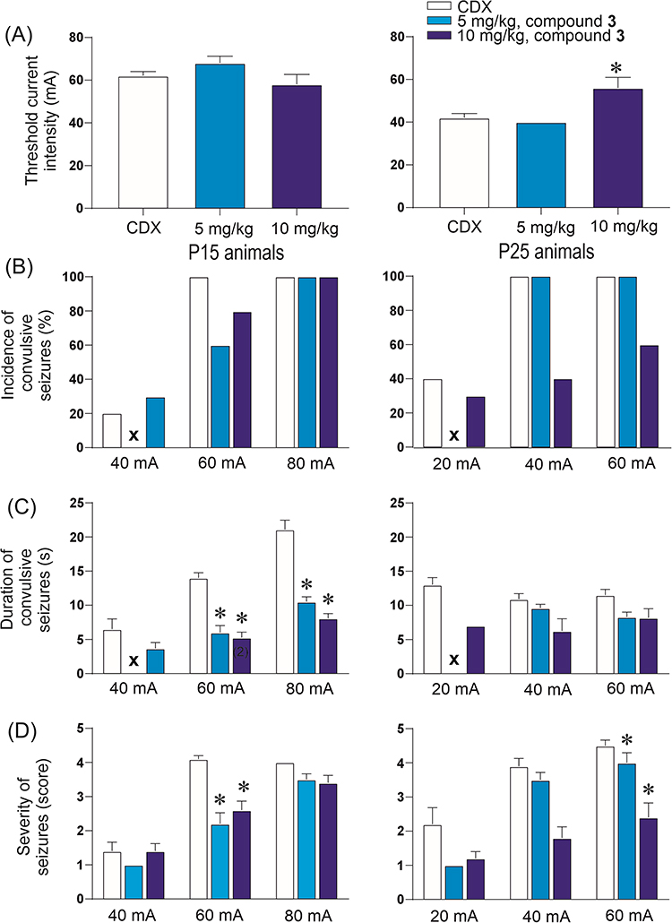 |
Figure 6 Effects of compound 3 on the 6-Hz model. The individual graphs show: (A) shows the threshold stimulation intensity necessary to induce the first convulsive seizure (severity score 3–5). The threshold intensity is expressed in mA (mean ± SEM; y-axis). (B) The incidence of motor seizures after stimulation at a given intensity (40, 60, and 80 mA in 15-day-old animals and 20, 40, and 60 mA in 25-day-old animals) is shown in the individual treatment groups (panel D; x-axis). Incidence is expressed as a percentage (y-axis); the total number of animals in the treatment group is taken as 100%. (C) The duration of convulsive seizures is expressed in seconds (y-axis). Only motor seizures (severity score 3–5) were used in the analysis, and seizure durations were statistically analyzed only in groups composed of at least three animals with a convulsive score of 3–5. The number of animals lower than three is shown in brackets. Panel (D) shows seizure severity, expressed as a score, on the y-axis. Scores 1 and 2 indicate no motor seizures were observed; scores 3–5 indicate motor seizures following stimulation. Abbreviations: x – no motor seizures observed in the group; * – discovery q < 0.05. The color codes used to mark the individual doses of the tested substances are used in all graphs: white for controls receiving CDX, grey for 0.5 mg/kg, ice blue for 1 mg/kg, sky blue for 5 mg/kg, royal blue for 10 mg/kg, and black for 20 mg/kg. The number of animals used in each experimental group is listed in Table S1 (Supporting Information file). |
 |
Figure 7 Effects of compound 4 on the 6-Hz model. The individual graphs show: (A) shows the threshold stimulation intensity necessary to induce the first convulsive seizure (severity score 3–5). The threshold intensity is expressed in mA (mean ± SEM; y-axis). (B) The incidence of motor seizures after stimulation at a given intensity (40, 60, and 80 mA in 15-day-old animals and 20, 40, and 60 mA in 25-day-old animals) is shown in the individual treatment groups (panel D; x-axis). Incidence is expressed as a percentage (y-axis); the total number of animals in the treatment group is taken as 100%. (C) The duration of convulsive seizures is expressed in seconds (y-axis). Only motor seizures (severity score 3–5) were used in the analysis, and seizure durations were statistically analyzed only in groups composed of at least three animals with a convulsive score of 3–5. The number of animals lower than three is shown in brackets. Panel (D) shows seizure severity, expressed as a score, on the y-axis. Scores 1 and 2 indicate no motor seizures were observed; scores 3–5 indicate motor seizures following stimulation. Abbreviations: x – no motor seizures observed in the group; * – discovery q < 0.05. The color codes used to mark the individual doses of the tested substances are used in all graphs: white for controls receiving CDX, grey for 0.5 mg/kg, ice blue for 1 mg/kg, sky blue for 5 mg/kg, royal blue for 10 mg/kg, and black for 20 mg/kg. The number of animals used in each experimental group is listed in Table S1 (Supporting Information file). |
 |
Figure 8 Effects of compound allopregnanolone on the 6-Hz model. The individual graphs show: (A) shows the threshold stimulation intensity necessary to induce the first convulsive seizure (severity score 3–5). The threshold intensity is expressed in mA (mean ± SEM; y-axis). (B) The incidence of motor seizures after stimulation at a given intensity (40, 60, and 80 mA in 15-day-old animals and 20, 40, and 60 mA in 25-day-old animals) is shown in the individual treatment groups (panel D; x-axis). Incidence is expressed as a percentage (y-axis); the total number of animals in the treatment group is taken as 100%. (C) The duration of convulsive seizures is expressed in seconds (y-axis). Only motor seizures (severity score 3–5) were used in the analysis, and seizure durations were statistically analyzed only in groups composed of at least three animals with a convulsive score of 3–5. The number of animals lower than three is shown in brackets. Panel (D) shows seizure severity, expressed as a score, on the y-axis. Scores 1 and 2 indicate no motor seizures were observed; scores 3–5 indicate motor seizures following stimulation. Abbreviations: x – no motor seizures observed in the group; * – discovery q < 0.05. The color codes used to mark the individual doses of the tested substances are used in all graphs: white for controls receiving CDX, grey for 0.5 mg/kg, ice blue for 1 mg/kg, sky blue for 5 mg/kg, royal blue for 10 mg/kg, and black for 20 mg/kg. The number of animals used in each experimental group is listed in Table S2 (Supporting Information file). |
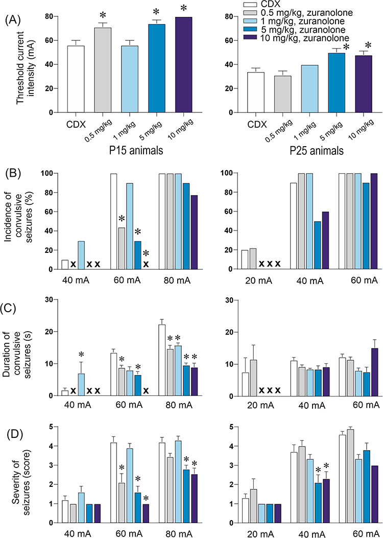 |
Figure 9 Effects of compound zuranolone on the 6-Hz model. The individual graphs show: (A) shows the threshold stimulation intensity necessary to induce the first convulsive seizure (severity score 3–5). The threshold intensity is expressed in mA (mean ± SEM; y-axis). (B) The incidence of motor seizures after stimulation at a given intensity (40, 60, and 80 mA in 15-day-old animals and 20, 40, and 60 mA in 25-day-old animals) is shown in the individual treatment groups (panel D; x-axis). Incidence is expressed as a percentage (y-axis); the total number of animals in the treatment group is taken as 100%. (C) The duration of convulsive seizures is expressed in seconds (y-axis). Only motor seizures (severity score 3–5) were used in the analysis, and seizure durations were statistically analyzed only in groups composed of at least three animals with a convulsive score of 3–5. The number of animals lower than three is shown in brackets. Panel (D) shows seizure severity, expressed as a score, on the y-axis. Scores 1 and 2 indicate no motor seizures were observed; scores 3–5 indicate motor seizures following stimulation. Abbreviations: x – no motor seizures observed in the group; * – discovery q < 0.05. The color codes used to mark the individual doses of the tested substances are used in all graphs: white for controls receiving CDX, grey for 0.5 mg/kg, ice blue for 1 mg/kg, sky blue for 5 mg/kg, royal blue for 10 mg/kg, and black for 20 mg/kg. The number of animals used in each experimental group is listed in Table S2 (Supporting Information file). |
In summary, compounds 1, 2, and 4 exhibited stronger anticonvulsant effects in 15-day-old rats, comparable to ALLO and zuranolone. Zuranolone was very potent at lower doses, which reflects its optimized GABAergic effect. ALLO, however, demonstrated mixed, dose-dependent effects on threshold intensities in 15-day-old rats. Compound 1 most robustly reduced seizure duration and severity across both age groups. Compound 2 also decreased seizure duration and incidence in younger rats, while compound 4 significantly reduced seizure duration and incidence only in 15-day-olds. Interestingly, in 25-day-old animals, only compound 3 and ALLO (20 mg/kg) significantly increased seizure thresholds, unlike the other compounds. The reason for this similarity remains unknown. From compounds 1–4, compound 1 appears most effective due to consistent effects on multiple seizure parameters across both ages. Overall, anticonvulsant activity was more pronounced in 15-day-old animals, indicating greater age-dependent sensitivity during early development.
The following is a summary of the detailed analysis of the effect of each compound. Pre-treatment of animals with 10 mg/kg of compound 1 (Figure 4) significantly increased the threshold intensity necessary to elicit seizures in 15-day-old rats. No changes in threshold were observed for either 5 mg/kg in 15-day-old rats or 25-day-old animals at either dose. The decrease in seizures elicited by the 60-mA intensity in 15-day-old animals was significant, in contrast to nonsignificant changes in seizure incidence after other stimulation intensities in either group. The duration and severity of seizures induced by stimulation currents of 60 and 80 mA were significantly reduced in a dose-dependent manner by both doses of compound 1 in both age groups.
The pre-treatment of animals with 10 mg/kg of compound 2 significantly increased the threshold current intensity in 15-day-old animals (Figure 5). The effect of the 5 mg/kg dose did not reach statistical significance. Neither dose altered the threshold in 25-day-old rats. The incidence of seizures decreased in 15-day-old animals that received either dose of compound 2 at a stimulation intensity of 60 mA. However, no change was observed at an intensity of 80 mA. The incidence of convulsions remained unchanged in 25-day-old animals. The duration of seizures was significantly shorter for both doses of compound 2 after 80-mA stimulation and for the 5-mg/kg dose of compound 2 after 60-mA stimulation in 15-day-old rats. Both doses shortened the duration of convulsions elicited by a 60-mA stimulation intensity, and the 5-mg/kg dose was efficient in 40-mA stimulation in 25-day-old rat pups. Similar tendencies, though not significant, were found in seizure severity after 60 and 80-mA stimulation current intensities in both age groups, but a statistically significant difference was found after either dose of compound 2 in 60/mA stimulation intensity in 15-day-old rats.
Pre-treatment with compound 3 did not change the seizure threshold in 15-day-old rats, but it significantly increased the threshold in 25-day-old rats at a dose of 10 mg/kg (Figure 6). In P25 animals, a 10 mg/kg dose reduced the incidence of seizures at stimulation intensities of 40 mA. The duration of seizures was significantly shorter for both doses of compound 2 after 60- and 80-mA stimulation in 15-day-old rats. Either dose tended to shorten the duration of convulsions elicited by the 40- and 60-mA stimulation intensities in 25-day-old animals, but the level of statistical significance was not reached. The severity of seizures was significantly lower after either dose of substance 3 in the 60-mA intensity groups in 15-day-old rats and in the 60-mA intensity groups in 25-day-old animals.
Pre-treating animals with compound 4 did not significantly affect the threshold in either group (Figure 7). All three doses tend to decrease the incidence of convulsions after 60-mA stimulus in 15-day-old animals; the decrease in the incidence of seizures after the highest dose after 40-mA stimulus in 25-day-old animals was significant. Both tested doses (5 and 10 mg/kg) significantly reduced the duration of seizures induced by 60-mA and 80-mA (respectively 40- and 60-mA) stimulus in either group. Severity of seizures increased in both groups by the lowest dose after the highest stimulation intensity, and decreased after the highest dose in the 40-mA stimulation group in 25-day-old rats.
A dose of 20 mg/kg of ALLO increased threshold current intensities in older age group (Figure 8). In contrast, the lower dose of 10 mg/kg significantly decreased the threshold in 15-day-old rats. The incidence of seizures remained consistent for both groups, with a significant decrease after the 20-mg/kg dose in the medium stimulation intensities. The duration of seizures elicited by an 80-mA stimulation current intensity in 15-day-old animals was shortened by a higher dose of ALLO. The 25-day-old group did not exhibit any significant changes. Severity of seizures in 15-day-old rat pups was decreased by both doses after the 60-mA stimulation and by a higher dose of ALLO after the 80-mA stimulation. The older age group exhibited lower seizure severity only after the 20-mg/kg dose and 40-mA stimulation intensity.
Zuranolone was tested in four doses of 0.5, 1, 5, and 10 mg/kg due to its more pronounced effect as compared with other compounds (Figure 9). Zuranolone demonstrated a significant anticonvulsant effect in 15-day-old rats at notably lower doses compared to compound 1 and its analogues. The 0.5, 5, and 10 mg/kg doses elevated the seizure threshold and significantly reduced seizure duration and severity. In 25-day-old rats, the significant effect was an increased threshold following the doses of 5 and 10 mg/kg and a decrease in seizure severity after the two high doses and 40-mA stimulation intensity.
Discussion
Compounds 1–4 were designed as analogues of the endogenous neurosteroid pregnanolone, building on previous work showing a potent anticonvulsant effect of pregnanolone glutamate and identifying pregnanolone and 17α‑hydroxy‑pregnanolone as its major in vivo metabolites (Figure 3).55 The feasibility of this prodrug approach was originally supported by an extensive series of analogues evaluated in the pentylenetetrazol seizure model, in which compounds 1–3 emerged as the most active in vivo, while compound 4 was prepared as a structural analogue of compound 3 mimicking the 5‑membered ring of pyroglutamic acid. The results of this study were described in a patent by Kudova et al.79
The metabolic instability of compound 1 in mouse and rat plasma, shown in this study, together with its efficient hydrolysis to endogenous pregnanolone, supports the rationale that designing and synthesizing additional pregnanolone prodrugs is a promising strategy for discovering new entities for further evaluation. At the same time, the complex metabolic profile of pregnanolone glutamate55 indicates that the resulting metabolites largely follow endogenous steroid metabolism, suggesting that tailored prodrug modifications for endogenous neurosteroids may generate molecules that act as transporters or reservoirs of pregnanolone, releasing it for further conversion to a “cocktail” of endogenous metabolites which may themselves contribute neuroprotective effects in addition to the parent prodrug. We have proposed that the in vivo effect of pregnanolone glutamate versus pregnanolone pyroglutamate (dose-dependent effect) is likely to be influenced also by the pattern and relative abundance of downstream metabolites and, consequently, their in vivo actions. For these reasons, we have prioritized the direct testing of prodrug compounds in an in vivo model such as the 6‑Hz seizure experiment, rather than first exhaustively characterizing all physicochemical, ADME properties, and potential mechanisms of action in vitro before in vivo testing.
Taken together, the strategy of generating a series of pregnanolone prodrugs is intended to identify the most potent compounds in vivo, then, for selected leads, to delineate their metabolic cascades, define the ADME profiles of key metabolites, and evaluate those metabolites in relevant seizure and neuroprotection models. Follow‑up work should also address species‑ and sex‑dependent differences in metabolism (eg, rat versus mouse), prepare additional prodrugs of major endogenous metabolites to test the central role of pregnanolone itself, and explore alternative formulations and routes of administration that may improve neurosteroid bioavailability and thereby enhance neuroprotective efficacy. This represents a broad research program that extends beyond the scope of a single publication; several of these aspects have already been investigated by the authors and will be reported separately. Consequently, the present results should be regarded as a proof‑of‑concept contribution that illustrates the viability of the pregnanolone‑prodrug approach rather than providing a complete account of all anticipated outcomes.
Recent clinical approvals of neuroactive steroids, including brexanolone and ganaxolone, underscore the therapeutic value of GABAARs modulation in CNS disorders, notably epilepsy. However, the significance of GABAergic modulation in immature rats and its potential as a primary target therapy remains unclear. Existing literature on neurosteroid mechanisms does not fully explain the age-dependent differences in anticonvulsant efficacy. In rodents, GABAergic neurotransmission, the primary target system of neurosteroids, matures gradually after birth. During the first postnatal week, GABA is predominantly excitatory, but by the end of this period, it becomes inhibitory in the hippocampus.95 In parallel, drugs that enhance GABAergic inhibition, such as benzodiazepines, show stable anticonvulsant efficacy from the first postnatal week onward, supporting a predominantly inhibitory GABA action at this stage.96 Data on the developmental profile of tonic inhibition mediated by extrasynaptic GABAARs, which are key targets of neurosteroids are limited, but an increase in tonic inhibition has recently been linked to reduced excitability and seizure-like activity in P8 animals.97
Neurosteroids exert their anti-seizure effects mainly through positive allosteric modulation and direct activation of GABAARs, particularly extrasynaptic δ-subunit-containing receptors that mediate tonic inhibition. The δ-subunit is expressed at low levels at birth, increases postnatally, and reaches adult-like levels around the third postnatal week, with strong region-specific timing.98
Additional mechanisms likely contribute to developmental differences in neurosteroid efficacy. These include the gradual postnatal maturation of pharmacokinetic systems, such as efflux transporters,99 and enzymes responsible for their elimination, notably phase-I biotransformation enzymes (cytochrome P450). Ontogeny differs substantially among individual CYP450 isoforms, with maximal activities often reached in juvenile or young adult rodents, while enzyme activity is lower in early stages of postnatal life.100
Patch-clamp recordings at extrasynaptic GABAARs would yield useful pharmacological data, but they would not necessarily clarify the main mechanisms underlying their anticonvulsant efficacy in the developmental context considered in this study. Because both extrasynaptic GABAergic signaling and neurosteroid metabolism change markedly with age and brain region, in vitro data from a single preparation are hard to generalize to complex seizure phenotypes, as they would capture only a small part of the overall pharmacology. In this situation, where both the developmental profile of extrasynaptic GABAARs and the relevant neurosteroid metabolites are insufficiently defined, available data for developmental epilepsy are too limited to establish a reliable mechanistic framework solely from in vitro studies. Given these uncertainties and the limited mechanistic information available for developmental epilepsy, direct testing of pregnanolone prodrugs in vivo currently offers the most efficient way to assess their anticonvulsant potential.
Given the prevalence of pharmacoresistant epilepsy, there is growing interest in novel treatments. The NIH Epilepsy Therapy Screening Program has recognized this challenge by employing the 6-Hz seizure model, where stimulation with the highest intensity current specifically represents pharmacoresistant temporal lobe epilepsy. Our study addresses a critical gap in the literature, as virtually no drug-development research studies have examined the 6-Hz model in immature animals, despite the importance of pediatric epilepsy. The present study addresses this gap by applying a prodrug‑based strategy targeting pregnanolone analogues to exploit, rather than avoid, the metabolic lability of pregnanolone prodrugs as a key design feature for novel compounds with robust in vivo activity, while leveraging the intrinsic safety associated with endogenous neurosteroids and their established physiological tolerance.
Exploratory in vitro evaluation of GABAergic activity of compounds 1–4 showed nanomolar-range GABAAR modulation for all compounds, suggesting their mechanism of action. Finally, compounds 1–4 were evaluated in the 6-Hz seizure model in immature rats. It should be noted that the evaluation of our in vivo results was based on the detailed description of the quantitative changes in the duration and severity of seizures, not only on the presence or absence of seizures. None of the tested neuroactive steroids, nor allopregnanolone or zuranolone, in any dose used, was able to completely suppress seizures.
Our data reveal quantitative and qualitative differences in the actions of individual drugs. Specifically, compounds 1, 2, 4, and zuranolone exhibited similar effects: anticonvulsant action in 15-day-old animals and almost no action in the 25-day-old group. Of compounds 1–4, compound 1 displayed the most consistent effects across multiple seizure parameters in both age groups. Compound 3 differs from all other neurosteroids in that it exhibits a more pronounced effect in 25-day-old animals than in 15-day-old animals concerning the threshold intensity of stimulation current. The reason for this difference should be analyzed in future experiments. These results also suggest that novel NAS targeting 15- or 25-day-old animals can be designed and developed.
Seizures induced by the highest stimulation intensity in mice (typically 44 mA) are commonly used as a model of pharmacoresistant epilepsy.101 By analogy, seizures elicited at the highest current in our rat model (80 mA) may similarly represent a refractory phenotype. This raises the possibility that compound 1 could be effective against seizures unresponsive to conventional antiseizure medications.
Given that only one published study to date has reported developmental data for the 6-Hz seizure model in rats, which is from our laboratory,56 we included PTZ-induced seizures for comparative purposes. The behavioral semiology observed following 6-Hz transcorneal stimulation in both age groups is identical to the minimal clonic seizures induced by PTZ from the third postnatal week onward.102,103 Also, identical semiology with minimal clonic seizures generated in the basal forebrain104 might indicate the site of origin of 6-Hz induced seizures in the same structures. Furthermore, seizure incidence is comparable between the two models: 100% of animals in both age groups exhibited convulsions following 60-mA transcorneal stimulation, while PTZ (100 mg/kg) produced seizures in 100% and 88% of 15- and 25-day-old rats, respectively.
In contrast, these models differ by their mechanism of action. While pentylenetetrazol acts at different parts of GABAA receptors (GABA binding site, benzodiazepine modulatory site, chloride channel105), the transcorneal electrical stimulation nonselectively activates all forebrain structures and systems, and therefore it may be efficient in all rats. We presume that 6-Hz stimulation might elicit seizures even in younger animals; we do not study rat pups that are younger than 15-day-old due to their closed eyelids.
Our study showed that 6-Hz seizures in 15-day-old rats may be affected by a 5 mg/kg dose and certainly by a 10 mg/kg dose. Despite the same semiology, the minimal clonic seizures elicited by pentylenetetrazol in 18-day-old and older rats are only moderately modified by high doses of neurosteroids (10 mg/kg and higher).63,80,82 Three steroids (1, 2, ALLO) were efficient against generalized pentylenetetrazol-induced seizures in 12- and 25-day-old rats, with a tendency to more marked effects in younger animals.63,79 Compound 3 had a similar effect on PTZ-induced seizures as on the 6-Hz model. It had a significant effect on 25-day-old rats but not on 12-day-old rats. Only the 5 mg/kg dose, not the 10 mg/kg dose, significantly suppressed the incidence of PTZ-induced generalized seizures.
In general, our study has shown that NAS are more efficient in the younger group than in 25-day-old animals (eg, in PTZ-induced convulsions63,82), and higher efficacy in the older group was seen with zuranolone,81 also in PTZ-induced seizures.
The observed age-dependent differences in anticonvulsant efficacy are not well supported by existing literature. Data on the developmental profile of tonic inhibition mediated by extrasynaptic GABAA receptors - the ideal targets of neurosteroids - remain limited. While the presence of these receptors has been demonstrated in the thalamus by the middle of the second postnatal week,106 comparable developmental data for forebrain regions implicated in minimal clonic seizure generation are currently lacking. This knowledge gap complicates the interpretation of the age-specific effects observed in our study.
Nanotechnology-based drug delivery systems represent innovative and promising strategies for the delivery of antiepileptic drugs,107,108 showing strong potential to enhance transport across the blood–brain barrier, increase brain concentrations, maintain therapeutic levels for longer periods, and reduce side effects compared to conventional formulations. Lipid-based nanosystems, including solid lipid nanoparticles and nanoliposomes, have been developed for antiepileptic drugs such as carbamazepine,109 lamotrigine,110 perampanel,111 phenytoin,112 and phenobarbital,113 and have been evaluated in various in vitro and in vivo models. Moreover, certain nanoformulations are designed to bypass or inhibit efflux transporters, such as P-glycoprotein, which are frequently upregulated in drug-resistant epilepsy.110 The action of ganaxolone has been addressed by enhancing its solubility through the use of cyclodextrins or suspension formulations. In our recent study, we evaluated the anticonvulsant effect of zuranolone formulated in our novel nanoformulation and compared it with its cyclodextrin-based counterpart.81 Given that a previously published study on zuranolone in nanoformulation exhibited a more pronounced effect in P12 animals, the development of neurosteroids in nanoformulated systems thus emerges as a particularly promising and innovative approach for future research. Such strategies may not only overcome pharmacokinetic limitations but also enable targeted brain delivery and sustained therapeutic action, offering new possibilities for improving efficacy and safety in the treatment of epilepsy and related neurological disorders.
In summary, our findings support a rational, endogenous prodrug strategy to expand the neurosteroid therapeutic toolkit. The compounds characterized here, particularly compound 1, represent promising leads for advancing pediatric and pharmacoresistant epilepsy treatment, with relevance for future translational development pipelines in neuropharmacology.
Conclusion
In conclusion, we demonstrate that rational C‑3 prodrug modification of the endogenous neurosteroid pregnanolone yields neuroactive steroids with proposed GABAA receptor modulation and measurable anticonvulsant activity in the 6‑Hz seizure model in immature rats, a paradigm relevant to pharmacoresistant pediatric epilepsy. Among the newly synthesized derivatives, pregnanolone pyroglutamate (compound 1) emerged as the lead candidate, combining nanomolar GABAA receptor potentiation with consistent effects across multiple seizure parameters in both early (P15) and later (P25) developmental stages. Anticonvulsant activity was generally more pronounced in younger animals, suggesting developmental differences in neurosteroid sensitivity. Plasma stability studies further support a mechanistic link between its anticonvulsant efficacy and in vivo biotransformation to pregnanolone, validating the concept of using enzymatically cleavable C‑3 promoiety to deliver endogenous neurosteroids. Together, these findings provide a proof‑of‑concept framework for developing for further preclinical development and highlight the broader potential of pregnanolone-based analogues as CNS-active therapeutics.
Declaration of Generative AI and AI-Assisted Technologies in the Writing Process
During the preparation of this work, the author (EK) used ChatGPT (GPT-5, OpenAI) and Perplexity AI (2025, Sept 8 version) to improve the language quality of the manuscript, as the authors are not native English speakers. After using this tool/service, the author reviewed and edited the content as needed and takes full responsibility for the content of the publication.
Ethical Approval Statement
All procedures involving animals and their care were conducted according to the ARRIVE guidelines in compliance with national (Act No 246/1992 Coll.) and international laws and policies (EEC Council Directive 86/609, OJ L 358, 1, December 12, 1987; Guide for the Care and Use of Laboratory Animals, US National Research Council, 1996). The Ethical Committee of the Czech Academy of Sciences approved the experimental protocol (Approval No. 35-2022P).
Acknowledgments
This work was supported by the Academy of Sciences of the Czech Republic (RVO 61388963 and RVO:67985823). This work was carried out as part of an ongoing research effort aimed at sustaining and developing the scientific long-term objectives of the research program “PharmaBrain”, No. CZ.02.1.01/0.0/0.0/16_025/0007444, funded by the European Regional Development Fund – ERDF/ESF.
Author Contributions
All authors made a significant contribution to the work reported, whether that is in the conception, study design, execution, acquisition of data, analysis and interpretation, or in all these areas; took part in drafting, revising or critically reviewing the article; gave final approval of the version to be published; have agreed on the journal to which the article has been submitted; and agree to be accountable for all aspects of the work.
Disclosure
Dr Eva Kudova reports equity in Sage Therapeutics till 08/2025, during the conduct of the study. All authors have no interest in conflict to be declared.
References
1. Kudova E. Rapid effects of neurosteroids on neuronal plasticity and their physiological and pathological implications. Neurosci Lett. 2021;750:135771. doi:10.1016/j.neulet.2021.135771
2. Sivcev S, Kudova E, Zemkova H. Neurosteroids as positive and negative allosteric modulators of ligand-gated ion channels: P2X receptor perspective. Neuropharmacology. 2023;234:109542. doi:10.1016/j.neuropharm.2023.109542
3. Kudova E, Chodounska H, Slavikova B, et al. A new class of potent N-Methyl-D-Aspartate receptor inhibitors: sulfated neuroactive steroids with lipophilic D-Ring modifications. J Med Chem. 2015;58(15):5950–23. doi:10.1021/acs.jmedchem.5b00570
4. Krausova B, Slavikova B, Nekardova M, et al. Positive modulators of the N-Methyl-d-aspartate receptor: structure-activity relationship study of steroidal 3-Hemiesters. J Med Chem. 2018;61(10):4505–4516. doi:10.1021/acs.jmedchem.8b00255
5. Bukanova JV, Solntseva EI, Kudova E. Neurosteroids as selective inhibitors of glycine receptor activity: structure-activity relationship study on endogenous androstanes and androstenes. Front Mol Neurosci. 2020;13:44. doi:10.3389/fnmol.2020.00044
6. Todorovic SM, Pathirathna S, Brimelow BC, et al. 5beta-reduced neuroactive steroids are novel voltage-dependent blockers of T-type Ca2+ channels in rat sensory neurons in vitro and potent peripheral analgesics in vivo. Mol Pharmacol. 2004;66(5):1223–1235. doi:10.1124/mol.104.002402
7. Joksimovic SM, Izumi Y, Joksimovic SL, et al. Novel neurosteroid hypnotic blocks T-type calcium channel-dependent rebound burst firing and suppresses long-term potentiation in the rat subiculum. Br J Anaesth. 2019;122(5):643–651. doi:10.1016/j.bja.2019.01.029
8. Joksimovic SL, Donald RR, Park JY, Todorovic SM. Inhibition of multiple voltage-gated calcium channels may contribute to spinally mediated analgesia by epipregnanolone in a rat model of surgical paw incision. Channels. 2019;13(1):48–61. doi:10.1080/19336950.2018.1564420
9. Dolejsi E, Chetverikov N, Szanti-Pinter E, et al. Neuroactive steroids, WIN-compounds and cholesterol share a common binding site on muscarinic acetylcholine receptors. Biochem Pharmacol. 2021;192:114699. doi:10.1016/j.bcp.2021.114699
10. Dolejsi E, Szanti-Pinter E, Chetverikov N, et al. Neurosteroids and steroid hormones are allosteric modulators of muscarinic receptors. Neuropharmacology. 2021;199:108798. doi:10.1016/j.neuropharm.2021.108798
11. Szczurowska E, Szanti-Pinter E, Chetverikov N, Randakova A, Kudova E, Jakubik J. Modulation of muscarinic signalling in the central nervous system by steroid hormones and neurosteroids. Int J Mol Sci. 2022;24(1). doi:10.3390/ijms24010507
12. Szczurowska E, Szanti-Pinter E, Randakova A, Jakubik J, Kudova E. Allosteric modulation of muscarinic receptors by cholesterol, neurosteroids and neuroactive steroids. Int J Mol Sci. 2022;23(21). doi:10.3390/ijms232113075
13. Wu YV, Burnham WM. Progesterone, 5a-dihydropogesterone and allopregnanolone’s effects on seizures: a review of animal and clinical studies. Seizure. 2018;63:26–36. doi:10.1016/j.seizure.2018.10.012
14. Miller PS, Scott S, Masiulis S, et al. Structural basis for GABA(A) receptor potentiation by neurosteroids. Nat Struct Mol Biol. 2017;24(11):986–992. doi:10.1038/nsmb.3484
15. Ciarlone SL, Wang X, Rogawski MA, Weeber EJ. Effects of the synthetic neurosteroid ganaxolone on seizure activity and behavioral deficits in an Angelman syndrome mouse model. Neuropharmacology. 2017;116:142–150. doi:10.1016/j.neuropharm.2016.12.009
16. Hawkins NA, Lewis M, Hammond RS, Doherty JJ, Kearney JA. The synthetic neuroactive steroid SGE-516 reduces seizure burden and improves survival in a dravet syndrome mouse model. Sci Rep. 2017;7(1):15327. doi:10.1038/s41598-017-15609-w
17. Izumi Y, Ishikawa M, Nakazawa T, et al. Neurosteroids as stress modulators and neurotherapeutics: lessons from the retina. Neural Regen Res. 2023;18(5):1004–1008. doi:10.4103/1673-5374.355752
18. Covey DF, Evers AS, Izumi Y, Maguire JL, Mennerick SJ, Zorumski CF. Neurosteroid enantiomers as potentially novel neurotherapeutics. Neurosci Biobehav Rev. 2023;149:105191. doi:10.1016/j.neubiorev.2023.105191
19. Zorumski CF, Covey DF, Izumi Y, Evers AS, Maguire JL, Mennerick SJ. New directions in neurosteroid therapeutics in neuropsychiatry. Neurosci Biobehav Rev. 2025;172:106119. doi:10.1016/j.neubiorev.2025.106119
20. Epperson CN, Rubinow DR, Meltzer-Brody S, et al. Effect of brexanolone on depressive symptoms, anxiety, and insomnia in women with postpartum depression: pooled analyses from 3 double-blind, randomized, placebo-controlled clinical trials in the HUMMINGBIRD clinical program. J Affect Disord. 2023;320:353–359. doi:10.1016/j.jad.2022.09.143
21. Coronel MF, Labombarda F, Gonzalez SL. Neuroactive steroids, nociception and neuropathic pain: a flashback to go forward. Steroids. 2016;110:77–87. doi:10.1016/j.steroids.2016.04.005
22. Joksimovic SL, Covey DF, Jevtovic-Todorovic V, Todorovic SM. Neurosteroids in pain management: a new perspective on an old player. Front Pharmacol. 2018;9:1127. doi:10.3389/fphar.2018.01127
23. Meyer L, Taleb O, Patte-Mensah C, Mensah-Nyagan AG. Neurosteroids and neuropathic pain management: basic evidence and therapeutic perspectives. Front Neuroendocrinol. 2019;55:100795. doi:10.1016/j.yfrne.2019.100795
24. Akwa Y. Steroids and Alzheimer’s disease: changes associated with pathology and therapeutic potential. Int J Mol Sci. 2020;21(13). doi:10.3390/ijms21134812
25. Vankova M, Velikova M, Vejrazkova D, et al. The role of steroidomics in the diagnosis of Alzheimer’s disease and type 2 diabetes mellitus. Int J Mol Sci. 2023;24(10). doi:10.3390/ijms24108575
26. Luchetti S, Huitinga I, Swaab DF. Neurosteroid and GABA-A receptor alterations in Alzheimer’s disease, Parkinson’s disease and multiple sclerosis. Neuroscience. 2011;191:6–21. doi:10.1016/j.neuroscience.2011.04.010
27. Hill MD, Blanco MJ, Salituro FG, et al. SAGE-718: a First-in-Class N-Methyl-d-Aspartate receptor positive allosteric modulator for the potential treatment of cognitive impairment. J Med Chem. 2022;65(13):9063–9075. doi:10.1021/acs.jmedchem.2c00313
28. Servi R, Akkoc RF, Aksu F, Servi S. Therapeutic potential of enzymes, neurosteroids, and synthetic steroids in neurodegenerative disorders: a critical review. J Steroid Biochem Mol Biol. 2025;251:106766. doi:10.1016/j.jsbmb.2025.106766
29. Diviccaro S, Cioffi L, Falvo E, Giatti S, Melcangi RC. Allopregnanolone: an overview on its synthesis and effects. J Neuroendocrinol. 2022;34(2):e12996. doi:10.1111/jne.12996
30. Levesque M, Biagini G, Avoli M. Neurosteroids and focal epileptic disorders. Int J Mol Sci. 2020;21(24). doi:10.3390/ijms21249391
31. Rogawski MA, Loya CM, Reddy K, Zolkowska D, Lossin C. Neuroactive steroids for the treatment of status epilepticus. Epilepsia. 2013;54(6):93–98. doi:10.1111/epi.12289
32. Lucchi C, Costa AM, Rustichelli C, Biagini G. Allopregnanolone and pregnanolone are reduced in the hippocampus of epileptic rats, but only allopregnanolone correlates with seizure frequency. Neuroendocrinology. 2021;111(6):536–541. doi:10.1159/000509093
33. Meletti S, Lucchi C, Monti G, et al. Decreased allopregnanolone levels in cerebrospinal fluid obtained during status epilepticus. Epilepsia. 2017;58(2):e16–e20. doi:10.1111/epi.13625
34. Vaitkevicius H, Husain AM, Rosenthal ES, et al. First-in-man allopregnanolone use in super-refractory status epilepticus. Ann Clin Transl Neurol. 2017;4(6):411–414. doi:10.1002/acn3.408
35. Walsh R, Doherty CP, Doran E. The use of steroids in adult epilepsy: a systematic review. Epilepsia Open. 2025;10(2):398–410. doi:10.1002/epi4.13019
36. Lamb YN. Ganaxolone: first Approval. Drugs. 2022;82(8):933–940. doi:10.1007/s40265-022-01724-0
37. Gould A, Amin S. An overview of ganaxolone as a treatment for seizures associated with cyclin-dependent kinase-like 5 deficiency disorder. Expert Rev Neurother. 2024;24(10):945–951. doi:10.1080/14737175.2024.2385937
38. Jankovic SM, Djesevic M, Jankovic SV. Experimental GABA A receptor agonists and allosteric modulators for the treatment of focal epilepsy. J Exp Pharmacol. 2021;13:235–244. doi:10.2147/JEP.S242964
39. Bialer M, Johannessen SI, Levy RH, Perucca E, Tomson T, White HS. Progress report on new antiepileptic drugs: a summary of the tenth eilat conference (EILAT X). Epilepsy Res. 2010;92(2–3):89–124. doi:10.1016/j.eplepsyres.2010.09.001
40. Reddy DS. Neurosteroid replacement therapy for catamenial epilepsy, postpartum depression and neuroendocrine disorders in women. J Neuroendocrinol. 2022;34(2):e13028. doi:10.1111/jne.13028
41. Ligsay A, Van Dijck A, Nguyen DV, et al. A randomized double-blind, placebo-controlled trial of ganaxolone in children and adolescents with fragile X syndrome. J Neurodev Disord. 2017;9(1):26. doi:10.1186/s11689-017-9207-8
42. Sperling MR, Klein P, Tsai J. Randomized, double-blind, placebo-controlled Phase 2 study of ganaxolone as add-on therapy in adults with uncontrolled partial-onset seizures. Epilepsia. 2017;58(4):558–564. doi:10.1111/epi.13705
43. Martinez Botella G, Salituro FG, Harrison BL, et al. Neuroactive steroids. 2. 3alpha-Hydroxy-3beta-methyl-21-(4-cyano-1H-pyrazol-1’-yl)-19-nor-5beta-pregnan-20-one (SAGE-217): a clinical next generation neuroactive steroid positive allosteric modulator of the (gamma-aminobutyric acid)(A) receptor. J Med Chem. 2017;60(18):7810–7819. doi:10.1021/acs.jmedchem.7b00846
44. Barnes KN, Vogl CM, Nelson LA. Zuranolone: the first FDA-Approved oral treatment option for postpartum depression. Ann Pharmacother. 2023;10600280231204953. doi:10.1177/10600280231204953
45. Clayton AH, Lasser R, Nandy I, Sankoh AJ, Jonas J, Kanes SJ. Zuranolone in major depressive disorder: results from MOUNTAIN-A phase 3, multicenter, double-blind, randomized, placebo-controlled trial. J Clin Psychiatry. 2023;84(2). doi:10.4088/JCP.22m14445
46. Oliveira JA, Eskandar K, Freitas MAA, Philip CE. Zuranolone for postpartum depression: a systematic review and meta-analysis of two randomized studies. Rev Bras Ginecol Obstet. 2024;46. doi:10.61622/rbgo/2024rbgo79.
47. Haris M, Mukhtar S, Mohiuddin M, et al. FDA-Approved zuranolone: first oral treatment for postpartum depression, ushering in a new era of hope; a narrative review. Health Sci Rep. 2025;8(3):e70513. doi:10.1002/hsr2.70513
48. Giannopoulos A, Singh J, Deligiannidis KM. Clinical utility of zuranolone for postpartum depression: a narrative review. Neuropsychiatr Dis Treat. 2025;21:93–105. doi:10.2147/NDT.S466260
49. Clayton AH, Suthoff E, Jain R, et al. The magnitude and sustainability of treatment benefit of zuranolone on function and well-being as assessed by the SF-36 in adult patients with MDD and PPD: an integrated analysis of 4 randomized clinical trials. J Affect Disord. 2024;351:904–914. doi:10.1016/j.jad.2024.01.268
50. Parikh SV, Aaronson ST, Mathew SJ, et al. Efficacy and safety of zuranolone co-initiated with an antidepressant in adults with major depressive disorder: results from the phase 3 CORAL study. Neuropsychopharmacology. 2024;49(2):467–475. doi:10.1038/s41386-023-01751-9
51. Gunduz-Bruce H, Silber C, Kaul I, et al. Trial of SAGE-217 in patients with major depressive disorder. N Engl J Med. 2019;381(10):903–911. doi:10.1056/NEJMoa1815981
52. Bullock A, Gunduz-Bruce H, Zammit GK, et al. A phase 1 double-blind, placebo-controlled study of zuranolone (SAGE-217) in a phase advance model of insomnia in healthy adults. Hum Psychopharmacol. 2022;37(1):e2806. doi:10.1002/hup.2806
53. Suthoff E, Kosinski M, Arnaud A, et al. Patient-reported health-related quality of life from a randomized, placebo-controlled phase 2 trial of zuranolone in adults with major depressive disorder. J Affect Disord. 2022;308:19–26. doi:10.1016/j.jad.2022.03.068
54. Deligiannidis KM, Citrome L, Huang MY, et al. Effect of zuranolone on concurrent anxiety and insomnia symptoms in women with postpartum depression. J Clin Psychiatry. 2023;84(1). doi:10.4088/JCP.22m14475
55. Kudova E, Mares P, Hill M, et al. The neuroactive steroid pregnanolone glutamate: anticonvulsant effect, metabolites and its effect on neurosteroid levels in developing rat brains. Pharmaceuticals. 2021;15(1). doi:10.3390/ph15010049
56. Mares P, Metcalf CS, Baloun J, Kubova H. Seizures elicited by transcorneal 6 Hz stimulation in developing rats. PLoS One. 2025;20(1):e0313681. doi:10.1371/journal.pone.0313681
57. Hill M, Cibula D, Havlikova H, et al. Circulating levels of pregnanolone isomers during the third trimester of human pregnancy. J Steroid Biochem Mol Biol. 2007;105(1–5):166–175. doi:10.1016/j.jsbmb.2006.10.010
58. Kancheva R, Hill M, Cibula D, et al. Relationships of circulating pregnanolone isomers and their polar conjugates to the status of sex, menstrual cycle, and pregnancy. J Endocrinol. 2007;195(1):67–78. doi:10.1677/JOE-06-0192
59. Hill M, Parizek A, Kancheva R, Jirasek JE. Reduced progesterone metabolites in human late pregnancy. Physiol Res. 2011;60(2):225–241. doi:10.33549/physiolres.932077
60. Hill M, Parizek A, Bicikova M, et al. Neuroactive steroids, their precursors, and polar conjugates during parturition and postpartum in maternal and umbilical blood: 1. Identification and simultaneous determination of pregnanolone isomers. J Steroid Biochem Mol Biol. 2000;75(4–5):237–244. doi:10.1016/s0960-0760(00)00192-8
61. Park-Chung M, Malayev A, Purdy RH, Gibbs TT, Farb DH. Sulfated and unsulfated steroids modulate gamma-aminobutyric acidA receptor function through distinct sites. Brain Res. 1999;830(1):72–87. doi:10.1016/s0006-8993(99)01381-5
62. Weir CJ, Ling AT, Belelli D, Wildsmith JA, Peters JA, Lambert JJ. The interaction of anaesthetic steroids with recombinant glycine and GABAA receptors. Br J Anaesth. 2004;92(5):704–711. doi:10.1093/bja/aeh125
63. Mares P, Haugvicova R, Kasal A. Action of two neuroactive steroids against motor seizures induced by pentetrazol in rats during ontogeny. Physiol Res. 2006;55(4):437–444. doi:10.33549/physiolres.930809
64. Moshe SL. Special considerations in treating children with epilepsy. Pharmacotherapy. 2000;20(8 Pt 2):171S–177S. doi:10.1592/phco.20.12.171s.35256
65. Maglalang PD, Wen J, Hornik CP, Gonzalez D. Sources of pharmacokinetic and pharmacodynamic variability and clinical pharmacology studies of antiseizure medications in the pediatric population. Clin Transl Sci. 2024;17(4):e13793. doi:10.1111/cts.13793
66. Lolam V, Roy A. Developmental mechanisms underlying pediatric epilepsy. Front Neurol. 2025;16:1586947. doi:10.3389/fneur.2025.1586947
67. Kwan P, Arzimanoglou A, Berg AT, et al. Definition of drug resistant epilepsy: consensus proposal by the ad hoc task force of the ILAE commission on therapeutic strategies. Epilepsia. 2010;51(6):1069–1077. doi:10.1111/j.1528-1167.2009.02397.x
68. Guerrini R, Zaccara G, la Marca G, Rosati A. Safety and tolerability of antiepileptic drug treatment in children with epilepsy. Drug Saf. 2012;35(7):519–533. doi:10.2165/11630700-000000000-00000
69. Ikonomidou C, Turski L. Antiepileptic drugs and brain development. Epilepsy Res. 2010;88(1):11–22. doi:10.1016/j.eplepsyres.2009.09.019
70. Pressler RM, Lagae L. Why we urgently need improved seizure and epilepsy therapies for children and neonates. Neuropharmacology. 2020;170:107854. doi:10.1016/j.neuropharm.2019.107854
71. Loscher W. Animal models of seizures and epilepsy: past, present, and future role for the discovery of antiseizure drugs. Neurochem Res. 2017;42(7):1873–1888. doi:10.1007/s11064-017-2222-z
72. Smith M, Wilcox KS, White HS. Discovery of antiepileptic drugs. Neurotherapeutics. 2007;4(1):12–17. doi:10.1016/j.nurt.2006.11.009
73. Bankstahl M, Bankstahl JP, Loscher W. Pilocarpine-induced epilepsy in mice alters seizure thresholds and the efficacy of antiepileptic drugs in the 6-Hertz psychomotor seizure model. Epilepsy Res. 2013;107(3):205–216. doi:10.1016/j.eplepsyres.2013.09.014
74. Zhang M, Glowaky RC; Inventors. Intravenous ganaxolone formulations and methods of use in treating status epilepticus and other seizure disorders. patent US20160228454A1. 2016.
75. Assessment report: ztalmy. European medicines agency, committee for medicinal products for human use (CHMP). Available from: https://www.ema.europa.eu/en/documents/assessment-report/ztalmy-epar-public-assessment-report_en.pdf.
76. Lillethorup IA, Hemmingsen AV, Qvortrup K. Prodrugs and their activation mechanisms for brain drug delivery. RSC Med Chem. 2025;16(3):1037–1048. doi:10.1039/d4md00788c
77. Rautio J, Laine K, Gynther M, Savolainen J. Prodrug approaches for CNS delivery. AAPS J. 2008;10(1):92–102. doi:10.1208/s12248-008-9009-8
78. Lavis LD. Ester bonds in prodrugs. ACS Chem Biol. 2008;3(4):203–206. doi:10.1021/cb800065s
79. Kudova E, Chodounska H, Mares P, Vales K; Inventors. 3Alpha, 5Beta-Neuroactive steroids for the treatment of epilepsy and seizure diseases. patent CZ309617. 2023;
80. Mares P, Mikulecka A, Haugvicova R, Kasal A. Anticonvulsant action of allopregnanolone in immature rats. Epilepsy Res. 2006;70(2–3):110–117. doi:10.1016/j.eplepsyres.2006.03.009
81. Chvíla S, Kubová H, Mareš P, Kudová E, Štěpánek F. A zuranolone nanocrystal formulation enables solubility-independent in vivo study of pentylenetetrazol-induced seizures in a rat model. RSC Pharmaceutics. 2024;1(1):37–46. doi:10.1039/D3PM00043E
82. Mares P, Kubova H, Kasal A. Anticonvulsant action of a new analogue of allopregnanolone in immature rats. Physiol Res. 2010;59(2):305–308. doi:10.33549/physiolres.931754
83. Racine RJ. Modification of seizure activity by electrical stimulation. II. Motor seizure. Electroencephalogr Clin Neurophysiol. 1972;32(3):281–294. doi:10.1016/0013-4694(72)90177-0
84. Lewin AH, de Costa BR, Rice KC, Skolnick P. meta- and para-isothiocyanato-t-butylbicycloorthobenzoate: irreversible ligands of the gamma-aminobutyric acid-regulated chloride ionophore. Mol Pharmacol. 1989;35(2):189–194.
85. Dillon GH, Im WB, Carter DB, McKinley DD. Enhancement by GABA of the association rate of picrotoxin and tert-butylbicyclophosphorothionate to the rat cloned alpha 1 beta 2 gamma 2 GABAA receptor subtype. Br J Pharmacol. 1995;115(3):539–545. doi:10.1111/j.1476-5381.1995.tb16368.x
86. Fleming FF, Yao L, Ravikumar PC, Funk L, Shook BC. Nitrile-containing pharmaceuticals: efficacious roles of the nitrile pharmacophore. J Med Chem. 2010;53(22):7902–7917. doi:10.1021/jm100762r
87. Callis TB, Garrett TR, Montgomery AP, Danon JJ, Kassiou M. Recent scaffold hopping applications in central nervous system drug discovery. J Med Chem. 2022;65(20):13483–13504. doi:10.1021/acs.jmedchem.2c00969
88. Srinivasan S, Sapp DW, Tobin AJ, Olsen RW. Biphasic modulation of GABA(A) receptor binding by steroids suggests functional correlates. Neurochem Res. 1999;24(11):1363–1372. doi:10.1023/a:1022524421464
89. Mares P, Zouhar A, Brozek G. Ontogenetic development of electrocorticogram in the rat. Act Nerv Super. 1979;21(4):218–225.
90. Hogenkamp DJ, Tahir SH, Hawkinson JE, et al. Synthesis and in vitro activity of 3 beta-substituted-3 alpha-hydroxypregnan-20-ones: allosteric modulators of the GABAA receptor. J Med Chem. 1997;40(1):61–72. doi:10.1021/jm960021x
91. Katona BW, Krishnan K, Cai ZY, et al. Neurosteroid analogues. 12. Potent enhancement of GABA-mediated chloride currents at GABAA receptors by ent-androgens. Eur J Med Chem. 2008;43(1):107–113. doi:10.1016/j.ejmech.2007.02.017
92. Durán FJ, Ghini AA, Coirini H, Burton G. Synthesis of 6-thia analogs of the natural neurosteroid allopregnanolone. Tetrahedron. 2006;62(20):4762–4768. doi:10.1016/j.tet.2006.03.025
93. Anderson A, Boyd AC, Clark JK, et al. Conformationally constrained anesthetic steroids that modulate GABA receptors. J Med Chem. 2000;43(22):4118–4125. doi:10.1021/jm000977e
94. Carter RB, Wood PL, Wieland S, et al. Characterization of the anticonvulsant properties of ganaxolone (CCD 1042; 3alpha-hydroxy-3beta-methyl-5alpha-pregnan-20-one), a selective, high-affinity, steroid modulator of the gamma-aminobutyric acid(A) receptor. J Pharmacol Exp Ther. 1997;280(3):1284–1295.
95. Ben-Ari Y, Tseeb V, Raggozzino D, Khazipov R, Gaiarsa JL. gamma-Aminobutyric acid (GABA): a fast excitatory transmitter which may regulate the development of hippocampal neurones in early postnatal life. Prog Brain Res. 1994;102:261–273. doi:10.1016/S0079-6123(08)60545-2
96. Kubova H, Mares P, Vorlicek J. Stable anticonvulsant action of benzodiazepines during development in rats. J Pharm Pharmacol. 1993;45(9):807–810. doi:10.1111/j.2042-7158.1993.tb05690.x
97. Liddiard GT, Suryavanshi PS, Glykys J. Enhancing GABAergic tonic inhibition reduces seizure-like activity in the neonatal mouse hippocampus and neocortex. J Neurosci. 2024;44(7). doi:10.1523/JNEUROSCI.1342-23.2023
98. Laurie DJ, Wisden W, Seeburg PH. The distribution of thirteen GABAA receptor subunit mRNAs in the rat brain. III. Embryonic and postnatal development. J Neurosci. 1992;12(11):4151–4172. doi:10.1523/JNEUROSCI.12-11-04151.1992
99. Koehn LM, Dziegielewska KM, Habgood MD, Huang Y, Saunders NR. Transfer of rhodamine-123 into the brain and cerebrospinal fluid of fetal, neonatal and adult rats. Fluids Barriers CNS. 2021;18(1):6. doi:10.1186/s12987-021-00241-8
100. Klinger W. Developmental pharmacology and toxicology: biotransformation of drugs and other xenobiotics during postnatal development. Eur J Drug Metab Pharmacokinet. 2005;30(1–2):3–17. doi:10.1007/BF03226403
101. Barton ME, Klein BD, Wolf HH, White HS. Pharmacological characterization of the 6 Hz psychomotor seizure model of partial epilepsy. Epilepsy Res. 2001;47(3):217–227. doi:10.1016/s0920-1211(01)00302-3
102. Mares P, Schickerova R. Seizures elicited by subcutaneous injection of metrazol during ontogenesis in rats. Act Nerv Super. 1980;22(4):264–268.
103. Velisek L, Kubova H, Pohl M, Stankova L, Mares P, Schickerova R. Pentylenetetrazol-induced seizures in rats: an ontogenetic study. Naunyn Schmiedebergs Arch Pharmacol. 1992;346(5):588–591. doi:10.1007/BF00169017
104. Browning RA, Nelson DK. Modification of electroshock and pentylenetetrazol seizure patterns in rats after precollicular transections. Exp Neurol. 1986;93(3):546–556. doi:10.1016/0014-4886(86)90174-3
105. Huang RQ, Bell-Horner CL, Dibas MI, Covey DF, Drewe JA, Dillon GH. Pentylenetetrazole-induced inhibition of recombinant gamma-aminobutyric acid type A (GABA(A)) receptors: mechanism and site of action. J Pharmacol Exp Ther. 2001;298(3):986–995.
106. Peden DR, Petitjean CM, Herd MB, et al. Developmental maturation of synaptic and extrasynaptic GABAA receptors in mouse thalamic ventrobasal neurones. J Physiol. 2008;586(4):965–987. doi:10.1113/jphysiol.2007.145375
107. Rai G, Sharma S, Bhasin J, Aggarwal K, Ahuja A, Dang S. Nanotechnological advances in the treatment of epilepsy: a comprehensive review. Nanotechnology. 2024;35(15). doi:10.1088/1361-6528/ad1c95
108. He S, Zheng L, Li J, Liu S. Epilepsy treatment and diagnosis enhanced by current nanomaterial innovations: a comprehensive review. Mol Neurobiol. 2025;62(1):946–961. doi:10.1007/s12035-024-04328-9
109. Scioli Montoto S, Sbaraglini ML, Talevi A, et al. Carbamazepine-loaded solid lipid nanoparticles and nanostructured lipid carriers: physicochemical characterization and in vitro/in vivo evaluation. Colloids Surf B Biointerfaces. 2018;167:73–81. doi:10.1016/j.colsurfb.2018.03.052
110. Liu J, He Y, Zhang J, et al. Functionalized nanocarrier combined seizure-specific vector with P-glycoprotein modulation property for antiepileptic drug delivery. Biomaterials. 2016;74:64–76. doi:10.1016/j.biomaterials.2015.09.041
111. Meirinho S, Rodrigues M, Ferreira CL, et al. Intranasal delivery of lipid-based nanosystems as a promising approach for brain targeting of the new-generation antiepileptic drug perampanel. Int J Pharm. 2022;622:121853. doi:10.1016/j.ijpharm.2022.121853
112. Guan Q, Wang X, Cao D, Li M, Luo Z, Mao X. Calcium phosphate-based nanoformulation selectively abolishes phenytoin resistance in epileptic neurons for ceasing seizures. Small. 2023;19(29):e2300395. doi:10.1002/smll.202300395
113. Scioli-Montoto S, Sbaraglini ML, Cisneros JS, et al. Novel phenobarbital-loaded nanostructured lipid carriers for epilepsy treatment: from QbD to in vivo evaluation. Front Chem. 2022;10:908386. doi:10.3389/fchem.2022.908386
 © 2026 The Author(s). This work is published and licensed by Dove Medical Press Limited. The
full terms of this license are available at https://www.dovepress.com/terms
and incorporate the Creative Commons Attribution
- Non Commercial (unported, 4.0) License.
By accessing the work you hereby accept the Terms. Non-commercial uses of the work are permitted
without any further permission from Dove Medical Press Limited, provided the work is properly
attributed. For permission for commercial use of this work, please see paragraphs 4.2 and 5 of our Terms.
© 2026 The Author(s). This work is published and licensed by Dove Medical Press Limited. The
full terms of this license are available at https://www.dovepress.com/terms
and incorporate the Creative Commons Attribution
- Non Commercial (unported, 4.0) License.
By accessing the work you hereby accept the Terms. Non-commercial uses of the work are permitted
without any further permission from Dove Medical Press Limited, provided the work is properly
attributed. For permission for commercial use of this work, please see paragraphs 4.2 and 5 of our Terms.