
Back to Journals » International Journal of General Medicine » Volume 18
Predictive Value of IL-6 and PDGF-AA for 28-Day Mortality Risk in Critical Ill Patients
Authors Wu L, Zhang Y, Gu L, Wang J, Wei B  , Liu Y
Received 17 January 2025
Accepted for publication 2 May 2025
Published 9 May 2025 Volume 2025:18 Pages 2477—2486
DOI https://doi.org/10.2147/IJGM.S512295
Checked for plagiarism Yes
Review by Single anonymous peer review
Peer reviewer comments 2
Editor who approved publication: Dr Vinay Kumar
Liyan Wu,1 Ye Zhang,2 Li Gu,1 Junyu Wang,2 Bing Wei,2 Yugeng Liu1
1Department of Infectious Diseases and Clinical Microbiology, Beijing Chaoyang Hospital, Capital Medical University, Beijing, 100043, People’s Republic of China; 2Emergency Medicine Clinical Research Center, Beijing Chaoyang Hospital, Capital Medical University, & Beijing Key Laboratory of Cardiopulmonary Cerebral Resuscitation, Clinical Center for Medicine in Acute Infection, Capital Medical University, Beijing, 100043, People’s Republic of China
Correspondence: Bing Wei, Emergency Medicine Clinical Research Center, Beijing Chaoyang Hospital, Capital Medical University, & Beijing Key Laboratory of Cardiopulmonary Cerebral Resuscitation, Clinical Center for Medicine in Acute Infection, Capital Medical University, Beijing, People’s Republic of China, Email [email protected] Yugeng Liu, Department of Infectious Diseases and Clinical Microbiology, Beijing Chaoyang Hospital, Capital Medical University, Beijing, People’s Republic of China, Email [email protected]
Background: Identification of prognostic biomarkers for critical illness are essential to improving mortality in the context of precision medicine. The purpose of this study was to evaluate the prognostic value of interleukin-6 (IL-6) and platelet-derived growth factor AA(PDGF-AA) in predicting 28-day mortality in critically ill patients.
Methods: 199 critically ill patients were recruited from the emergency department of the Beijing Chaoyang Hospital, Capital Medical University, between October 2020 and April 2021. IL-6, PDGF-AA and other markers were tested immediately, and the Sequential Organ Failure Assessment (SOFA) and Acute Physiology and Chronic Health Evaluation II (APACHE II) scores were calculated within 24h of admission to the emergency department. Patients were divided into survival and non-survival groups according to clinical outcomes for 28 days. The quantitative detections of IL-6 and PDGF-AA were performed using the Luminex assay. Spearman correlation, logistic regression, and receiver operating characteristic curve (ROC) analyses were conducted for comparison.
Results: Among 199 patients, 139 died and 60 survived within 28 days, IL-6 and PDGF-AA levels were higher in the non-survival group (P< 0.05). IL-6 levels correlated with PDGF-AA levels in the non-survival group (P< 0.001). IL-6 and PDGF-AA were independent predictors off 28-day mortality in critically ill patients (OR=1.003, 1.002). Combination of IL-6 and SOFA can make an AUROC of 0.892 with a specificity of 91.4%. Combination of IL-6, PDGF-AA and SOFA can make an AUROC of 0.905 with a specificity of 91.5%.
Conclusion: This study highlights the importance of monitoring serum levels of IL-6 and PDGF-AA in critically ill patients. Compared with the marker alone, combinations with other conventional risk factors have better predictive values.
Keywords: IL-6, PDGF-AA, SOFA, APACHE II, Critical illness
Introduction
The complexity of a patient’s disease and severity of organ dysfunction are associated with mortality in critically ill patients. The clinician’s challenge is to make decisions regarding the daily practice of intensive care. Clinicians must simultaneously identify, diagnose, and provide definitive treatment for critically ill patients with rapid deterioration or risk of death. They must prevent, recognize, and treat complications in order to support failing organs.
Critical illness syndromes cannot be diagnosed with a single test, unlike HIV serology or malaria with blood smears.1 Compared with some chronic diseases, such as asthma, chronic obstructive pulmonary disease (COPD), and cancer, the prodrome of critical illness is brief and short-term mortality is high.
The most easily induced acute phase proteins in humans include C-reactive protein (CRP), serum amyloid A (SAA) and fibrinogen.2 Functionally, some acute phase proteins are components of the complement and coagulation cascade.2 Clinically, procalcitonin (PCT), lactate (LAC), C-reactive protein (CRP), sequential organ failure assessment (SOFA) score, and Acute Physiology and Chronic Health Evaluation II (APACHE II) score, which are usually used to measure the extent of critical illness. Recently, IL-6, a key cytokine with multiple physiological functions, was found to be elevated in critical diseases such as sepsis, acute respiratory distress syndrome (ARDS), and, most recently, corona virus disease 2019 (COVID-19).3
IL-6, which was discovered in 1986 as a B-cell differentiation factor, is a multifunctional cytokine that regulates immune response, hematopoiesis, and acute phase response.4 IL-6 is a four-helical cytokine of 184 amino acids that can be secreted by a wide range of cell types, such as T-cells, B-cells, monocytes, fibroblasts, endothelial cells, adipocytes and some tumor cells under the conditions of infection, inflammation, or neoplastic disease.5,6 Although the circulating IL-6 concentration is normally around 1–5pg/mL, serum IL-6 concentration can easily increase in the ng/mL range under pathological conditions.7 Platelet-derived growth factor (PDGF) family (PDGF-A, -B) and two receptors (PDGFR-α and PDGFR-β), PDGF binds to PDGFR and triggers dimerization, activates a variety of downstream pathways, participates in a variety of physiological and pathological processes, such as embryonic development, angiogenesis, tumor growth, stem cell regulation, and metabolism.8 PDGF-AA can promote cell proliferation, survival and migration. PDGF-BB plays a beneficial cardioprotective role in cardiac ischemia-reperfusion injury model.9 PDGF has been shown to induce the expression of IL-6 gene.10 This study focused on the value of IL-6 and PDGF-AA levels of 28-day mortality in critically ill patients.
Materials and Methods
Patients
This study was conducted in accordance with the Declaration of Helsinki and all methods were performed in accordance with the relevant guidelines and regulations. This study was approved by the Clinical Research Ethics Committee of Beijing Chaoyang Hospital (2021-S-636). Prior to the study, we obtained informed consent from patients in normal states of consciousness and informed consent from immediate family members of patients in comatose states.
A total of 199 critically ill patients from the emergency department of the Affiliated Beijing Chaoyang Hospital, Capital Medical University, were included in our study from October 2020 to April 2021. The inclusion method was consecutive sampling. The inclusion criteria were based on the diagnostic criteria recommended by the American College of Critical Care Medicine and expert consensus on emergency pre-examination and triage:11,12 1) adults aged≥18 years; 2) respiratory failure, 3) hemodynamic disorders, and 4) other organ dysfunction. The exclusion criteria were as follows: 1) acute or chronic renal insufficiency; 2) trauma; 3) previous malignant tumors, blood diseases, or connective tissue diseases; and 4) less than 12 h of emergency treatment or transfer from another hospital. The patients were divided into non-survival and survival groups based on clinical outcome within 28 days.
Clinical and Laboratory Profiles
The following information was included in the studies: sex; age; vital signs of the patients within an hour of entering the emergency department stay, including respiratory rate, heart rate, systolic blood pressure, diastolic blood pressure, laboratory values: such as routine blood tests, liver and kidney function, arterial blood gas analysis, procalcitonin and C-reactive protein. The laboratory values were measured in a clinical laboratory. IL-6 and PDGF-AA levels were measured using the Human XL Cytokine Luminex® Performance Assay 46-plex Fixed Panel (LKTM014B, R&D Systems), and all clinical events and data, as well as non-survival and survival at 28 days.
The APACHE II and SOFA score were calculated according to the vital signs of the patients, clinical data and laboratory results to evaluate the severity of critically ill patient’s condition.12–14
Statistical Analysis
The SPSS software (version 26.0) was used for all statistical comparisons. Normally and non-normally distributed continuous variables were presented as mean ± standard deviation and median ± interquartile range respectively. An independent-sample nonparametric test was used for non-normally distributed continuous variables. Discontinuous variables were compared using the chi-square test. The Pearson correlation coefficient was used to analyze correlations among the factors. Binary logistic regression analysis was used to analyze the independent risk factors of non-survivors at 28 days. Receiver operating characteristic (ROC) curves for different markers or scores were calculated to determine the area under the curve (AUCs), sensitivity, and specificity. The cut-off values were calculated according to Youden indices. AUC comparisons were performed using the Z-test. The final multiple regression models included all the potential variables that were significantly related to the results. P was set than 0.05.
Results
Comparison of Non-Survival and Survival Patients
A total of 199 eligible critically ill patients were included in this study, of which 139 died and 60 survived for 28 days. The baseline characteristics, vital signs, laboratory parameters and patient outcomes are summarized in Table 1. There were no significant differences in sex, age, or underlying diseases between the survival and non-survival groups (P>0.05). The median value of systolic blood pressure (SBP) (132 (115, 143) vs 140 (126, 153)) was significantly different in the non-survival group and survival group (P< 0.05). Among the laboratory parameters measured, the median values of platelet (PLT) (177 (136, 247) vs 209 (152.5, 302)), albumin (ALB) (33.0 (24.7, 38.0) vs 36.30 (31.9, 41.5)), HDL (1.18 (0.89, 1.43) vs 1.48 (1.03, 1.75)) were significantly different in the non-survival group and survival group (P< 0.05). IL-6 (109.45 (25.62, 357.99) vs 12.69 (6.64, 36.21)) and PDGF-AA (4217.39 (2419.35, 6502.67) vs 3174.34 (2583.09, 3968.04)) were both significantly different in the non-survival group and survival group (P<0.05). The non-survival group had a higher medians SOFA score (8 (6, 10) vs.5 (3, 5)) and APACHE II score (22 (16.75, 27) vs.15 (11.0, 18.0)) than the survival group; the differences were statistically significant (P<0.01). No statistically significant differences were observed in the other indices.
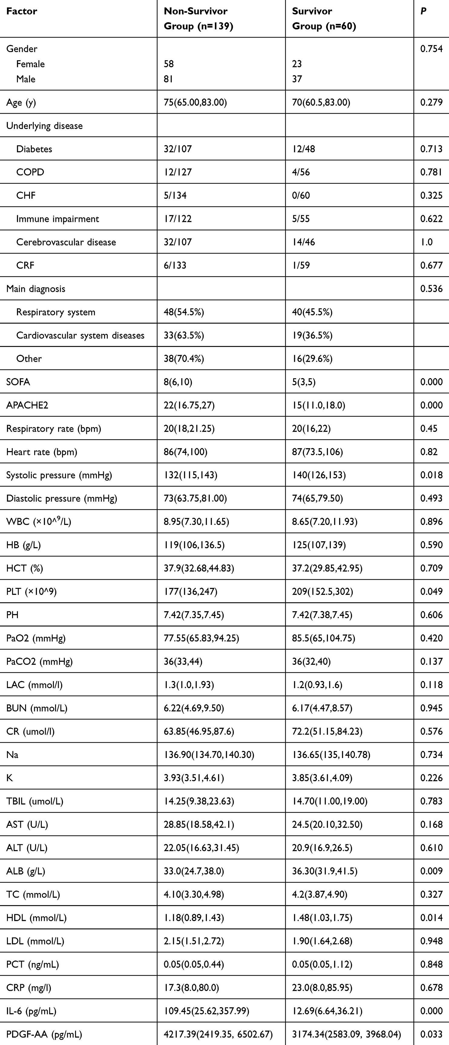 |
Table 1 Baseline Characteristics of the Patients |
Correlation Analysis of IL-6 and Other Indicators
Spearman correlation analysis showed that IL-6 was correlated with PDGF-AA in the non-survival group (p=0.000), but not in the survival group (p=0.853, Table 2). Coefficient of variation (CV) for IL-6 was 60.4%, CV for PDGF-AA was 176.7%. No correlations were found between IL-6 and other factors such as PLT, ALB, HDL, CRP, PCT, SBP, SOFA and APACHE II in both the survival and non-survival groups (p>0.05, Table 2).
 |
Table 2 Spearman Correlation Analysis Between IL-6 and Other Indicators |
Results from Logistic Regression and ROC Curve Analysis
The logistic regression analysis revealed that IL-6 and PDGF-AA were both independent risk factors associated with 28-day mortality (OR=1.003, 95% CI (1.001–1.005), OR=1.002, 95% CI (1.001–1.003)). The 28-day mortality in critically ill patients was also independently associated with factors such as SBP (OR=0.967, 95% CI (0.944–0.990), P=0.005), PLT (OR=0.997(0.994–1.000), P=0.057) and ALB (OR=0.959 (0.926–0.994), P=0.021) (Table 3).
 |
Table 3 Binary Logistic Regression Analysis of the Risk Factors for 28-Day Prognosis in Critically Ill Patients |
The AUROCs for IL-6, PDGF-AA, SOFA and APACHE II scores were 0.736, 0.596, 0.856, and 0.767, respectively. The cut-off values for IL-6 and PDGF-AA were 24.41 and 4182.5 pg/mL. The cut-off values of SOFA and APACHE II were 5.5 and 20.5, respectively (Table 4). The ROC curves of IL-6 and SOFA were significantly different from those of IL-6 (Z =3.668, p=0.0002), SOFA(Z= 2.298, p=0.0216), or APACHE II (Z=2.923, p=0.0035)(Figure 1).The ROC curves for IL-6, PDGF-AA, and SOFA were significantly different from those of IL-6 (Z=4.030, p=0.0001) and SOFA (Z=2.633, p=0.0085). The AUROC calculated by combining IL-6 and SOFA can reach 0.892 (95% CI (0.848,0.936)) with a sensitivity of 91.4% and the specificity of 71.7%, respectively. The combination of IL-6, PDGF-AA and SOFA can make an AUROC of 0.905 (95% CI (0.864,0.946)) and increased the specificity to 91.5%.
 |
Table 4 Predictive Value of IL-6 and PDGF-AA Together with Other Factors for 28-Day Prognosis in Critically Ill Patients |
 |
Figure 1 ROC curve of IL-6 and PDGF-AA together with other factors for 28-day prognosis in critically ill patients. |
Discussion
Critical illness is a common clinical manifestation in emergency patients, which is characterized by unstable vital signs and complex and rapid disease changes, often accompanied by more than two organ dysfunctions, and is life-threatening. Several factors can affect patient prognosis. Simply and quickly identifying the severity of the disease and assessing the risk of death after a patient arrives at the emergency department can help physicians make appropriate clinical interventions.
Activation of the acute phase response causes the liver to secrete a variety of proteins, including CRP, fibrinogen, and haptoglobin. In terms of mechanism, acute phase proteins can be induced mainly by IL-6.15 IL-6 is secreted by the endothelial cells and macrophages in response to inflammatory stimuli. IL-6 is a pivotal cytokine with a diverse repertoire of physiological functions, including regulation of immune cell proliferation and differentiation. IL-6 has been identified as a driving signal in several inflammatory diseases. Clinically, high IL-6 levels are positively associated with disease severity and poor outcomes in sepsis patients.16
Among primary prevention patients, IL-6 levels consistently predict incident metabolic syndrome and type 2 diabetes.17 In cardiovascular disease studies, IL-6, a marker of upstream inflammation, was independently associated with the risk of major adverse cardiovascular events, cardiovascular and all-cause mortality, myocardial infarction, heart failure, and cancer mortality in patients with stable coronary heart disease.18 Fanola et al showed that IL-6 concentration was associated with adverse cardiovascular outcomes in patients with ACS, independent of established risk predictors and biomarkers.19 IL-6 levels are significantly elevated in patients with advanced chronic kidney disease (CKD) and independently predict overall mortality in a cohort of patients with different CKD stages compared to the other three acute-phase proteins (CRP, TNF-α, and albumin).20 Milenkovic et al found that IL-6 ≥ 74.98 pg/mL could effectively predict in-hospital mortality in COVID-19 patients.21 A higher IL-6 (the highest quartile≥ 3.19 pg/mL) level was associated with a twofold greater risk of death compared with the lowest quartile of 1.9 pg/mL; higher circulating levels of IL-6 were associated with mortality in the sample of healthy older persons.22 In a study of patients with sepsis, Yu et al found that IL-6 was an independent risk factor for sepsis diagnosis; however IL-6 could not predict patient mortality.23 In our study, we found that IL-6 levels in the non-survival group were higher than those in the survival group (109.45 (25.62, 357.99) vs 12.69 (6.64, 36.21), p=0.000) and IL-6 was an independent risk factor for 28-day mortality in critically ill patients by logistic analysis (OR 1.004 (1.001–1.006), P= 0.002). The ROC showed that the cut-off value of IL-6 was 24.41, the sensitivity to prognosis was 77%, and the specificity was 66.7%.
We also examined routine indices and found differences in SBP, PLT, albumin and HDL levels between survivors and non-survivors. Our results demonstrated significantly lower SBP, PLT, albumin and HDL levels in critically ill patients in the non-survival group than in those in the survival group.
Logistic analysis showed that SBP, PLT count and albumin level were independent risk factors for 28-day mortality in critically ill patients. Hu et al showed a nonlinear relationship between SBP and outcomes, with increased risk observed at both low and high blood pressure levels. An SBP of 120–140 mm Hg was associated with decreased adverse outcomes.24 Thrombocytopenia is common among critically ill patients.25 A decrease in platelet count may indicate ongoing coagulation activation, which contributes to microvascular failure and organ dysfunction.26 A decrease in platelet count of 30% or more, rather than thrombocytopenia itself, is independently associated with intensive care unit death.25 ALB levels have been proven to be a predictor of poor outcomes in critically ill patients.27 In critically ill patients, low serum ALB levels were associated with poorer clinical outcomes, particularly increased mortality; While supplementing with ALB may positively affect mortality, especially when it is administered to patients with low ALB levels and those with sepsis.28 Our study showed that the HDL levels were lower in the non-survival group than in the survival group of critically ill patients. This funding is consistent with that of a previous report of critically ill patients with sepsis.29–31 HDL appears to be an inflammatory marker because reduced levels reflect the intensity of the underlying inflammatory process.32 Low HDL-C levels are independently associated with higher all-cause mortality in critically ill patients.33
We also found that the PDGF-AA level was an independent risk factor for 28-day mortality in critically ill patients. PDGF-AA is a potent stimulator of smooth muscle cells (SMCs), fibroblast growth, and motility of SMCs and fibroblasts.34 Fibroblasts, endothelial cells and macrophages secrete PDGF-AA to participate in inflammatory response and histiocytic repair.35,36
No correlations were found between levels of IL-6 levels and other factors, such as PLT, ALB, HDL, CRP, PCT, SBP, SOFA and APACHE II, in either the survival or non-survival groups in our study. However, we found that IL-6 levels were correlated with PDGF-AA levels in the non-survivor group. Three isoforms of PDGF (-AA, -AB and -BB) have been shown to induce IL-6 expression and vascular smooth muscle cells (VSMC).10 Thus, there may be a synergistic effect of IL-6 and PDGF in critically ill patients.
The severity of organ dysfunction has been assessed using a variety of scoring systems that quantify abnormalities based on clinical presentation, laboratory data, or therapeutic interventions; The main score currently used is the SOFA.37,38 APACHE II score involves multiple systems that can synthesize a patient’s overall condition and predict prognosis. It has been widely used to assess the condition of critically ill patients39,40 and in our study, SOFA and APACHE scores II also had clinical significance in predicting the 28-day mortality of critically ill patients by logistic analysis. Statistically, there was no difference in the diagnostic value of IL-6 compared with the APACHE II score (Z=0.571, P=0.568). However, IL-6 (77%) was more sensitive than APACHE II score (57.6%).
The AUROCs for IL-6, PDGF-AA, SOFA and APACHE II scores were 0.736, 0.596, 0.856 and 0.767, respectively. The AUROC curves of IL-6 and SOFA were significantly different from those of IL-6, SOFA, and APACHE II scores. The diagnostic value of IL-6 combined SOFA was better than that of SOFA alone, with an AUROC of 0.892, a sensitivity of 91.4%, and a specificity of 71.7%. Further combined use of IL-6, PDGF-AA and SOFA can increase the AUROC to 0.905 (95% CI (0.864,0.946) and specificity to 91.5%. These data indicate that the addition of IL-6 and PDGF-AA to SOFA can effectively improve the sensitivity or specificity for predicting short-term mortality in critically ill patients.
Overall, our data showed that serum IL-6 and PDGF-AA levels are significant for the diagnosis and prognosis assessment of critically ill patients. After admission, the IL-6, PDGF-AA and SOFA scores can be quickly assessed and clinicians can perform early diagnosis and treatment of patients.
This study has some limitations. First, the relatively small number of patients included limited the reliability of the results. Second, we only detected the serum IL-6 and PDGF-AA within an hour of admission and did not dynamically monitor their changes. Third, further studies could improve the role of this marker in evaluating the prognosis of critical diseases with different etiologies.
Conclusions
In conclusion, IL-6 and PDGF-AA levels are independent risk factors associated with the 28-day mortality in critically ill patients. This study highlights the importance of monitoring serum levels of IL-6 and PDGF-AA in critically ill patients. Compared with the marker alone, combinations with other conventional risk factors have better predictive values.
Abbreviations
IL-6, interleukin-6; PDGF-AA, platelet-derived growth factor AA; SOFA, sequential organ failure assessment score; APACHE II, Acute Physiology and Chronic Health Evaluation II; ROC, receiver operating characteristic curve; SAA, serum amyloid A; SBP, systolic blood pressure; AUC, area under curve; LAC, Lactate; PCT, procalcitonin; CRP, C-reactive protein; PLT, platelet; ALB, albumin; HDL, high-density lipoprotein; COPD, chronic obstructive pulmonary disease; ARDS, acute respiratory distress syndrome; COVID-19, corona virus disease 2019; CKD, chronic kidney disease; VSMC, vascular smooth muscle cells.
Acknowledgments
This paper has been uploaded to Research Square as a preprint: https://www.researchsquare.com/article/rs-3477822/v1.
Disclosure
The authors report no conflicts of interest in this work.
References
1. Adhikari NK, Fowler RA, Bhagwanjee S, Rubenfeld GD. Critical care and the global burden of critical illness in adults. Lancet. 2010;376(9749):1339–1346. doi:10.1016/S0140-6736(10)60446-1
2. Gabay C, Kushner I. Acute-phase proteins and other systemic responses to inflammation. N Engl J Med. 1999;340(6):448–454. doi:10.1056/NEJM199902113400607
3. McElvaney OJ, Curley GF, Rose-John S, McElvaney NG. Interleukin-6: obstacles to targeting a complex cytokine in critical illness. Lancet Respir Med. 2021;9(6):643–654. doi:10.1016/S2213-2600(21)00103-X
4. Hirano T, Yasukawa K, Harada H, et al. Complementary DNA for a novel human interleukin (BSF-2) that induces B lymphocytes to produce immunoglobulin. Nature. 1986;324(6092):73–76. doi:10.1038/324073a0
5. Heinrich PC, Castell JV, Andus T. Interleukin-6 and the acute phase response. Biochem J. 1990;265(3):621–636. doi:10.1042/bj2650621
6. Mihara M, Hashizume M, Yoshida H, Suzuki M, Shiina M. IL-6/IL-6 receptor system and its role in physiological and pathological conditions. Clin Sci (Lond) 2012;122(4):143–159. doi:10.1042/CS20110340
7. Calabrese LH, Rose-John S. IL-6 biology: implications for clinical targeting in rheumatic disease. Nat Rev Rheumatol. 2014;10(12):720–727. doi:10.1038/nrrheum.2014.127
8. Kelly JD, Haldeman BA, Grant FJ, et al. Platelet-derived growth factor (PDGF) stimulates PDGF receptor subunit dimerization and intersubunit trans-phosphorylation. J Biol Chem. 1991;266(14):8987–8992. doi:10.1016/S0021-9258(18)31541-2
9. Hamid T, Xu Y, Ismahil MA, et al. Cardiac mesenchymal stem cells promote fibrosis and remodeling in heart failure: role of PDGF signaling. JACC Basic Transl Sci. 2022;7(5):465–483. doi:10.1016/j.jacbts.2022.01.004
10. Roth M, Nauck M, Tamm M, et al. Intracellular interleukin 6 mediates platelet-derived growth factor-induced proliferation of nontransformed cells. Proc Natl Acad. 1995;92(5):1312–1316. doi:10.1073/pnas.92.5.1312
11. Shi DL, Liu XY, Zhou Y. Guidelines on the emergency triage. Chin J Emerg Med. 2018;27:599–604.
12. Evans L, Rhodes A, Alhazzani W, et al. Surviving sepsis campaign: international guidelines for management of sepsis and septic shock 2021. Intensive Care Med. 2021;47(11):1181–1247. doi:10.1007/s00134-021-06506-y
13. Lambden S, Laterre PF, Levy MM, Francois B. The SOFA score-development, utility and challenges of accurate assessment in clinical trials. Crit Care. 2019;23(1):374. doi:10.1186/s13054-019-2663-7
14. Godinjak A, Iglica A, Rama A, et al. Predictive value of SAPS II and APACHE II scoring systems for patient outcome in a medical intensive care unit. Acta Med Acad. 2016;45(2):97–103. doi:10.5644/ama2006-124.165
15. Garbers C, Heink S, Korn T, Rose-John S. Interleukin-6: designing specific therapeutics for a complex cytokine. Nat Rev Drug Discov. 2018;17(6):395–412. doi:10.1038/nrd.2018.45
16. Zhang Y, Li B, Ning B. Evaluating IL-6 and IL-10 as rapid diagnostic tools for Gram-negative bacteria and as disease severity predictors in pediatric sepsis patients in the intensive care unit. Front Immunol. 2022;13:1043968. doi:10.3389/fimmu.2022.1043968
17. Pradhan AD, Manson JE, Rifai N, Buring JE, Ridker PM. C-reactive protein, interleukin 6, and risk of developing type 2 diabetes mellitus. JAMA. 2001;286(3):327–334. doi:10.1001/jama.286.3.327
18. Held C, White HD, Stewart RAH, et al. Inflammatory biomarkers interleukin-6 and C-reactive protein and outcomes in stable coronary heart disease: experiences from the STABILITY (stabilization of atherosclerotic plaque by initiation of darapladib therapy) trial. J Am Heart Assoc. 2017;6(10):e005077. doi:10.1161/JAHA.116.005077
19. Fanola CL, Morrow DA, Cannon CP, et al. Interleukin-6 and the risk of adverse outcomes in patients after an acute coronary syndrome: observations from the SOLID-TIMI 52 (stabilization of plaque using darapladib-thrombolysis in myocardial infarction 52) trial. J Am Heart Assoc. 2017;6(10):e005637. doi:10.1161/JAHA.117.005637
20. Barreto DV, Barreto FC, Liabeuf S, et al. Plasma interleukin-6 is independently associated with mortality in both hemodialysis and pre-dialysis patients with chronic kidney disease. Kidney Int. 2010;77(6):550–556. doi:10.1038/ki.2009.503
21. Milenkovic M, Hadzibegovic A, Kovac M, et al. D-dimer, CRP, PCT, and IL-6 levels at admission to ICU can predict in-hospital mortality in patients with COVID-19 pneumonia. Oxid Med Cell Longev. 2022;2022:8997709. doi:10.1155/2022/8997709
22. Harris TB, Ferrucci L, Tracy RP, et al. Associations of elevated interleukin-6 and C-reactive protein levels with mortality in the elderly. Am J Med. 1999;106(5):506–512. doi:10.1016/s0002-9343(99)00066-2
23. Yu B, Chen M, Zhang Y, et al. Diagnostic and prognostic value of interleukin-6 in emergency department sepsis patients. Infect Drug Resist. 2022;15:5557–5566. doi:10.2147/IDR.S384351
24. Hu AM, Hai C, Wang HB, et al. associations between elevated systolic blood pressure and outcomes in critically ill patients: a retrospective cohort study and propensity analysis. Shock. 2021;56(4):557–563. doi:10.1097/SHK.0000000000001774
25. Strauss R, Wehler M, Mehler K, Kreutzer D, Koebnick C, Hahn EG. Thrombocytopenia in patients in the medical intensive care unit: bleeding prevalence, transfusion requirements, and outcome. Crit Care Med. 2002;30(8):1765–1771. doi:10.1097/00003246-200208000-00015
26. Levi M. Platelets in Critical Illness. Semin Thromb Hemost. 2016;42(3):252–257. doi:10.1055/s-0035-1570080
27. Atrash AK, de Vasconcellos K. Low albumin levels are associated with mortality in the critically ill: a retrospective observational study in a multidisciplinary intensive care unit. South Afr J Crit Care. 2020;36(2):74. doi:10.7196/SAJCC.2020.v36i2.422
28. Rabi R, Alsaid RM, Matar AN, Dawabsheh Y, Abu Gaber D. The role of serum albumin in critical illness, predicting poor outcomes, and exploring the therapeutic potential of albumin supplementation. Sci Prog. 2024;107(3):368504241274023. doi:10.1177/00368504241274023
29. Zhou Z, Liu Q, Zheng M, et al. Comparative study on the predictive value of TG/HDL-C, TyG and TyG-BMI indices for 5-year mortality in critically ill patients with chronic heart failure: a retrospective study. Cardiovasc Diabetol. 2024;23(1):213. doi:10.1186/s12933-024-02308-w
30. Tanaka S, Labreuche J, Drumez E, et al. Low HDL levels in sepsis versus trauma patients in intensive care unit. Ann Intensive Care. 2017;7(1):60. doi:10.1186/s13613-017-0284-3
31. Gordon BR, Parker TS, Levine DM, et al. Relationship of hypolipidemia to cytokine concentrations and outcomes in critically ill surgical patients. Crit Care Med. 2001;29(8):1563–1568. doi:10.1097/00003246-200108000-00011
32. Golucci APBS, Marson FAL, Ribeiro AF, Nogueira RJN. Lipid profile associated with the systemic inflammatory response syndrome and sepsis in critically ill patients. Nutrition. 2018;55-56:7–14. doi:10.1016/j.nut.2018.04.007
33. Li S, Zhang W, Liu H. Association between lipid levels and all-cause and cause-specific mortality in critically ill patients. Sci Rep. 2023;13(1):5109. doi:10.1038/s41598-023-32209-z
34. Chen CP, Hung CF, Lee SC, Lo HM, Wu PH, Wu WB. Lycopene binding compromised PDGF-AA/-AB signaling and migration in smooth muscle cells and fibroblasts: prediction of the possible lycopene binding site within PDGF. Naunyn Schmiedebergs Arch Pharmacol. 2010;381(5):401–414. doi:10.1007/s00210-010-0501-1
35. Nagaoka I, Trapnell BC, Crystal RG. Upregulation of platelet-derived growth factor-A and -B gene expression in alveolar macrophages of individuals with idiopathic pulmonary fibrosis. J Clin Invest. 1990;85(6):2023–2027. doi:10.1172/JCI114669
36. Demaria M, Ohtani N, Youssef SA, et al. An essential role for senescent cells in optimal wound healing through secretion of PDGF-AA. Dev Cell. 2014;31(6):722–733. doi:10.1016/j.devcel.2014.11.012
37. Vincent JL, de Mendonça A, Cantraine F, et al. Use of the SOFA score to assess the incidence of organ dysfunction/failure in intensive care units: results of a multicenter, prospective study. Working group on “sepsis-related problems of the European Society of Intensive Care Medicine”. Crit Care Med. 1998;26(11):1793–1800. doi:10.1097/00003246-199811000-00016
38. Singer M, Deutschman CS, Seymour CW, et al. The third international consensus definitions for sepsis and septic shock (sepsis-3). JAMA. 2016;315(8):801–810. doi:10.1001/jama.2016.0287
39. Wang J, Wang H, Liu W, Zhang D, Guo S. Assessment values of procalcitonin, lactic acid, and disease severity scores in patients with sepsis. Zhonghua Wei Zhong Bing Ji Jiu Yi Xue. 2019;31(8):938–941. doi:10.3760/cma.j.issn.2095-4352.2019.08.005
40. Zhang Y, Wang J, Yu BZ, et al. Predictive value of heart-type fatty acid-binding protein for mortality risk in critically ill patients. Dis Markers. 2022;2022:1720414. doi:10.1155/2022/1720414
 © 2025 The Author(s). This work is published and licensed by Dove Medical Press Limited. The
full terms of this license are available at https://www.dovepress.com/terms
and incorporate the Creative Commons Attribution
- Non Commercial (unported, 4.0) License.
By accessing the work you hereby accept the Terms. Non-commercial uses of the work are permitted
without any further permission from Dove Medical Press Limited, provided the work is properly
attributed. For permission for commercial use of this work, please see paragraphs 4.2 and 5 of our Terms.
© 2025 The Author(s). This work is published and licensed by Dove Medical Press Limited. The
full terms of this license are available at https://www.dovepress.com/terms
and incorporate the Creative Commons Attribution
- Non Commercial (unported, 4.0) License.
By accessing the work you hereby accept the Terms. Non-commercial uses of the work are permitted
without any further permission from Dove Medical Press Limited, provided the work is properly
attributed. For permission for commercial use of this work, please see paragraphs 4.2 and 5 of our Terms.
Recommended articles
Based on the Dual Pathway of Interaction-Mediated NF-κB in Cell Apoptosis and Immune Inflammation to Study the Effect of Danzhi Xiaoyao Powder on the Learning and Cognitive Ability of AD Model Rats
Wang HP, Li MC, Yang J, Zhou J, Meng ZP, Hu YY, Lyu YJ, Chen YQ, Han YM, Pei WL
Degenerative Neurological and Neuromuscular Disease 2025, 15:41-64
Published Date: 14 April 2025