")
Back to Journals » Diabetes, Metabolic Syndrome and Obesity » Volume 15
Potential Molecular Mechanism of Yishen Capsule in the Treatment of Diabetic Nephropathy Based on Network Pharmacology and Molecular Docking
Authors Hu Y, Liu S, Liu W, Zhang Z, Liu Y , Li S, Sun D, Zhang G, Fang J
Received 25 November 2021
Accepted for publication 1 March 2022
Published 29 March 2022 Volume 2022:15 Pages 943—962
DOI https://doi.org/10.2147/DMSO.S350062
Checked for plagiarism Yes
Review by Single anonymous peer review
Peer reviewer comments 5
Editor who approved publication: Professor Ming-Hui Zou
Yaling Hu,1,2 Shuang Liu,3 Wenyuan Liu,2 Ziyuan Zhang,2 Yuxiang Liu,1 Sufen Li,2 Dalin Sun,2 Guang Zhang,2 Jingai Fang2
1Shanxi Medical University, Taiyuan, Shanxi, 030001, People’s Republic of China; 2Department of Nephrology, First Hospital of Shanxi Medical University, Taiyuan, Shanxi, 030001, People’s Republic of China; 3Department of Urology, First Hospital of Shanxi Medical University, Taiyuan, Shanxi, 030001, People’s Republic of China
Correspondence: Jingai Fang, Department of Nephrology, First Hospital of Shanxi Medical University, 85 Jiefangnan Road, Taiyuan, Shanxi, 030001, People’s Republic of China, Email [email protected]
Purpose: Using network pharmacology and molecular docking to explore the mechanism of Yishen Capsule in the treatment of diabetic nephropathy.
Materials and Methods: Active components of Yishen Capsule were obtained using database such as TCMSP and TCMID. UniProt protein database was used to screen and standardize the human-derived targets of the active chemical components. Diabetic nephropathy (DN) targets were obtained from databases such as GeneCards, OMIM, TTD, DisGeNET and DrugBank. A network of “Yishen Capsule Components-diabetic nephropathy Targets-Pathways” was constructed by analyzing data above to screening out core targets for molecular docking verification. DN is induced by streptozocin in rats after left nephrectomy. Renal tubular epithelial cells (RTECs) was isolated and cultured under high glucose conditions. Based on these experimental models, key pathway target genes screened by network pharmacology were verified both in vitro and in vivo.
Results: The main active components of Yishen Capsule in the treatment of DN include quercetin, kaempferol, gallic acid, astragaloside IV, etc. Some key targets (such as AR, AKT1, TP53, ESR1, JUN) and important signal pathways (such as AGE-RAGE, HIF-1 and JAK-STAT signal pathway) were included in the treatment of DN with Yishen Capsule. Molecular docking assay showed that most of the targets have good binding activity with the components of Yishen Capsule. Based on the results of network pharmacology, key target proteins in HIF-1α and JAK2/STAT3 signaling pathways were selected for experimental verification. Results presented that HIF-1α, JAK2, STAT3, TGF-β and MCP-1 were increased under high glucose environment. With the treatment of Yishen Capsule, the expression of HIF-1α further increased, while the expression of JAK2, STAT3, MCP-1 and TGF-β was decreased.
Conclusion: This study revealed the mechanism of Yishen Capsule in the treatment of DN, which possesses the characteristics of multi-component, multi-target, and multi-pathway. Further experiments confirmed that Yishen Capsule interfered with HIF-1α and JAK/STAT signaling pathways to reduce inflammation and fibrosis damage in the kidney tissue of rats with diabetic nephropathy.
Keywords: diabetic nephropathy, network pharmacology, molecular docking, HIF-1α, JAK/STAT
Introduction
Diabetes is a complex chronic metabolic disorder. Complications include cardiovascular and cerebrovascular diseases and microvascular diseases, such as retinopathy, peripheral neuropathy, and diabetic nephropathy.1–3 Diabetic nephropathy (DN) is one of the most common microvascular pathologies of diabetes, and further development of it may progress to the stage of end-stage renal disease, which requires renal replacement therapy and seriously affects the patient’s health and quality of life.4
The pathogenesis of DN is complex. Reported evidences showed it is related to changes in glomerular hemodynamics, excessive activation of Renin-Angiotensin-Aldosterone System (RAAS), luxuriant generation of reactive oxygen species (ROS), inflammation, mitochondrial dysfunction etc. However, its exact mechanism is not yet clear.5
The current clinical treatments for DN mainly include blood glucose control, blood pressure control, correction of metabolic disorders, and improvement of microcirculation, while the clinical treatment has not yet achieved satisfactory results.6 The application of special Chinese medicine treatments for DN possesses the advantages of multi-target, multi-level treatment mechanism, fewer adverse reactions and high safety and the unique advantages for the treatment of DN have been continuously highlighted.
Diabetic nephropathy belongs to kidney deficiency. Diabetes persists for a long time, consumes qi and injures yin, damages organs, and also contains phlegm, heat, depression and blood stasis, and eventually leads to diabetic nephropathy. Diabetic nephropathy, persists for a long time, can cause deficiency of both qi and yin, deficiency of yin of liver and kidney, deficiency of spleen and kidney, internal stop of blood stasis, and dampness stagnation, resulting in a syndrome of mixed deficiency and excess. Therefore, in order to treat the symptoms of proteinuria associated with diabetic nephropathy, according to the principle of TCM syndrome differentiation and treatment, clinical treatment methods such as promoting blood circulation and removing blood stasis, clearing heat and removing dampness, strengthening spleen and nourishing qi, nourishing yin and nourishing kidney are often used.7
The Chinese medicine preparation Yishen Capsule is composed of Astragalus, Angelica, Gordon Euryale, Alisma, Schisandra, Rhodiola and other Chinese medicines. Yishen Capsule is a hospital preparation, the preparation number is “97 Bing Wei Preparation with Zi f01-2005”. The formula has the functions of invigorating kidney qi, diuretic and swelling, regulating and enhancing the immunity of the body. Astragalus in the prescription is the monarch drug, which has the functions of diuresis and swelling, antiperspirant, invigorating qi and nourishing yin. Alisma is a minister drug, which can eliminate dampness and diuresis, namely purging the water of the kidney meridian, and purging the dampness of the bladder meridian. Angelica is also a minister drug, which benefit blood and camp. Gorgon is an assistant drug, which has the functions of invigorating the spleen and stopping diarrhea, invigorating the kidney and strengthening the essence, removing dampness and stopping the leucorrhea.Schisandra chinensis is used as a guide medicine, which nourishing the kidney, solidifying essence, stopping diarrhea, promoting saliva and abstaining sweat. Rhodiola clears heat, detoxifies, nourishes the lungs, and regulates immunity. The combination of the whole agents has the functions of nourishing kidney qi, promoting diuresis and relieving stranguria, promoting blood circulation and removing blood stasis, regulating and enhancing the immunity of the body. It has been confirmed by acute and chronic toxicity experiments that this preparation has no toxicity and side effects.8 Previous studies have indicated that Yishen Capsule can improve kidney function through multiple targets and mechanisms, which effectively delays the progression of DN.9–15
Network pharmacology is based on bioinformatics and computer technology, integrating a large amount of biological information and data. It systematically and comprehensively reflects the intervention mechanism of drugs on the disease network, which is similar to the mechanism that the complex components of traditional Chinese medicines interfere with diseases through multiple channels and multiple targets.16 In recent years, network pharmacology approaches have provided new insights into the mechanisms by which TCM treats diseases. The network pharmacology method can provide technical support for the research on the mechanism of action of traditional Chinese medicine compounds in the diagnosis and treatment of diseases. It is helpful to screen the active ingredients of drugs, predict the action targets in the ingredients, and study the mechanism of action of drugs. Thereby revealing the scientific connotation of traditional Chinese medicine compounds and guiding the research and development of new traditional Chinese medicines.17,18 This study intends to explore the targets of Yishen Capsule in the treatment of DN and to elucidate the therapeutic mechanism of Yishen Capsule on DN from many aspects. Further, some target proteins of key signaling pathway were verified through experiment both in vitro and in vivo, which may provide evidence for the treatment of DN using Yishen Capsule.
Materials and Methods
Network Pharmacology Analysis
Screening of Active Chemical Components and Acquisition of Targets
Using the TCMSP database (http://tcmspw.com/) to retrieve all the chemical components of the 6 traditional Chinese medicines (Astragalus, Angelica, Gordon Euryale, Alisma, Schisandra and Rhodiola) in Yishen Capsule. According to the two ADME attribute values of oral bioavailability (≥ 30%) and drug-likeness (≥ 0.18) of these ingredients, preliminary screening was performed to obtain the active compounds, as well as their protein targets. According to the TCMID database (http://www.megabionet.org/tcmid/) and published literature reports,19 some unpredicted active compounds and reported targets have been supplemented. In the UniProt protein database (https://www.uniprot.org), human-derived targets were screened and standardized.
Target Screening of Diabetic Nephropathy
Searching the GeneCards database (https://www.genecards.org), OMIM database (http://www.omim.org), TTD database (http://bidd.nus.edu.sg/group/cjttd) and DisGeNET database (https://www.disgenet.org) for potential targets of diabetic nephropathy, by using “Diabetic nephropathy” and “Diabetic Kidney Disease” as keywords. And some target sites for disease were also supplemented through searching the DrugBank database (https://www.drugbank.ca). According to experience, the target points with Score ≥30 in the GeneCards database and Score ≥0.3 in the DisGeNET database were selected as potential targets for diabetic nephropathy.
Intersection of Chinese Medicine and Disease Target Genes and Construction of PPI Network
The intersection of the drug target of Yishen Capsule and the target of diabetic nephropathy were selected to draw the Venn diagram, using Venny 2.1.0 (https://bioinfogp.cnb.csic.es/tools/venny/index.html). Submitting the intersection target to the STRING11.0 database (https://string-db.org) to construct a protein interaction (PPI) network.20 The PPI network was visually analyzed by Cytoscape 3.7.2 software, and PPI core network proteins were screened out according to the degree value≥ 2 times the median.
GO and KEGG Pathway Enrichment Analysis
Using Metascape platform (http://metascape.org/gp/index.html) for GO and KEGG pathway enrichment analysis. Setting P<0.01 is statistically significant, and the analysis results were visualized.
Construction of Components of Yishen Capsule-Targets of Diabetic Nephropathy Disease- Pathway Network Diagram
Using CytoScape 3.7.2 to construct a network diagram of Yishen Capsule components-diabetic nephropathy disease targets-pathway network. Then, using CytoScape 3.7.2 built-in tools to analyze the network topology parameters of active ingredients and targets, which helps to determine core targets and main active ingredients that exert efficacy. The core targets of Cytoscape network analysis and the core target proteins of PPI network were intersected to obtain the key targets of Yishen Capsule in the treatment of diabetic nephropathy, according to which the Venn diagram was drawn.
Verification by Molecular Docking
Downloading the crystal structure of the target protein from the PDB database (https://www.rcsb.org/), and using PyMOL and AutoDock software to remove water, remove the original ligand of the active center, and to hydrogenate the target protein. Downloading the 3D structure file of the core components of traditional Chinese medicine from the PubChem database (https://www.ncbi.nlm.nih.gov/pccompound/). And then loading it to the AutoDock Tools 1.5.6 software to add atomic charges, assign atomic types. All flexible bonds were set as rotatable Running AutoDock Vina for molecular docking. The conformation with the highest score is selected as the docking conformation, and the PyMOL program was used to visualize the results.
Experimental Verification
Reagents
Yishen Capsule was prepared at Department of Pharmacy of the First Hospital of Shanxi Medical University. Boiling the herbs in water thrice for 1 h. The decoctions were combined and filtered through a membrane, then concentrated through vacuum evaporation to a density of 1.20–1.24 at 70 °C. The concentrated decoction was further spray-dried into particles that were subsequently filled into capsule.
Streptozotocin (STZ, S8050), Penicillin,-streptomycin antibiotic (P1400), D-glucosum anhydricum (G8150) and Horseradish peroxidase-conjugated goat anti-rabbit IgG (SE134) were bought from Solarbio Company (China). PAS staining kit (BA-4114) and MASSON staining kit (BA-4079) were purchased from Baso Biotechnology (China). Fetal bovine serum (FBS, 10100147), DMEM low glucose medium (1185092) and DMEM high glucose medium (1196007) were purchased from Thermo Fisher Scientific (USA). Tris-glycine-SDS running buffer (AR1139-10) and transfer buffer (AR1149-10), SDS-PAGE protein loading buffer (AR1112) were purchased from Boster Biological Technology (China). PVDF membrane (AR0136-02), WB transfer filter paper (AR0172), hypersensitive ECL chemiluminescence solution (AR1191), and 4% paraformaldehyde fixative solution (AR1068) were purchased from Boster Biological Technology (China). Rabbit antibody of HIF-1α (ab51608), JAK2 (ab32101), STAT3 (ab68153), TGF-β (ab215715) and MCP-1 (ab7202) were purchased from Abcam (England). Both DAB chromogenic reagent (AR1022) and hematoxylin-eosin staining solution (BA-4025) were purchased from Boster Biological Technology (China).
Animal Experiment and Preparation of Plasma Containing Yishen Components
Forty SPF-grade healthy male SD rats (185–215g, 40) were purchased from the Animal Experimental Center of Shanxi Medical University, with the animal certificate number of SCXK (Jin) 2015-0001. Ten rats were randomly selected to give Yishen Capsule by gavage, with the frequency of once a day for 7 days. Two hours after the last gavage, the rats were anesthetized by intraperitoneal injection of 3% pentobarbital sodium. Blood was collected from the abdominal aorta. Blood was centrifugation and the upper serum was collected and stored after inactivation and sterilization.
The remaining 30 rats were randomly divided into the control group (N group, 10 rats) and the model group (20 rats). After one week of adaptive feeding, the model group was injected intraperitoneally with streptozotocin (50 mg/kg) after left nephrectomy. After 72 hours of injection, the rats with random blood glucose> 16.7 mmol/L and 24-hour urine protein quantification> 30 mg are the successful model rats. The successfully modeled DN rats were randomly divided into diabetic nephropathy group (DN) and Yishen capsule group (YS). The YS group was given Yishen capsule gavage solution at 1.25 g/kg/d by gavage (The previous study of the research has carried out different doses of intervention treatment, and it is confirmed that 1.25 g/kg/d is the effective dose of gavage treatment),9 and the N group and DN group were given the same amount of normal saline gavage for 8 consecutive weeks. After 8 weeks of treatment, the right kidney specimens of each group of rats were collected for subsequent immunohistochemistry and Western Blotting detection. All animal experiment protocols were reviewed and approved by the Animal Ethics Committee of Shanxi Medical University (batch number: 2019LL242), and the study was conducted in accordance with the guidelines for the care and use of laboratory animals.
Cell Culture and Treatment
Rat renal tubular epithelial cells (NRK-52E) were got from National Model and Characteristic Experimental Cell Resource Bank/Chinese Academy of Sciences Cell Bank (China) and the CSTR number is 19375.09.3101RATGNR8. And it was cultured in a standard medium (DMEM high glucose medium, containing fetal bovine serum and penicillin- streptomycin antibiotic), and in a 5% CO2 atmosphere at 37 °C. The experimental groups were as follows: normal group (5.5mmol/L glucose), high glucose group (30mmol/L glucose) and Yishen containing-serum treatment group (30 mmol/L glucose + 10% Yishen containing-serum). After each group was cultured for 24 hours, cell proteins were collected for Western Blotting detection.
Pathology Examination of Kidney
Specimens of right kidney were collected, fixed in 4% paraformaldehyde solution, embedded in paraffin, and sectioned routinely. After section, slides were stained with HE, PAS, and Masson. The pathological changes of the renal tissue were observed under light microscope.
Immunohistochemistry
Kidney tissues were embedded in paraffin, conventionally sectioned, deparaffinized, sealed, and incubated at room temperature. Slides were incubated with rabbit anti-HIF-1α, rabbit anti-JAK2, and rabbit anti-STAT3 antibody, respectively overnight at 4°C. Then Slides were incubated with biotin Labeled with goat anti-rabbit IgG (secondary antibody) at 37°C for 15 min. After that, Slides were orderly incubated with DAB reagent and hematoxylin (for 2 min). Then, slides were dehydrated and transparent, and mount with neutral gum. The tissue sections were examined under an optical microscope and quantitative analysis was performed using Image-Pro Plus 6.0 medical pathology graphic color analysis software.
Western Blotting
Extracting and collecting total protein from cell and kidney tissue and determining concentration of the total protein. Denatured protein samples (20 μL) were loaded into the wells, and the proteins were separated by electrophoresis with 8% SDS-PAGE.21–23 Then, the protein in the gel was transferred to the PVDF membrane using a Bio-Rad electrotransmitter. Subsequently, 5% skimmed milk powder was used to seal the PVDF membrane at room temperature. After blocking, adding the primary antibody and incubating overnight at 4°C. The next day, the washed PVDF membrane was incubated with horseradish peroxidase-labeled secondary antibody at room temperature. After incubating for 1 hour, the strips on the PVDF membrane were developed with ECL chemiluminescence reagent in a dark room, and the strips were scanned and analyzed by Alpha-view software.
Data Analysis and Statistics
SPSS 22.0 statistical software was used for data analysis. If the measurement data obey a normal distribution, it is expressed as the mean ± SD. One-way analysis of variance was used for comparison between groups. LDS-t-test was used for multiple comparisons. For non-normal distribution or uneven variance, a non-parametric test of multiple independent samples is used. Extended t-test was used for multiple comparisons. P<0.05 indicates that the difference is statistically significant.
Results
Network Pharmacology Analysis
Screening of Active Chemical Components and Acquisition of Targets
Through searching the TCMSP and TCMID database and published literature, a total of 53 active ingredients of traditional Chinese medicine were obtained. Among them, there were 20, 7, 1, 7, 8 and 10 ingredients belonging to Astragalus, Angelica, Gordon Euryale, Alisma, Schisandra, and Rhodiola respectively (see Table 1). There are 3 repeated active ingredients in each traditional Chinese medicine. 571, 106, 21, 9, 27, and 562 targets of effective active compounds of Astragalus, Angelica, Gordon Euryale, Alisma, Schisandra, and Rhodiola were obtained respectively. After merging and deleting duplicate values, a total of 516 targets were obtained.
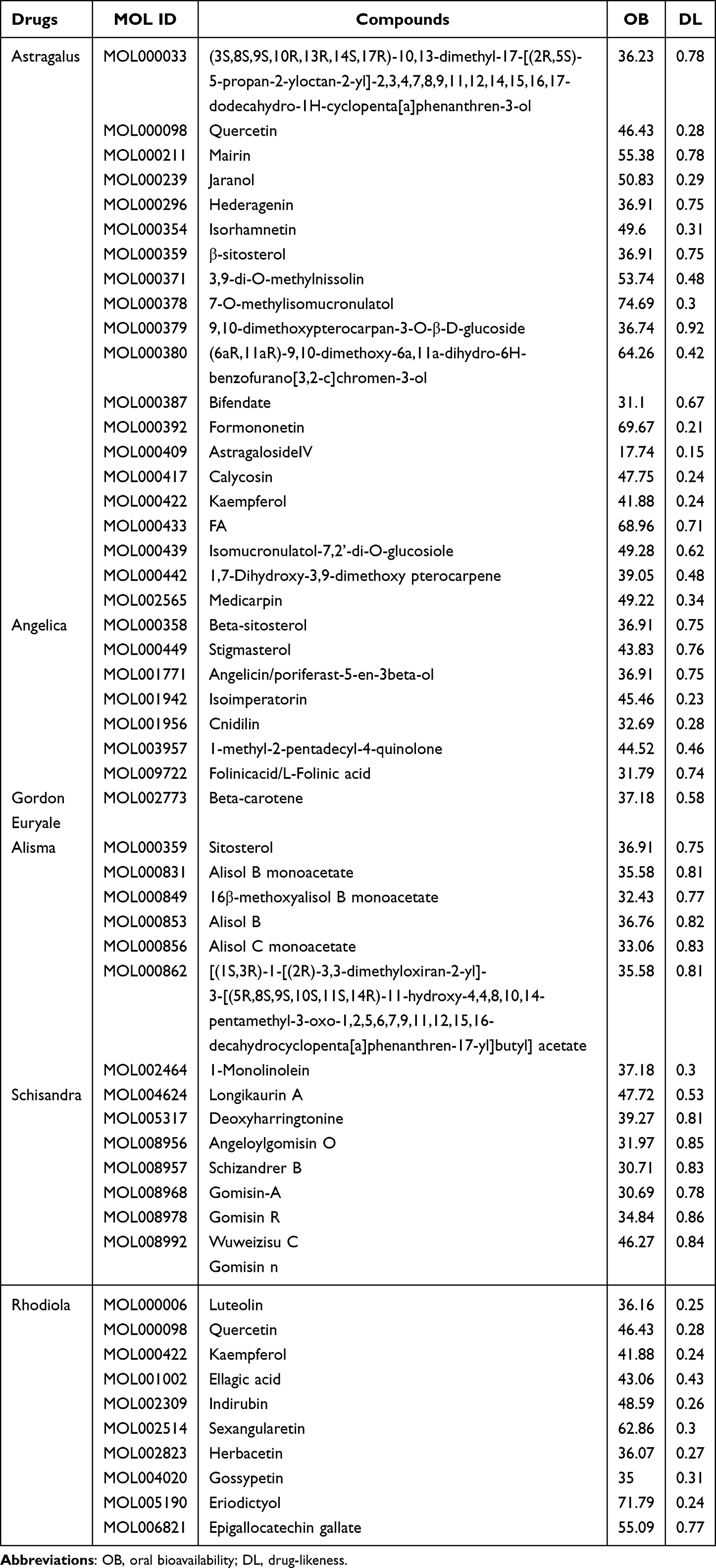 |
Table 1 Main Ingredients of Yishen Capsule |
Acquisition of Target Genes in Diabetic Nephropathy
Diabetic nephropathy disease targets were collected through GeneCards, OMIM, TTD, DisGeNET and DrugBank databases. After merging and deleting duplicate values, a total of 1324 disease targets were obtained.
PPI Network
Taking the intersection of the diabetic nephropathy targets and the drug action targets of the Yishen capsule, and obtaining a total of 150 intersection targets. And a Venn diagram was drawn based on the above data (see Figure 1A). The intersection targets were submitted to the STRING11.0 platform to obtain the PPI network of the components of the Yishen capsule-diabetic nephropathy disease targets (see Figure 1B). The PPI network was visually analyzed by Cytoscape software (see Figure 1C). Setting the degree value ≥ 2 times the median, and 26 core network proteins were obtained using this as a threshold value to screen out. The core network proteins screened out include STAT3, AGT, TP53, SRC, CAV1, EGFR, CTNNB1, ESR1, STAT1, PTEN, JAK2, AKT1, MAPK1, JUN, TNF, AR, CCND1, FGF2, TLR4, CDH1, MYC, VEGFA, MAPK8, EGF, FOS, HSPB1, and the above target proteins are the core nodes in the PPI network.
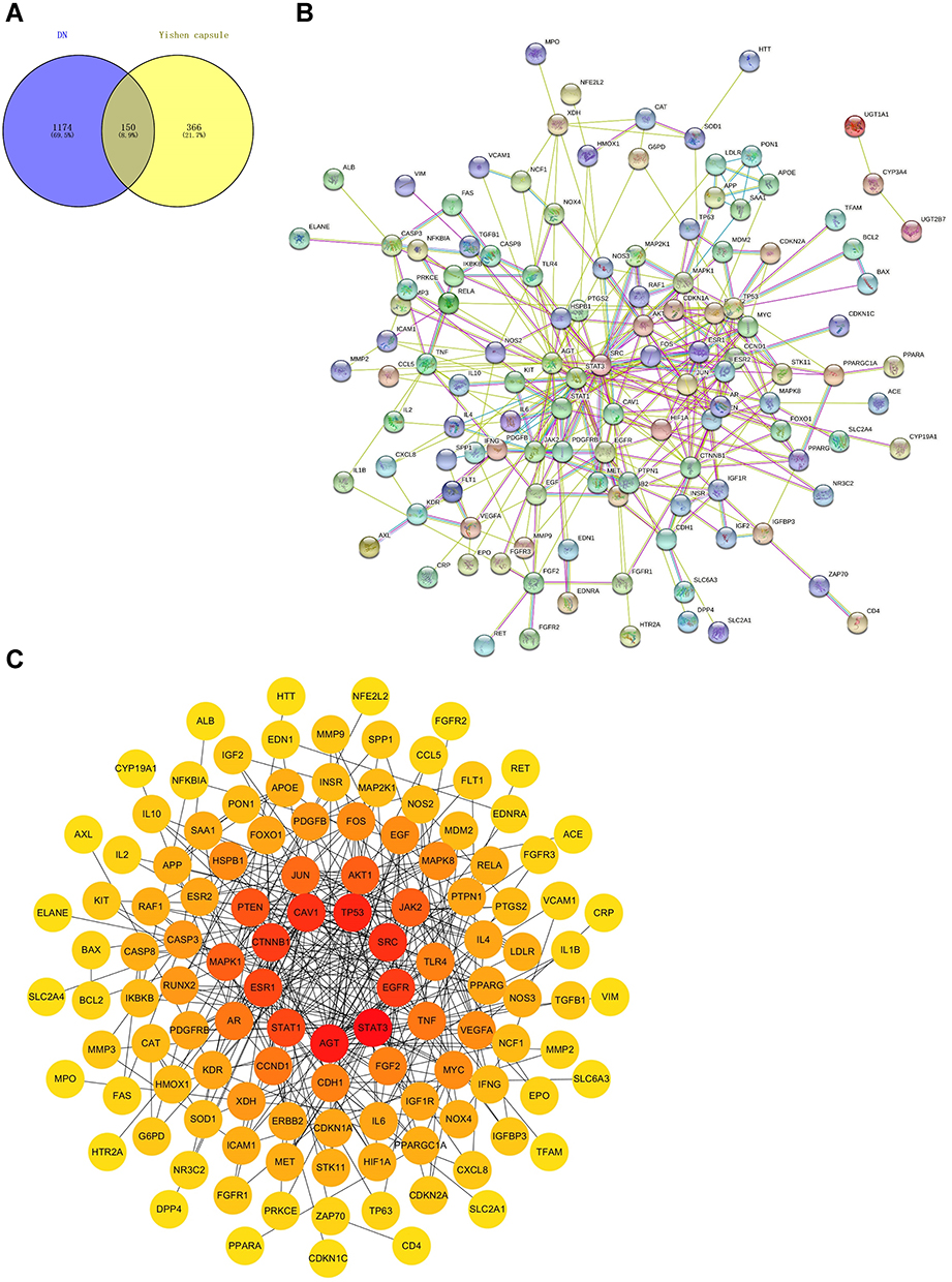 |
Figure 1 Common targets of Yishen capsule ingredients and diabetic nephropathy. (A) Venn diagram. Venn diagram shows the intersection of Yishen Capsule target genes and DN disease target genes, and the overlapping part is the number of intersection target genes. (B) PPI network of the common targets of Yishen Capsule and diabetic nephropathy. (C) PPI protein interaction network diagram. The nodes in the picture represent the common target proteins, and the color depth is positively correlated with the degree value of the node. |
GO and KEGG Pathway Enrichment Analysis
GO and KEGG enrichment analysis were performed on the targets of Yishen Capsule in the treatment of diabetic nephropathy. A total of 255 significantly related biological processes were screened, including MAPK cascade regulation, DNA binding transcription factor activity regulation, lipopolysaccharide response, reactive oxygen metabolism and apoptosis signaling pathway (see Figure 2A). A total of 74 significant related cell components were screened, mainly including various cell structures such as membrane rafts, nucleoli, myelin sheaths, endoplasmic reticulum lumen, and extracellular matrix (see Figure 2B). 96 significant related molecular functions were obtained, mainly related to protein tyrosine kinase activity, receptor regulatory activity, kinase binding, protein homodimerization activity, transcription factor binding and integrin binding (see Figure 2C). KEGG enrichment screening resulted in 159 main signal pathways, mainly including AGE-RAGE signal pathway, PI3K-Akt signal pathway, HIF-1 signal pathway, JAK-STAT signal pathway, insulin signal pathway and other signal pathways. Pathways ranking the top 20 was visualized (see Figure 2D).
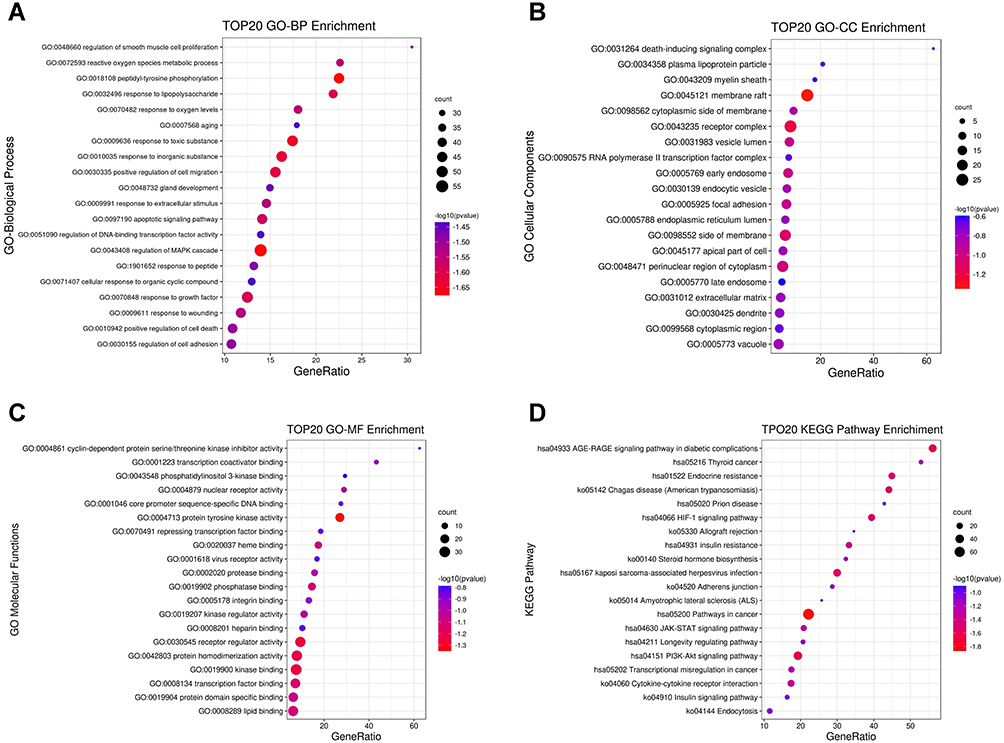 |
Figure 2 GO and KEGG pathway enrichment analysis. (A–C) The BP, CC and MF of GO enrichment analysis, respectively. (D) The KEGG signal pathway analysis. The size of the dot indicates the number of enriched genes. The color from blue to red indicates significance. The smaller the p.value, the more significant the difference. |
Construction of Components of Yishen Capsule-Targets of Diabetic Nephropathy Disease- Pathway Network Diagram
CytoScape 3.7.2 was used to construct a network diagram of Yishen Capsule components-diabetic nephropathy disease target-pathway network. The network has a total of 586 nodes and 1748 component-target protein-pathway relationships (see Figure 3A). Analyzing network topology parameters through CytoScape’s built-in Network Analyzer to obtain core components and core targets. According to analysis and prediction, Quercetin was the main component of Yishen Capsule in the treatment of diabetic nephropathy. Kaempferol (Kaempferol), gallic acid (Epigallocatechin gallate), and astragaloside IV (Astragaloside IV) were minor components (see Table 2). It was predicted that PTGS2 was the main target of Yishen Capsule in the treatment of diabetic nephropathy, and PTGS1, AR, AKT1, NOS2, and PPARG were the secondary targets (see Table 3). Taking the intersection of the above targets and the core targets of the PPI network and drawing the Venn diagram (see Figure 3B). Ten intersection targets were obtained, namely AR, AKT1, TP53, ESR1, JUN, TNF, MAPK1, EGFR, CCND1, VEGFA.
 |
Table 2 Network Node Characteristic Parameters of the Main Active Ingredients of Yishen Capsule |
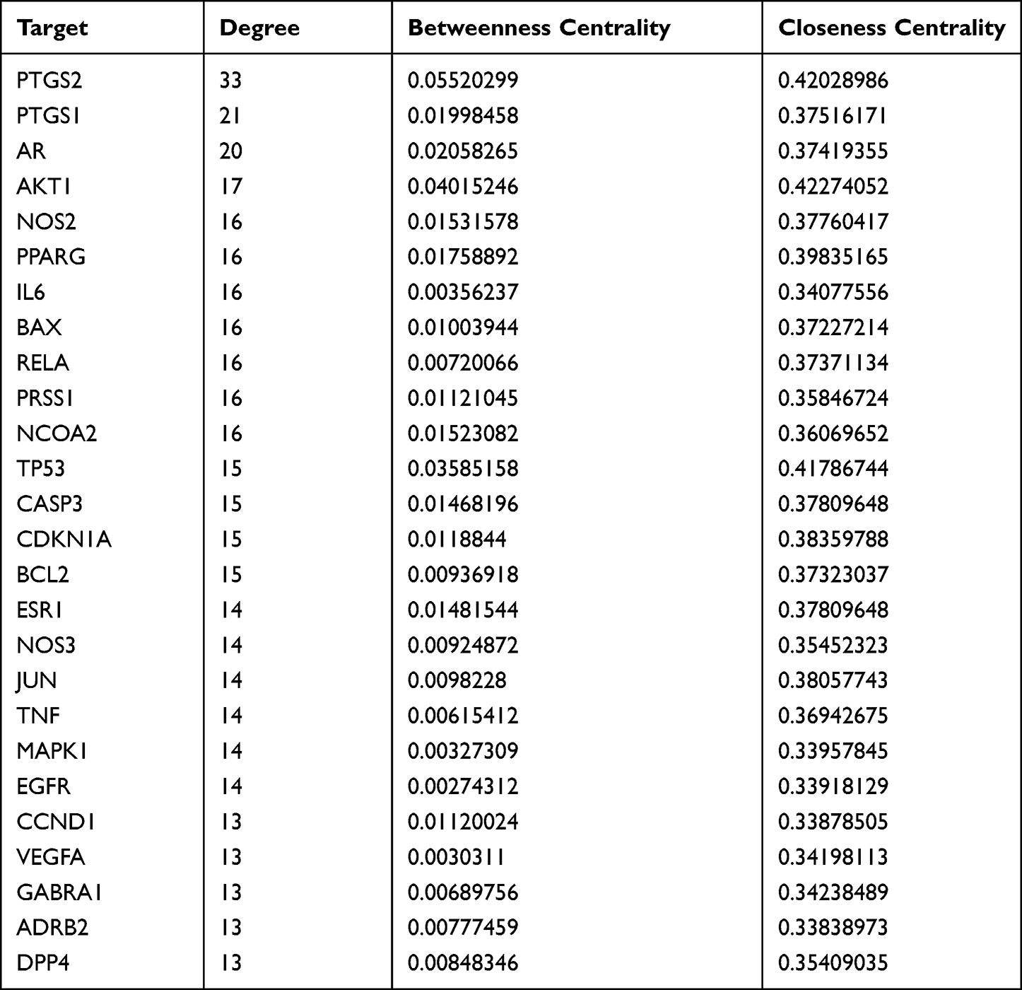 |
Table 3 The Characteristic Parameters of the Target Network Node of the Main Active Ingredients of Yishen Capsule |
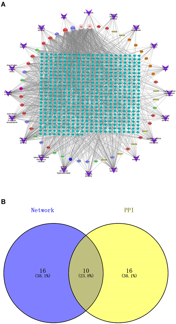 |
Figure 3 Network diagram and Venn diagram. (A) The inverted triangle node represents the name of the pathway, the octagonal node represents the active ingredient of traditional Chinese medicine, and the diamond node represents the gene. The larger the node area of the traditional Chinese medicine component, the more the number of genes enriched by this component, or the more this component is enriched in the pathway. (B) Venn diagram shows the intersection of the key target genes screened by the drug-disease-pathway network diagram and the key target genes screened by the PPI network. |
Verification with Molecular Docking
Selecting the key genes AR, AKT1, TP53 obtained in the previous steps, and the core targets HIF-1α, JAK2, and STAT3 in the key signal pathway, and carrying out molecular docking with the main active ingredients of the Yishen capsule. The docking results were shown in Figure 4A. Among the 60 docking results, 57 suggested a better docking activity (binding energy less than −1.2kcal/mol). Among them, β-sitosterol binded best to the core target protein, and TP53 was the target protein with the best binding activity to the key chemical components in the Yishen capsule.
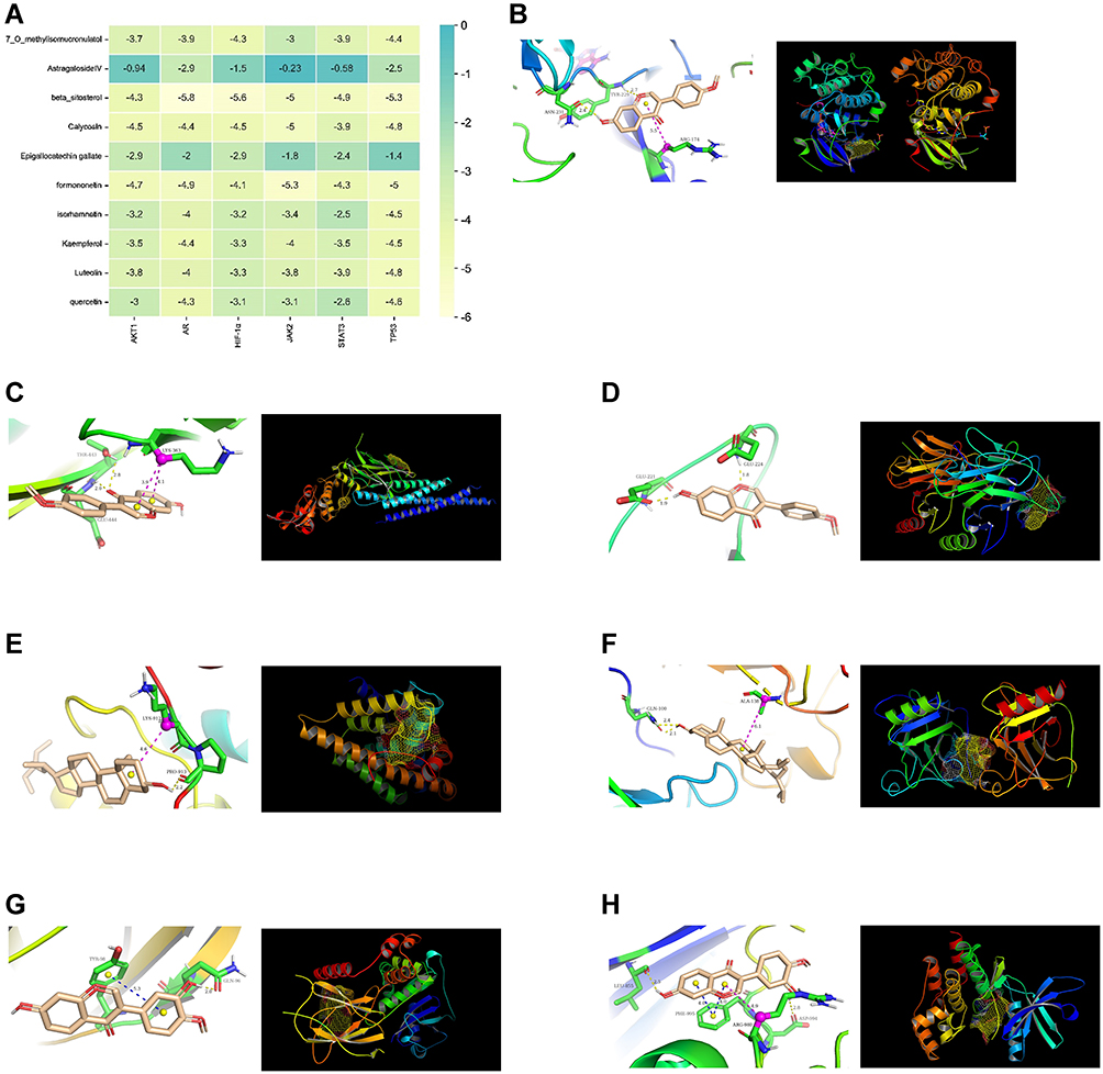 |
Figure 4 The molecule docking heat map of the key active compounds in the Yishen capsule and the core target proteins. (A) The vertical axis represents the effective active ingredient of Yishen capsule and the horizontal axis represents the core target protein receptor. Each value represents the total score of molecular docking, and the darker the color, the higher the score. (B) Docking analysis of formononetin with AKT1 receptor. (C) Docking analysis of formononetin with STAT3 receptor. (D) Docking analysis of formononetin with TP53 receptor. (E) Docking analysis of β-sitosterol with AR receptor. (F) Docking analysis of β-sitosterol with TP53 receptor. (G) Docking analysis of Calycosin with HIF-1α receptor. (H) Docking analysis of Calycosin with JAK2 receptor. For all the docking analysis results, the right panel shows the global diagram of the simulated docking of each ligand molecule and the receptor protein. The colored ribbon model, the protein structure. The yellow grid structure, the ligand molecule. The left panel is a partial three-dimensional image of simulated docking. Wheat, small ligand molecules. Green, amino acid residues that have interaction with active molecules. The yellow dashed line, interacting hydrogen bonds. The blue dashed line, Pi-Pi Stacked. The magentas dashed line, Pi-Alkyl interaction. |
The docking results with better binding energy was shown in Figure 4. Formononetin formed a hydrogen bond with the AKT1 receptor target protein through the amino acid residue ASN-231 and TYR-229 and formed Pi-Alkyl interaction through the amino acid residue ARG-174 (see Figure 4B). Formononetin formed two hydrogen bonds with the STAT3 receptor target through the amino acid residues GLU-444 and THR-443, formed Pi-Alkyl interaction through the amino acid residue LYS-363 (see Figure 4C). Formononetin formed two hydrogen bonds with the TP53 receptor target through the amino acid residues GLU-221 and THR-224 (see Figure 4D). β-sitosterol formed a hydrogen bond with the AR receptor target protein through the amino acid residue PRO-913, formed Pi-Alkyl interaction through the amino acid residue LYS-912 (see Figure 4E). β-sitosterol binded to the TP53 receptor target protein formed hydrogen bonds through the amino acid residue GLN-100, formed Pi-Alkyl interaction through the amino acid ALA-138 (see Figure 4F). Calycosin binded to the HIF-1α receptor target protein through amino acid residue GLN-96 to form hydrogen bond and through amino acid residue TYR-98 to form Pi-Pi Stacked (see Figure 4G). Calycosin formed two hydrogen bonds through amino acid residues ASP-994 and LEU-855 with the JAK2 receptor target protein, formed Pi-Pi Stacked through amino acid residue PHE-995, and formed Pi-Alkyl interaction through amino acid residue ARG-980 (see Figure 4H). The above-mentioned ligand compounds could be well embedded in the active pocket of the receptor target protein.
In vitro and in vivo Experimental Verification
Pathological Changes of Rat Kidney Tissues
The results of HE staining of kidney tissue specimens (see Figure 5A) showed that the glomerulus in group N was normal, and the structure of mesangium and basement membrane was normal. In the DN group, the glomerular capillary loops were hypertrophy, the basement membrane was thickened, the mesangial cells and mesangial matrix were proliferated, and the renal tubular epithelial cells were hypertrophy. However, there was no glomerular sclerosis or interstitial fibrosis. Compared with the DN group, the YS group had increased glomerular volume, mesangial hyperplasia, basement membrane thickening, and renal tubular epithelial cell hypertrophy to varying degrees. The results of PAS staining (see Figure 5B) showed that the glomeruli and tubules of rats in group N were normal in structure and morphology. PAS-positive protein deposits were seen in the glomerular mesangial area and renal tubular epithelial cells of rats in the DN group. Compared with the DN group, the YS group had less PAS-positive protein deposition. The results of Masson staining (see Figure 5C) showed no abnormal changes in the morphology and structure of the kidney tissue in the normal group, and blue staining of collagen tissue was rarely seen in Masson staining. The glomerulus volume of rats in the DN group increased, the renal tubular epithelial cells were obviously edema, and the lumen became narrow. Masson staining showed blue-stained collagen tissue. Compared with the DN group, the YS group had increased glomerular volume, reduced renal tubular epithelial cell edema, and decreased blue-stained collagen tissue.
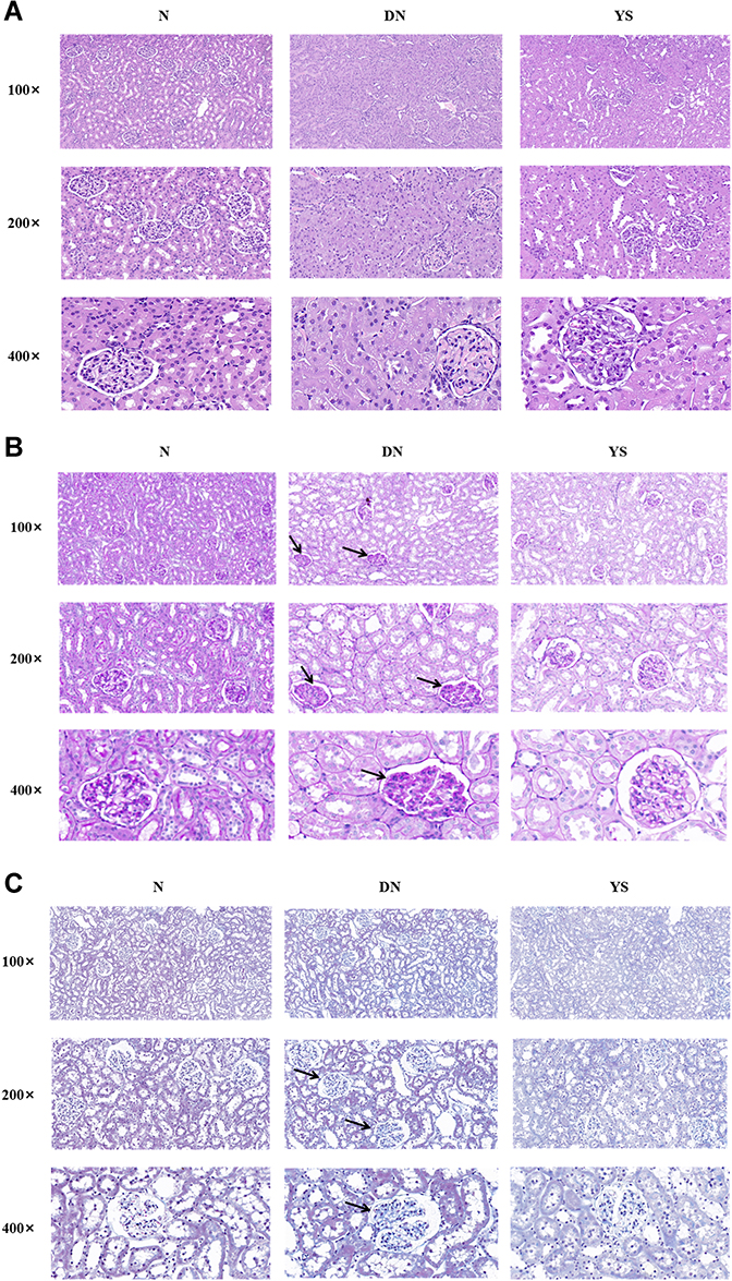 |
Figure 5 Pathological analysis of rat kidney tissues in each group (×100,×200,×400). (A) HE staining of rat kidney tissue. In the DN group, the glomerular capillary loops were hypertrophy, the basement membrane was thickened, the mesangial cells and mesangial matrix were proliferated, and the renal tubular epithelial cells were hypertrophy, while there was no glomerular sclerosis or interstitial fibrosis. Compared with the DN group, the above-mentioned lesions in the YS group were reduced to varying degrees. (B) PAS staining. PAS-positive protein deposits can be seen in the glomerular mesangial area and renal tubular epithelial cells of rats in the DN group (Arrows in the figure). Compared with the DN group, the YS group had less PAS-positive protein deposition. (C) Masson staining. There were no abnormal changes in the morphology and structure of the kidney tissue in the normal group. Masson staining rarely showed blue staining of collagen tissue. The glomerulus volume of rats in the DN group increased, the renal tubular epithelial cells were obviously edema, and the lumen became narrow. Masson staining showed blue-stained collagen tissue (Arrow in the figure). Compared with the DN group, the YS group had increased glomerular volume, reduced renal tubular epithelial cell edema, and decreased blue-stained collagen tissue. |
Immunohistochemical Detection of Key Proteins in the Signal Pathway in Rat Kidney Tissue
After 8 weeks of rearing, the kidney tissues of rats in each group were collected for immunohistochemical examination. The results showed that HIF-1α was expressed in the nucleus and cytoplasm of renal tubular cells. There was a certain amount of HIF-1α expression in the kidney tissue of rats in the N group. The expression of HIF-1α in the DN group was higher than that in the N group. Compared with the DN group, the expression of HIF-1α in the YS group was further increased (P<0.05, see Figure 6A). JAK-2 and STAT3 were expressed in the nucleus and cytoplasm of renal tubules. There was a small amount of JAK-2 and STAT3 expression in the kidney tissue of rats in the N group. The expression of JAK-2 and STAT3 in the DN group was higher than that in the N group, while the expression in the YS group was lower than that in the DN group (P<0.05, see Figure 6B and C). The above results indicated that the expressions of HIF-1α, JAK-2 and STAT3 in diabetic nephropathy were elevated. Yishen Capsule could interfere with HIF-1α and JAK/STAT signaling pathways and improved the glomerulus and renal tubular interstitial damage in rats with diabetic nephropathy.
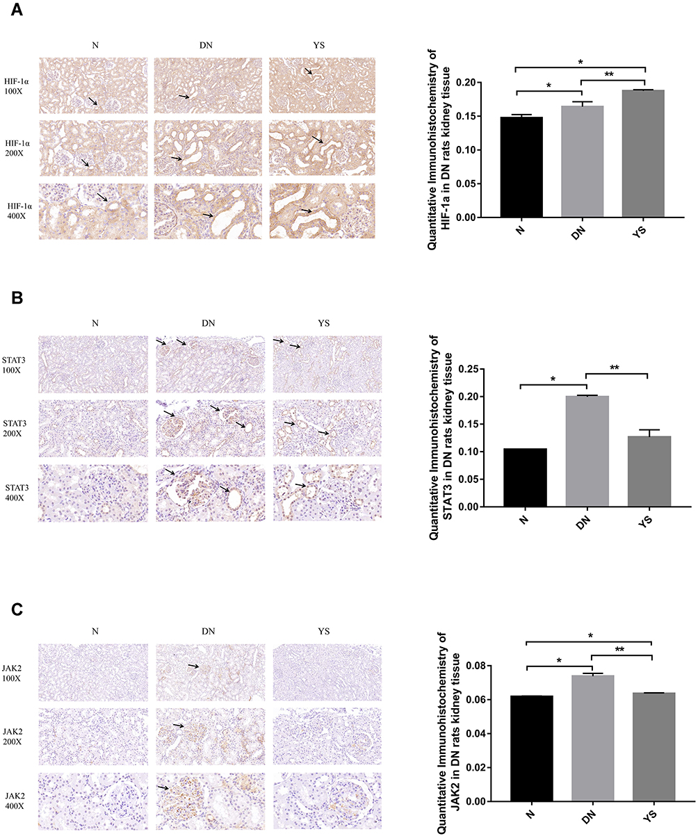 |
Figure 6 Immunohistochemical detection and quantitative analysis of HIF-1α (A), STAT3 (B) and JAK2 (C) in kidney tissues of rats in each group. The expression of HIF-1α, STAT3 and JAK-2 in the DN group was higher than that in the N group. The expression of HIF-1α was further up-regulated in the YS group (P<0.05). The expression of STAT3 and JAK-2 in the YS group were decreased compared with the DN group (P<0.05). *Compared with the N group P<0.05, **Compared with the DN group P<0.05. |
Effects of High Glucose and Yishen Capsule Intervention on Renal Signaling Pathway, Inflammation and Fibrosis Index Expression
Western Blotting indicates that, there was a trace amount of HIF-1α expression in kidney tissue and NRK-52E cells in the normal group. After 24 hours of high glucose stimulation, results showed that the expression of HIF-1α, JAK-2, STAT3, TGF-β and MCP-1 protein in NRK-52E cells and renal tissues all increased compared with the N group (P<0.05). Compared with the DN group, the expression of HIF-1α in kidney tissue and NRK-52E cells increased after the treatment of Yishen capsule (P<0.05), while the expression of JAK2, STAT3, MCP-1 and TGF-β were all decreased (P<0.05). The above results were shown in Figure 7A–D.
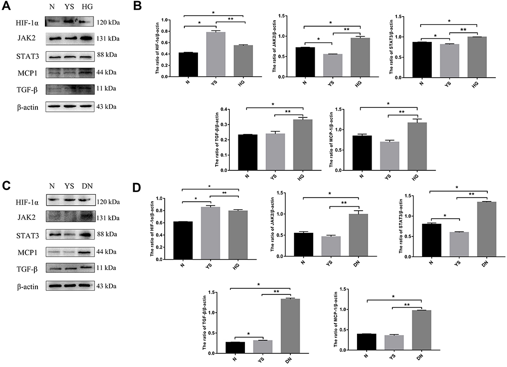 |
Figure 7 Western blotting and quantitative analysis. (A and B) The expression and quantitative analysis of key proteins in NRK-52E cells. After HG stimulation, the expression of HIF-1α, JAK-2, STAT3, TGF-β and MCP-1 protein increased in NRK-52E cells. After Yishen Capsule treatment, compared with the HG group, the expression of HIF-1α in NRK-52E further increased, while the expression of JAK2, STAT3, MCP-1 and TGF-β decreased. (C and D) The expression and quantitative analysis of key proteins in kidney tissue. Compared with the normal group (N), the expression of HIF-1α, JAK-2, STAT3, TGF-β and MCP-1 protein in the kidney tissue of the DN group increased. After treatment with Yishen Capsule, the expression of HIF-1α increased compared with the DN group. Compared with the DN group, the expressions of JAK2, STAT3, MCP-1 and TGF-β all decreased after treatment with Yishen capsule. *Compared with the N group P<0.05, **Compared with the DN group P<0.05. |
Discussion
DN has become the leading cause of end-stage renal disease worldwide. At present, the treatment of DN mainly involves reducing cardiovascular risk, controlling blood glucose, controlling blood pressure and inhibiting the renin-angiotensin system (RAS).24,25 With the in-depth exploration of the pathogenesis of DN, many studies are devoted to finding new drugs to treat DN.26 Over the past few decades, Chinese herbal medicine is emerging as an effective drug candidate for the treatment of diabetes and its complications.4 Traditional Chinese medicine has certain advantages in treating DN. Yishen Capsule is composed of Astragalus, Angelica, Gordon Euryale, Alisma, Schisandra, Rhodiola and other Chinese medicines. It possesses the effect of improving the pathological changes of DN kidney tissue and delaying the progression of the disease, while its specific mechanism of action is not completely clear.
This study applied network pharmacology methods to discover the key active ingredients of Yishen Capsule in the treatment of DN. Our results showed that Quercetin, Kaempferol, Epigallocatechin gallate, Astragaloside IV, Luteolin and Calycosin were important components in the treatment of DN, which may act on multiple pharmacology targets of DN treatment. Lei et al found that quercetin inhibits the proliferation of glomerular mesangial cells induced by high glucose through activating the Hippo pathway.27 The study of Alshehri confirmed that kaempferol exerts an antioxidant effect by activating the Nrf-2/Ho-1 axis to reduce DN damage.28 Clinical and animal experiments have confirmed that gallic acid targets the activation of Notch signal by inhibiting the TGFβ/Smad3 pathway in diabetic mice, leading to the improvement of renal fibrosis.29 Astragaloside IV is a saponin extracted from Astragalus. Astragaloside IV reduces endoplasmic reticulum stress, inhibits mitochondrial division and increases autophagy activity through mechanisms of anti-fibrosis, anti-oxidation, and anti-apoptosis, which is benefit to the improvement of DN symptoms.30 Recent studies have found that astragaloside IV may inhibit oxidative stress and reduce DN podocyte apoptosis by activating the PPARγ-Klotho-FoxO1 signaling pathway.31 Luteolin, a natural flavonoid compound, may inhibit the activation of STAT3, inhibit inflammation and oxidative stress, and improve glomerular sclerosis and renal interstitial fibrosis in DN mice.32 In addition, mullein, β-sitosterol, formononetin may also act on DN disease targets and signal pathways through a variety of biological processes.
The potential target genes and signal pathways of Yishen capsule in the treatment of DN were predicted based on network pharmacology. Our results showed that AR, AKT1, TP53, ESR1, JUN, TNF, MAPK1, EGFR, CCND1 and VEGFA were the potential key targets for the treatment of DN. AGE-RAGE, PI3K-Akt, HIF-1, JAK-STAT may be the key signaling pathways for the treatment of DN. Some of the above results have been confirmed in the preliminary research of our group. Yishen capsule gavage treatment can reduce urinary protein excretion in DN rats, improve renal function, reduce foot process fusion, improve podocyte Podocin expression, and reduce DN podocyte damage.10 Treated with Yishen capsule-containing serum, the expression of p-JAK2 and p-STAT3 in high glucose cultured podocytes were decreased.11 These results initially suggest that Yishen Capsule can alleviate the pathological damage of the kidney of DN and improve the podocyte damage induced by high glucose. Cytokine suppressor (SOCS) is a classic JAK/STAT signaling pathway inhibitor.33 Yishen Capsule may reduce the degree of glomerular sclerosis and tubular interstitial fibrosis in DN rats by up-regulating the expression of SOCS-3.12,13 In addition, Yishen Capsule can also inhibit the overexpression of TLR4, NF-κB14 and other inflammatory factors and Vascular Endothelial Growth Factor (VEGF)15 in the kidney tissue of DN rats. The reduction of inflammatory response can inhibit the damage of vascular epithelial cells caused by proliferative inflammatory response, which leads to the delay of DN progression. Recent research team found that Yishen Capsule has the function of improving diabetic nephropathy by promoting autophagy and inhibiting inflammation.9
Based on the preliminary research foundation of our group and the results of this study, HIF-1α and JAK2/STAT3 signaling pathway were selected for further experimental verification. JAK/STAT signaling pathway is involved in the signal transduction of IL-10, IL-6, TNF-α and other cytokines and activates the expression of genes related to inflammation and cell proliferation,34 which promotes the progression of DN. This study suggests that Yishen Capsule may inhibit the downstream inflammation cascade by inhibiting the JAK/STAT signaling pathway, and improve DN kidney injury.
HIF-1α is a regulatory protein that monitors the body’s sensitivity to oxygen.35 Hypoxia exists in the subclinical stage of DN, and hypoxic environment may induce the expression of HIF-1α.36 In addition to hypoxia, factors such as inflammation and stress can also up-regulate the expression of HIF-1α.37,38 Studies have suggested that enhanced HIF-1α activity is one of the reasons for the dysfunction of podocytes, glomerular mesangial cells, and tubular epithelial cells (RTECs) and for the activation of pro-inflammatory pathways.39,40 Thus, HIF-1α may be related to glomerulosclerosis and renal interstitial fibrosis. Diabetes hyperglycemia environment and AGEs directly affect the transcription of HIF-1α and lead to HIF-1α activation.41 Inhibiting the HIF-1α/VEGF signaling pathway in RTECs can reduce the expression of extracellular matrix (ECM) markers in DN.41,42 However, studies have also found that the diabetic environment activates the atypical proteasome-dependent pathway of HIF-1α degradation in human renal tubular epithelial cells (HK-2), and reduces the basic expression of HIF-1α.43 HIF-1α can promote autophagy, which reduces kidney damage caused by oxidative stress, ischemia and diabetes.44,45 Conditional knockout of HIF-1α aggravates DN renal tubular damage, promotes mitochondrial rupture in HK-2 cells cultured under hypoxia, promotes ROS generation and promotes mitochondrial membrane potential loss and apoptosis. However, overexpression of HIF-1α or HO- 1 agonist treatment can reverse the above changes.46 The hypoglycemic drug SGLT2 inhibitor empagliflozin can reduce the proximal tubular epithelial cell damage induced by high glucose, through up-regulation of HIF-1α.47 Under physiological conditions, when the oxygen concentration of the body’s tissues or cells decreases, the expression of HIF-1α and HIF-2α increases, thereby mediating the adaptive response of cells to hypoxia to maintain the body’s homeostasis.48 HIF-1α and HIF-2α are important regulatory proteins in the body’s adaptive regulation of hypoxia. Studies have found that both of them can promote autophagy, thereby reducing diabetic kidney damage.49 HIF-1α promotes clearance of damaged mitochondria,50 whereas HIF-2α promotes lysosomal dysfunction-mediated peroxisomal clearance.51 The controversial results may be due to the intensity of the HIF response in DN that regulates the progression of kidney injury in a different subtype and time-dependent manner. The results of this study showed that the expression of HIF-1α increased after the intervention of high glucose. And the expression of HIF-1α further increased after the intervention of Yishen Capsule. HIF-1α activation under high glucose conditions may have a protective effect on the kidneys. After the intervention of Yishen Capsule, the expression of HIF-1α was further increased, which may improve the renal injury of diabetic nephropathy by activating autophagy and other mechanisms.49
Molecular docking is mainly used to study the interaction between molecules.52–55 The more stable the binding conformation of the ligand and the receptor, the stronger the binding and the lower the energy.56 The screened active ingredients and key targets were verified by molecular docking technology.β-sitosterol binds best to the core target protein, suggesting that it plays the greatest role in the treatment of DN by Yishen capsule. According to the optimal composite structure of key targets and active ingredients, it is found that formononetin has better binding ability with AKT1, STAT3 and TP53. In addition, β-sitosterol has good binding ability with AR and TP53, and mullein has good binding ability with HIF-1α and JAK2. The above data suggest that the key chemical components of Yishen Capsule have good binding activity with key targets of diseases.
Conclusion
This study used bioinformatics to initially analyze the mechanism of action of Yishen capsule in the treatment of DN, and selected key pathways for experimental verification both in vitro and in vivo. Results showed that the identical compound of Yishen Capsule may regulate different targets, and the identical target may interfere with different biological processes and signal pathways. It reflects the combined effect characteristics of multi-pathway and multi-target of Yishen Capsule. This study provides a scientific basis for Yishen Capsule in the treatment of DN, and also provides a new direction for exploring the potential mechanism of Yishen Capsule. Based on the network pharmacology analysis, the key components and core targets of Yishen Capsule were obtained. In the future, the effect of the key monomer components in Yishen Capsule on the core targets and the internal mechanism of the compatibility of traditional Chinese medicine components can be further verified.
Acknowledgments
Thanks to all project participants for their dedication to this research.
Author Contributions
YLH and SL conducted bioinformatics data analysis. YLH, ZYZ, and SFL conducted in vivo experiments. YXL and DLS conducted in vitro experiments. YLH, ZYZ, and YXL conducted molecular biological analysis. YLH and SL prepared the manuscript. WYL, GZ and JAF conceptualized this research and finalized this manuscript. All authors contributed to data analysis, drafting or revising the article, have agreed on the journal to which the article will be submitted, gave final approval of the version to be published, and agree to be accountable for all aspects of the work.
Funding
This study has been supported by the National Natural Science Foundation of China [81873159], Shanxi Key Research and Development Project (International Science and Technology Cooperation) [No. 201903D421057], Shanxi Applied Basic Research Project (Natural Science Foundation Project) [No. 201801D121341], Scientific research project [No. 2018040, No. 2017040] funded by Health and Family Planning Commission of Shanxi Province.
Disclosure
The authors report no conflicts of interest in this work.
References
1. Sever B, Altntop MD, Demir Y, et al. An extensive research on aldose reductase inhibitory effects of new 4H-1,2,4-triazole derivatives. J Mol Struct. 2020;1224(2013):129446. doi:10.1016/j.molstruc.2020.129446
2. Sever B, Altintop MD, Demir Y, et al. Design, synthesis, in vitro and in silico investigation of aldose reductase inhibitory effects of new thiazole-based compounds. Bioorg Chem. 2020;102:104110. doi:10.1016/j.bioorg.2020.104110
3. Sever B, Altıntop MD, Demir Y, et al. Identification of a new class of potent aldose reductase inhibitors: design, microwave-assisted synthesis, in vitro and in silico evaluation of 2-pyrazolines. Chem Biol Interact. 2021;345:109576. doi:10.1016/j.cbi.2021.109576
4. Li KX, Ji MJ, Sun HJ. An updated pharmacological insight of resveratrol in the treatment of diabetic nephropathy. Gene. 2021;780:145532. doi:10.1016/j.gene.2021.145532
5. Chuang SM, Shih HM, Chien MN, et al. Risk factors in metabolic syndrome predict the progression of diabetic nephropathy in patients with type 2 diabetes. Diabetes Res Clin Pract. 2019;153:6–13. doi:10.1016/j.diabres.2019.04.022
6. Andrade LS, Jornayvaz FR, De Seigneux S. Chronic kidney disease and new antidiabetic drugs: an overview in 2019. Rev Med Suisse. 2019;15(653):1106–1111.
7. Chinese Association of Chinese Medicine. [Guidelines for the prevention and treatment of diabetic nephropathy with traditional Chinese medicine]. Chin Med Mod Distance Educ China. 2011;9(4):151–153. Chinese.
8. Fang J, Deng A, Liu J. A clinical study of Yishe capsule in treating early diabetic nephropathy. Chin J Integr Trad West Nephrol. 2005;2005(08):457–459.
9. Liu YX, Liu WY, Zhang ZY, et al. Yishen capsule promotes podocyte autophagy through regulating SIRT1/NF-κB signaling pathway to improve diabetic nephropathy. Ren Fail. 2021;43(1):128–140. doi:10.1080/0886022X.2020.1869043
10. Zhang XD, Fang JA, Sun YY, et al. Effect of yishen capsule on podocin in renal tissue of rats with diabetic nephropathy. Chin J Integr Trad West Nephrol. 2011;12(03):199–201.
11. Liu YX, Hu YL, Shi QW, et al. Effect of Yishen capsule on the expression of SOCS3, p-JAK2 and p-STAT3 in mouse podocyte of high glucose. Chin J Integr Trad West Nephrol. 2019;20(10):854–858.
12. Dong HT, Fang JA, Zhang XD, et al. Effects of Yishen capsule on the expression of SOCS-3 and TGF-β1 in renal tubular epithelial cells under high glucose condition. Chin J Integr Trad West Nephrol. 2016;17(04):327–329.
13. Chai B, Fang JA, Sun YY, et al. The effect of Yishen capsule on SOCS-3, collagen type I and IV in diabetic nephropathy rats. Chin J Integr Trad West Nephrol. 2013;14(07):573–575.
14. Zhang XD, Fang JA, Li XX, et al. Effect of Yishen capsule on tubulointerstitial expression of TLR4 and NF-κB in experimental diabetic nephropathy. Chin J Integr Trad West Nephrol. 2013;2(04):192–196.
15. Wang RH, Fang JA, Yang SY. Effect of Yishen Capsule on expression of vascular endothelial growth factor in renal tissue of diabetic nephropathy rats. Chin J Integr Trad West Nephrol. 2010;8(07):832–833.
16. Hookins AL. Network pharmacology. Nat Biotechnol. 2007;25(10):1110–1111. doi:10.1038/nbt1007-1110
17. Liang G, Zhang L, Jiang G, et al. Effects and components of herb pair Huanglian-Banxia on diabetic gastroparesis by network pharmacology. Biomed Res Int. 2021;2021:8257937. doi:10.1155/2021/8257937
18. Luo Y, Li D, Liao Y, et al. Systems pharmacology approach to investigate the mechanism of Kai-Xin-San in Alzheimer’s disease. Front Pharmacol. 2020;11:381. doi:10.3389/fphar.2020.00381
19. Chen YY. Systems-pharmacology investigation of the mechanisms of traditional Chinese medicine Rhodiola rosea L. for the remission of fatigue. Doctoral dissertation. Northwest Sci-Tech University of Agriculture and Forestry; 2018.
20. Szklarczyk D, Gable AL, Lyon D, et al. STRING v11: protein-protein association networks with increased coverage, supporting functional discovery in genome-wide experimental datasets. Nucleic Acids Res. 2019;47(D1):D607–D613. doi:10.1093/nar/gky1131
21. Demir Y, Kotan MŞ, Dikbaş N, et al. Phytase from Weissella halotolerans: purification, partial characterisation and the effect of some metals. Int J Food Properties. 2017;1–11. doi:10.1080/10942912.2017.1368547
22. Demir Y, Duran HE, Durmaz L, et al. The influence of some nonsteroidal anti-inflammatory drugs on metabolic enzymes of aldose reductase, sorbitol dehydrogenase, and α-glycosidase: a perspective for metabolic disorders. Appl Biochem Biotechnol. 2020;190(2):437–447. doi:10.1007/s12010-019-03099-7
23. Demir Y. The behaviour of some antihypertension drugs on human serum paraoxonase-1: an important protector enzyme against atherosclerosis. J Pharm Pharmacol. 2019;71(10):1576–1583. doi:10.1111/jphp.13144
24. Umanath K, Lewis JB. Update on diabetic nephropathy: core curriculum 2018. Am J Kidney Dis. 2018;71(6):884–895. doi:10.1053/j.ajkd.2017.10.026
25. Sever B, Altıntop M, Demir Y, et al. A new series of 2,4-thiazolidinediones endowed with potent aldose reductase inhibitory activity. Open Chem. 2021;19(1):347–357. doi:10.1515/chem-2021-0032
26. Demir Y, Ozaslan MS, Duran HE, et al. Inhibition effects of quinones on aldose reductase: antidiabetic properties. Environ Toxicol Pharmacol. 2019;70:103195. doi:10.1016/j.etap.2019.103195
27. Lei D, Chengcheng L, Xuan Q, et al. Quercetin inhibited mesangial cell proliferation of early diabetic nephropathy through the Hippo pathway. Pharmacol Res. 2019;146:104320. doi:10.1016/j.phrs.2019.104320
28. Alshehri AS. Kaempferol attenuates diabetic nephropathy in streptozotocin-induced diabetic rats by a hypoglycaemic effect and concomitant activation of the Nrf-2/Ho-1/antioxidants axis. Arch Physiol Biochem. 2021;1–14. doi:10.1080/13813455.2021.1890129
29. Zhu QQ, Yang XY, Zhang XJ, et al. EGCG targeting Notch to attenuate renal fibrosis via inhibition of TGFbeta/Smad3 signaling pathway activation in streptozotocin-induced diabetic mice. Food Funct. 2020;11(11):9686–9695. doi:10.1039/D0FO01542C
30. Wang H, Zhuang Z, Huang YY, et al. Protective effect and possible mechanisms of astragaloside IV in animal models of diabetic nephropathy: a preclinical systematic review and meta-analysis. Front Pharmacol. 2020;11:988. doi:10.3389/fphar.2020.00988
31. Xing L, Fang J, Zhu B, et al. Astragaloside IV protects against podocyte apoptosis by inhibiting oxidative stress via activating PPARgamma-Klotho-FoxO1 axis in diabetic nephropathy. Life Sci. 2021;269:119068. doi:10.1016/j.lfs.2021.119068
32. Zhang M, He L, Liu J, et al. Luteolin attenuates diabetic nephropathy through suppressing inflammatory response and oxidative stress by inhibiting STAT3 pathway. Exp Clin Endocrinol Diabetes. 2020;129:729–739. doi:10.1055/a-0998-7985
33. Nicholson SE, De Souza D, Fabri LJ, et al. Suppressor of cytokine signaling-3 preferentially binds to the SHP-2-binding site on the shared cytokine receptor subunit gp130. Proc Natl Acad Sci U S A. 2000;97(12):6493–6498. doi:10.1073/pnas.100135197
34. Pace J, Paladugu P, Das B, et al. Targeting STAT3 signaling in kidney disease. Am J Physiol Renal Physiol. 2019;316(6):F1151–F1161. doi:10.1152/ajprenal.00034.2019
35. Zbytek B, Peacock DL, Seagroves TN, et al. Putative role of HIF transcriptional activity in melanocytes and melanoma biology. Dermatoendocrinol. 2013;5(2):239–251. doi:10.4161/derm.22678
36. Takiyama Y, Harumi T, Watanabe J, et al. Tubular injury in a rat model of type 2 diabetes is prevented by metformin: a possible role of HIF-1alpha expression and oxygen metabolism. Diabetes. 2011;60(3):981–992. doi:10.2337/db10-0655
37. Ortega A, Fernandez A, Arenas MI, et al. Outcome of acute renal injury in diabetic mice with experimental endotoxemia: role of hypoxia-inducible factor-1 alpha. J Diabetes Res. 2013;2013:254529. doi:10.1155/2013/254529
38. Laemmle A, Lechleiter A, Roh V, et al. Inhibition of SIRT1 impairs the accumulation and transcriptional activity of HIF-1alpha protein under hypoxic conditions. PLoS One. 2012;7(3):e33433. doi:10.1371/journal.pone.0033433
39. Nayak BK, Shanmugasundaram K, Friedrichs WE, et al. HIF-1 mediates renal fibrosis in OVE26 type 1 diabetic mice. Diabetes. 2016;65(5):1387–1397. doi:10.2337/db15-0519
40. Hu J, Wang W, Zhang F, et al. Hypoxia inducible factor-1alpha mediates the profibrotic effect of albumin in renal tubular cells. Sci Rep. 2017;7(1):15878. doi:10.1038/s41598-017-15972-8
41. Baumann B, Hayashida T, Liang X, et al. Hypoxia-inducible factor-1alpha promotes glomerulosclerosis and regulates COL1A2 expression through interactions with Smad3. Kidney Int. 2016;90(4):797–808. doi:10.1016/j.kint.2016.05.026
42. Pang X, Zhang Y, Shi X, et al. Hirudin reduces the expression of markers of the extracellular matrix in renal tubular epithelial cells in a rat model of diabetic kidney disease through the hypoxia-inducible factor-1alpha (HIF-1alpha)/Vascular Endothelial Growth Factor (VEGF) signaling pathway. Med Sci Monit. 2020;26:e921894. doi:10.12659/MSM.921894
43. Garcia-Pastor C, Benito-Martinez S, Moreno-Manzano V, et al. Mechanism and consequences of the impaired Hif-1alpha response to hypoxia in human proximal tubular HK-2 cells exposed to high glucose. Sci Rep. 2019;9(1):15868. doi:10.1038/s41598-019-52310-6
44. Lv B, Hua T, Li F, et al. Hypoxia-inducible factor 1 alpha protects mesenchymal stem cells against oxygen-glucose deprivation-induced injury via autophagy induction and PI3K/AKT/mTOR signaling pathway. Am J Transl Res. 2017;9(5):2492–2499.
45. Xie Y, Jiang D, Xiao J, et al. Ischemic preconditioning attenuates ischemia/reperfusion-induced kidney injury by activating autophagy via the SGK1 signaling pathway. Cell Death Dis. 2018;9(3):338. doi:10.1038/s41419-018-0358-7
46. Jiang N, Zhao H, Han Y, et al. HIF-1alpha ameliorates tubular injury in diabetic nephropathy via HO-1-mediated control of mitochondrial dynamics. Cell Prolif. 2020;53(11):e12909. doi:10.1111/cpr.12909
47. Ndibalema AR, Kabuye D, Wen S, et al. Empagliflozin protects against proximal renal tubular cell injury induced by high glucose via regulation of hypoxia-inducible factor 1-alpha. Diabetes Metab Syndr Obes. 2020;13:1953–1967. doi:10.2147/DMSO.S243170
48. Schonenberger MJ, Kovacs WJ. Hypoxia signaling pathways: modulators of oxygen-related organelles. Front Cell Dev Biol. 2015;3:42. doi:10.3389/fcell.2015.00042
49. Yeh CH, Hsu SP, Yang CC, et al. Hypoxic preconditioning reinforces HIF-alpha-dependent HSP70 signaling to reduce ischemic renal failure-induced renal tubular apoptosis and autophagy. Life Sci. 2010;86(3–4):115–123. doi:10.1016/j.lfs.2009.11.022
50. Han B, Cui H, Kang L, et al. Metformin inhibits thyroid cancer cell growth, migration, and EMT through the mTOR pathway. Tumour Biol. 2015;36(8):6295–6304. doi:10.1007/s13277-015-3315-4
51. Walter KM, Schonenberger MJ, Trotzmuller M, et al. Hif-2alpha promotes degradation of mammalian peroxisomes by selective autophagy. Cell Metab. 2014;20(5):882–897. doi:10.1016/j.cmet.2014.09.017
52. Istrefi Q, Türkeş C, Arslan M, et al. Sulfonamides incorporating ketene N,S-acetal bioisosteres as potent carbonic anhydrase and acetylcholinesterase inhibitors. Arch Pharm (Weinheim). 2020;353(6):e1900383. doi:10.1002/ardp.201900383
53. Türkeş C, Demir Y, Beydemir Ş. Calcium channel blockers: molecular docking and inhibition studies on carbonic anhydrase I and II isoenzymes. J Biomol Struct Dyn. 2021;39(5):1672–1680. doi:10.1080/07391102.2020.1736631
54. Demir Y. Naphthoquinones, benzoquinones, and anthraquinones: molecular docking, ADME and inhibition studies on human serum paraoxonase-1 associated with cardiovascular diseases. Drug Dev Res. 2020;81(5):628–636. doi:10.1002/ddr.21667
55. Demir Y, Taslimi P, Koçyiğit ÜM, et al. Determination of the inhibition profiles of pyrazolyl-thiazole derivatives against aldose reductase and α-glycosidase and molecular docking studies. Arch Pharm. 2020;353(12):e2000118. doi:10.1002/ardp.202000118
56. Hsin KY, Ghosh S, Kitano H. Combining machine learning systems and multiple docking simulation packages to improve docking prediction reliability for network pharmacology. PLoS One. 2013;8(12):e83922. doi:10.1371/journal.pone.0083922
 © 2022 The Author(s). This work is published and licensed by Dove Medical Press Limited. The full terms of this license are available at https://www.dovepress.com/terms.php and incorporate the Creative Commons Attribution - Non Commercial (unported, v3.0) License.
By accessing the work you hereby accept the Terms. Non-commercial uses of the work are permitted without any further permission from Dove Medical Press Limited, provided the work is properly attributed. For permission for commercial use of this work, please see paragraphs 4.2 and 5 of our Terms.
© 2022 The Author(s). This work is published and licensed by Dove Medical Press Limited. The full terms of this license are available at https://www.dovepress.com/terms.php and incorporate the Creative Commons Attribution - Non Commercial (unported, v3.0) License.
By accessing the work you hereby accept the Terms. Non-commercial uses of the work are permitted without any further permission from Dove Medical Press Limited, provided the work is properly attributed. For permission for commercial use of this work, please see paragraphs 4.2 and 5 of our Terms.