")
Back to Journals » Cancer Management and Research » Volume 15
Potential Diagnostic Utility of microRNAs in Gastrointestinal Cancers
Received 3 June 2023
Accepted for publication 2 August 2023
Published 22 August 2023 Volume 2023:15 Pages 863—871
DOI https://doi.org/10.2147/CMAR.S421928
Checked for plagiarism Yes
Review by Single anonymous peer review
Peer reviewer comments 2
Editor who approved publication: Dr Antonella D'Anneo
Wojciech Jelski,1 Barbara Mroczko1,2
1Department of Biochemical Diagnostics, Medical University, Bialystok, Poland; 2Department of Neurodegeneration Diagnostics, Medical University, Bialystok, Poland
Correspondence: Wojciech Jelski, Department of Biochemical Diagnostics, Medical University, Waszyngtona 15 A, Bialystok, 15– 269, Poland, Tel +48 85 746 8587, Fax +48 85 746 8585, Email [email protected]
Abstract: Early detection of gastrointestinal cancers is beneficial for patient survival and prognosis. MiRNAs have been shown to be potential cancer biomarkers that can be used to diagnose cancers. MiRNAs are single-stranded, small non-coding RNAs that are involved in the post-transcriptional regulation of the expression of different oncogenes. Cancer tissues contain miRNAs that play a special role in the etiology of cancer development or limiting cancer suppression. Dysregulation of miRNAs occurs in a variety of malignancies, including gastrointestinal cancers. MiRNAs are stable and protected against degradation by RNase, which enables their detection in tissues and biological fluids. The results of many studies suggest that miRNAs have a relatively higher diagnostic efficiency in distinguishing cancer patients from healthy people. The researchers have identified many miRNA signature in the blood for the detection of gastrointestinal cancers. This review focuses on the role and potential utility of miRNAs in the early detection, prognosis and evaluation of the treatment effectiveness of gastrointestinal cancers.
Keywords: miRNA, gastric cancer, colorectal cancer, esophageal cancer, diagnosis
Introduction
Early diagnosis and careful monitoring of tumor development is a prerequisite for reducing the consequences of the disease and reducing the number of deaths, and to improve management cancer patients. The most important features of early tumor detection markers are the highest sensitivity and specificity. Recent research to identify tumors based on biomarkers in the blood has led to the identification a wide variety of cancer-related molecules. In recent years, microRNAs (miRNAs) have been shown to be effective candidates for the molecular diagnosis of many cancers.
MiRNAs are endogenous small molecule single-chain non-coding regulatory RNAs with a length of 17–25 nucleotides that play a crucial role in post-transcriptional gene regulation. There are currently about 30,000 entries in the miR Registry Database, which means over forty thousand mature miRNA products, and their number may increase significantly in an exponential manner in the coming years. Non-coding RNA includes all RNA chains that are not translated into proteins. They are classified based on structure and length and include miRNAs, long non-coding RNAs (lncRNAs) and circular RNAs (circRNAs). These different classes of ncRNAs play important roles in intricate networks of interaction with each other, miRNAs and various signaling pathways to effect changes in cells, surrounding tissues and entire organ systems. MiRNAs have highly conservative, temporal and tissue-specific characteristics. The biogenesis of miRNAs begins with their transcription from miRNA genes in the nucleus, transcripted into pri-miRNAs by RNA polymerase II. The resulting pri-miRNAs is then processed into pre-miRNA by the RNase III enzyme Drosha and its dsRNA-binding domain (dsRBD), DGCR8. Next, the mature miRNAs can arise from the stem of the pre-miRNA by the Dicer after be exported to the cytoplasm by XPO5.1 Numerous studies have proven that miRNAs regulate over 30% of human genes, where one miRNA controls several hundred RNAs. Approximately 30–60% of all human proteins can be targeted via miRNAs that play a role in various cell development procedures. Gene regulation by miRNAs plays important roles in development, physiology, and disease. miRNAs are an abundant class of noncoding RNAs that are generated through multistep biosynthetic pathways and typically repress gene expression through target destabilization and translational inhibition.2,3 MiRNAs are common in eukaryotes and play a regulatory role in cell proliferation, differentiation, and apoptosis. MiRNAs are regulators of gene expression and lead to the development of inflammation and carcinogenicity. The rapid progress of next-generation sequencing makes miRNA interposition possible change related physiological functions, causing inflammation cell penetration, neurological diseases, infectious disorders, immune disorders, cancers and other diseases.4 Deregulation of miRNAs in cancer cells constitutes result of disturbed miRNA biogenesis process, epigenetic and genomic transformation which leads to protooncogenic or suppressive effects of miRNAs in carcinogenesis. In the case of oncogenic miRNAs, a miRNA inhibitor blocks its miRNA function; while tumor-suppressing miRNAs, reconstitution of the miRNA precursor produces an anticancer effect. Because miRNAs are highly specific for different tissue types as well as for cell types within a given tissue, numerous studies have profiled miRNA patterns in different types of cancer, demonstrating the diagnostic and prognostic importance of miRNAs in clinical practice. In cancer diagnosis, a single miRNA usually has low specificity and/or sensitivity, but a full suite of miRNA expression is very specific, can show the evolutionary lineage and differentiation of a tumor, and be helpful in conducting a diversity study. By horizontal comparison of the tumor tissue with the surrounding healthy cells, the specific expression of miRNAs in tumors is detected. In contrast, longitudinal comparison of individual cancer stages identified different miRNAs at all stages up to fully reliable diagnosis and tumor staging.5 A single type of miRNA molecule regulates multidirectional gene expression and multiple pathways, influencing tumor development. For this reason, miRNAs act more effectively as biological regulation particles than gene-encoding molecules. miRNA analysis allows for the precise identification of the origin of cancer cells in various cancers.6 Methods for detecting miRNA and miRNA expression in tumors may have clinical applications.7 There are currently several different platforms to explore miRNA expression, for example RT-PCR, Northern blot, microarray or primer extension. Microarrays are most commonly used for miRNA research, and not only provide a user-friendly platform but also have significant throughput allowing profiling of the entire miRNA genome.8
Increasing evidence suggests that miRNAs can serve as noninvasive biomarkers for gastrointestinal cancers (Figure 1). In the current review, we discuss the diagnostic usefulness of miRNA in gastrointestinal cancers.
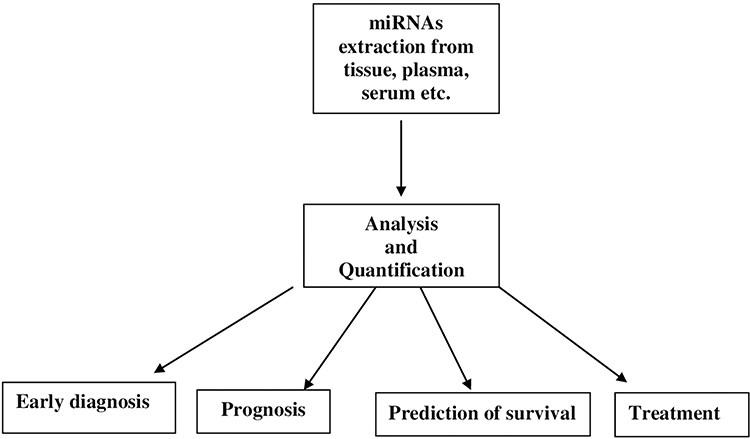 |
Figure 1 Application of miRNAs as biomarkers for gastrointestinal cancers. |
The comprehensive online search about articles from three databases, PubMed, Web of Science and Embase was performed by two independent authors (W.J. and B.M.). A comprehensive search was conducted employing the subject terms: “miRNA” and “gastric cancer”, “colorectal cancer”, “esophageal cancer”. We came up with inclusion criteria for qualified articles that were analyzed by our full-text estimation: articles concerning the pertinence between miRNA level and diagnosis of gastrointestinal cancer diagnosis; full-text articles published in English.
MiRNA in Gastric Cancer Diagnosis
In recent years, it has been proven that miRNAs, due to their durability and typicality of expression in tissues and in the blood, can be considered as new biomarkers with a potential tool of clinical importance for the outcome of patients with gastric cancer (GC). Many researchers have analyzed the miRNA signature of the serum of gastric cancer patients as diagnostic and prognostic indicators. MiRNAs are released from tumor cells into not only blood but also into various body fluids: urine, tears and, most importantly, into gastric juice through the secretion of exosome particles.9 Yu et al evaluated the diagnostic value of miRNA 129-1/2 in gastric secretions as a new screening tool for gastric cancer. After examining the secretions of more than 140 patients with gastric cancer, significant reductions in miRNA-129-1 and miRNA-129-2 levels were found. Thus, gastric juice may be a suitable material for the molecular diagnosis of gastric cancer.9
Chinese scientists describe many types of miRNAs that play a predictive role in gastric cancer. Among others, Li et al showed that the signature of several miRNAs (miRNA-10b, miRNA-21, miRNA-126, miRNA-223, and miRNA-338) is an independent predictor of overall survival and relapse-free survival.10 Moreover, miRNA-25 has been shown also to be overexpressed in the plasma and tumor tissue of patients with gastric cancer with lymph node metastases. Inhibition of miRNA-25 significantly reduced the rate of metastasis, invasiveness and proliferation in vitro and reduced the metastatic capacity of gastric cancer cells in vivo by repressing the expression of the ERBB2 transducer. In addition, patients with high blood level of miRNA-25 had a poor prognosis.11 MiRNA-199a-3p has also been shown to be a potential biomarker for detection and monitoring of gastric cancer in the early stage. Level of miRNA-199a-3p is significantly elevated in the serum of patients with gastric cancer and decreases after resection of the primary tumor. During the validation phase in a very large cohort, miRNA-199a-3p levels were high in cancer patients compared to controls, with a large area under the ROC curve (0.837) and was significantly associated with lymph node metastases, tumor depth and tumor stage.12 Increased expression of miRNA-630 was found in the tumor tissue of the stomach. It was significantly associated with tumor depth, lymph node metastases, distant metastases, and poor overall survival. This indicates that miRNA-630 could be a potential biomarker of gastric cancer initiation and progression.13 In addition, many researchers have discovered numerous miRNAs that play a role as biomarkers in gastric cancer. For example, very high expression of miRNA-20b, miRNA-214, miRNA-142-5p, miRNA-375 and miRNA-150 and low expression of miRNA-451, miRNA-125-5p and miRNA-433 is correlated with short survival time.14,15 Low expressions of miRNA-148, miRNA-126, miRNA-146a, miRNA-439, miRNA-218, and miRNA-335 and high levels of miRNA-650 and miRNA-27a indicates lymph node metastasis.16,17 Thus, various authors have demonstrated the usefulness of different miRNAs in the diagnosis of gastric cancer, which allows to predict cancers with a worse prognosis and can be used as markers of progression risk. Table 1 lists miRNAs that may function as gastric cancer biomarkers.
 |
Table 1 Potential miRNA Biomarkers of Gastric Cancer |
MiRNA in Colorectal Cancer Diagnosis
MiRNAs play an important role in the carcinogenesis and development of colorectal cancer (CRC), but there is also evidence that miRNAs are good biomarkers for cancer. A non-invasive miRNA-based test for the early detection of colorectal cancer is an attractive pre-colonoscopy screening tool. Dysregulated miRNAs were detected in the serum of patients with colorectal cancer using sequencing-based analysis. In the earliest studies of serum miRNA profiling in colorectal cancer, 69 serum miRNAs were found to be differentially expressed in cancer patients but not in healthy controls. Of these, 14 miRNAs were rarely expressed in colorectal cancer patients, and the researchers believe that these miRNAs have a high degree of specificity for CRC.18 Subsequently, the researchers found that the expression of miRNA-92a and miRNA-17-3p was significantly overexpressed in the blood of cancer patients. They reported miR-17-3p and miR-92 with about 90% sensitivity and about 71% specificity in colorectal cancer with healthy controls. Interestingly, the expression level of these miRNAs was down regulated after surgical tumor surgery, suggesting that the expression levels of these miRNAs in the blood were a reflection of the underlying CRC. In addition, the expression of miR-92a also distinguishes colorectal cancer from other gastrointestinal diseases and cancers, emphasizing the specificity of miRNA biomarkers for various diseases and suggesting that serum miRNA-92a expression levels may be an important diagnostic biomarker for non-invasive CRC screening. The results of these studies have led to significant advance in the study of diagnostic markers in the blood of patients with CRC. Diagnosis at an early stage of the disease, including the presence of precancerous lesions in CRC, is a key issue to improve patient survival in CRC treatment.19 Since early diagnosis of colorectal cancer is crucial, identifying markers of precancerous adenoma is highly beneficial. Kanaan et al found that a panel of 8 plasma miRNAs, including miRNA-532-3p, miRNA-195, miRNA-17, miRNA-331, miRNA-142-3p, miRNA-652, and miRNA-15b, were able to recognize adenomas with a sensitivity of about 90% and a specificity of about 65%.20 Other research tested 12 selected miRNAs using blood samples from 157 patients with advanced colorectal carcinoma and adenoma and 59 control samples. Based on the obtained results, miRNA-29a and miRNA-92a were found to be important in cancer diagnosis values with a combined area under ROC Curve of 0.883 with about 85% specificity and about 84% sensitivity. For detection of advanced adenomas, AUC power is 0.773 with a sensitivity of 73% and about 80% specificity.21 In the last decade, with the help of a large number of serum samples and relevant tissue sections, it was also discovered that miRNA-21 may be a marker for early detection and prognosis in patients with CRC. Serum miRNA-21 expression status increased by stage in patients with CRC stages I, II, III and IV, drawing attention to its possible use as a new prognostic marker in patients with colorectal cancer.22
Metastases to other organs are the leading cause of death in patients with advanced colorectal cancer. Most of the articles published so far have focused on evaluating miRNA expression in primary tumor samples, but there are also studies on metastatic tissues. Pizzini et al in a study of 78 tissue samples (23 healthy colon mucosa, 31 primary tumor tissues and 24 liver metastases) from 46 colorectal cancer patients showed that three miRNAs, miRNA-210, miRNA-146a and miRNA-122, were dysregulated in metastatic and primary cancer tissues.23 In another study, Toiyama et al examined serum miRNA-200c expression from colorectal cancer patients and showed that the elevated level of miRNA-200c in metastatic cancer reflects the state of expression in plasma samples. High expression of miRNA-200c in the blood was strongly correlated with distant and lymph node metastases and was an independent factor of tumor recurrence and an independent prognostic factor of colorectal cancer.24
A large number of miRNAs show variability expression in colorectal cancer tissues with reduced miRNAs (Table 2), and this is particularly relevant for cell proliferation, apoptosis and metastasis, but further extensive research in a large number of independent cohorts is necessary to determine their practical applications.
 |
Table 2 Potential miRNA Biomarkers of Colorectal Cancer |
MiRNA in Esophageal Cancer Diagnosis
Esophageal cancer (EC) is the sixth most common cancer and the sixth leading cause of cancer-related deaths in human around the world. Histologically, it can be divided into two subtypes, adenocarcinoma and squamous cell carcinoma, each with a specific cell type, epidemiology, origin, pathogenesis. Today’s gold standards in EC diagnostics, such as biopsy or endoscopy, are unfortunately invasive. There are several proposals for potential EC biomarkers. But the pace of their implementation into practical use in the clinic was unsatisfactory. As high-throughput assays developed, more and more non-coding RNAs associated with cancer were recognized.25 Several potential EC biomarkers have been investigated. However, their clinical relevance was unsatisfactory. As high-throughput assays have developed, an increasing number of miRNAs associated with esophageal cancer have been recognized.26 Since Zhang et al in 2010 first described blood miRNA levels in human with esophageal cancer, numerous researchers have investigated the differentiation of miRNA expression and their potential utility in EC has been investigated.27 Study results confirmed that miRNAs as biomarkers can contribute to the early diagnosis of EC (Table 3). A review of eight manuscripts by Wang et al looked at a total of 16 various types of miRNAs in the blood and saliva of patients with esophageal squamous cell carcinoma. The clinical use of miRNAs has also been studied by Fu et al who reported 39 potentially prognostic miRNAs in 25 studies.28 Most of the diagnostically useful miRNA types were identified by a single study and only three miRNAs (miRNA-145, 375, 21) were reported by more researchers. Researchers found relatively high levels of specificity and sensitivity for combinations and single-stranded miRNAs. For example, Wang et al, analyzing the diagnostic value of miR-21, showed that it has a high specificity and sensitivity for squamous cell carcinoma of the esophagus, 71.0% and 96.9%, respectively.29 As a potential diagnostic biomarker for esophageal cancer, miRNA-21 has several valuable advantages. Serum miRNA-21 expression levels are both reproducible and stable. In addition, miRNA-21 is minimally invasive and convenient when compared to histopathology in serum or plasma, and it is easily detected by Quantitative Reverse Transcription PCR method. Finally, its level is particularly correlated with various types of cancer. MiRNA-21 alters the expression of many cancer-related target genes, including PDCD, TPM1, PTEN. Significant overexpression of miRNA-21 in plasma has been reported also in patients with early-stage esophageal cancer.30 Chen et al showed that plasma microRNA-375 was significantly different between esophageal cancer patients and controls, but found no difference in gender or age. They evaluated the diagnostic value of miRNA-375 37 EC patients and 26 healthy controls. The AUC of miRNA-375 in plasma was 0.707. Short survival time in patients with esophageal cancer positively correlated with low expression of miRNA-375. This is likely due to hypermethylation of the miRNA-375 promoter region in cancer cells, which slows down motility, cell proliferation, cell colony formation, and tumor metastasis.31 Other researchers (Zhang et al) found that the expression of miRNA-26a in esophageal adenocarcinoma tissues was lower than in non-cancerous tissue, and miRNA-26a expression was significantly lower in metastatic lymph nodes than in primary esophageal tumors.32
 |
Table 3 Potential miRNA Biomarkers of Esophageal Cancer |
In addition, most studies have examined the alteration of expression levels of multiple tumor suppressor oncomiRNAs and miRNAs in esophageal adenocarcinoma and esophageal squamous cell carcinoma, thus demonstrating how miRNAs can alter key regulatory pathways of the carcinogenesis process. Given these revelations, miRNAs may function not only as a prognostic and diagnostic tool but may also be a predictive biomarker of response to anti-cancer therapies and as potential therapeutic targets. Moreover, considering the role of miRNAs in the development of esophageal cancer and the fact that miRNAs can be quickly and successfully detected in body fluids, are very good candidates for diagnostic biomarkers, prognostic and predictive stratification of patients.33 In addition, a new understanding of the role of miRNAs in cancer pathology has created miRNAs as an attractive tool for new therapies.
Summary
MiRNAs have an important function in various processes of cell biology, including the formation of cancer. These small non-coding RNAs may play a role as diagnostic and prognostic biomarkers. They can be related to various cancers and distinctive miRNA expression profiles; they are also known for their functional and therapeuthic roles of miRNAs. Recently, miRNAs have been the subject of interest of numerous researchers. New types of miRNAs have been discovered and research techniques have been modernized. Therefore, this study reviews the role of miRNAs in relation to cancer prognosis and diagnosis. Although some human tissues exhibit malignant features, individual miRNAs are over- or under-expressed in different cancers and at different stages, correlating with cancer development, prevalence, and prognosis.34 Further studies of the relationship between miRNAs and gastrointestinal cancers may provide further applications in early cancer detection, prognosis, monitoring, gene therapy and avoiding chemotherapy resistance. In recent years, miRNAs are in progress clinical phases and significant progress has been made in miRNA drug research and advancement patents. Additionally, an expression profile study revealed distinctive miRNA signatures that could predict the clinical outcome of cancer.35 Recently, the detection of miRNAs secreted in exosomes, ie, in membrane vesicles, and in blood and other body fluids, indicate that miRNAs are important in intracellular communication, both in an endocrine and paracrine way. Moreover, individual miRNAs have been identified as tumor suppressor genes and oncogenes in cancers.36,37 A wide range of body fluids are currently being studied for their suitability as non-invasive miRNA-based biomarkers to detect different types of cancer in its early stages. Given that blood is one of the most readily available body fluids, it is most commonly used as a diagnostic fluid to test surrogate cancer biomarkers.38 Microarrays and real-time RT-PCR are analytical techniques that are widely used in screening and validation studies. Due to the ability to target many genes, miRNAs can be a much more effective therapeutic tool than monogenic therapy.
However, new miRNAs have been discovered and research techniques constantly updated and in the future, high-quality research into the diagnostic function of miRNAs should be carried out. There are also other types of miRNAs that, due to editorial constraints, have not been described in this work. The vast majority of studies published in recent years have involved biomarkers predictive of distant metastases using resected primary tissues or pre-surgical serum/plasma samples, but there are no studies on the importance of monitoring metachronous biomarkers using large numbers of blood samples. Further studies are required in the future to accurately determine the clinical relevance of already published miRNAs as recurrence monitoring biomarkers such as antigen carcinoembryonic (CEA) and antigen CA19-9 for colorectal cancer and pancreatic cancer.
Numerous new miRNAs have been discovered for early prognosis, radiotherapy and chemotherapy efficacy evaluation. Unfortunately, there are numerous problems in trials of clinical application. Recognition of downstream miRNA targets is complicated. In addition, in research based on the practical application of miRNAs in cancer, there is still no reliable and precise information from large-scale multicentre studies. Unified standards and test platforms are necessary for an accurate diagnosis. Effective treatment requires modern micromolecular drugs based on a well-chosen appropriate drug carrier without side effects. There is a growing need for miRNA profiling with the extension of its application to differentiation subtypes and predict treatment response in cancer. The stability of miRNA in the blood makes it suitable as a diagnostic marker in various cancers. In addition, it is likely to obtain a miRNA signature instead of a single miRNA to provide useful information so clinicians can decide to personalize disease management. A better understanding of these miRNAs and their target genes will open up new prospects for more efficient diagnostic methods for gastrointestinal cancers. We hope that further research on miRNAs will increase their clinical significance in diagnosis and treatment of gastrointestinal cancers.
Abbreviations
AUC, area under curve; CEA, antigen carcinoembryonic; circRNAs, circular RNAs; CRC, colorectal cancer; EC, esophageal cancer; GC, gastric cancer; lncRNAs, long non-coding RNAs; ncRNAs, non-coding miRNAs; RT-PCR, reverse-transcription polymerase chain reaction.
Disclosure
The authors report no conflicts of interest in this work.
References
1. Zhao Y, Li X, Ye C, Huang C, Lv X, Li J. The biogenesis, mechanism and function of the tRNA-derived small RNA (tsRNA): a review compared with microRNA. Am J Cancer Res. 2023;13(5):1656–1666.
2. Matsuyama H, Suzuki HI. Systems and synthetic microRNA biology: from biogenesis to disease pathogenesis. Int J Mol Sci. 2019;21(1):132–154. doi:10.3390/ijms21010132
3. Suzuki HI. Roles of microRNAs in disease biology. JMA J. 2023;6:104–113.
4. Komatsu S, Kitai H, Suzuki HI. Network regulation of microRNA biogenesis and target interaction. Cells. 2023;12(2):306–318. doi:10.3390/cells12020306
5. Babaeenezhad E, Naghibalhossaini F, Rajabibazl M, et al. The roles of microRNA miR-185 in digestive tract cancers. Noncoding RNA. 2022;8:83 67–84.
6. Heneghan HM, Miller N, Kerin MJ. MiRNAs as biomarkers and therapeutic targets in cancer. Curr Opin Pharmacol. 2010;10(5):543–550. doi:10.1016/j.coph.2010.05.010
7. Nuovo GJ, Elton TS, Nana-Sinkam P, Volinia S, Croce CM, Schmittgen TD. A methodology for the combined in situ analyses of the precursor and mature forms of microRNAs and correlation with their putative targets. Nat Protoc. 2009;4(1):107–115. doi:10.1038/nprot.2008.215
8. Mei Q, Li X, Meng Y, et al. A facile and specific assay for quantifying microRNA by an optimized RT-qPCR approach. PLoS One. 2012;7(10):e46890. doi:10.1371/journal.pone.0046890
9. Yu X, Luo L, Wu Y, et al. Gastric juice miR-129 as a potential biomarker for screening gastric cancer. Med Oncol. 2013;30(1):365. doi:10.1007/s12032-012-0365-y
10. Li X, Zhang Y, Zhang Y, Ding J, Wu K, Fan D. Survival prediction of gastric cancer by a seven-microRNA signature. Gut. 2010;59(5):579–585. doi:10.1136/gut.2008.175497
11. Li BS, Zuo QF, Zhao YL, et al. MicroRNA-25 promotes gastric cancer migration, invasion and proliferation by directly targeting transducer of ERBB2, 1 and correlates with poor survival. Oncogene. 2015;34(20):2556–2565. doi:10.1038/onc.2014.214
12. Li C, Li JF, Cai Q, et al. miRNA-199a-3p in plasma as a potential diagnostic biomarker for gastric cancer. Ann Surg Oncol. 2013;20(Suppl 3):397–405. doi:10.1245/s10434-012-2600-3
13. Chu D, Zhao Z, Li Y, et al. Increased microRNA-630 expression in gastric cancer is associated with poor overall survival. PLoS One. 2014;9(3):e90526. doi:10.1371/journal.pone.0090526
14. Xiong X, Ren HZ, Li MH, Mei JH, Wen JF, Zheng CL. Down regulatedmiRNA-214 induces a cell cycle G1 arrest in gastric cancer cells by up-regulating the PTEN protein. Pathol Oncol Res. 2011;17:931–937.
15. Nishida N, Mimori K, Fabbri M, et al. MicroRNA-125a-5p is an independent prognostic factor in gastric cancer and inhibits the proliferation of human gastric cancer cells in combination with trastuzumab. Clin Cancer Res. 2011;17(9):2725–2733. doi:10.1158/1078-0432.CCR-10-2132
16. Feng R, Chen X, Yu Y, et al. miR-126 functions as a tumour suppressor in human gastric cancer. Cancer Lett. 2010;298:50–63. doi:10.1016/j.canlet.2010.06.004
17. Zheng B, Liang L, Wang C, et al. MicroRNA-148a suppresses tumor cell invasion and metastasis by downregulating ROCK1 in gastric cancer. Clin Cancer Res. 2011;17(24):7574–7583. doi:10.1158/1078-0432.CCR-11-1714
18. Chen X, Ba Y, Ma L, et al. Characterization of microRNAs in serum: a novel class of biomarkers for diagnosis of cancer and other diseases. Cell Res. 2008;18(10):997–1006. doi:10.1038/cr.2008.282
19. Ng EK, Chong WW, Jin H, et al. Differential expression of microRNAsin plasma of patients with colorectal cancer: a potential marker for colorectal cancer screening. Gut. 2009;58(10):1375–1381. doi:10.1136/gut.2008.167817
20. Kanaan Z, Roberts H, Eichenberger MR, et al. A plasma microRNA panel for detection of colorectal adenomas: a step toward more precise screening for colorectal cancer. Ann Surg. 2013;258(3):400–408. doi:10.1097/SLA.0b013e3182a15bcc
21. Huang Z, Huang D, Ni S, Peng Z, Sheng W, Du X. Plasma microRNAs are promising novel biomarkers for early detection of colorectal cancer. Int J Cancer. 2010;127(1):118–126. doi:10.1002/ijc.25007
22. Huang Z, Huang D, Ni S, Peng Z, Sheng W, Du X. Serum miR-21 as a diagnostic and prognostic biomarker in colorectal cancer. J Nat Cancer Inst. 2013;105(12):849–859. doi:10.1093/jnci/djt101
23. Pizzini S, Bisognin A, Mandruzzato S, et al. Impact of microRNAs on regulatory networks and pathways in human colorectal carcinogenesis and development of metastasis. BMC Genomics. 2013;14(1):589–602. doi:10.1186/1471-2164-14-589
24. Toiyama Y, Hur K, Tanaka K, et al. Serum miR-200c Is a novel prognostic and metastasis-predictive biomarker in patients with colorectal cancer. Ann Surg. 2014;259(4):735–743. doi:10.1097/SLA.0b013e3182a6909d
25. Bird-Lieberman EL, Fitzgerald RC. Early diagnosis ofoesophageal cancer. Br J Cancer. 2009;101(1):1–6. doi:10.1038/sj.bjc.6605126
26. Lopez-Camarillo C, Marchat LA, Arechaga-Ocampo E, et al. MetastamiRs: non-coding microRNAs driving cancer invasion and metastasis. Int J Mol Sci. 2012;13(2):1347–1379. doi:10.3390/ijms13021347
27. Zhang C, Wang C, Chen X, et al. Expression profile of microRNAs in serum: a fingerprint for esophageal squamous cell carcinoma. Clin Chem. 2010;56(12):1871–1879. doi:10.1373/clinchem.2010.147553
28. Fu W, Pang L, Chen Y, Yang L, Zhu J, Wei Y. The microRNAs as prognostic biomarkers for survival in esophageal cancer: a metaanalysis. Sci World J. 2014;2014:523979. doi:10.1155/2014/523979
29. Wang K, Chen D, Meng Y, Xu J, Zhang Q. Clinical evaluation of 4 types of microRNA in serum as biomarkers of esophageal squamous cell carcinoma. Oncol Lett. 2018;16(1):1196–1204. doi:10.3892/ol.2018.8720
30. Zheng H, Lingbo P, Bangjie L. MicroRNA-21 as a potential biomarker for detecting esophageal carcinoma in Asian populations: a meta-analysis. Peer J. 2002;10:e14048.
31. Chen J, Cai Z, Hu J, Zhou L, Zhang P, Xu X. MicroRNA-375 in extracellular vesicles – novel marker for esophageal cancer diagnosis. Medicine. 2023;102(5):e32826. doi:10.1097/MD.0000000000032826
32. Zhang YF, Zhang AR, Zhang BC, et al. MiR-26a regulates cell cycle and anoikis of human esophageal adenocarcinoma cells through Rb1-E2F1 signaling pathway. Mol Biol Rep. 2013;40(2):1711–1720. doi:10.1007/s11033-012-2222-7
33. Angerilli V, Galuppini F, Businello G, et al. MicroRNAs as predictive biomarkers of resistance to targeted therapies in gastrointestinal tumors. Biomedicines. 2021;9(3):318–336. doi:10.3390/biomedicines9030318
34. He L, Hannon GJ. MicroRNAs: small RNAs with a big role in gene regulation. Nat Rev Genet. 2004;5(7):522–531. doi:10.1038/nrg1379
35. Skog J, Würdinger T, van Rijn S, et al. Glioblastoma microvesicles transport RNAand proteins that promote tumour growth and provide diagnostic biomarkers. Nat Cell Biol. 2008;10(12):1470–1476. doi:10.1038/ncb1800
36. Lawrie CH, Gal S, Dunlop HM, et al. Detection of elevated levels of tumour-associated microRNAs in serum of patients with diffuse large B-cell lymphoma. Br J Haematol. 2008;141(5):672–675. doi:10.1111/j.1365-2141.2008.07077.x
37. Chang KH, Miller N, Kheirelseid EA, et al. MicroRNAsignature analysis in colorectal cancer: identification of expression profiles in stage II tumors associated with aggressive disease. Int J Colorectal Dis. 2011;26(11):1415–1422. doi:10.1007/s00384-011-1279-4
38. Gilad S, Meiri E, Yogev Y, et al. Serum microRNAsare promising novel biomarkers. PLoS One. 2008;3(9):e3148. doi:10.1371/journal.pone.0003148
 © 2023 The Author(s). This work is published and licensed by Dove Medical Press Limited. The full terms of this license are available at https://www.dovepress.com/terms.php and incorporate the Creative Commons Attribution - Non Commercial (unported, v3.0) License.
By accessing the work you hereby accept the Terms. Non-commercial uses of the work are permitted without any further permission from Dove Medical Press Limited, provided the work is properly attributed. For permission for commercial use of this work, please see paragraphs 4.2 and 5 of our Terms.
© 2023 The Author(s). This work is published and licensed by Dove Medical Press Limited. The full terms of this license are available at https://www.dovepress.com/terms.php and incorporate the Creative Commons Attribution - Non Commercial (unported, v3.0) License.
By accessing the work you hereby accept the Terms. Non-commercial uses of the work are permitted without any further permission from Dove Medical Press Limited, provided the work is properly attributed. For permission for commercial use of this work, please see paragraphs 4.2 and 5 of our Terms.