
Back to Journals » Breast Cancer: Targets and Therapy » Volume 18
Plasma Vitamin C Levels in Distinguishing Benign from Malignant Breast Tumors and Clinical Correlates in Chinese Patients: A Cross-Sectional Study
Authors Jiang S, Gao D, Yin Y, Sun Z, Ding T, He Y, Zhang Y, Li Y, Dong M
Received 6 November 2025
Accepted for publication 23 April 2026
Published 27 May 2026 Volume 2026:18 579640
DOI https://doi.org/10.2147/BCTT.S579640
Checked for plagiarism Yes
Review by Single anonymous peer review
Peer reviewer comments 2
Editor who approved publication: Professor Pranela Rameshwar
Shuai Jiang,1,2,* Dongqing Gao,1,* Yichuang Yin,1,3,* Zhonghua Sun,1 Tingting Ding,1 Ying He,1,4 Yifei Zhang,1,4 Yuexin Li,1 Mei Dong1,2
1Department of Pharmacy, Harbin Medical University Cancer Hospital, Harbin, Heilongjiang, People’s Republic of China; 2State Key Laboratory of Neurology and Oncology Drug Development, Nanjing, Jiangsu, People’s Republic of China; 3Department of Pharmacy, Beidahuang Industry Group General Hospital, Harbin, Heilongjiang, People’s Republic of China; 4College of Pharmacy, Jiamusi University, Jiamusi, Heilongjiang, People’s Republic of China
*These authors contributed equally to this work
Correspondence: Mei Dong, Email [email protected] Yuexin Li, Email [email protected]
Background: Breast cancer is the most common malignancy in women globally, with rising incidence and earlier onset. Vitamin C, an essential micronutrient, plays critical roles in immune regulation and antioxidation, and cancer patients often exhibit lower plasma levels.
Aim: To assess plasma vitamin C as a predictive biomarker for breast cancer and its clinical relevance.
Methods: From January to July 2025, 115 patients with benign breast tumors and 131 breast cancer patients were enrolled. Inclusion criteria were age > 18 years and no regular vitamin C supplementation in the prior three months. Exclusion criteria included recent high-dose vitamin C supplementation and pregnancy. Fasting blood samples were collected prior to surgery for benign tumor patients, and 21 days after discharge (immediately before the next chemotherapy cycle) for breast cancer patients. Plasma vitamin C concentrations were quantified using UPLC-MS/MS. Clinical data, including demographics, tumor characteristics, and treatment-related parameters, were recorded. Statistical analyses were performed using SPSS 27.0.
Results: Plasma vitamin C concentrations were significantly lower in breast cancer patients (median 0.72 μg/mL) compared to benign tumor patients (mean 4.06 μg/mL). In benign patients, vitamin C levels negatively correlated with BMI. In breast cancer patients, vitamin C levels were significantly associated with TNM stage, pathological type, and number of metastatic sites. Hematocrit, platelet distribution width, triglycerides, and hemoglobin significantly correlated with plasma vitamin C levels. Patients receiving anthracycline-based chemotherapy had the lowest vitamin C levels, and severe adverse effects (grade ≥ 3) were linked to further reductions.
Conclusion: Plasma vitamin C levels are significantly lower in breast cancer patients than in benign tumor cases, correlating with disease severity, chemotherapy regimens, and adverse effects. Our findings identify plasma vitamin C as a potential dual-utility biomarker for distinguishing malignant tumors and predicting chemotherapy-related toxicity, suggesting its therapeutic potential as a nutritional modulator.
Keywords: vitamin C, breast tumors, breast cancer, plasma biomarker, clinical diagnosis and treatment
Introduction
The 2022 global cancer data report indicates that breast cancer is the most common malignancy among women, with an increasing incidence and a trend toward younger age groups.1 Early symptoms are often subtle, leading to late-stage diagnoses and poor prognosis, with low survival rates and high recurrence rates.2–4 Breast cancer treatment involves multidisciplinary approaches, including surgery, radiotherapy, neoadjuvant, and adjuvant therapies. However, each treatment is accompanied by side effects, which can affect treatment efficacy and quality of life.5
Vitamin C, an essential micronutrient, plays a vital role in immune regulation, inflammation reduction, and enhancing the utilization of other vitamins.6,7 Its bioavailability and tissue distribution are tightly regulated by sodium-dependent vitamin C transporters (SVCTs): SVCT1 mediates intestinal absorption and renal reabsorption, while SVCT2 facilitates cellular uptake in peripheral tissues, including the mammary gland.8 Notably, reduced expression of SVCT2 has been observed in breast cancer cells, potentially contributing to localized vitamin C deficiency within the tumor microenvironment.9 Compared to healthy individuals, patients with hematologic malignancies or solid tumors, such as melanoma, lung, head and neck squamous cell carcinoma, hepatocellular carcinoma, and breast cancer, often exhibit lower plasma vitamin C levels. This may be due to reduced intake, increased metabolism, and tumor cell absorption of lipid peroxides. Plasma vitamin C levels, indicative of overall health, may correlate with cancer prognosis, either positively or negatively. Studies have shown a relationship between plasma vitamin C levels and cancer risk, suggesting that optimal levels may have significant clinical implications.10,11 Recent meta-analyses have further consolidated these findings, demonstrating that lower circulating vitamin C is associated with poorer prognosis in breast cancer patients, with particularly pronounced deficiencies observed in aggressive subtypes such as triple-negative breast cancer (TNBC) and Her-2 positive tumors.12
Breast cancer is a heterogeneous disease with various molecular subtypes. Although various circulating antioxidants, including vitamin E and carotenoids, have been explored as candidate biomarkers for breast cancer, their clinical applicability is constrained by inconsistent research findings and weak associations with disease progression.13 Vitamin C offers distinct advantages as a biomarker: it is highly sensitive to oxidative stress, directly influenced by transporter-mediated uptake, and readily modifiable through dietary intervention.14
However, the underlying mechanisms linking vitamin C depletion to breast cancer remain unclear. Low vitamin C levels in cancer patients could reflect several non-mutually exclusive processes: (1) tumor biology, such as reduced SVCT2 expression in aggressive subtypes leading to impaired cellular uptake;15 (2) systemic inflammation and oxidative stress, which increase vitamin C consumption and turnover; or (3) nutritional compromise, resulting from poor dietary intake or cancer-related cachexia. Elucidating which of these mechanisms predominates is essential for interpreting the clinical significance of low vitamin C and for designing rational therapeutic strategies.
In this study, we hypothesized that plasma vitamin C levels are differentially associated with tumor burden (TNM stage, metastatic sites), aggressive tumor biology (molecular subtypes), and treatment-related factors (chemotherapy regimens, adverse effects). By systematically evaluating these associations, we aimed to determine whether vitamin C depletion primarily reflects tumor biology, systemic inflammation, nutritional status, or a combination of these factors, and to assess its potential as a clinical indicator in breast cancer patients.
Furthermore, confounding factors including dietary habits, regional supplementation practices, and lifestyle variations can significantly influence baseline vitamin C status, with these factors being particularly relevant in the Chinese population. To address this, our study employed strict exclusion criteria such as no regular vitamin C supplementation in the prior three months and the absence of conditions affecting nutrient absorption, thereby minimizing these confounders and isolating disease-specific effects.16
This study aims to measure plasma vitamin C levels in benign breast tumor and breast cancer patients, exploring its potential as a predictive biomarker and its clinical relevance, providing significant clinical insights for personalized treatment and rational drug use.
Subjects and Methods
Patient Characteristics
From January 2025 to July 2025, 115 benign breast tumor patients and 131 breast cancer patients were recruited. Inclusion criteria were age over 18, no significant vitamin C supplementation, good treatment compliance, and absence of severe diseases in other vital organs. Exclusion criteria included recent high-dose vitamin C supplementation, pregnancy, poor physical or mental health, significant heart, liver, or kidney dysfunction, history of liver or spleen surgery, hypertension, other malignancies, infections, autoimmune diseases, or participation in other drug clinical trials within the past three months. The study was approved by the ethics committee, and all patients provided informed consent. Baseline data are shown in Table 1.
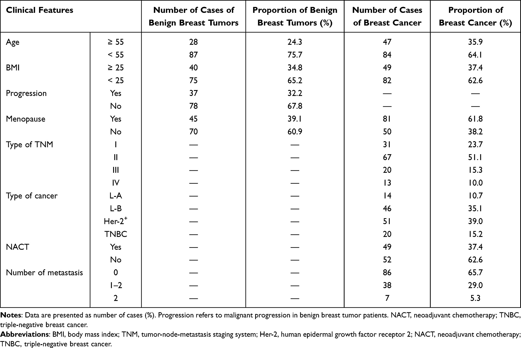 |
Table 1 Basic Clinical Characteristics of Patients with Benign Breast Tumors and Breast Cancer Patients |
Procedures
Blood samples were collected from 115 benign breast tumor patients and 131 breast cancer patients. Plasma vitamin C concentrations were measured, and clinical data, including laboratory indicators, age, BMI, TNM stage, neoadjuvant chemotherapy, number of metastatic sites, menopausal status, and adverse reactions, were collected. The correlation between plasma vitamin C levels and these factors was analyzed. Differences in plasma vitamin C levels among different patient groups, pathological types, and chemotherapy regimens were examined, along with correlations with clinical laboratory indicators and adverse reaction severity.
Concentration Measurement
Fasting blood samples were collected in the morning using EDTA-K2 anticoagulant tubes. To ensure vitamin C stability, all samples were processed within 30 minutes of collection and protected from light using amber tubes or aluminum foil wrapping during handling. Samples were centrifuged at 3000 rpm at 4°C for 10 minutes, and plasma was immediately separated and stored at −80°C until analysis. Plasma samples were thawed only once immediately prior to analysis to avoid degradation from repeated freeze-thaw cycles.
Plasma vitamin C concentrations were measured using a validated UPLC-MS/MS method with an ACQUITY UPLC HSS T3 column (100 mm × 2.1 mm, 1.8 μm). The mobile phase consisted of 0.1% formic acid in water (A) and acetonitrile (B), with gradient elution at a flow rate of 0.3 mL·min−1, column temperature of 35°C, and injection volume of 2 μL. The run time was 5 minutes, and L-Ascorbic Acid-1-13C was used as the internal standard. The mass spectrometer operated in multiple reaction monitoring (MRM) mode with positive electrospray ionization (ESI+).
The method was validated according to bioanalytical guidance. The assay exhibited linearity over 0.5–15 μg/mL (R2 > 0.99), with a lower limit of quantification (LLOQ) of 0.5 μg/mL. The intra-day and inter-day precision (coefficient of variation, CV) were were <5% and <8%, respectively. Accuracy (recovery) ranged from 95% to 105%. Stability tests confirmed that vitamin C was stable in plasma for 4 hours at room temperature (protected from light), 4 hours at 4°C, and 5 days at −80°C. Sample preparation was optimized using 10% metaphosphoric acid as the diluent and acetonitrile as the precipitant. The entire process was completed within the stable period of vitamin C to minimize errors and ensure accuracy. Detailed UPLC-MS/MS method validation data are provided in the Supplementary Materials (Supplementary Figures 2 and 3 and Supplementary Tables 1–3).
Each plasma sample was measured in triplicate (technical replicates), and the mean value was used for statistical analysis. A total of 246 biological replicates (115 benign, 131 breast cancer) were included, each with three technical replicates.
Statistical Analysis
Data were entered into an Excel database and analyzed using SPSS 27.0. Normality was tested using the Shapiro–Wilk test. For normally distributed data (P > 0.05), means (M) ± standard deviations (SD) were used, for non-normally distributed data, medians and interquartile ranges were used. For comparisons of plasma vitamin C levels between two groups, Mann–Whitney U-test was used; for comparisons among multiple groups, Kruskal–Wallis test was used. Correlations between plasma vitamin C and continuous laboratory parameters were assessed using Spearman’s rank correlation. For categorical clinical variables, differences in vitamin C levels were compared using the non-parametric tests described above.
A post-hoc power analysis was performed using G*Power software (version 3.1.9.7) to determine whether the sample size was sufficient to detect the observed differences in plasma vitamin C levels between benign and malignant groups. With an effect size of 1.2, α = 0.05, and a total sample size of 246, the achieved power exceeded 0.95, indicating adequate statistical power.
For subgroup analyses involving multiple groups (TNM stage, molecular subtype, metastatic sites), P-values are presented without adjustment for multiple comparisons due to the exploratory nature of these analyses (Table 2). Findings should be interpreted as hypothesis-generating rather than confirmatory.
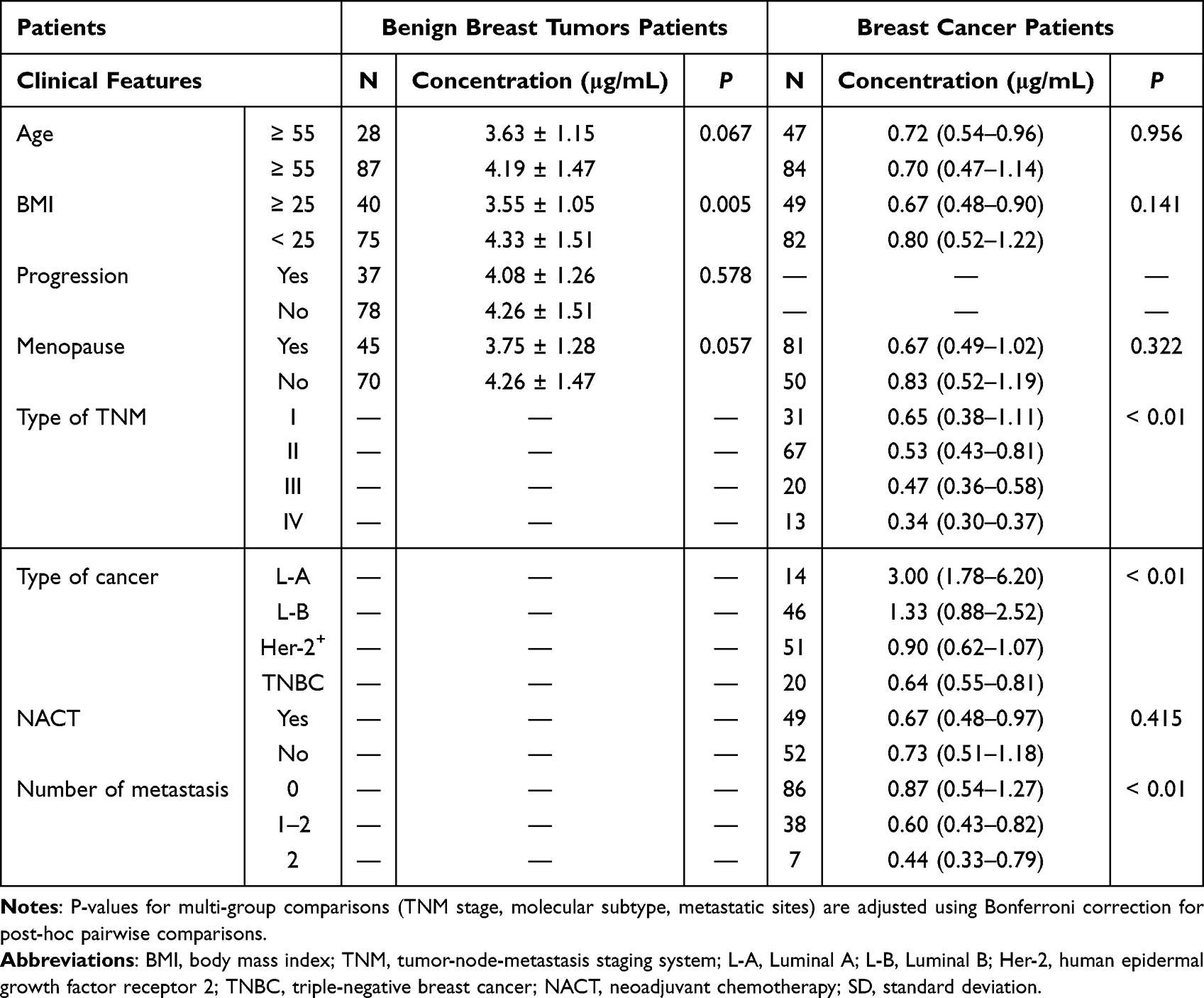 |
Table 2 Correlation Analysis Between Plasma Vitamin C Concentration and Clinical Characteristics in Patients with Benign Breast Tumors and Breast Cancer |
To account for multiple comparisons in the multivariate analyses (Table 3), Bonferroni correction was applied. With 14 laboratory parameters examined, the significance threshold was adjusted to P < 0.0036 (0.05/14). Results that remained significant after this correction are indicated in the corresponding tables.
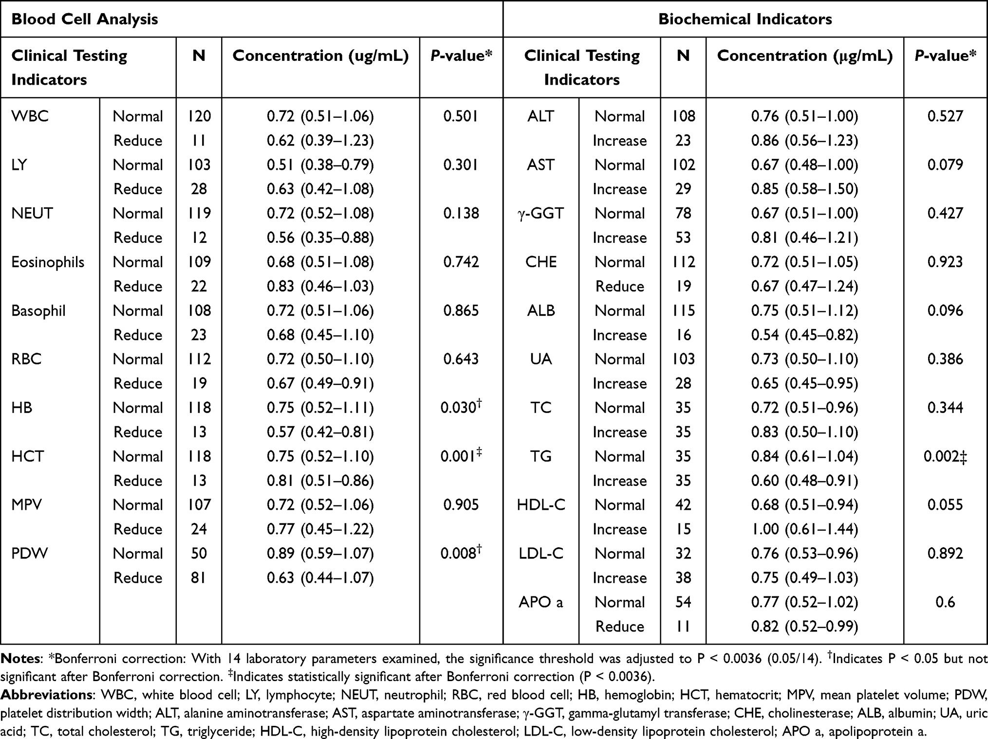 |
Table 3 Associations Between Plasma Vitamin C Concentration and Clinical Laboratory Parameters |
Sensitivity analyses were conducted to assess the robustness of our findings. First, to evaluate the potential confounding effect of BMI, patients were stratified by BMI category (<25 vs. ≥25 kg/m2), and subgroup analyses were repeated. Second, to assess the influence of extreme vitamin C values, analyses were repeated after excluding outliers (values outside mean ± 3 SD). The results remained consistent with the primary analyses, confirming the robustness of our conclusions.
P<0.05 was considered statistically significant, unless otherwise specified for Bonferroni-corrected thresholds.
Results
Plasma Vitamin C Concentrations in Benign Breast Tumors Patients and Breast Tumor Patients
Our study included a total of 115 patients with benign breast tumors and 131 patients with breast cancer. In the benign group, plasma vitamin C concentrations followed a normal distribution (P>0.05), ranging from 0.70 to 8.15 µg/mL, with a mean of 4.06 µg/mL, median of 3.89 µg/mL, and standard deviation of 1.414 µg/mL (Figure 1A and B). In contrast, plasma vitamin C levels in the breast cancer group were not normally distributed (P<0.05), with a range of 0.31–6.33 µg/mL, a mean of 0.81 µg/mL, a median of 0.72 µg/mL, and a standard deviation of 0.653 µg/mL (Figure 1C and D). Comparative analysis (Figure 1E) revealed a significant difference (P<0.05) between the two groups: the benign group had a higher mean vitamin C level (4.06 µg/mL) compared to the malignant group (0.72 µg/mL).
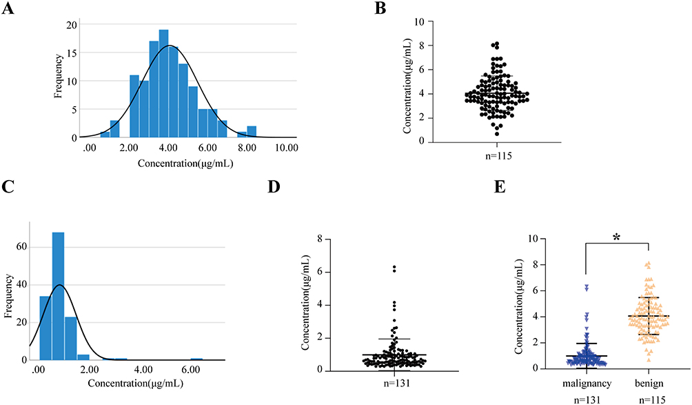 |
Figure 1 Plasma vitamin C concentration in different patient populations. (A) Normal distribution graph of the plasma vitamin C concentration in patients with benign breast tumors; (B) Scatter plot of the distribution of the plasma vitamin C concentration in patients with benign breast tumors; (C) Non-normal distribution graph of the plasma vitamin C concentration in patients with breast cancer; (D) Distribution graph of the plasma vitamin C concentration in patients with breast cancer; (E) Plasma vitamin C concentration in different patient populations (*P < 0.05). |
Correlation Between Plasma Vitamin C Concentrations and Clinical Characteristics with Benign Breast Tumors Patients
A total of 115 patients with benign breast tumors were enrolled in our study, including 28 patients aged ≥55 years (mean plasma vitamin C: 3.63 ± 1.15 µg/mL) and 87 patients aged <55 years (mean: 4.19 ± 1.47 µg/mL). Among them, 40 patients had Body Mass Index (BMI) ≥25 (3.55 ± 1.05 µg/mL) and 70 had BMI <25 (4.33 ± 1.51 µg/mL). The mean vitamin C concentrations were 4.08 ± 1.26 µg/mL in 37 patients with malignant progression and 4.26 ± 1.51 µg/mL in 78 patients without progression, while postmenopausal patients had lower levels (3.75 ± 1.28 µg/mL) than premenopausal patients (4.26 ± 1.47 µg/mL).
Statistical analysis revealed a significant negative correlation between plasma vitamin C and BMI in benign breast tumor patients (P<0.05), suggesting that higher body surface area was associated with lower vitamin C levels (Supplementary Figure 1). However, no significant differences were found for age, malignant progression, or menopausal status (P>0.05).
In 131 breast cancer patients, plasma vitamin C was significantly correlated with TNM stage, pathological type of breast cancer, and the number of metastatic sites (P<0.05), but not with age, BMI, menopausal status, or neoadjuvant chemotherapy (P>0.05).
Correlation Between Plasma Vitamin C Concentrations and Clinical Characteristics with Breast Tumors Patients
A total of 131 breast cancer patients were tested for plasma vitamin C concentrations. The breast cancer patients were stratified into ≥ 55 years group (n=47) and < 55 years group (n=84), and the median vitamin C concentrations were 0.72 μg/mL and 0.70 μg/mL, respectively. There were 49 patients with BMI ≥ 25 and 82 patients with BMI<25. The median vitamin C concentration was 0.67 μg/mL and 0.80 μg/mL, respectively. There were 31 patients with TNM stage I, 67 patients with TNM stage II, 13 patients with TNM stage III and 20 patients with TNM stage IV, and the median plasma vitamin C concentration was 0.65 μg/mL, 0.53 μg/mL, 0.47 μg/mL and 0.34 μg/mL. There were 14 cases of L-A breast cancer, 46 cases of L-B breast cancer, 51 cases of Her-2 positive breast cancer and 20 cases of triple negative breast cancer according to pathological classification. The median plasma vitamin C concentrations were 3.00 μg/mL, 1.33 μg/mL, 0.90 μg/mL and 0.64 μg/mL respectively. Forty-nine patients received neoadjuvant chemotherapy and 82 patients did not. The median plasma vitamin C concentration was 0.67 μg/mL and 0.73 μg/mL, respectively. There were 86 patients with no metastasis, 38 patients with 1–2 metastatic sites, and 7 patients with more than 2 metastatic sites. The median plasma vitamin C concentration was 0.87 μg/mL, 0.60 μg/mL and 0.44 μg/mL, respectively. The mean concentration of vitamin C was 0.67 μg/mL in 81 postmenopausal patients and 0.83 μg/mL in 50 premenopausal patients.
The results showed that the plasma vitamin C concentration of breast cancer patients was significantly correlated with tumor TNM stage, pathological type of breast cancer, and the number of metastatic sites (P < 0.05). However, there was no significant difference between plasma vitamin C concentration and clinical characteristics of breast cancer patients such as age, BMI, menopause and neoadjuvant chemotherapy (P > 0.05), as shown in Table 2.
Kruskal–Wallis tests revealed significant associations between plasma vitamin C concentrations and clinical parameters in breast cancer patients. Vitamin C levels differed significantly across TNM stages (Figure 2A). Stage IV patients showed markedly lower concentrations compared to stages I–II (P < 0.05), while stage III patients had reduced levels versus stage I (P < 0.05). Significant variations were observed among different pathological types (Figure 2B). Triple-negative and Her-2 positive patients exhibited significantly lower vitamin C levels compared to Luminal A/B subtypes (*P < 0.05 for both comparisons). Vitamin C concentrations progressively decreased with increasing metastatic sites (Figure 2C). Patients with 1–2 metastases had significantly lower levels than non-metastatic cases (P<0.05).
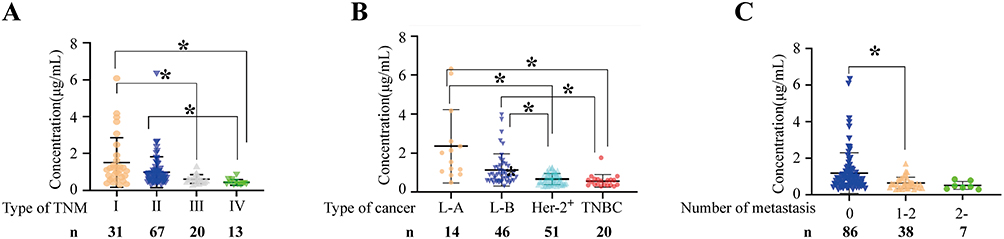 |
Figure 2 Differences in plasma vitamin C concentration among patients with (A) different TNM stages, (B) different pathological types and (C) different numbers of transfer sites of breast cancer. Bold numbers indicate sample size per group. (*P < 0.05). |
To assess the robustness of our findings, sensitivity analyses were performed. After stratifying patients by BMI (<25 vs. ≥25 kg/m2), the significant differences in plasma vitamin C between benign and malignant groups persisted in both strata (P<0.05), indicating that BMI did not confound the primary comparison. Similarly, after excluding outliers (n=4) with extreme vitamin C values (> mean ± 3 SD), all significant associations with TNM stage, pathological type, and metastatic sites remained unchanged (P<0.05), confirming that the results were not driven by extreme observations.
Associations of Plasma Vitamin C Concentrations with Clinical Laboratory Parameters
Our study comprehensively analyzed plasma vitamin C concentrations in 131 breast cancer patients along with detailed clinical laboratory parameters, including complete blood count and comprehensive biochemical profiles. Blood cell analysis included white blood cell count, absolute lymphocyte count, neutrophil count, eosinophils, basophils, red blood cell count, hemoglobin, hematocrit, platelet count, and platelet distribution width. Biochemical tests included alanine aminotransferase (ALT), aspartate aminotransferase (AST), glutamyltransferase γ-GGT, cholinesterase, serum albumin, uric acid, cholesterol, triglyceride, high density lipoprotein cholesterol, low density lipoprotein cholesterol, apolipoprotein a. The correlation analysis between the above indicators and the plasma vitamin C concentration of the patients showed that there were significant correlations between the four test indicators of hematocrit, platelet distribution width, triglyceride and hemoglobin and plasma vitamin C (P < 0.05). There was no significant correlation between the other test indexes and plasma vitamin C concentration (P > 0.05). The results are presented in Table 3 below.
Mann–Whitney U-tests revealed significant correlations between plasma vitamin C concentrations and hematological/biochemical parameters in breast cancer patients. After applying Bonferroni correction for multiple comparisons (adjusted significance threshold: P<0.0036), the correlations with hematocrit (negative) and triglycerides (negative) remained statistically significant (P = 0.001 and P = 0.002, respectively). The correlations with platelet distribution width (positive, P = 0.008) and hemoglobin (positive, P = 0.015) showed trends but did not reach the corrected significance level.
Patients with reduced hematocrit exhibited higher plasma vitamin C levels (0.81 vs. 0.75 µg/mL in normal hematocrit, P<0.05), indicating a negative correlation. Conversely, platelet distribution width (PDW) and hemoglobin showed positive correlations with vitamin C: patients with normal PDW had higher vitamin C levels (0.89 vs. 0.63 µg/mL in reduced PDW, P<0.05), as did those with normal hemoglobin (0.75 vs. 0.57 µg/mL in decreased hemoglobin, P<0.05). Additionally, elevated triglyceride levels were associated with lower vitamin C concentrations (0.61 vs. 0.84 µg/mL in normal triglycerides, P<0.05), suggesting a negative relationship.
Associations of Plasma Vitamin C Concentrations with Chemotherapy Regimens
According to the chemotherapy regimen, the patients were divided into anthracyclines + taxanes, anthracyclines, taxanes, dual targeted therapy and other groups, with 75, 14, 33 and 9 patients in each group. The mean plasma vitamin C concentrations of each group were 0.68, 1.19, 0.65, 0.54 μg/mL, respectively. The results showed that there was a significant difference in plasma vitamin C concentration among patients receiving different chemotherapy regimens (P < 0.05).
Kruskal–Wallis test was used to analyze the relationship between different chemotherapy regimens and plasma vitamin C concentration, and pairwise test was performed for each group of patients. The plasma vitamin C concentration of patients receiving different chemotherapy regimens was significantly different, and the analysis results are shown in the Figure 3. The plasma vitamin C level of patients treated with dual targeted drugs and other chemotherapy regimens was significantly lower than that of patients treated with anthracyclines (P < 0.05). However, given that HER2+ status itself is associated with lower vitamin C (Table 2), this finding may reflect tumor biology rather than a direct treatment effect.
 |
Figure 3 Differences in plasma vitamin C concentration among patients with different chemotherapy regimens. Bold numbers indicate sample size per group. (*P < 0.05). |
Associations of Plasma Vitamin C Concentrations with Adverse Reactions
In this study, the adverse reactions of patients after chemotherapy were collected by querting case information or clinical follow-up, and the adverse reactions were graded according to NCI-CTCAE 4.03. Patients were categorized based on the worst grade experienced: no adverse events (n=21), grade 1–2 (low-to-moderate) adverse events (n=79), and grade 3–4 (severe) adverse events (n=31). By system organ class, adverse events included: gastrointestinal disorders (nausea, n=60; vomiting, n=60; abdominal pain, n=5; dysphagia, n=2; oral mucositis, n=1; anorexia, n=9); hematologic disorders (neutropenia, n=39; anemia, n=39; thrombocytopenia, n=39; febrile neutropenia, n=16); hepatobiliary disorders (ALT/AST increased, n=35; hepatotoxicity, n=2); nervous system disorders (dizziness, n=13; syncope, n=2; leukoencephalopathy, n=1); and general disorders (fatigue, n=13; pyrexia, n=16). Severe adverse events (grade 3–4) included grade 3–4 myelosuppression, febrile neutropenia, severe hepatotoxicity, syncope, leukoencephalopathy, severe oral mucositis, and dysphagia requiring intervention. As shown in Figure 4, vitamin C concentrations were significantly different among groups (P < 0.05), with progressively lower levels associated with increasing severity of adverse events.
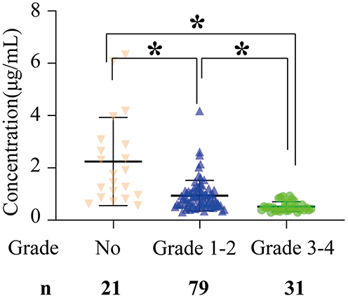 |
Figure 4 Differences in plasma vitamin C concentration in patients with different degrees of adverse reactions. Bold numbers indicate sample size per group. (*P < 0.05). |
Discussion
Vitamin C is an essential micronutrient with antioxidant and immune-regulatory functions, absorbed via SVCT1 and distributed to tissues via SVCT2.17–20 Reduced SVCT2 expression in breast cancer cells may contribute to localized vitamin C deficiency, particularly in aggressive subtypes.21 Chemotherapy-related adverse effects (eg, nausea, dysphagia) may further exacerbate depletion by reducing dietary intake.22
In this study, plasma vitamin C was significantly lower in breast cancer patients than in benign tumor cases, with the most pronounced depletion observed in advanced TNM stage, aggressive subtypes (HER2-positive and triple-negative breast cancer), and patients with multiple metastases (Figures 1 and 2). Vitamin C also correlated negatively with hematocrit and triglycerides, and positively with hemoglobin and platelet distribution width (Table 3). Patients receiving anthracycline-based or dual targeted therapy exhibited the lowest levels, and those with severe adverse events showed further reductions (Figures 3 and 4).
This study has several limitations. First, the single-center design limits generalizability. Second, dietary vitamin C intake was not quantitatively assessed. Third, multiple subgroup comparisons (Table 2) were performed without adjustment, increasing Type I error risk; these findings should be considered hypothesis-generating. Fourth, analysis of chemotherapy regimens is subject to confounding by indication, as regimen choice reflects tumor biology and stage rather than random assignment. Fifth, vitamin C is a non-specific marker that may reflect global nutritional and inflammatory status rather than cancer-specific mechanisms. Finally, the cross-sectional design precludes causal inferences.
Prospective studies with treatment-naive patients, standardized blood collection timing, and concurrent measurement of inflammatory and nutritional biomarkers are needed to clarify whether observed differences reflect tumor biology or treatment-related effects. Future research should also employ multivariable adjustment or propensity score matching to control for confounders, and include multicenter cohorts to validate generalizability.
Conclusion
In this cross-sectional study, plasma vitamin C levels were significantly lower in patients with breast cancer than in those with benign tumors, with the most pronounced deficiency observed in patients with advanced stage, aggressive subtypes (HER2-positive and triple-negative breast cancer), and multiple metastases. Importantly, based solely on these findings, plasma vitamin C cannot be regarded as a diagnostic, prognostic, or predictive biomarker. Future prospective studies involving treatment-naive patients, with standardized timing of blood collection and concurrent measurement of inflammatory and nutritional biomarkers, are warranted to determine whether the observed differences reflect tumor biology or are attributable to treatment-related systemic effects.
Data Sharing Statement
The authors are willing to unconditionally provide the raw data that support the conclusions of the article. The data that support the findings of this study are available from the corresponding authors upon reasonable request.
Ethical Approval and Consent to Participate
The study was approved by the Ethics Committee of Harbin Medical University (KY2025-02) on January 10, 2025. Written informed consent was obtained from all participants prior to participation.
Consent for Publication
All participants provided consent for data to be published.
Acknowledgments
The authors are grateful to the patients and their families for their participation in this study.
Author Contributions
All authors made a significant contribution to the work reported, whether that is in the conception, study design, execution, acquisition of data, analysis and interpretation, or in all these areas; took part in drafting, revising or critically reviewing the article; gave final approval of the version to be published; have agreed on the journal to which the article has been submitted; and agree to be accountable for all aspects of the work.
Funding
This work was supported by Wu Jieping Medical Foundation Clinical Research Special Fund (320.6750.2024-18-89 and 320.6750.2024-6-107), General Project of Postdoctoral Fund in Heilongjiang Province (LBH-Z23216), 2024 Heilongjiang Province “Outstanding Young Teachers Basic Research Support Plan” project and State Key Laboratory of Neurology and Oncology Drug Development Fund (SKLSIM-2024141 and 2024142).
Disclosure
The authors report no conflicts of interest in this work.
References
1. Bray F, Laversanne M, Sung H, et al. Global cancer statistics 2022:GLOBOCAN estimates of incidence and mortality worldwide for 36 cancers in 185 countries. CA Cancer J Clin. 2024;74(3):229–12. doi:10.3322/caac.21834
2. Zou T, Zou F, Sun Y, et al. Expression of nicotinamide phosphoribosyltransferase and silent information regulator 2 homologous protein 1 in breast cancer and its relationship with clinicopathological characteristics of patients. Shaanxi Med J. 2023;52(8):1093–1096, 1101.
3. Liu X-N, Hao N, Ma -X-X, et al. Clinical study of Sanhuang decoction combined with pirarubicin, docetaxel and cyclophosphamide neoadjuvant chemotherapy in the treatment of triple-negative breast cancer. Shaanxi Trad Chin Med. 2021;42(10):1367–1370.
4. Shao Z, Yang B, Wu J. Results and latest progress of important clinical trials on breast cancer in China in 2022. Chin J Cancer. 2023;33(2):103–109.
5. Fisusi, Akala EO, Funmilola A, et al. Drug combinations in breast cancer therapy. Pharm Nanotechnol. 2019;7(1):3–23. doi:10.2174/2211738507666190122111224
6. Mohajer MH, Brummer RJM, Rastll A, et al. The role of the microbiome for human healthfrom basic science to clincal appicationsLJ. Eur J Nur. 2018;57(Supl 1):1–14.
7. Hefenan A, Evans C, Holmes M, et al. The reguation of ietay lron bcavala ity by viamin C a systematic review and meta-analysis. Proc Nutr Soc. 2017;76:E182.
8. Sant DW, Mustafi S, Gustafson CB, Chen J, Slingerland JM, Wang G. Vitamin C promotes apoptosis in breast cancer cells by increasing TRAIL expression. Sci Rep. 2018;8(1):5306. doi:10.1038/s41598-018-23714-7
9. Spielholz C, Golde DW, Houghton AN, Nualart F, Vera JC. Increased facilitated transport of dehydroascorbic acid without changes in sodium-dependent ascorbate transport in human melanoma cells. Cancer Res. 1997;57(12):2529–2537.
10. Yuan S, Zheng JS, Mason AM, Burgess S, Larsson SC. Genetically predicted circulating vitamin c in relation to cardiovascular disease. Eur J Prev Cardiol. 2022;28(16):1829–1837. doi:10.1093/eurjpc/zwab081
11. Zheng JS, Luan J, Sofianopoulou E, et al. Plasma vitamin c and type 2 diabetes: genome-wide association study and mendelian randomization analysis in European populations. Diabetes Care. 2021;44(1):98–106. doi:10.2337/dc20-1328
12. Pal SK, Childs BH, Pegram M. Triple negative breast cancer: unmet medical needs. Breast Cancer Res Treat. 2011;125(3):627–636. doi:10.1007/s10549-010-1293-1
13. Tarighati E, Keivan H, Mahani H. A review of prognostic and predictive biomarkers in breast cancer. Clin Exp Med. 2023;23(1):1–16. doi:10.1007/s10238-021-00781-1
14. Levine M, Padayatty SJ, Espey MG. Vitamin C: a concentration-function approach yields pharmacology and therapeutic discoveries. Adv Nutr. 2011;2(2):78–88. PMID: 22332036; PMCID: PMC3065766. doi:10.3945/an.110.000109
15. Muñoz-Montesino C, Peña E, Roa FJ, Sotomayor K, Escobar E, Rivas CI. Transport of Vitamin C in Cancer. Antioxid Redox Signal. 2021;35(1):61–74. PMID: 33607936. doi:10.1089/ars.2020.8166
16. Nechuta S, Lu W, Chen Z, et al. Vitamin supplement use during breast cancer treatment and survival: a prospective cohort study. Cancer Epidemiol Biomarkers Prev. 2011;20(2):262–271. PMID: 21177425; PMCID: PMC3057050. doi:10.1158/1055-9965.EPI-10-1072
17. Cai J. Comparative analysis of the value of ultrasound elastography and conventional ultrasound in the diagnosis of breast cancer. Imaging Res Med Appl. 2020;6(15):89–91.
18. Lykkesfeldt J, Tveden-Nyborg P. The pharmacokinetics of vitamin C. Nutrients. 2019;11(10):2412. doi:10.3390/nu11102412
19. Mikkelsen SU, Gillberg L, Lykkesfeldt J, et al. The role of vitamin c inepigenetic cancer therapy. Free Radic Biol Med. 2021;170:179–193. doi:10.1016/j.freeradbiomed.2021.03.017
20. Mikkelsen SU, Gillberg L, Lykkesfeldt J, et al. New Insights on Vitamin C and Cancer. Springer Briefs in Cancer Research; 2014.
21. Mustafi S, Camarena V, Qureshi R, et al. Vitamin C supplementation expands the therapeutic window of BETi for triple negative breast cancer. EBioMedicine. 2019;43:201–210. doi:10.1016/j.ebiom.2019.04.006
22. Lykkesfeldt J, Carr AC. Vitamin C. Adv Nutr. 2024;15(1):100155. doi:10.1016/j.advnut.2023.100155
 © 2026 The Author(s). This work is published and licensed by Dove Medical Press Limited. The
full terms of this license are available at https://www.dovepress.com/terms
and incorporate the Creative Commons Attribution
- Non Commercial (unported, 4.0) License.
By accessing the work you hereby accept the Terms. Non-commercial uses of the work are permitted
without any further permission from Dove Medical Press Limited, provided the work is properly
attributed. For permission for commercial use of this work, please see paragraphs 4.2 and 5 of our Terms.
© 2026 The Author(s). This work is published and licensed by Dove Medical Press Limited. The
full terms of this license are available at https://www.dovepress.com/terms
and incorporate the Creative Commons Attribution
- Non Commercial (unported, 4.0) License.
By accessing the work you hereby accept the Terms. Non-commercial uses of the work are permitted
without any further permission from Dove Medical Press Limited, provided the work is properly
attributed. For permission for commercial use of this work, please see paragraphs 4.2 and 5 of our Terms.