
Back to Journals » Drug Design, Development and Therapy » Volume 20
Pharmacological Treatment for Diabetic Macular Edema: A 2025 Update on Durability and Multi-Target Therapies
Authors Tao Y  , Zhang Y , Xu W, Liu G, Liu H
Received 23 November 2025
Accepted for publication 25 January 2026
Published 3 February 2026 Volume 2026:20 583837
DOI https://doi.org/10.2147/DDDT.S583837
Checked for plagiarism Yes
Review by Single anonymous peer review
Peer reviewer comments 2
Editor who approved publication: Professor Anastasios Lymperopoulos
Yunhan Tao,1,2 Yiteng Zhang,1,2 Wei Xu,1,2 Guina Liu,1,2 Hongtao Liu1,2
1Department of Ophthalmology, West China Second University Hospital, Sichuan University, Chengdu, Sichuan, People’s Republic of China; 2Key Laboratory of Birth Defects and Related Diseases of Women and Children (Sichuan University), Ministry of Education, Chengdu, Sichuan, People’s Republic of China
Correspondence: Hongtao Liu, Department of Ophthalmology, West China Second University Hospital, Sichuan University, No. 20, Section 3, Renmin South Road, Wuhou District, Chengdu, Sichuan, People’s Republic of China, Email [email protected]
Abstract: Diabetic macular edema (DME) remains a leading cause of vision loss in the working-age population, yet its management is undergoing a fundamental paradigm shift from high-frequency anti-VEGF monotherapy toward more durable and multi-mechanism therapeutic strategies. This narrative review provides an up-to-date overview of the pharmacological landscape of DME as of 2025, synthesizing evidence from pivotal clinical trials, recent regulatory approvals, and available real-world outcomes. We examine the transition toward injection-sparing approaches and discuss the clinical rationale for targeting alternative biological pathways beyond VEGF inhibition, including angiopoietin-2 (eg, faricimab), the plasma kallikrein–kinin system, and tyrosine kinase–mediated signaling. Collectively, emerging evidence suggests that the future management of DME will be defined by a paradigm shift toward injection-sparing, multi-pathway therapeutic strategies, extending beyond short-term fluid suppression toward a more personalized, precision-medicine framework that prioritizes long-term durability, safety, and functional outcomes.
Keywords: diabetic macular edema, pharmacological treatment, anti-VEGF, corticosteroids, long-acting therapy, multi-target, gene therapy
Introduction
Diabetic macular edema (DME) is one of the most common microvascular complications of diabetes mellitus (DM) and a leading cause of vision loss among individuals with diabetes, posing a particular burden on the working-age population.1 It is estimated that approximately 19 million people worldwide were affected by DME in 2020, and the number is expected to rise in parallel with the global increase in DM prevalence, underscoring DME as a growing public health challenge.2,3 This review aims to provide a comprehensive overview of the pathophysiology of DME, current therapeutic options, and advances in emerging pharmacologic treatments.
Methods
This narrative review was conducted in accordance with the Scale for the Assessment of Narrative Review Articles (SANRA)4 to ensure methodological rigor, balance, and transparency.
Literature Search and Data Sources
A structured literature search was performed using PubMed, Embase, and the Cochrane Library, covering publications from January 1, 2010, to October 31, 2025. Search terms included Medical Subject Headings and free-text keywords related to diabetic macular edema, anti-VEGF therapy, corticosteroids, angiopoietin-2, tyrosine kinase inhibitors, and gene therapy. ClinicalTrials.gov and the World Health Organization International Clinical Trials Registry Platform were additionally searched to identify ongoing or unpublished studies, particularly for emerging therapeutic agents.
Study Selection and Narrative Synthesis
Priority was given to Phase III randomized controlled trials for approved therapies and Phase I/II studies for investigational agents. Real-world and retrospective studies were included where available to contextualize long-term safety, durability, and treatment burden. Studies were selected based on predefined criteria, including stage of clinical development, relevance to therapeutic pathways, and availability of efficacy and safety data, while preclinical studies without clear translational relevance were not prioritized.
Given substantial heterogeneity in mechanisms of action, trial design, outcome measures, and development stages, quantitative meta-analysis was not performed. Instead, a critical narrative synthesis was applied, incorporating thematic grouping by mechanism of action, contextual comparison with established clinical benchmarks, and focused assessment of safety signals and reasons for trial discontinuation.
Pathophysiology of DME
The pathogenesis of DME involves a complex network of metabolic, vascular, and inflammatory disturbances driven by chronic hyperglycemia. A key event is dysfunction of the retinal microcirculation leading to breakdown of the blood–retinal barrier (BRB). Compromise of the BRB permits leakage of plasma fluid and macromolecules into the neurosensory retina, resulting in macular thickening and cystoid edema. Early hyperglycemia-induced alterations include transient vasodilation and hemodynamic changes,5 followed by structural damage6 such as pericyte loss,7,8 endothelial apoptosis, and basement membrane thickening,9,10 all of which collectively weaken capillary integrity.
Hyperglycemia activates several pathological pathways11— including the polyol pathway,12 accumulation of advanced glycation end products (AGEs),13,14 protein kinase C (PKC) activation, and increased hexosamine flux15–17— which amplify oxidative stress, promote inflammation, and impair cellular function. Additional dysregulation of the renin–angiotensin–aldosterone system (RAAS),18 the kallikrein–kinin system (KKS),19 and aquaporins (AQPs)20 further contributes to BRB disruption and fluid imbalance.
Vascular endothelial growth factor (VEGF), particularly VEGF-A, is a central mediator of DME.21 Capillary nonperfusion and tissue hypoxia activate hypoxia-inducible factor-1 (HIF-1), which markedly upregulates VEGF expression.22,23 VEGF increases vascular permeability by inducing phosphorylation and degradation of tight-junction proteins via VEGF receptor (VEGFR)-2, promotes endothelial proliferation, and possesses pro-inflammatory activity—all of which exacerbate BRB compromise.24–26 The pivotal role of VEGF forms the basis for anti-VEGF therapy as the first-line treatment for DME in most guidelines/clinical practice.27
Inflammation also plays a crucial role.28 Elevated intraocular cytokines such as TNF-α, IL-1β, IL-6, IL-8, IL-17A, and MCP-129,30 enhance vascular permeability and promote leukostasis, further damaging the BRB.31 Activation of microglia and Müller cells in a hyperglycemic and inflammatory milieu sustains a vicious cycle of cytokine release, oxidative stress, and VEGF overproduction.32,33
Finally, dysfunction of the retinal neurovascular unit—comprising neurons, glia, endothelial cells, and pericytes—contributes to disease progression.34 Diabetic retinal neurodegeneration, characterized by neuronal apoptosis and reactive gliosis, often precedes microvascular damage,35 highlighting the need to consider neuroprotection in DME management.
Current Pharmacologic Therapies for DME
Anti-VEGF Agents
Anti-VEGF agents inhibit the binding of VEGF to its receptors, thereby reducing VEGF-mediated vascular permeability and neovascularization. They remain the first-line therapy for DME.36 Currently approved or commonly used anti-VEGF agents include ranibizumab, aflibercept, conbercept, brolucizumab, and bevacizumab (Table 1).
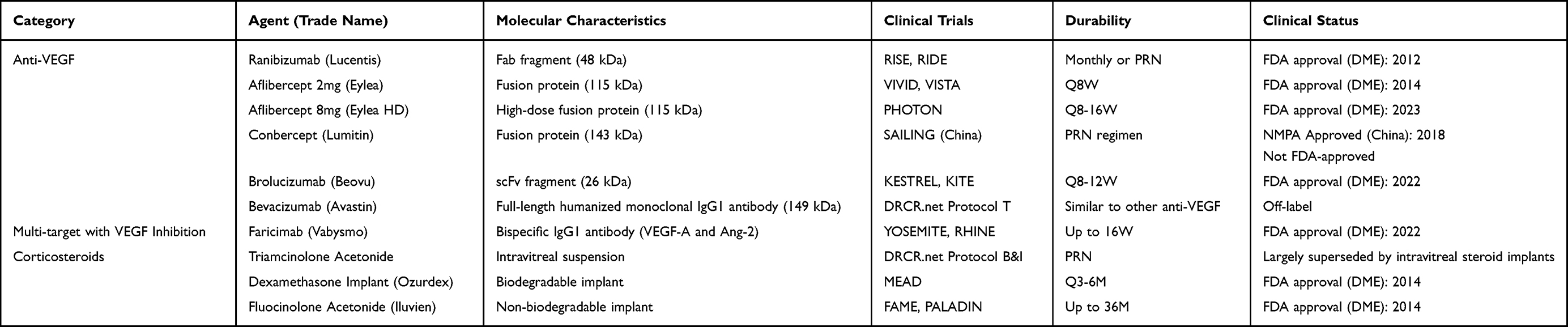 |
Table 1 Overview of Current Pharmacological Therapies for Diabetic Macular Edema |
Ranibizumab
Ranibizumab (Lucentis) is a humanized recombinant Fab fragment (48 kDa) that specifically binds VEGF-A and blocks its interaction with VEGFR-1/2, thus suppressing endothelial proliferation, vascular leakage, and neovascularization.37 It was the first FDA-approved anti-VEGF agent for DME.38 Several biosimilars, including Byooviz (ranibizumab-nuna) and Cimerli (ranibizumab-eqrn), have also received FDA approval for retinal vascular diseases.39
Aflibercept
Aflibercept (Eylea) is a 115-kDa recombinant fusion protein comprising the extracellular domains of VEGFR-1 and VEGFR-2 fused to the Fc portion of human IgG1. It binds VEGF-A, VEGF-B, and placental growth factor (PLGF).40 The PHOTON trial demonstrated that high-dose Eylea HD (8 mg) effectively extended treatment intervals while maintaining outcomes compared with the 2-mg formulation, reducing treatment burden.41 Multiple aflibercept biosimilars, including Yesafili, Opuviz, Ahzantive, Enzeevu, and Pavblu, have also gained FDA approval.
Conbercept
Conbercept (Lumitin) is a 143-kDa recombinant fusion protein developed in China. It contains the second extracellular domain of VEGFR-1 and the third and fourth domains of VEGFR-2 fused to human IgG1 Fc, enabling high-affinity binding to VEGF-A/B/C and PLGF.42 It received the China National Medical Products Administration approval for DME in 2018. However, global Phase III trials (PANDA-1/2) were terminated early in 2021 due to the inability to meet the primary endpoint, limiting regulatory approval to specific regions outside the United States and Europe.
Brolucizumab
Brolucizumab (Beovu) is a 26-kDa single-chain variable fragment (scFv) with high molar concentration per injection due to its small size.43 It binds VEGF-A and inhibits its biological activity. The KESTREL and KITE trials showed non-inferior visual and anatomical outcomes to aflibercept in DME, with the possibility of extending dosing intervals to 3–4 months.44,45 Beovu received FDA approval for DME in 2022.
Bevacizumab
Bevacizumab (Avastin) is a 149-kDa full-length monoclonal antibody initially approved for metastatic solid tumors.46 Although not formally approved for ophthalmic use, its low cost has led to widespread off-label use in retinal diseases.47 In 2024, the European Commission approved Lytenava, the first ophthalmic bevacizumab formulation, for age-related macular degeneration (AMD).48
Limitations of Anti-VEGF Agents
Despite their effectiveness, anti-VEGF agents have notable limitations:
- High treatment burden: Monthly injections during initiation and long-term frequent dosing result in substantial time, financial, and logistical burdens, contributing to reduced adherence and poorer real-world outcomes;49
- Suboptimal responders: A significant proportion of patients exhibit persistent or refractory DME despite adequate anti-VEGF therapy,50–52 highlighting that VEGF is not the only pathway driving DME and underscoring the need for additional targets;
- Potential complications: Ocular adverse events are primarily related to the intravitreal injection procedure, while severe complications such as endophthalmitis or retinal detachment are rare. Because these ocular events are attributable to the route of administration rather than to specific pharmacological targets, similar procedural safety considerations apply across intravitreal therapies and are addressed where applicable in this review. Systemic adverse events are uncommon; however, caution is warranted in patients with cardiovascular risk factors due to the theoretical risk of arterial thromboembolic events associated with anti-VEGF therapy.42,53
Multi-Target Agents Combined with VEGF Inhibition
Faricimab (Vabysmo) is a bispecific IgG1 antibody that inhibits both VEGF-A and angiopoietin-2 (Ang-2),54 a stress-induced cytokine elevated in DME that destabilizes retinal vasculature by antagonizing the Ang-1/Tie2 pathway, promoting endothelial dysfunction, and enhancing VEGF-mediated leakage.55 Dual inhibition provides broader vascular stabilization than targeting VEGF alone.
In the YOSEMITE and RHINE trials,56,57 faricimab achieved visual and anatomical outcomes comparable to aflibercept while enabling extended dosing intervals of up to 12–16 weeks for many patients, substantially reducing treatment burden with a similar safety profile. Approved by the FDA in 2022, faricimab introduces a novel dual-pathway strategy for DME, with the potential to improve adherence and long-term disease control. Further real-world data and phenotype-specific analyses remain needed.
Corticosteroids
Corticosteroids exert potent anti-inflammatory, anti-permeability, and anti-proliferative effects, making them an important therapeutic option for DME,58 particularly in cases with suboptimal or transient response to anti-VEGF therapy.59 Currently available corticosteroids for DME include triamcinolone acetonide (TA), dexamethasone, and fluocinolone acetonide (FA), administered via intravitreal injection or sustained-release implants.
Triamcinolone Acetonide
TA is a potent corticosteroid with strong anti-inflammatory and anti-edematous activity and was one of the earliest intravitreal agents used for DME. DRCR.net Protocol B60 demonstrated that intravitreal TA provided more rapid visual improvement than focal/grid laser photocoagulation, although with higher rates of cataract formation and intraocular pressure (IOP) elevation. Due to its suspension nature, limited duration, and relatively high complication rate, TA has largely been replaced by anti-VEGF therapy and newer steroid implants. However, TA remains valuable in resource-limited settings or in patients who require rapid reduction of inflammation or cannot afford long-term anti-VEGF therapy.
Dexamethasone Implant
The dexamethasone intravitreal implant (Ozurdex) encloses 0.7 mg of dexamethasone within the NOVADUR biodegradable polymer system, allowing sustained release for up to 6 months and reducing the need for frequent injections. The MEAD trials61,62 demonstrated significant improvements in retinal thickness and visual acuity. However, adverse events—particularly cataract progression (65% vs 20% in phakic eyes) and IOP elevation (40% vs 10%)—were markedly more common.63 Given its long-acting profile, Ozurdex is particularly suitable for pseudophakic eyes, vitrectomized eyes, patients intolerant to anti-VEGF therapy, or those unwilling to undergo monthly injections.
Fluocinolone Acetonide Implant
The FA implant (Iluvien) is a non-biodegradable device containing 0.19 mg FA that delivers 0.25 μg/day over 36 months. The FAME and PALADIN trials showed sustained anatomical and visual gains with significant reduction in treatment burden—76% of eyes required no additional therapy during the study.64,65 However, IOP elevation and cataract formation remain major considerations. The FDA approved FA implants in 2014 for DME patients previously exposed to corticosteroids without significant IOP response.
Limitations of Corticosteroids
The use of corticosteroids in DME requires balancing efficacy and safety. While they offer important benefits for patients with inadequate anti-VEGF response and significantly reduce treatment frequency through sustained-release formulations, their risk of cataract and IOP elevation restricts use mainly to second-line therapy or carefully selected patient populations.
Emerging Therapies for DME
Despite major advances, current DME treatments remain limited by high injection burden, suboptimal response in a substantial proportion of patients, and long-term risks. To address these challenges, multiple new therapeutic strategies are under active investigation (Table 2).
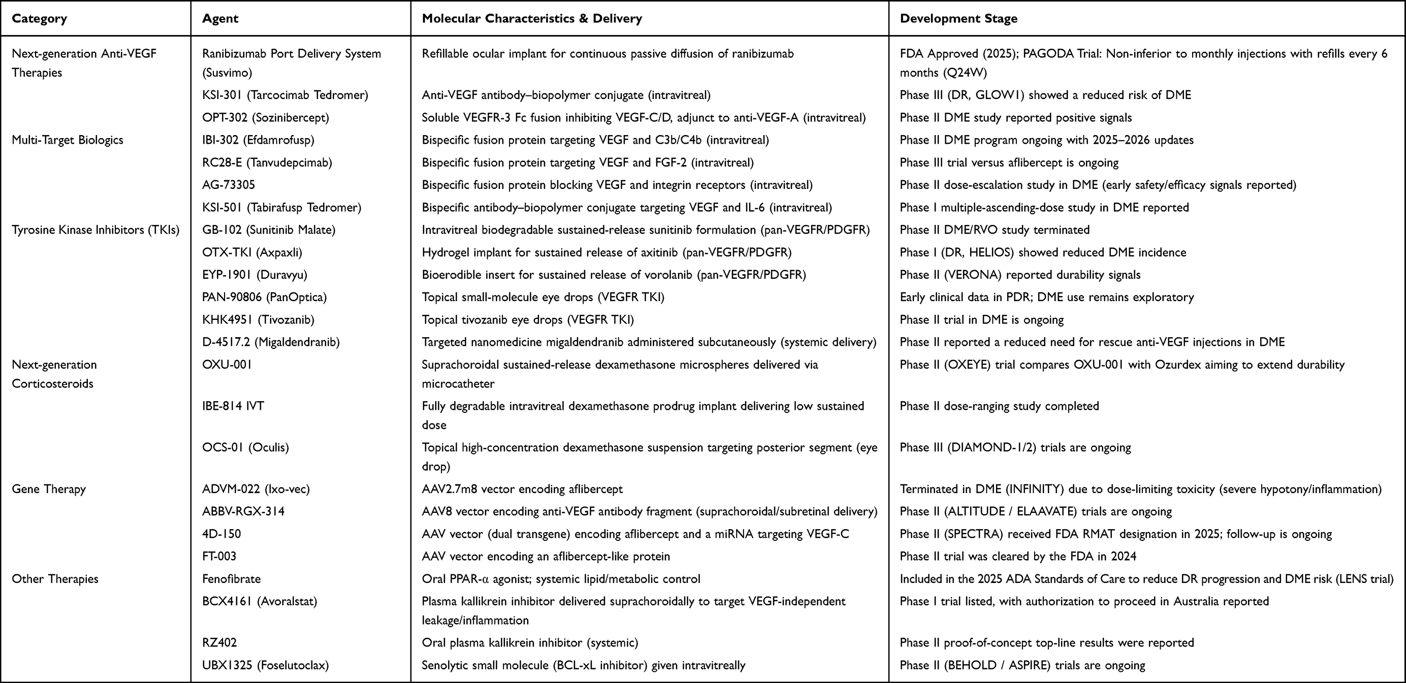 |
Table 2 Emerging and Investigational Therapies for Diabetic Macular Edema |
Next-Generation Anti-VEGF Therapies
Ranibizumab Port Delivery System (Susvimo)
Susvimo is a small, refillable intraocular implant comprising a 0.02 mL drug reservoir and a titanium release-control element that regulates passive diffusion of ranibizumab (100 mg/mL) into the vitreous cavity. This system maintains therapeutic drug concentrations while substantially reducing the frequency of conventional intravitreal injections.66,67 The PAGODA study68 compared Susvimo refilled every six months with monthly intravitreal ranibizumab injections, demonstrating comparable one-year visual gains. While offering durability, the implant requires vigilance for device-related adverse events such as endophthalmitis, which have been observed in the broader PDS program. In 2025, the FDA approved Susvimo for DME patients previously responsive to at least two anti-VEGF treatments, offering an innovative option to reduce treatment burden while maintaining visual benefits.
KSI-301 (Tarcocimab Tedromer)
KSI-301 is a 950 kDa antibody-biopolymer conjugate combining a humanized IgG1 anti-VEGF antibody with a phosphorylcholine polymer. The antibody binds VEGF-A with high affinity, blocking VEGFR-1/2 interactions, while the large hydrophilic polymer prolongs intravitreal half-life, enabling 4–6 month dosing intervals for sustained inhibition.69 The GLOW1 study70 demonstrated that KSI-301 significantly slows diabetic retinopathy (DR) progression and reduces the risk of vision-threatening complications, including DME, although its specific efficacy in DME requires further evaluation.
OPT-302 (Sozinibercept)
OPT-302 is a 150 kDa fusion protein composed of human IgG1-Fc and extracellular domains 1–3 of VEGFR-3. It binds VEGF-C/D with high affinity, preventing VEGFR-2/3 activation, and is designed to complement anti-VEGF-A therapy for comprehensive VEGF pathway inhibition.71 Phase II trials in DME patients showed that OPT-302 combined with aflibercept provided significant improvements in BCVA and retinal thickness compared with aflibercept monotherapy, suggesting that VEGF-C/D blockade may confer additional anatomical and functional benefits.72
Multi-Target Biologics Combined with Anti-VEGF Activity
IBI-302 (Efdamrofusp Alfa)
IBI-302 is a dual-specific fusion protein targeting VEGF and complement components C3b/C4b. Its N-terminal includes VEGFR-1/2 extracellular domains, and its C-terminal comprises human complement receptor-1 (CR1) fused to an IgG1-Fc. CR1 selectively binds C3b/C4b, inhibiting both classical and alternative complement pathways, reducing complement-mediated inflammation, while neutralizing all VEGF isoforms to decrease vascular permeability and neurovascular damage.73 Phase I studies demonstrated favorable safety, significant improvement in visual acuity and macular edema, and the potential to extend dosing intervals to 12–16 weeks. A Phase II trial is ongoing to directly compare IBI-302 (4 mg and 8 mg) with faricimab (6 mg) in DME treatment.74
RC28-E (Tanvudepcimab)
RC28-E is a fusion protein targeting VEGF and fibroblast growth factor (FGF), comprising VEGFR-1 domain 2, VEGFR-2 domain 3, FGFR1 domain 3, and IgG1-Fc.75 FGF2 binding to FGFR extracellular domains promotes angiogenesis and alters vascular permeability.76 Phase II trials indicated that RC28-E provides superior outcomes in DME patients, particularly those with suboptimal glycemic control, with fewer injections during maintenance and comparable safety to conbercept.77 A Phase III trial comparing RC28-E (2 mg) with aflibercept (2 mg) is underway.
AG-73305
AG-73305 is a fusion protein targeting VEGF and integrins.78 Integrins are transmembrane receptors mediating cell-cell and cell-matrix adhesion, influencing vascular leakage, inflammation, neovascularization, and fibrosis.79 Phase II intravitreal studies (0.5–4 mg) demonstrated good safety and tolerability, with over 50% of DME patients showing significant BCVA and CST improvements after a single injection, maintained for 12–24 weeks.80
KSI-501 (Tabirafusp Tedromer)
KSI-501 targets VEGF and IL-6, conjugated to a phosphorylcholine polymer, binding one VEGF and two IL-6 molecules simultaneously.81 IL-6, produced by monocytes, macrophages, and microglia, disrupts the BRB, induces VEGF, and increases vascular permeability, contributing to macular edema.29 Phase I studies indicated potential for visual and structural improvement with sustained effect.82
Tyrosine Kinase Inhibitors (TKIs)
GB-102 (Sustained-Release Sunitinib Malate)
GB-102 is a biodegradable intravitreal sustained-release formulation of sunitinib. By inhibiting multiple receptor tyrosine kinases (RTKs), including VEGFR and platelet-derived growth factor receptor (PDGFR), it suppresses both angiogenesis and inflammatory responses. This multi-targeted mechanism positions GB-102 as a potential therapy for ocular neovascular diseases such as DME.83 The sustained-release design aims to extend injection intervals up to six months. However, clinical trials in AMD patients showed inferior visual outcomes compared with aflibercept, and subsequent studies in DME have been discontinued.
OTX-TKI (Axpaxli)
OTX-TKI is an intravitreal implant of axitinib developed using the biodegradable Elutyx hydrogel platform.84 Axitinib inhibits VEGFR-1/2/3 and PDGFR, among other RTKs, and the hydrogel enables stable intraocular release for 6–12 months, markedly reducing injection frequency.85 The HELIOS study in DR patients demonstrated that OTX-TKI delays DR progression and lowers DME incidence, though dedicated trials targeting DME are yet to be conducted.
EYP-1901 (Duravyu)
EYP-1901 is a sustained-release TKI implant based on the Durasert E biodegradable platform, with vorolanib as the active ingredient. Vorolanib simultaneously inhibits VEGFR-1/2/3 and PDGFR-β, providing broad-spectrum anti-VEGF/PDGF effects for 6–9 months following a single injection.86 The mid-term report of the VERONA study indicated that a higher proportion of EYP-1901-treated eyes did not require supplemental injections at 16 weeks compared with aflibercept (82% vs 50%), with stable BCVA and CST improvements. These results support progression to Phase III trials to further evaluate the feasibility of a “half-yearly” dosing regimen in terms of efficacy and safety.
PAN-90806 (PanOptica)
PAN-90806 is a small-molecule TKI eye drop administered once daily. It diffuses through the sclera and choroid to reach the retina and inhibit RTKs.87 Phase I/II studies in AMD and proliferative DR patients demonstrated biological activity, and reversible punctate keratopathy reported as an adverse event has been mitigated by reformulation as a particulate suspension. Whether PAN-90806 can ultimately provide an injection-free option for DME patients awaits results from ongoing Phase II/III trials.
KHK4951 (Tivozanib)
KHK4951 is a nanocrystalline TKI eye drop containing tivozanib, which inhibits VEGFR-1/2/3 and PDGFR. Tivozanib is a clinically approved, safe agent, with an oral formulation already FDA-approved for renal cell carcinoma.88 Phase I studies have demonstrated its efficacy and favorable safety profile in AMD patients.89 Phase II trials in DME are currently ongoing, with outcomes yet to be reported.
D-4517.2 (Migaldendranib)
D-4517.2 is a hydroxyl dendrimer therapeutic formulation delivering the small-molecule TKI migaldendranib. It can cross the blood-retinal barrier via subcutaneous or oral administration and is selectively taken up by macrophages, microglia, and hypertrophic RPE cells, maintaining therapeutic levels in target tissues for at least one month after a single dose.90 Interim Phase II data indicate significant improvements in visual acuity and retinal edema in DME patients, with overall good safety and tolerability.
Next-Generation Corticosteroids
OXU-001
OXU-001 is a long-acting dexamethasone microsphere (Dexaspheres) formulation delivered via the Oxulumis luminescent microcatheter into the suprachoroidal space (SCS). This approach maintains sustained drug concentrations in the retina/choroid while markedly minimizing exposure to anterior segment structures such as the lens and trabecular meshwork. The OXEYE study is comparing the safety, tolerability, efficacy, and durability of OXU-001 with intravitreal dexamethasone implants (Ozurdex). Designed as a “once-yearly” therapy, it aims to reduce frequent intravitreal injections and short-acting steroid implant limitations, offering a next-generation long-acting corticosteroid strategy for DME.
IBE-814 IVT
IBE-814 IVT is a long-acting dexamethasone prodrug implant based on the Epidel platform, composed entirely of dexamethasone dimer prodrug. Following a single intravitreal injection, it undergoes surface erosion at a zero-order rate, releasing active dexamethasone continuously for 6–9 months. The RIPPLE-1 study demonstrated significant BCVA and CST improvements at 6 and 9 months, with an 82% reduction in treatment burden, and a safety profile comparable to other corticosteroids.91,92 Phase III trials are ongoing to further establish its clinical potential in DME.
OCS-01 (Oculis)
OCS-01 is a high-concentration dexamethasone ocular suspension formulated via the OPTIREACH nanonization technology, using cyclodextrins as solubilizing excipients. Administered topically to the conjunctival sac, it penetrates the blood-retinal barrier to reach the posterior retina.93 Preliminary Phase I results from the DIAMOND-1 trial demonstrated statistically significant BCVA and CST improvements. Phase III DIAMOND-1/2 trials are ongoing, with topline data expected in 2026.
Gene Therapy
ADVM-022 (Ixo-Vec)
ADVM-022 uses an intravitreal AAV2.7m8 vector encoding aflibercept under proprietary promoter/enhancer elements, enabling long-term retinal ganglion and RPE cell secretion of aflibercept. This continuous VEGF and PLGF blockade reduces capillary permeability and macular edema.94 The INFINITY trial evaluated its efficacy in newly diagnosed DME patients.95 Early data suggested reduced treatment frequency; however, dose-limiting toxicities, including severe intraocular inflammation and hypotony at higher doses, led to discontinuation of further DME development. ADVM-022 illustrates a “single injection, long-acting aflibercept” paradigm, but the INFINITY findings highlight the critical importance of safety in high-inflammation-prone populations. Research has pivoted to low-dose AMD strategies, which, if successful, may reopen the potential for DME applications.
ABBV-RGX-314
ABBV-RGX-314 employs a NAV-AAV8 vector carrying a gene encoding a ranibizumab-like anti-VEGF Fab fragment under a retina-specific promoter. Following subretinal or SCS injection, transduced retinal cells continuously secrete the antibody, providing durable VEGF neutralization and BRB stabilization.96 The ALTITUDE trial in DR patients demonstrated significant slowing of disease progression and reduced vision-threatening events with good tolerability.97 Based on these findings, the ELAAVATE study in DME patients began enrollment in 2025, aiming to deliver up to one year of therapeutic effect from a single administration.
4D-150
4D-150 utilizes an engineered AAV R100 vector carrying two gene sequences: aflibercept and a miRNA targeting VEGF-C, aiming to inhibit VEGF-A/B/C and PLGF simultaneously with a single intravitreal injection, reducing injection burden while preserving vision.98 Interim results from the SPECTRA study demonstrated favorable visual, anatomical, and safety outcomes, with an 86% reduction in injection frequency compared to aflibercept. Consequently, 4D-150 received FDA Regenerative Medicine Advanced Therapy designation, accelerating further clinical development.
FT-003
FT-003, developed by a Chinese research team, employs a modified AAV vector encoding an aflibercept-like protein. After intravitreal administration, transduced retinal cells stably express the anti-VEGF protein.99 Phase I data in China indicated good safety and preliminary efficacy in DME patients, with more than 80% reduction in supplemental anti-VEGF injection frequency.100 Building on these results, the FDA has approved Phase II trials in AMD and DME, to be conducted in both China and the US.
Other Therapies
Fenofibrate
Fenofibrate, a widely used oral fibrate, improves triglyceride and high density lipid-cholesterol levels more effectively than statins.101 It activates PPAR-α, suppressing pro-inflammatory cytokines (IL-1β, TNF-α), downregulating VEGF, and inhibiting retinal neuronal apoptosis.102 Large trials (FIELD, ACCORD-Eye, LENS) demonstrated its protective effect in diabetic eyes, reducing DR progression, need for laser therapy, and DME risk.102–105 Accordingly, the 2025 ADA Standards of Care recommend fenofibrate as a therapeutic option for type 2 diabetic patients with DR to mitigate disease progression.106
BCX4161 (Avoralstat)
BCX4161 is a plasma kallikrein inhibitor (PKI) initially developed for hereditary angioedema.107 The plasma kallikrein-kinin system promotes retinal vascular leakage and inflammation independently of VEGF. BCX4161 inhibits plasma kallikrein activity, reducing bradykinin production and downstream inflammation.108 Preclinical studies show that SCS administration achieves sustained high tissue concentrations with minimal systemic exposure.109 In 2025, an I/IIa clinical trial commenced in Australia to evaluate efficacy and tolerability in DME patients.
RZ402
RZ402, another PKI, demonstrated in animal studies that oral administration suppresses retinal vascular leakage and leukostasis without exposure-related adverse effects.110,111 Phase II topline data indicate that daily oral RZ402 significantly improves CST with good tolerability.112 Larger studies are needed to optimize dosing and confirm efficacy, potentially offering a non-invasive, bilateral therapy for patients with suboptimal anti-VEGF response.
UBX1325 (Foselutoclax)
UBX1325 is a BCL-xL small-molecule inhibitor, administered intravitreally, which selectively eliminates senescent cells in retinal microvasculature via a senolytic mechanism, thereby reducing inflammation and vascular leakage.113 Single injections showed durable 48-week visual benefits in the BEHOLD study.114 In the ASPIRE trial for refractory DME, UBX1325 provided visual gains comparable to aflibercept with favorable safety and tolerability.115
Discussion
Pharmacological management of DME is shifting from frequent, burden-intensive anti-VEGF monotherapy toward strategies that prioritize durability and multi-pathway inhibition. The therapies reviewed here are not simply incremental additions; collectively, they point to a restructuring of care delivery centered on sustained disease control with fewer interventions.
Quantifying the Shift in Durability
The most immediate impact of emerging therapies is the extension of treatment intervals. Clinical practice is transitioning from a standard of 8–12 injections in the first year (typical of ranibizumab or aflibercept 2 mg monotherapy)116 toward a realistic expectation of approximately 3–4 treatments per year with high-dose aflibercept 8 mg41 and faricimab56 during maintenance phases. Delivery innovations, including the ranibizumab port delivery system (Susvimo)68 and potentially one-time gene therapies (eg, ABBV-RGX-314),96,97 further advance this trajectory, offering the goal of reducing treatment to 1–2 interventions annually or even zero maintenance injections in selected responders. By alleviating injection fatigue, this durability paradigm may finally narrow the well-documented gap between clinical trial efficacy and real-world visual outcomes.117
Systemic and Non-Pharmacological Factors
The pursuit of potent intraocular therapy should not obscure systemic drivers of disease. Apparent pseudoresistance to anti-VEGF therapy is frequently observed in patients with uncontrolled systemic risk factors, and durable control is unlikely when poor glycemic control, hypertension, and dyslipidemia continue to compromise the blood–retinal barrier.118,119 Higher HbA1c and poorer renal function have also been associated with less favorable anti-VEGF responses, reinforcing the need for systemic optimization as part of routine DME care.120,121
In parallel, pharmacotherapy is not sufficient for all DME phenotypes. Vitreoretinal interface abnormalities can limit the efficacy of pharmacologic inhibition; in eyes with vitreomacular traction or epiretinal membrane, pars plana vitrectomy remains the definitive option, particularly when injections or laser are inadequate.122 Although laser photocoagulation is no longer first-line for most cases, subthreshold micropulse laser retains an adjunctive role in extrafoveal edema and non–center-involving DME, with potential to reduce injection burden without permanent retinal injury.123,124 Future management will likely follow a hybrid model integrating systemic control, targeted pharmacotherapy, and phenotype-specific mechanical or laser interventions.
Critical Appraisal of Safety
Innovation must be matched by rigorous safety appraisal. High-dose or molecularly engineered anti-VEGF agents have introduced rare but serious adverse events, including retinal vasculitis and occlusive inflammation.125 Similarly, implant-based durability may be offset by device-related complications such as implant-associated endophthalmitis.126 Clinicians must weigh low-frequency, high-severity risks against the clinical value of reduced injection burden, particularly in monocular patients or in settings with limited access to urgent ophthalmic care. As efficacy gains converge across therapies, safety, tolerability, and delivery reliability are likely to become the principal determinants of adoption.
Towards Precision Medicine
The one-drug-fits-all approach to DME is increasingly untenable. Growing evidence supports phenotype-guided therapy, with treatment selection informed by biomarkers and disease characteristics. A central priority is to define clinically meaningful DME endotypes and to identify imaging or molecular biomarkers that indicate which pathways are dominant in an individual patient, thereby guiding selection of pathway-targeted therapies.127,128 Such an approach can maximize efficacy, reduce avoidable risk, and lower costs by minimizing trial-and-error treatment.
Conclusion
The therapeutic landscape for DME has evolved from simple VEGF blockade into a complex system of durability and precision. This review leads to three critical conclusions:
- Durability is the New Benchmark: The primary clinical goal has shifted from maximizing visual gains to maintaining those gains with a sustainable frequency (moving from monthly to quarterly or bi-annual dosing).
- Safety Dictates Adoption: As efficacy plateaus across agents, the distinct safety profiles of novel drugs and delivery systems will drive therapeutic selection.
- Holistic Integration is Essential: Pharmacotherapy cannot succeed in isolation. Optimal visual preservation requires integrating advanced therapeutics with strict systemic metabolic control and timely surgical intervention for mechanical pathology.
By embracing these principles, clinicians can navigate the expanding array of DME treatment options and deliver care that is not only cutting-edge but also patient-centered and sustainable for the long term.
Abbreviations
AGEs, advanced glycation end products; AMD, age-related macular degeneration; Ang-2, angiopoietin-2; AQPs, aquaporins; BRB, blood – retinal barrier; CR1, complement receptor-1; DM, diabetes mellitus; DME, diabetic macular edema; DR, diabetic retinopathy; FA, fluocinolone acetonide; FGF, fibroblast growth factor; HIF-1, hypoxia-inducible factor-1; IOP, intraocular pressure; KKS, kallikrein–kinin system; PDGFR, platelet-derived growth factor receptor; PKC, protein kinase C; PKI, plasma kallikrein inhibitor; PLGF, placental growth factor; RAAS, renin–angiotensin–aldosterone system; RTKs, receptor tyrosine kinases; scFv, single-chain variable fragment; SCS, suprachoroidal space; TA, triamcinolone acetonide; TKIs, Tyrosine Kinase Inhibitors; VEGF, vascular endothelial growth factor; VEGFR, vascular endothelial growth factor receptor.
Author Contributions
All authors made a significant contribution to the work reported, whether that is in the conception, study design, execution, acquisition of data, analysis and interpretation, or in all these areas; took part in drafting, revising or critically reviewing the article; gave final approval of the version to be published; have agreed on the journal to which the article has been submitted; and agree to be accountable for all aspects of the work.
Funding
This research received no external funding.
Disclosure
The authors declare no conflicts of interest.
References
1. Im JHB, Jin Y-P, Chow R, Yan P. Prevalence of diabetic macular edema based on optical coherence tomography in people with diabetes: a systematic review and meta-analysis. Surv Ophthalmol. 2022;67:1244–16. doi:10.1016/j.survophthal.2022.01.009
2. Teo ZL, Tham Y-C, Yu M, et al. Global prevalence of diabetic retinopathy and projection of burden through 2045: systematic review and meta-analysis. Ophthalmology. 2021;128:1580–1591. doi:10.1016/j.ophtha.2021.04.027
3. Wong TY, Tan T-E. The diabetic retinopathy “pandemic” and evolving global strategies: the 2023 friedenwald lecture. Invest Ophthalmol Visual Sci. 2023;64:47. doi:10.1167/iovs.64.15.47
4. Baethge C, Goldbeck-Wood S, Mertens S. SANRA—a scale for the quality assessment of narrative review articles. Res Integrity Peer Rev. 2019;4:5. doi:10.1186/s41073-019-0064-8
5. Bek T. Diameter changes of retinal vessels in diabetic retinopathy. Curr Diab Rep. 2017;17:82. doi:10.1007/s11892-017-0909-9
6. Yun J-H, Park SW, Kim K-J, et al. Endothelial STAT3 activation increases vascular leakage through downregulating tight junction proteins: implications for diabetic retinopathy. J Cell Physiol. 2017;232:1123–1134. doi:10.1002/jcp.25575
7. Romeo G, Liu W-H, Asnaghi V, Kern TS, Lorenzi M. Activation of nuclear factor-kappaB induced by diabetes and high glucose regulates a proapoptotic program in retinal pericytes. Diabetes. 2002;51:2241–2248. doi:10.2337/diabetes.51.7.2241
8. Naruse K, Nakamura J, Hamada Y, et al. Aldose reductase inhibition prevents glucose-induced apoptosis in cultured bovine retinal microvascular pericytes. Exp Eye Res. 2000;71:309–315. doi:10.1006/exer.2000.0882
9. Beltramo E, Porta M. Pericyte loss in diabetic retinopathy: mechanisms and consequences. Curr Med Chem. 2013;20:3218–3225. doi:10.2174/09298673113209990022
10. Elshaer SL, Lemtalsi T, El-Remessy AB. High glucose-mediated tyrosine nitration of pi3-kinase: a molecular switch of survival and apoptosis in endothelial cells. Antioxidants. 2018;7:47. doi:10.3390/antiox7040047
11. Brownlee M. The pathobiology of diabetic complications: a unifying mechanism. Diabetes. 2005;54:1615–1625. doi:10.2337/diabetes.54.6.1615
12. Vedantham S, Ananthakrishnan R, Schmidt AM, Ramasamy R. Aldose reductase, oxidative stress and diabetic cardiovascular complications. Cardiovasc Hematol Agents Med Chem. 2012;10:234–240. doi:10.2174/187152512802651097
13. Curtis TM, Hamilton R, Yong P-H, et al. Müller glial dysfunction during diabetic retinopathy in rats is linked to accumulation of advanced glycation end-products and advanced lipoxidation end-products. Diabetologia. 2011;54:690–698. doi:10.1007/s00125-010-1971-x
14. Santos HO, Penha-Silva N. Translating the advanced glycation end products (ages) knowledge into real-world nutrition strategies. Eur J Clin Nutr. 2022;76:922–928. doi:10.1038/s41430-021-01028-8
15. Shiba T, Inoguchi T, Sportsman JR, Heath WF, Bursell S, King GL. Correlation of diacylglycerol level and protein kinase C activity in rat retina to retinal circulation. Am J Physiol Endocrinol Metab. 1993;265:E783–E793. doi:10.1152/ajpendo.1993.265.5.E783
16. Yuan T, Yang T, Chen H, et al. New insights into oxidative stress and inflammation during diabetes mellitus-accelerated atherosclerosis. Redox Biol. 2019;20:247–260. doi:10.1016/j.redox.2018.09.025
17. Chen Y, Wang W, Liu F, Tang L, Tang R, Li W. 9-Cis-retinoic acid improves sensitivity to platelet-derived growth factor-BB via RXRα and SHP-1 in diabetic retinopathy. Biochem Biophys Res Commun. 2015;465:810–816. doi:10.1016/j.bbrc.2015.08.093
18. Hackett SF, Ozaki H, Strauss RW, et al. Angiopoietin 2 expression in the retina: upregulation during physiologic and pathologic neovascularization. J Cell Physiol. 2000;184:275–284.
19. Gao -B-B, Clermont A, Rook S, et al. Extracellular carbonic anhydrase mediates hemorrhagic retinal and cerebral vascular permeability through prekallikrein activation. Nat Med. 2007;13:181–188. doi:10.1038/nm1534
20. Cui B, Sun J-H, Xiang -F-F, Liu L, Li W-J. Aquaporin 4 knockdown exacerbates streptozotocin-induced diabetic retinopathy through aggravating inflammatory response. Exp Eye Res. 2012;98:37–43. doi:10.1016/j.exer.2012.02.013
21. Peach CJ, Mignone VW, Arruda MA, et al. Molecular pharmacology of VEGF-a isoforms: binding and signalling at VEGFR2. Int J Mol Sci. 2018;19:1264. doi:10.3390/ijms19041264
22. Huang H, He J, Johnson D, et al. Deletion of placental growth factor prevents diabetic retinopathy and is associated with Akt activation and HIF1α-VEGF pathway inhibition. Diabetes. 2015;64:200–212. doi:10.2337/db14-0016
23. Sarkar S. Anti-VEGF therapy for diabetic macular edema (DME): a narrative review. Int J Appl Res. 2024;10:232–235. doi:10.22271/allresearch.2024.v10.i11d.12164
24. Cong X, Kong W. Endothelial tight junctions and their regulatory signaling pathways in vascular homeostasis and disease. Cell Signalling. 2020;66:109485. doi:10.1016/j.cellsig.2019.109485
25. Rousseau S, Houle F, Landry J, Huot J. P38 MAP kinase activation by vascular endothelial growth factor mediates actin reorganization and cell migration in human endothelial cells. Oncogene. 1997;15:2169–2177. doi:10.1038/sj.onc.1201380
26. Failla CM, Carbone ML, Ramondino C, Bruni E, Orecchia A. Vascular endothelial growth factor (VEGF) family and the immune system: activators or inhibitors? Biomedicines. 2024;13:6. doi:10.3390/biomedicines13010006
27. Cheema AA, Cheema HR. Diabetic macular edema management: a review of anti-vascular endothelial growth factor (VEGF) therapies. Cureus. 2024;16:e52676. doi:10.7759/cureus.52676
28. Wang W, Lo ACY. Diabetic retinopathy: pathophysiology and treatments. Int J Mol Sci. 2018;19:1816. doi:10.3390/ijms19061816
29. Starace V, Battista M, Brambati M, et al. The role of inflammation and neurodegeneration in diabetic macular edema. Ther Adv Ophthalmol. 2021;13:25158414211055963. doi:10.1177/25158414211055963
30. Kwon J-W, Kim B, Jee D, Cho YK. Aqueous humor analyses of diabetic macular edema patients with subretinal fluid. Sci Rep. 2021;11:20985. doi:10.1038/s41598-021-00442-z
31. Barouch FC, Miyamoto K, Allport JR, et al. Integrin-mediated neutrophil adhesion and retinal leukostasis in diabetes. Invest Ophthalmol Visual Sci. 2000;41:1153–1158.
32. Sorrentino FS, Allkabes M, Salsini G, Bonifazzi C, Perri P. The importance of glial cells in the homeostasis of the retinal microenvironment and their pivotal role in the course of diabetic retinopathy. Life Sci. 2016;162:54–59. doi:10.1016/j.lfs.2016.08.001
33. Abcouwer SF. Müller cell-microglia cross talk drives neuroinflammation in diabetic retinopathy. Diabetes. 2017;66:261–263. doi:10.2337/dbi16-0047
34. Stitt AW, Curtis TM, Chen M, et al. The progress in understanding and treatment of diabetic retinopathy. Prog Retin Eye Res. 2016;51:156–186. doi:10.1016/j.preteyeres.2015.08.001
35. Simó R, Hernández C. European consortium for the early treatment of diabetic retinopathy (EUROCONDOR) neurodegeneration in the diabetic eye: new insights and therapeutic perspectives. Trends Endocrinol Metab. 2014;25:23–33. doi:10.1016/j.tem.2013.09.005
36. Kim EJ, Lin WV, Rodriguez SM, Chen A, Loya A, Weng CY. Treatment of diabetic macular edema. Curr Diab Rep. 2019;19:68. doi:10.1007/s11892-019-1188-4
37. Narayanan R, Kuppermann BD, Jones C, Kirkpatrick P. Ranibizumab. Nat Rev Drug Discov. 2006;5:815–816. doi:10.1038/nrd2157
38. Lucentis® (Ranibizumab) ABBREVIATED UK PRESCRIBING INFORMATION. Eye. 2017;31:S18–S20. doi:10.1038/eye.2017.149
39. Kim E, Han J, Chae Y, et al. Evaluation of the structural, physicochemical, and biological characteristics of SB11, as Lucentis® (Ranibizumab) biosimilar. Ophthalmol Ther. 2022;11:639–652. doi:10.1007/s40123-022-00453-7
40. Stewart MW, Grippon S, Kirkpatrick P. Aflibercept. Nat Rev Drug Discov. 2012;11:269–270. doi:10.1038/nrd3700
41. Brown DM, Boyer DS, Do DV, et al. Intravitreal AFLIBERCEPT 8 Mg in diabetic macular oedema (PHOTON): 48-week results from a randomised, double-masked, non-inferiority, phase 2/3 trial. Lancet. 2024;403:1153–1163. doi:10.1016/S0140-6736(23)02577-1
42. Cai S, Yang Q, Li X, Zhang Y. The efficacy and safety of aflibercept and conbercept in diabetic macular edema. Drug Des Dev Ther. 2018;12:3471–3483. doi:10.2147/dddt.s177192
43. Markham A. Brolucizumab: first approval. Drugs. 2019;79:1997–2000. doi:10.1007/s40265-019-01231-9
44. Brown DM, Emanuelli A, Bandello F, et al. KESTREL and KITE: 52-week results from two phase iii pivotal trials of brolucizumab for diabetic macular edema. Am J Ophthalmol. 2022;238:157–172. doi:10.1016/j.ajo.2022.01.004
45. Wykoff CC, Garweg JG, Regillo C, et al. KESTREL and KITE phase 3 studies: 100-week results with brolucizumab in patients with diabetic macular edema. Am J Ophthalmol. 2024;260:70–83. doi:10.1016/j.ajo.2023.07.012
46. Muhsin M, Graham J, Kirkpatrick P. Bevacizumab. Nat Rev Drug Discov. 2004;3:995–996. doi:10.1038/nrd1601
47. Grisanti S, Ziemssen F. Bevacizumab: off-Label Use in Ophthalmology. Indian J Ophthalmol. 2007;55:417–420. doi:10.4103/0301-4738.36474
48. Rahhal FM, Hu A, Humayun M, et al. ONS-5010 (Bevacizumab-Vikg) safety and efficacy in subfoveal choroidal neovascularization secondary to age-related macular degeneration. Ophthalmic Surg Lasers Imaging Retina. 2025;56:178–189. doi:10.3928/23258160-20240924-01
49. Choi K, Park SJ, Yoon H, et al. Patient-centered economic burden of diabetic macular edema: retrospective cohort study. JMIR Public Health Surveill. 2024;10:e56741. doi:10.2196/56741
50. Wells JA, Glassman AR, Ayala AR, et al. Aflibercept, bevacizumab, or ranibizumab for diabetic macular edema: two-year results from a comparative effectiveness randomized clinical trial. Ophthalmology. 2016;123:1351–1359. doi:10.1016/j.ophtha.2016.02.022
51. Elman MJ, Aiello LP, Beck RW, et al; Diabetic Retinopathy Clinical Research Network. Randomized trial evaluating ranibizumab plus prompt or deferred laser or triamcinolone plus prompt laser for diabetic macular edema. Ophthalmology. 2010;117:1064–1077.e35. doi:10.1016/j.ophtha.2010.02.031
52. Maturi RK, Bleau L, Saunders J, Mubasher M, Stewart MW. A 12-month, single-masked, randomized controlled study of eyes with persistent diabetic macular edema after multiple anti-vegf injections to assess the efficacy of the dexamethasone-delayed delivery system as an adjunct to bevacizumab compared with continued bevacizumab monotherapy. Retina. 2015;35:1604–1614. doi:10.1097/IAE.0000000000000533
53. Malik D, Tarek M, Caceres Del Carpio J, et al. Safety profiles of anti-VEGF drugs: bevacizumab, ranibizumab, aflibercept and ziv-aflibercept on human retinal pigment epithelium cells in culture. Br J Ophthalmol. 2014;98:i11–i16. doi:10.1136/bjophthalmol-2014-305302
54. Shirley M. Faricimab: first Approval. Drugs. 2022;82:825–830. doi:10.1007/s40265-022-01713-3
55. Nicolò M, Ferro Desideri L, Vagge A, Traverso CE. Faricimab: an investigational agent targeting the tie-2/angiopoietin pathway and VEGF-A for the treatment of retinal diseases. Expert Opin Invest Drugs. 2021;30:193–200. doi:10.1080/13543784.2021.1879791
56. Wong TY, Haskova Z, Asik K, et al. Faricimab treat-and-extend for diabetic macular edema: two-year results from the randomized phase 3 YOSEMITE and RHINE trials. Ophthalmology. 2024;131:708–723. doi:10.1016/j.ophtha.2023.12.026
57. Wykoff CC, Abreu F, Adamis AP, et al. Efficacy, durability, and safety of intravitreal faricimab with extended dosing up to every 16 weeks in patients with diabetic macular oedema (YOSEMITE and RHINE): two randomised, double-masked, phase 3 trials. Lancet. 2022;399:741–755. doi:10.1016/S0140-6736(22)00018-6
58. Silva PS, Sun JK, Aiello LP. Role of steroids in the management of diabetic macular edema and proliferative diabetic retinopathy. Semin Ophthalmol. 2009;24:93–99. doi:10.1080/08820530902800355
59. Al Dhibi HA, Arevalo JF. Clinical trials on corticosteroids for diabetic macular edema. World J Diabetes. 2013;4:295–302. doi:10.4239/wjd.v4.i6.295
60. Diabetic Retinopathy Clinical Research Network. Three-year follow up of a randomized trial comparing focal/grid photocoagulation and intravitreal triamcinolone for diabetic macular edema. Arch Ophthalmol. 2009;127:245–251. doi:10.1001/archophthalmol.2008.610
61. Boyer DS, Yoon YH, Belfort R, et al. Three-year, randomized, sham-controlled trial of dexamethasone intravitreal implant in patients with diabetic macular edema. Ophthalmology. 2014;121:1904–1914. doi:10.1016/j.ophtha.2014.04.024
62. Malclès A, Dot C, Voirin N, et al. Real-life study in diabetic macular edema treated with dexamethasone implant: the reldex study. Retina. 2017;37:753–760. doi:10.1097/IAE.0000000000001234
63. Maturi RK, Pollack A, Uy HS, et al. Intraocular pressure in patients with diabetic macular edema treated with dexamethasone intravitreal implant in the 3-year mead study. Retina. 2016;36:1143–1152. doi:10.1097/IAE.0000000000001004
64. Campochiaro PA, Brown DM, Pearson A, et al. Long-term benefit of sustained-delivery fluocinolone acetonide vitreous inserts for diabetic macular edema. Ophthalmology. 2011;118:626–635.e2. doi:10.1016/j.ophtha.2010.12.028
65. Singer MA, Sheth V, Mansour SE, Coughlin B, Gonzalez VH. Three-year safety and efficacy of the 0.19-mg fluocinolone acetonide intravitreal implant for diabetic macular edema: the PALADIN study. Ophthalmology. 2022;129:605–613. doi:10.1016/j.ophtha.2022.01.015
66. Khanani AM, Aziz AA, Weng CY, et al. Port delivery system: a novel drug delivery platform to treat retinal diseases. Expert Opin Drug Deliv. 2021;18:1571–1576. doi:10.1080/17425247.2021.1968826
67. Carlà MM, Savastano MC, Boselli F, Giannuzzi F, Rizzo S. Ranibizumab port delivery system in neovascular age-related macular degeneration: where do we stand? Overview of pharmacokinetics, clinical results, and future directions. Pharmaceutics. 2024;16:314. doi:10.3390/pharmaceutics16030314
68. Khanani AM, Campochiaro PA, Graff JM, et al. Continuous ranibizumab via port delivery system vs monthly ranibizumab for treatment of diabetic macular edema: the pagoda randomized clinical trial. JAMA Ophthalmol. 2025;143:326–335. doi:10.1001/jamaophthalmol.2025.0006
69. Chandrasekaran PR, Madanagopalan VG. KSI-301: antibody biopolymer conjugate in retinal disorders. Ther Adv Ophthalmol. 2021;13:25158414211027708. doi:10.1177/25158414211027708
70. Wong RW, Janer D, Zawadzki R, Perlroth DV, Velazquez-Martin P. Tarcocimab tedromer (KSI-301) 5mg: outcomes of the phase 3 GLOW study in patients with non-proliferative diabetic retinopathy. Invest Ophthalmol Visual Sci. 2024;65:1773.
71. Jackson TL, Slakter J, Buyse M, et al. A randomized controlled trial of OPT-302, a VEGF-C/D inhibitor for neovascular age-related macular degeneration. Ophthalmology. 2023;130:588–597. doi:10.1016/j.ophtha.2023.02.001
72. Boyer DS. Phase 1b/2a DME study results of OPT-302 to block VEGF-C/-D in combination with aflibercept. In:
73. Ren X, Li J, Xu X, Wang C, Cheng Y. IBI302, a promising candidate for AMD treatment, targeting both the VEGF and complement system with high binding affinity in vitro and effective targeting of the ocular tissue in healthy rhesus monkeys. Exp Eye Res. 2016;145:352–358. doi:10.1016/j.exer.2016.02.004
74. Newswire PR. Bispecific vs. bispecific: innovent announces first patient dosed in the phase 2 clinical study of Efdamrofusp Alfa (IBI302), a first-in-class anti-VEGF and anti-complement bispecific fusion protein for the treatment of diabetic macular edema.
75. Yao X, Qi G, Qu Y, et al. Structural characterization of RC28-E, a recombinant fusion protein with dual targets on VEGF and FGF2. Nat Prod Commun. 2022;17:1934578X221086989. doi:10.1177/1934578X221086989
76. Giavazzi R, Sennino B, Coltrini D, et al. Distinct role of fibroblast growth factor-2 and vascular endothelial growth factor on tumor growth and angiogenesis. Am J Pathol. 2003;162:1913–1926. doi:10.1016/S0002-9440(10)64325-8
77. Zhang W, Cheng S, Gu X, et al. Simultaneous inhibition of fibroblast growth factor-2 and vascular endothelial growth factor-a with RC28-E in diabetic macular edema: a phase 2 randomised trial. Br J Ophthalmol. 2025:bjo–2024–326006. doi:10.1136/bjo-2024-326006
78. Cherukury M, Wu A, Chang B, Nguyen T, Wheeler LA. AG-73305, a novel multi-specific fc-fusion protein for the treatment of diabetic macular edema. Invest Ophthalmol Visual Sci. 2019;60:3663.
79. Pang X, He X, Qiu Z, et al. Targeting integrin pathways: mechanisms and advances in therapy. Signal Transduct Target Ther. 2023;8:1. doi:10.1038/s41392-022-01259-6
80. Nguyen T, Patel S, Cheetham JK, et al. A multicenter, open-labeled, phase 2a study of AG-73305, a novel bi-specific fc-fusion protein for the treatment of diabetic macular edema. Invest Ophthalmol Visual Sci. 2024;65:1759.
81. Lin J, Correa F, Prasad N, et al. KSI-501 is a novel anti-VEGF and Anti-IL-6 bispecific biopolymer conjugate to simultaneously address neovascularization and inflammation in retinal diseases. Invest Ophthalmol Visual Sci. 2023;64:1153.
82. Wykoff CC, Janer D, Zawadzki R, Perlroth DV, Velazquez-Martin P. KSI-501: a bispecific fusion protein antibody inhibiting both interleukin-6 and vascular endothelial growth factor. first-in human-trial results of multiple ascending doses in patients with diabetic macular edema. Invest Ophthalmol Visual Sci. 2024;65:6252.
83. Yang M, Peterson W, Yu Y, et al. Modifications of sunitinib-loaded GB-102 microparticles that lengthen drug release: 9-months ocular tolerability and PK in rabbit following IVT dosing. Invest Ophthalmol Visual Sci. 2017;58.
84. Jarrett PK, Elhayek RF, Kahn E, Takach S, Metzinger JL, Goldstein MH. Efficacy & tolerability of OTX-TKI, a sustained hydrogel delivery system for a tyrosine kinase inhibitor, in a VEGF induced retinal leakage model: 1 year results. Invest Ophthalmol Visual Sci. 2019;60:372.
85. Moshfeghi AA, Khanani AM, Eichenbaum DA, et al. US phase 1 study of intravitreal axitinib implant (OTX-TKI) for neovascular age-related macular degeneration. Invest Ophthalmol Visual Sci. 2023;64:936.
86. Kuppermann BD, Howard-Sparks M, Lynch J, et al. Design and function of EYP-1901, a sustained-release platform for retinal/choroidal diseases: pan–vascular endothelial growth factor receptor inhibitor vorolanib in a bioerodible intravitreal insert. Invest Ophthalmol Visual Sci. 2024;65:1938.
87. Ravichandran P, Canizela C, Sayed A, Hussain RM. Tyrosine kinase inhibitors: the next chapter in reducing treatment burden for exudative retinal diseases? Int Ophthalmol Clin. 2025;65:9. doi:10.1097/IIO.0000000000000551
88. Salgia NJ, Zengin ZB, Pal SK. Tivozanib in renal cell carcinoma: a new approach to previously treated disease. Ther Adv Med Oncol. 2020;12:1758835920923818. doi:10.1177/1758835920923818
89. Gomi F, Iida T, Mori R, et al. Phase I study of tivozanib eye drops in healthy volunteers and patients with neovascular age-related macular degeneration. Ophthalmol Sci. 2024;4:100553. doi:10.1016/j.xops.2024.100553
90. Ashvattha Therapeutics I. A study to evaluate the safety, tolerability and pharmacokinetics of d‐4517.2 after subcutaneous administration in subjects with neovascular (Wet) age‐related macular degeneration (AMD) or subjects with diabetic macular edema (DME)(Tejas). 2023.
91. Mehta H, Wootton K, Simpson M, Mackey G, Naimark W. Efficacy and safety of a low dose dexamethasone implant for diabetic macular edema and retinal vein occlusion: results of the first-in-human phase 2 RIPPLE-1 trial. Invest Ophthalmol Visual Sci. 2024;65:6253.
92. Wootton K, Mackey G, Simpson M, Reiz J, Naimark W. IBE-814 IVT implants reduce treatment burden in subjects with dme and rvo due to sustained-release of dexamethasone: an analysis of the first-in-human phase 2 RIPPLE-1 trial. Invest Ophthalmol Visual Sci. 2024;65:3121.
93. Stefansson E, Loftsson T, Larsen M, et al. Dx, −211 study group topical treatment of diabetic macular edema using dexamethasone ophthalmic suspension: a randomized, double-masked, vehicle-controlled study. Acta Ophthalmol. 2023;101:22–33. doi:10.1111/aos.15215
94. Gelfman CM, Grishanin R, Bender KO, et al. Comprehensive preclinical assessment of ADVM-022, an intravitreal Anti-VEGF gene therapy for the treatment of neovascular AMD and diabetic macular edema. J Ocul Pharmacol Ther. 2021;37:181–190. doi:10.1089/jop.2021.0001
95. Wykoff C, Emanueli A, Barakat M. Intravitreal gene therapy for diabetic macular edema with ADVM-022: first-time data presentation of prospective, randomized phase 2 INFINITY trial. 2021.
96. Campochiaro PA, Avery R, Brown DM, et al. Gene therapy for neovascular age-related macular degeneration by subretinal delivery of RGX-314: a Phase 1/2a dose-escalation study. Lancet. 2024;403:1563–1573. doi:10.1016/S0140-6736(24)00310-6
97. Dhoot DS. Suprachoroidal delivery of RGX-314 for diabetic retinopathy: the phase II ALTITUDETM study. Invest Ophthalmol Visual Sci. 2022;63:1152.
98. Kay C, Khanani A, Hershberger V. Interim results for the Phase 1/2 PRISM Trial evaluating 4D-150, a dual-transgene intravitreal genetic medicine in individuals with neovascular (wet) age-related macular degeneration. Invest Ophthalmol Vis Sci. 2023;64:5055.
99. Rowe LW, Ciulla TA. Gene therapy for non-hereditary retinal disease: age-related macular degeneration, diabetic retinopathy, and beyond. Genes. 2024;15:720. doi:10.3390/genes15060720
100. Frontera therapeutics frontera to present innovative gene therapy advances at the 2025 ARVO and ASGCT annual meetings. Available from: https://fronteratherapeutics.com/news/224.html.
101. McKeage K, Keating GM. Fenofibrate: a Review of Its Use in Dyslipidaemia. Drugs. 2011;71:1917–1946. doi:10.2165/11208090-000000000-00000
102. Preiss D, Logue J, Sammons E, et al. Effect of fenofibrate on progression of diabetic retinopathy. NEJM Evid. 2024;3. doi:10.1056/EVIDoa2400179
103. Keech AC, Mitchell P, Summanen PA, et al. Effect of fenofibrate on the need for laser treatment for diabetic retinopathy (FIELD Study): a randomised controlled trial. Lancet. 2007;370:1687–1697. doi:10.1016/S0140-6736(07)61607-9
104. Wright AD, Dodson PM. Medical Management of Diabetic Retinopathy: fenofibrate and ACCORD Eye Studies. Eye. 2011;25:843–849. doi:10.1038/eye.2011.62
105. Matthews DR. Fenofibrate and statin therapy, compared with placebo and statin, slows the development of retinopathy in type 2 diabetes patients of 10 years duration: the ACCORD study. Evid -Based Med. 2011;16. doi:10.1136/ebm1155
106. ElSayed NA, McCoy RG, Aleppo G, American Diabetes Association Professional Practice Committee. 12. retinopathy, neuropathy, and foot care: standards of care in diabetes—2025. Diabetes Care. 2024;48:S252–S265. doi:10.2337/dc25-S012
107. Cornpropst M, Collis P, Collier J, et al. Safety, pharmacokinetics, and pharmacodynamics of avoralstat, an oral plasma kallikrein inhibitor: phase 1 study. Allergy. 2016;71:1676–1683. doi:10.1111/all.12930
108. Kita T, Clermont AC, Murugesan N, et al. Plasma Kallikrein-Kinin system as a VEGF-independent mediator of diabetic macular edema. Diabetes. 2015;64:3588–3599. doi:10.2337/db15-0317
109. Muya L, El-Kattan Y, Kansara V, Babu Y, Ciulla TA. Long-acting potential of suprachoroidally delivered BCX4161, a selective plasma kallikrein inhibitor, for diabetic macular edema. Invest Ophthalmol Visual Sci. 2021;62:2194.
110. Clermont A, Chilcote TJ, Kita T, et al. Plasma kallikrein mediates retinal vascular dysfunction and induces retinal thickening in diabetic rats. Diabetes. 2011;60:1590–1598. doi:10.2337/db10-1260
111. Mantripragada S, Wang SX, Chilcote T, Sinha S. Nonclinical safety and pharmacology of RZ402, a plasma kallikrein inhibitor, for the treatment of diabetic macular edema as a daily oral therapy. Invest Ophthalmol Visual Sci. 2020;61:307.
112. Rezolute reports positive topline results from phase 2 Proof of concept study of RZ402 in patients with diabetic macular edema (DME). Available from: https://ir.rezolutebio.com/news/detail/330/rezolute-reports-positive-topline-results-from-phase-2-proof-of-concept-study-of-rz402-in-patients-with-diabetic-macular-edema-dme.
113. Tsuruda P, Chaney S, Dejda A, et al. UBX1325, a small molecule inhibitor of BCL-xl, attenuates vascular dysfunction in two animal models of retinopathy. Invest Ophthalmol Visual Sci. 2021;62:1163.
114. Klier S, Dananberg J, Masaki L, et al. Safety and efficacy of senolytic UBX1325 in diabetic macular edema. NEJM Evid. 2025;4. doi:10.1056/EVIDoa2400009
115. Unity Biotechnology, Inc. UNITY biotechnology announces complete 36-week results from the ASPIRE Phase 2b study of UBX1325 in diabetic macular edema and provides corporate updates. Available from: https://www.globenewswire.com/news-release/2025/05/05/3074007/0/en/UNITY-Biotechnology-Announces-Complete-36-Week-Results-from-the-ASPIRE-Phase-2b-Study-of-UBX1325-in-Diabetic-Macular-Edema-and-Provides-Corporate-Updates.html.
116. Kiss S, Malangone-Monaco E, Wilson K, et al. Real-world injection frequency and cost of ranibizumab and aflibercept for the treatment of neovascular age-related macular degeneration and diabetic macular edema. J Manag Care Spec Pharm. 2020;26:253–266. doi:10.18553/jmcp.2020.19245
117. Ciulla TA, Bracha P, Pollack J, Williams DF. Real-world outcomes of anti–vascular endothelial growth factor therapy in diabetic macular edema in the United States. Ophthalmol Retina. 2018;2:1179–1187. doi:10.1016/j.oret.2018.06.004
118. Chew EY, Davis MD, Danis RP, et al. The effects of medical management on the progression of diabetic retinopathy in persons with type 2 diabetes: the ACCORD eye study. Ophthalmology. 2014;121:2443–2451. doi:10.1016/j.ophtha.2014.07.019
119. UK Prospective Diabetes Study Group*. Tight blood pressure control and risk of macrovascular and microvascular complications in type 2 diabetes: UKPDS 38. BMJ. 1998;317:703–713. doi:10.1136/bmj.317.7160.703
120. Debourdeau E, Medard R, Chamard C, et al. Does HbA1c level or glomerular filtration rate affect the clinical response to endothelial growth factor therapy (Ranibizumab or Aflibercept) in diabetic macular edema? A real-life experience. Ophthalmol Ther. 2023;12:2657–2670. doi:10.1007/s40123-023-00758-1
121. Wong WM, Chee C, Bhargava M, et al. Systemic factors associated with treatment response in diabetic macular edema. J Ophthalmol. 2020;2020:1875860. doi:10.1155/2020/1875860
122. Diabetic Retinopathy Clinical Research Network. Diabetic retinopathy clinical research network* vitrectomy outcomes in eyes with diabetic macular edema and vitreomacular traction. Ophthalmology. 2010;117:1087–1093.e3. doi:10.1016/j.ophtha.2009.10.040
123. Ma -C-C, Chen P-H, Hsieh Y-H. Optimizing diabetic macular edema treatment: a meta-analysis of subthreshold micropulse laser and anti-vascular endothelial growth factor combination therapy. J Clin Med. 2024;13:4782. doi:10.3390/jcm13164782
124. Lim JI, Kim SJ, Bailey ST, et al. American academy of ophthalmology preferred practice pattern retina/vitreous committee diabetic retinopathy preferred practice pattern®. Ophthalmology. 2025;S0161-6420(24):784–X. doi:10.1016/j.ophtha.2024.12.020
125. Monés J, Srivastava SK, Jaffe GJ, et al. Risk of inflammation, retinal vasculitis, and retinal occlusion-related events with brolucizumab: post hoc review of HAWK and HARRIER. Ophthalmology. 2021;128:1050–1059. doi:10.1016/j.ophtha.2020.11.011
126. Regillo C, Berger B, Brooks L, et al. Archway phase 3 trial of the port delivery system with ranibizumab for neovascular age-related macular degeneration 2-year results. Ophthalmology. 2023;130:735–747. doi:10.1016/j.ophtha.2023.02.024
127. Midena E, Lupidi M, Toto L, et al. AI-assisted OCT clinical phenotypes of diabetic macular edema: a large cohort clustering study. J Clin Med. 2025;14(7893):7893. doi:10.3390/jcm14227893
128. Suciu C-I, Suciu V-I, Nicoara S-D. Optical coherence tomography (Angiography) biomarkers in the assessment and monitoring of diabetic macular edema. J Diabetes Res. 2020;2020:6655021. doi:10.1155/2020/6655021
 © 2026 The Author(s). This work is published and licensed by Dove Medical Press Limited. The
full terms of this license are available at https://www.dovepress.com/terms
and incorporate the Creative Commons Attribution
- Non Commercial (unported, 4.0) License.
By accessing the work you hereby accept the Terms. Non-commercial uses of the work are permitted
without any further permission from Dove Medical Press Limited, provided the work is properly
attributed. For permission for commercial use of this work, please see paragraphs 4.2 and 5 of our Terms.
© 2026 The Author(s). This work is published and licensed by Dove Medical Press Limited. The
full terms of this license are available at https://www.dovepress.com/terms
and incorporate the Creative Commons Attribution
- Non Commercial (unported, 4.0) License.
By accessing the work you hereby accept the Terms. Non-commercial uses of the work are permitted
without any further permission from Dove Medical Press Limited, provided the work is properly
attributed. For permission for commercial use of this work, please see paragraphs 4.2 and 5 of our Terms.
Recommended articles
Brolucizumab versus Aflibercept in Patients with Diabetic Macular Edema: A Meta-Analysis of Randomized Controlled Trials
Justino LB, Justino GB, Graffunder FP, Binotti WW, Khodor A, Caranfa JT
Clinical Ophthalmology 2024, 18:3679-3690
Published Date: 10 December 2024

Outcomes of Anti-VEGF Therapy in Eyes with Diabetic Macular Edema, Vein Occlusion-Related Macular Edema, and Neovascular Age-Related Macular Degeneration: A Systematic Review
Aldokhail LS, Alhadlaq AM, Alaradi LM, Alaradi LM, AlShaikh FY
Clinical Ophthalmology 2024, 18:3837-3851
Published Date: 17 December 2024
Association Between Serum Vitamin D3 Levels and Diabetic Macular Edema in Patients with Type 2 Diabetes: A Retrospective Case-Control Study
Al-latayfeh M, Shatnawi RA, Abu-Ain MS, Alotaibi TM, Albazee E, Al-balawi M, Alnifise MH
Diabetes, Metabolic Syndrome and Obesity 2026, 19:588850
Published Date: 20 February 2026