
Back to Journals » International Journal of Nanomedicine » Volume 20

Peri-Lymph Node Tattooing with Carbon Nanoparticles Suspension: A Novel Strategy for Targeted Axillary Dissection in Breast Cancer
Authors Li J, Zhang N, Yan Z, Luo Y, Feng G, Hou Z, Li T, Lv J 
Received 16 May 2025
Accepted for publication 2 September 2025
Published 12 September 2025 Volume 2025:20 Pages 11119—11126
DOI https://doi.org/10.2147/IJN.S541016
Checked for plagiarism Yes
Review by Single anonymous peer review
Peer reviewer comments 2
Editor who approved publication: Professor Jie Huang
Video abstract presented by Lv.
Views: 48
Jiayang Li,1,* Nengying Zhang,2,3,* Zhongliang Yan,2 Yi Luo,2 Guoli Feng,2 Zeyu Hou,2 Taolang Li,2 Junyuan Lv2
1Office of Drug Clinical Trial Institution, The Affiliated Hospital of Zunyi Medical University, Zunyi, 563009, People’s Republic of China; 2Department of General Surgery, The Affiliated Hospital of Zunyi Medical University, Zunyi, 563009, People’s Republic of China; 3Department of Breast and Thyroid Surgery, The Second Affiliated Hospital of Zunyi Medical University, Zunyi, 563000, People’s Republic of China
*These authors contributed equally to this work
Correspondence: Junyuan Lv, Email [email protected]
Background: Breast cancer is the most common malignancy in women, with neoadjuvant systemic therapy (NST) increasingly used for management. Targeted axillary dissection (TAD) is a promising approach post-NST, but current marker technologies have limitations. This study evaluated the feasibility and diagnostic efficacy of peri-lymph node tattooing with carbon nanoparticles suspension (CNS) for TAD in breast cancer patients following NST.
Methods: Thirty breast cancer patients with suspicious axillary lymph nodes were prospectively included. Each underwent fine needle aspiration biopsy (FNAB) of a suspected lymph node, followed by CNS tattooing under ultrasound guidance. Patients were categorized into cN(-) and cN(+) groups based on FNAB results. Post-NST, they underwent sentinel lymph node biopsy (SLNB) with TAD or axillary lymph node dissection (ALND), with histopathological examination of removed lymph nodes. Interactive 3D surface plots were generated and the proportion of CNS-stained area was quantified to assess its distribution.
Results: CNS successfully marked lymph nodes without adverse events, primarily in surrounding soft tissues. In the cN(-) group, all SLNB and TAD cases were negative for TLN. In the cN(+) group, 10/18 patients had negative TLN post-NST, while 9 had positive TLN. The method showed 88.9% sensitivity, 100% specificity, 95.5% NPV, and 100% PPV, with 96.7% overall diagnostic accuracy. Quantification of the proportion of CNS-stained area revealed 0.10 ± 0.09% in the TLN and 42.61 ± 13.45% in the surrounding tissue (p < 0.05), consistent with the interactive 3D surface plot analysis.
Conclusion: Peri-lymph node tattooing with CNS is a feasible and effective strategy for TAD post-NST, improving surgical precision and reducing morbidity. Further large-scale, multi-center trials are needed to validate these findings and assess long-term outcomes.
Keywords: carbon nanoparticles suspension, nanomedicine, breast cancer, targeted axillary dissection
Introduction
Breast cancer remains the most common malignancy among women worldwide, necessitating the continuous evolution of treatment strategies to improve patient outcomes.1 Neoadjuvant systemic therapy (NST) is increasingly utilized to downstage tumors, facilitate breast-conserving surgery, and address micrometastatic disease early in treatment.2 However, accurately assessing the axillary lymph node status following NST presents a significant clinical challenge.
Targeted axillary dissection (TAD) is a specialized surgical technique that involves the selective removal of specific axillary lymph nodes identified as containing cancer, particularly after NST.3 This procedure typically targets lymph nodes that have been previously identified as positive for cancer cells, either through biopsy or imaging, and are marked using various methods, such as clips,4 wires,5 magnetic markers,6 or dyes.7 The technical challenges of TAD include precise lymph node mapping and localization, maintaining surgical precision to avoid tissue damage, and minimizing complications such as lymphedema, while effectively removing the targeted nodes.
Carbon nanoparticles suspension (CNS), a 150 nm nanoparticle lymphatic tracer comprising polymeric carbon granules, has been approved for clinical usage by the Chinese Food and Drug Administration (CFDA). CNS selectively populates the lymphatic system (diameter: 120–500 nm) over the vascular system (diameter: 20–50 nm) due to its permeability and molecular size.8 CNS has been used to inject suspected metastatic lymph nodes prior to neoadjuvant therapy and perform TAD.9,10 However, when CNS is injected into the lymph nodes, it results in staining of the secondary lymph nodes, making it difficult to identify during surgery which node is the suspected metastatic lymph node marked prior to neoadjuvant therapy (Figure 1A).
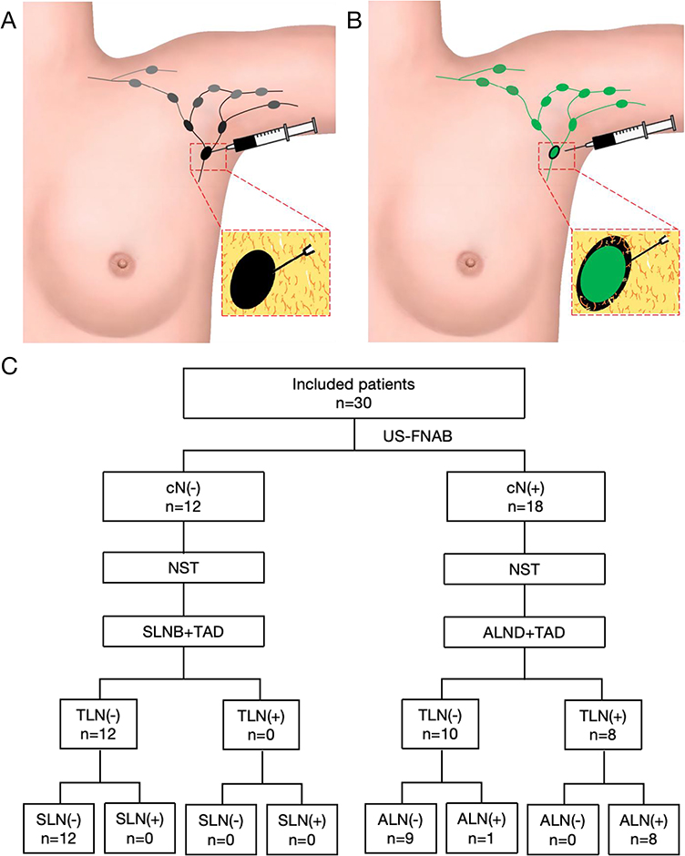 |
Figure 1 Lymph node tattooing technique diagram and experimental flowchart. (A) Intranodal injection of the CNS results in black staining of secondary lymph nodes; (B) The novel approach involving perinodal injection of the CNS prevents secondary lymph node staining, achieving selective black staining exclusively in the targeted lymph node. (C) The research flowchart. |
In clinical practice, we have observed that nanocarbon suspensions can remain stable in peri-lymph node tissues for extended periods. Therefore, we propose a novel strategy of injecting CNS around the suspected metastatic lymph node rather than into it, which not only avoids secondary lymph node staining but also makes it easier to identify the tattooed lymph node (TLN) during surgery (Figure 1B). The purpose of this study is to evaluate the feasibility of a novel TAD strategy—peri-lymph node tattooing with CNS—following NST in breast cancer.
Materials and Methods
Ethics and patients
This prospective single-center feasibility study was approved by the Ethics Committee of Affiliated Hospital of Zunyi Medical University (No.20230517). All patients provided written informed consent prior to participation, in accordance with the Declaration of Helsinki. The study included all consecutive breast cancer patients with clinically and/or radiologically suspicious axillary lymph nodes at the time of diagnosis who were considered appropriate candidates for NST. In all cases, a single suspected lymph node was subjected to fine needle aspiration biopsy (FNAB) and promptly tattooed with CNS. The study workflow is illustrated in Figure 1C.
CNS Injection and Localization
A high-frequency linear-array ultrasound probe (7.5–15 MHz) was used to identify and localize suspicious axillary lymph nodes, particularly those exhibiting malignant sonographic features such as a round shape (low long-to-short axis ratio), cortical thickening, and loss of the fatty hilum (Figure 2A). To ensure standardization, the target lymph node was selected based on the presence of at least one of these suspicious ultrasound characteristics. Under real-time ultrasound guidance, carbon nanoparticle suspension (CNS; Chongqing Lummy Pharmaceutical Co., Ltd., China) was injected into the soft tissue immediately adjacent to the capsule of the target lymph node. The needle tip position was carefully confirmed under ultrasound to ensure accurate placement while avoiding direct intranodal injection. A total volume of 0.2 mL was administered per site using a 1 mL syringe with a 20-gauge needle. The needle was advanced in-plane with the ultrasound probe to maintain continuous visualization of the needle tip throughout the procedure (Figure 2B).
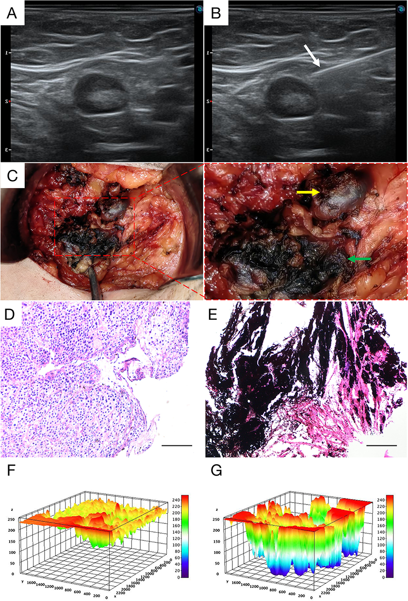 |
Figure 2 Clinical Application of Peri-lymph Node Tattooing with CNS for Targeted Axillary Dissection. (A) Ultrasonographic detection of metastatic axillary lymph nodes. (B) The CNS was injected around the targeted lymph node under ultrasound guidance, with the white arrow indicating the injection needle. (C) In the surgery of the CNS-labeled targeted axillary lymph node, the yellow arrow points to the lymph node, while the green arrow indicates the surrounding soft tissue of the lymph node stained black. (D) H&E staining results of the CNS-labeled lymph node showed no black nanocarbon particles within the lymph node. (E) H&E staining of the surrounding soft tissue of the lymph node revealed a large number of black nanocarbon particles. (F and G) Interactive 3D surface plots corresponding to (D) and (E), generated from grayscale images to represent the distribution of CNS-positive regions. ×200, scale bar = 50 μm. |
Treatment and Surgical Procedure
All patients received neoadjuvant systemic therapy in accordance with the Chinese Anti-Cancer Association (CACA) guidelines for breast cancer with reference to the National Comprehensive Cancer Network (NCCN) guidelines. The regimens included TAC, TCbHP, and THP, all of which were administered every 21 days for a total of 6 cycles. Within two weeks after the completion of NST, all patients underwent surgery (Figure 2C). Patients with or without lymph node metastases according to FNAB results were divided into two groups, cN(-) and cN(+). Patients in the cN(-) group underwent sentinel lymph node biopsy (SLNB) and TAD, while patients in the cN(+) group underwent axillary lymph node dissection (ALND) and TAD. After surgery, all removed lymph nodes were sent for histopathological examination to assess the correlation between TLN and axillary lymph nodes. All cases were de-identified, and clinicopathological data were accessed anonymously.
Histopathological Examination
Tissue samples containing TLN and its surrounding tissues were paraffin-embedded, sectioned, and stained with hematoxylin and eosin (H&E) to observe the distribution characteristics of CNS particles. ImageJ (version 1.54g) was used to generate 3D surface plots and to quantify the proportion of CNS-positive area. The macro code used for analysis is provided in Supplementary Material 1.
Statistical Analysis
The data were analyzed using SPSS version 29.0 (IBM Corp., Armonk, NY, USA) and were presented as mean ± SD. Statistical significance was determined by unpaired two-tailed t-test between two groups. p < 0.05 was considered statistically significant.
Results
Clinicopathological Characteristics of Patients
A total of 30 patients were prospectively enrolled between June 2023 and May 2024. The clinicopathologic characteristics of the patients are summarized in Table 1.
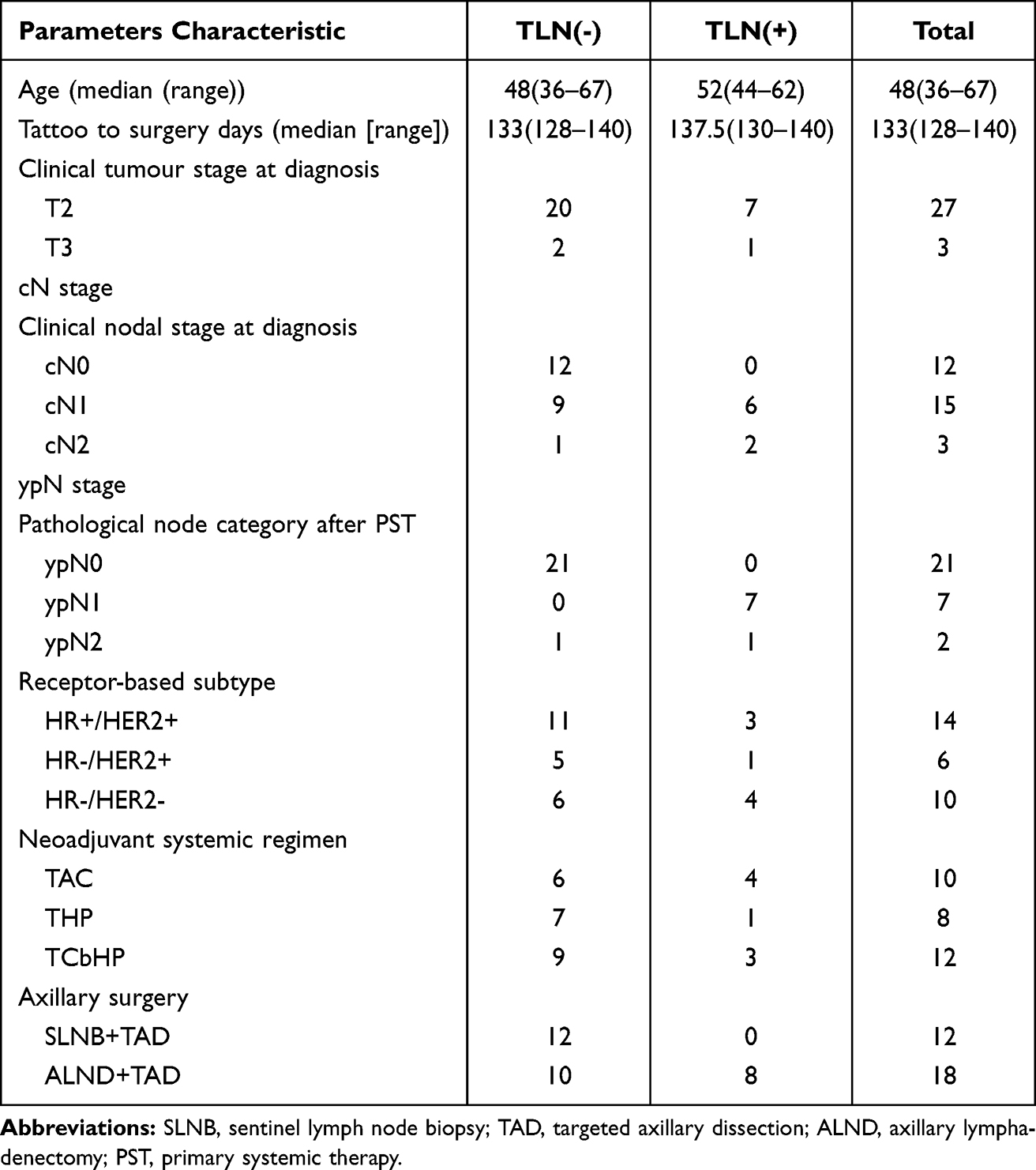 |
Table 1 Clinicopathologic Characteristics of the Patients with Axillary Surgery; PST, Primary Systemic Therapy |
Feasibility of Peri-Lymph Node Tattooing with CNS for TAD
The median time from CNS application to the surgical procedure was 133 days. No adverse events were reported. All CNS-marked lymph nodes were successfully identified and evaluated (Figure 2C). Pathological analysis revealed that CNS was prevented from penetrating the interior of TLN, with CNS predominantly located in the soft tissues surrounding the lymph node (Figure 2D and E). This observation was corroborated by 3D surface plots which demonstrated a lower grayscale signal for CNS deposition in surrounding tissues (Figure 2F and G). Quantification of the proportion of CNS-stained area revealed 0.10 ± 0.09% in the TLN and 42.61 ± 13.45% in the surrounding tissue (p < 0.05).
Diagnostic Efficacy of Peri-Lymph Node Tattooing with CNS for TAD
In the cN(-) group, SLNB and TAD were performed in 12 cases, with all SLN and TLN being negative. In the cN(+) group, comprising a total of 18 patients, 10 patients exhibited negative TLN status following neoadjuvant therapy, while the remaining 8 patients demonstrated positive TLN status. Notably, 1 of the 10 patients with negative TLN still had axillary lymph node metastases, the patients had high tumor burden with a staging of T3N2M0. The results showed that the peri-lymph node tattooing strategy with CNS for TAD exhibited a sensitivity of 88.9%, a specificity of 100%, a negative predictive value (NPV) of 95.5%, and a positive predictive value (PPV) of 100%, resulting in an overall diagnostic accuracy of 96.7%.
Discussion
This study demonstrated that peri-lymph node tattooing with CNS is an effective approach for TAD following neoadjuvant therapy. All CNS-marked target lymph nodes were successfully identified intraoperatively, with a high identification rate and an overall diagnostic accuracy of 96.7%. No adverse events were observed. Overall, compared with traditional localization methods, peri-lymph node tattooing with CNS offers a visual, cost-effective, and reliable strategy for the precise localization of target axillary lymph nodes in the TAD setting.
Traditional ALND provides comprehensive pathological assessment of the axilla but is associated with significant morbidity, including lymphedema, sensory nerve damage, and restricted shoulder mobility. SLNB is a minimally invasive procedure used for axillary staging in breast cancer patients, particularly after NST. Clinical trials such as ACOSOG Z107111 and SENTINA12 have evaluated its efficacy post-NAT, reporting false-negative rates of 12.6% and 14.2%. Although SLNB post-NST offers reduced morbidity compared to ALND, it has inherent limitations, with an FNR exceeding 10%, making it unsuitable for safely staging breast cancer patients.
TAD has emerged as a valuable approach for managing axillary lymph nodes post-NST, offering a way to reduce the extent of surgery while maintaining diagnostic accuracy, with a globally reported false-negative rate (FNR) of less than 9%.13 Traditional TAD methods utilize various markers, such as clips, wires, radioactive seeds, magnetic markers, or dyes. Each of these markers has distinct advantages and limitations.
The use of clips or wires for the marking of lymph nodes is a reliable and non-radioactive method that has a relatively low FNR for clip localization in TAD.14 However, the shortcomings of these two methods are that migration, patient discomfort, and limited real-time visualization, which can complicate localization during surgery. The application of 125I-labelled radioactive seeds for TAD offers high localization accuracy, with identification rates of 97% and a reduced FNR of 3.5%.15 Nevertheless, it involves safety concerns related to radiation exposure, necessitating strict handling protocols, and is more costly compared to other techniques. Moreover, the requirement for specialized facilities and training limits its availability in some centers. Magnetic markers also provide a lower FNR and a higher identification rate.16 As the technique’s non-radioactive nature eliminates radiation exposure risks, making it safer for both patients and healthcare providers. The primary challenge of magnetic markers is the potential for signal interference from metal instruments during surgery, which can complicate node localization. Additionally, the cost of magnetic markers can be higher than traditional wire localization, impacting overall cost-effectiveness. Dyes such as methylene blue, indocyanine green or carbon suspension has been demonstrated to provide a high identification rate, offering a cost-effective and relatively straightforward approach that does not require the use of specialized equipment.10 However, the FNR for dyes localization can be relatively high, ranging from 10–15%, which is less favorable compared to other localization methods.
Compared to traditional dyes such as indocyanine green and methylene blue, which lack long-term stability in vivo and are therefore unsuitable for TLN labeling before neoadjuvant therapy, CNS demonstrated excellent persistence. In our cohort, the average interval between CNS injection and surgery was 133 days, and black staining of the perinodal tissues remained clearly visible in all patients during the operation. This prolonged visibility supports the use of CNS as a reliable marker for pre-neoadjuvant lymph node localization. Compared with conventional markers such as clips and wires, which may elicit foreign body reactions, CNS demonstrates superior biocompatibility. Its biosafety has been validated through both preclinical and clinical studies. In particular, a meta-analysis encompassing 2,171 patients reported no incidences of local inflammation, fat necrosis, skin necrosis, or allergic reactions.8,17 Additionally, CNS is a more cost-effective option and does not require the use of specialized medical equipment.
In a previous study, clips were placed in the cortex of the lymph nodes following confirmation of the presence of metastatic disease, then CNS was injected around the clips on the day preceding the surgical procedure.10 A limitation of this approach is that the nanocarbon suspension injected into the lymph nodes will flow through the lymphatic vessels to secondary lymph nodes, resulting in black staining of additional lymph nodes beyond the targeted ones for labelling. In addition, patients are required to undergo multiple invasive procedures—one for clip placement and another for CNS injection—potentially increasing both physical discomfort and procedural burden. We present a novel approach that modifies the site of injection of CNS, demonstrating a substantial benefit to the patient. This modification not only enhances visualization and precision in the labelling of TLN, but also mitigates discomfort and reduces healthcare expenditure, which achieves twice the result with half the effort. From a surgical technical perspective, CNS injection should be performed under ultrasound guidance, ensuring that the needle tip is positioned outside the lymph node before administration. During surgery, TLN are identified based on the black-stained surrounding tissue indicating the labeled site.
More promisingly, CNS was found to effectively adsorb a range of pharmaceuticals, demonstrating competitive performance at the cellular level. Of particular interest was its selective adsorption of doxorubicin, a chemotherapeutic agent used to treat breast cancer. Using high-performance liquid chromatography (HPLC), Huang et al demonstrated that CNS can efficiently adsorb and release doxorubicin (DOX), with an initial adsorption rate of 86.0% and cumulative release rates of 56.8% at pH 5.0 versus 41.4% at pH 7.0 after 72 hours.18 The acidic nature of the tumor microenvironment facilitates greater DOX release at tumor sites compared to normal tissues, enhancing local delivery and minimizing systemic toxicity. Additionally, they demonstrated that CNS has similar photothermal conversion capacity and can be used for tumor photothermal therapy.19 These findings indicate that CNS may not only serve as a label for metastatic lymph nodes, but also potentially offer “off-label” anti-tumor effects through the inherent properties of nanomaterials.
Reducing the incidence of postoperative lymphedema is a key reason for the implementation of TAD as an alternative to ALND. However, the current large-scale studies of TAD are mostly focusing on oncologic results and accuracy of marker identification with a limited focus on lymphedema.14,20,21 A small prospective study by Sedky et al reported a lymphedema rate of 8% following TAD, providing initial safety data, though the sample size and follow-up duration were limited.22 Lymphedema was not monitored as an endpoint in the current study since the relatively low incidence within a short follow-up period and limited sample size. This study aimed to assess the feasibility and diagnostic capability of CNS as TAD markers. The promising results support the design of a future randomized controlled trial comparing CNS with traditional localization markers. In the next planned trial of CNS, systematic long-term safety evaluations including those for lymphedema will be included to further confirm the clinical effectiveness of CNS.
Conclusions
Peri-lymph node tattooing with CNS represents a promising advancement in TAD for breast cancer surgery following NST. By addressing limitations of current TAD techniques, this approach has the potential to improve surgical precision and reduce morbidity. However, given the limited sample size and single-center design of the present study, continued research and larger prospective trials with long-term follow-up are essential to validate these findings and optimize the clinical application of CNS.
Acknowledgments
This study was supported by National Natural Science Foundation of China (No. 82204692), Science and Technology Innovation Talent Team Training Program of Zunyi [No. 2023(4)], Medical Research Union Found for High-quality health development of Guizhou Province (No. 2024GZYXKYJJXM0049), and Zunyi Municipal Science and Technology Program Project [No. (2024) 177]. The authors acknowledge the use of DeepSeek AI for assistance with English language editing during manuscript preparation. All content was subsequently reviewed and verified by the authors to ensure accuracy and integrity.
Disclosure
The authors declare no conflicts of interest in this work.
References
1. Bray F, Laversanne M, Sung H, et al. Global cancer statistics 2022: GLOBOCAN estimates of incidence and mortality worldwide for 36 cancers in 185 countries. CA Cancer J Clin. 2024;74(3):229–263. doi:10.3322/caac.21834
2. Zhang J, Wang T, Wang Z, et al. Clinical practice guidelines for neoadjuvant therapy in patients with early-stage breast cancer: Chinese society of breast surgery practice guidelines 2022. Chin Med J. 2024;137(12):1387–1388. doi:10.1097/CM9.0000000000003093
3. De Wild SR, Koppert LB, van Nijnatten TJA, et al. Systematic review of targeted axillary dissection in node-positive breast cancer treated with neoadjuvant systemic therapy: variation in type of marker and timing of placement. Br J Surg. 111(3). doi:10.1093/bjs/znae071
4. Switalla KM, Boughey JC, Dimitroff K, et al. Clipping the positive lymph node in patients with clinically node positive breast cancer treated with neoadjuvant chemotherapy: impact on axillary surgery in the ISPY-2 clinical trial. Ann Surg Oncol. 2024. doi:10.1245/s10434-024-15792-x
5. Balasubramanian R, Morgan C, Shaari E, et al. Wire guided localisation for targeted axillary node dissection is accurate in axillary staging in node positive breast cancer following neoadjuvant chemotherapy. Eur J Surg Oncol. 2020;46(6):1028–1033. doi:10.1016/j.ejso.2019.12.007
6. Alamoodi M, Wazir U, Sakr RA, Venkataraman J, Mokbel K, Mokbel K. Evaluating magnetic seed localization in targeted axillary dissection for node-positive early breast cancer patients receiving neoadjuvant systemic therapy: a comprehensive review and pooled analysis. J Clin Med. 2024;13(10). doi:10.3390/jcm13102908
7. Hartmann S, Kühn T, de Boniface J, et al. Carbon tattooing for targeted lymph node biopsy after primary systemic therapy in breast cancer: prospective multicentre TATTOO trial. Br J Surg. 2021;108(3):302–307. doi:10.1093/bjs/znaa083
8. Jiang Y, Li J, Chen B, et al. Sentinel lymph node biopsy mapped with carbon nanoparticle suspensions in patients with breast cancer: a systematic review and meta-analysis. Front Oncol. 2022;12:818812. doi:10.3389/fonc.2022.818812
9. Li J, Jia S, Wang Y, et al. Long-term tracing and staining of carbon nanoparticles for axillary lymph nodes in patients with locally advanced breast cancer treated with neoadjuvant chemotherapy. Asian J Surg. 2022;45(1):89–96. doi:10.1016/j.asjsur.2021.03.020
10. Yang X, Li Y, Ren XT, Fan L, Hua B. Carbon nanoparticles localized clipped node dissection combined with sentinel lymph node biopsy with indocyanine green and methylene blue after neoadjuvant therapy in node positive breast cancer in China: initial results of a prospective study. World J Surg Oncol. 2023;21(1):214. doi:10.1186/s12957-023-03120-8
11. Boughey JC, Ballman KV, Le-Petross HT, et al. Identification and resection of clipped node decreases the false-negative rate of sentinel lymph node surgery in patients presenting with node-positive breast cancer (T0-T4, N1-N2) who receive neoadjuvant chemotherapy: results from ACOSOG Z1071 (Alliance). Ann Surg. 2016;263(4):802–807. doi:10.1097/SLA.0000000000001375
12. Kuehn T, Bauerfeind I, Fehm T, et al. Sentinel-lymph-node biopsy in patients with breast cancer before and after neoadjuvant chemotherapy (SENTINA): a prospective, multicentre cohort study. Lancet Oncol. 2013;14(7):609–618. doi:10.1016/s1470-2045(13)70166-9
13. Gante I, Maldonado JP, Figueiredo Dias M. Marking techniques for targeted axillary dissection among patients with node-positive breast cancer treated with neoadjuvant chemotherapy. Breast Cancer. 2023;17:11782234231176159. doi:10.1177/11782234231176159
14. Wu SY, Li JW, Wang YJ, et al. Clinical feasibility and oncological safety of non-radioactive targeted axillary dissection after neoadjuvant chemotherapy in biopsy-proven node-positive breast cancer: a prospective diagnostic and prognostic study. Int J Surg. 2023;109(7):1863–1870. doi:10.1097/JS9.0000000000000331
15. Munck F, Andersen IS, Vejborg I, et al. Targeted axillary dissection with (125)i seed placement before neoadjuvant chemotherapy in a vanish multicenter cohort. Ann Surg Oncol. 2023;30(7):4135–4142. doi:10.1245/s10434-023-13432-4
16. Hartmann S, Banys-Paluchowski M, Stickeler E, et al. Applicability of magnetic seeds for target lymph node biopsy after neoadjuvant chemotherapy in initially node-positive breast cancer patients: data from the AXSANA study. Breast Cancer Res Treat. 2023;202(3):497–504. doi:10.1007/s10549-023-07100-0
17. Xie P, Yang ST, He T, Yang S, Tang XH. Bioaccumulation and Toxicity of Carbon Nanoparticles Suspension Injection in Intravenously Exposed Mice. Int J Mol Sci. 2017;18(12). doi:10.3390/ijms18122562
18. Huang Y, Xie P, Yang ST, et al. Carbon nanoparticles suspension injection for the delivery of doxorubicin: comparable efficacy and reduced toxicity. Mater Sci Eng C Mater Biol Appl. 2018;92:416–423. doi:10.1016/j.msec.2018.07.012
19. Huang Y, Zeng G, Xin Q, et al. Carbon nanoparticles suspension injection for photothermal therapy of xenografted human thyroid carcinoma in vivo. MedComm. 2020;1(2):202–210. doi:10.1002/mco2.28
20. Kuemmel S, Heil J, Rueland A, et al. A prospective, multicenter registry study to evaluate the clinical feasibility of Targeted Axillary Dissection (TAD) in node-positive breast cancer patients. Ann Surg. 2022;276(5):e553–e562. doi:10.1097/sla.0000000000004572
21. Žatecký J, Coufal O, Zapletal O, et al. Ideal marker for targeted axillary dissection (IMTAD): a prospective multicentre trial. World J Surg Oncol. 2023;21(1):252. doi:10.1186/s12957-023-03147-x
22. Sedky MK, Mostafa AA, AboElazm HA, Zaid MH. Evaluation of targeted axillary dissection in node positive locally advanced breast cancer patients with complete pathological response to neoadjuvant chemotherapy. Egy J Surg. 2024;43(2):383–388. doi:10.4103/ejs.ejs_292_23
 © 2025 The Author(s). This work is published and licensed by Dove Medical Press Limited. The
full terms of this license are available at https://www.dovepress.com/terms
and incorporate the Creative Commons Attribution
- Non Commercial (unported, 4.0) License.
By accessing the work you hereby accept the Terms. Non-commercial uses of the work are permitted
without any further permission from Dove Medical Press Limited, provided the work is properly
attributed. For permission for commercial use of this work, please see paragraphs 4.2 and 5 of our Terms.
© 2025 The Author(s). This work is published and licensed by Dove Medical Press Limited. The
full terms of this license are available at https://www.dovepress.com/terms
and incorporate the Creative Commons Attribution
- Non Commercial (unported, 4.0) License.
By accessing the work you hereby accept the Terms. Non-commercial uses of the work are permitted
without any further permission from Dove Medical Press Limited, provided the work is properly
attributed. For permission for commercial use of this work, please see paragraphs 4.2 and 5 of our Terms.