Back to Journals » Journal of Inflammation Research » Volume 15
PD-1-mAb Plus Regimen in the First and Second Lines of Advanced and Unresectable Biliary Tract Carcinoma: A Real-World, Multicenter Retrospective Analysis
Authors Wang F, Wang FH, Sun K, Jiang C, Peng S, Xu LX, Kuang M , Guo GF, Chen SL
Received 6 March 2022
Accepted for publication 19 August 2022
Published 31 October 2022 Volume 2022:15 Pages 6031—6046
DOI https://doi.org/10.2147/JIR.S364303
Checked for plagiarism Yes
Review by Single anonymous peer review
Peer reviewer comments 3
Editor who approved publication: Professor Ning Quan
Fang Wang,1,* Feng-Hua Wang,2,3,* Kaiyu Sun,4,* Chang Jiang,2,5 Sui Peng,6 Li-Xia Xu,1 Ming Kuang,7 Gui-Fang Guo,2,5,8 Shu-Ling Chen9
1Department of Oncology, The First Affiliated Hospital of Sun Yat-Sen University, Guangzhou, People’s Republic of China; 2State Key Laboratory of Oncology in South China, The Sun Yat-Sen University Cancer Center, Guangzhou, People’s Republic of China; 3Department of Oncology, The Sun Yat-Sen University Cancer Center, Guangzhou, People’s Republic of China; 4Department of Gastrointestinal Surgery, The First Affiliated Hospital of Sun Yat-Sen University, Guangzhou, People’s Republic of China; 5Collaborative Innovation Center for Cancer Medicine, The Sun Yat-Sen University Cancer Center, Guangzhou, People’s Republic of China; 6Institute of Precision Medicine, The First Affiliated Hospital of Sun Yat-Sen University, Guangzhou, People’s Republic of China; 7Department of Liver Surgery, Center of Hepato-Pancreato-Biliary Surgery, The First Affiliated Hospital, Sun Yat-sen University, Guangzhou, People’s Republic of China; 8VIP Department, Sun Yat-sen University Cancer Center, Guangzhou, People’s Republic of China; 9Institute of Diagnostic and Interventional Ultrasound, The First Affiliated Hospital of Sun Yat-Sen University, Guangzhou, People’s Republic of China
*These authors contributed equally to this work
Correspondence: Shu-Ling Chen; Gui-Fang Guo, Email [email protected]; [email protected]
Introduction: Advanced biliary tract carcinoma (BTC) has a poor prognosis and few treatment options. We compared the efficacy of the PD-1 monoclonal antibody (PD-1-mAb) combined regimens with the standard chemotherapy in the first-line and second-line treatment of advanced BTC.
Methods: We retrospectively assessed the patients with advanced BTC, who received treatment at the First Affiliated Hospital of Sun Yat-Sen University and the Sun Yat-Sen University Cancer Center. The patients were treated with PD-1-mAb combined regimens or standard chemotherapy at the first line or treated with PD-1-mAb combined regimens or systematic therapy at the second line. Further subgroup analyses were assessed to identify superior regimens.
Results: This study included 210 patients. The first-line PD-1-mAb combination group (n = 83) achieved longer median PFS (mPFS) (7.3 vs 5.3 months, p=0.001) and median OS (mOS) (15.6 vs 11.4 months, p=0.002) than the first-line standard chemotherapy group (n=76). Similarly, the second-line PD-1-mAb combination group (n=50) yielded longer mPFS (6.1 vs 2.6 months, p< 0.001) and mOS (11.7 vs 7.2 months, p=0.008) than the second-line systematic therapy group (n=51). Subgroup analyses showed that the PD-1-mAb combined with TKI group achieved better mPFS than the chemotherapy group whether in the first-line (HR = 0.468, p=0.005) or the second-line setting (HR = 0.45, p=0.009), but did not achieve superiority in mOS (both p> 0.05). Compared with the chemotherapy group, the PD-1-mAb combined with chemotherapy group achieved longer mOS (HR = 0.53, p=0.023) in the first-line setting and longer mPFS in the second-line setting (HR = 0.54, p=0.044).
Conclusion: The PD-1-mAb combination therapy is superior to the standard chemotherapy in advanced or unresectable BTC, whether as a first-line or second-line treatment. Among the combination therapy, both the PD-1-mAb combined with TKI and combined with standard chemotherapy were promising options for advanced BTC patients.
Keywords: biliary tract carcinoma, PD-1-mAb, PD-1 plus anti-angiogenesis TKI, PD-1 plus chemotherapy, the first-line chemotherapy, the second-line chemotherapy
Introduction
Biliary tract carcinoma (BTC) is mainly composed of intrahepatic cholangiocarcinoma, extrahepatic cholangiocarcinoma, and gallbladder carcinoma. The incidence of BTC is high in East Asia and shows an upward trend.1 Surgery is the only possible curative treatment for BTC;2 however, most BTC is unresectable or advanced at diagnosis. Although gemcitabine or fluorouracil-based chemotherapy can be used as the standard treatment for patients with metastatic or unresectable BTC, the median overall survival (OS) only ranges from 11.6 to 13.4 months, and the 5-year OS rate only ranges from 10% to 20%.3–6 Therefore, more effective treatment strategies are needed for advanced BTC.7
In recent years, immunotherapeutic approaches have become the standard of care for many kinds of cancers for their durable efficacy.8–10 But the role of immunotherapy in BTC remains to be elucidated. The PD-1 monoclonal antibody (PD-1-mAb) was only approved in BTC with high microsatellite instability (MSI-H) or mismatch repair defect (dMMR).11 However, the dMMR/MSI-H phenotype was observed in only 7.1% of BTC patients.12
For PD-1-mAb monotherapy, the Keynote-028 study reported that 24 patients with PD-L1 positive advanced BTC were treated with pembrolizumab. The objective response rate (ORR) was 13%, mOS was 5.7 months, and median progression-free survival (mPFS) was 1.8 months.13 At the same time, the pembrolizumab monotherapy achieved an ORR of just 5.8% in the Keynote-158 basket trial. The PD-1-mAb monotherapy delivered restricted effectiveness to advanced BTC.13
In terms of PD-1-mAb combined with antiangiogenic TKI, Zhou et al reported that when treprizumab combined with lenvatinib was used in unresectable ICC, the ORR was 32.3%, and the disease control rate (DCR) was 74.2%. The 6-month OS rate was 87.1%, and the PFS and OS were not met.14 Regarding PD-1-mAb combined with chemotherapy, a Phase I clinical trial in Japan found that when nivolumab was combined with gemcitabine plus cisplatin (GP), the mPFS was 4.2 months; the mOS was 15.4 months; and the partial response (PR) rate was 36.6% (11/30).15 For PD-1-mAb combined with antiangiogenic TKI and chemotherapy, Zhou et al reported that 30 patients with advanced ICC were treated with lenvatinib combined with GEMOX chemotherapy and treprizumab. The ORR was 80%, DCR was 93.3%, mPFS was 10.0 months, median duration of response (DOR) was 9.8 months, and mOS had not yet reached.16
Until now, there is no first-line Phase III clinical trial of PD-1-mAb in BTC.17 The recent TOPAZ-1 phase III clinical trial reported the efficacy of dovalizumab combined with GP versus placebo combined with GP in the first-line treatment of BTC. The dovalizumab combined with GP achieved longer mPFS (7.2 vs 5.7 months) and mOS (12.8 vs 11.5 months) than the placebo combined with GP group.18
According to the abovementioned previous works, the efficacy of the PD-1-mAb monotherapy is restricted, but the PD-1-mAb combined regimens bring a wealth of potential in the treatment of BTC with promising response rates and survival data. However, all the above researches are phase I to II clinical studies with very limited study participants. The efficacy of the PD-1-mAb combination regimens for BTC in the real world and which regimen is more superior in a head-to-head comparison remains unknown.
To the best of our knowledge, there are no studies comparing the efficacy of anti-PD-1 immunotherapy plus TKI or chemotherapy to chemotherapy alone in advanced BTC. The purpose of this study is to compare the efficacy of the combination immunotherapy of anti-PD-1 with TKI or chemotherapy to the standard first-line chemotherapy or second-line treatment, in the first or second-line setting of advanced BTC.
Patients and Methods
Patients
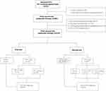
We included the BTC patients who had received treatments at the First Affiliated Hospital of Sun Yat-Sen University and the Sun Yat-Sen University Cancer Center between January 1, 2014, and September 27, 2020. The inclusion criteria were as follows: (1) histologically confirmed unresectable or metastatic BTC; (2) or received at least 2 cycles of PD-1 plus regimen or the standard chemotherapy of the first line and has post-treatment imaging evaluation; (3) accepted PD-1 plus regimen, chemotherapy or anti-angiogenesis TKI at the second line. The exclusion criteria were as follows: (1) patients without biopsy confirming BTC; (2) treated with the first-line anti-PD-1 monotherapy; (3) treated with a regimen containing anti-PD-1 after the second line; (4) radiotherapy was applied to any of the target lesions before receiving the anti-PD-1 regimen; (5) received the treatment of anti-PD-1 combined with TKI and chemotherapy. Figure 1 illustrates the patient screening and inclusion process.
The data of this study was reviewed and approved by the ethics committee of the First Affiliated Hospital of Sun Yat-Sen University ([2022]069) and the Sun Yat-Sen University Cancer Center (SYSUCC; Guangzhou, China, B2020-190-01). Our study is retrospective without influencing patients’ interests, and strictly complied with the Declaration of Helsinki and ensured patients’ data confidentiality. Therefore, both ethics committees agreed to exempt patients’ informed consent.
Data Extraction and Efficacy Assessment
All research subjects’ clinical characteristics, treatment strategies, and efficacy information were collected from medical records of the two centers. The research group’s statisticians perform the data quality control regularly to guarantee the data accuracy. Two oncologists examined the treatment efficacy according to the Response Evaluation Criteria in Solid Tumors (RESIST) criteria (1.1).19 Any inconsistency in assessment results was resolved by consensus. The ORR was defined as the percentage of patients achieving complete response (CR) or partial response (PR), and the DCR as the percentage of patients achieving CR, PR, or stable disease (SD).
The researcher conducted a standard follow-up, and the follow-up intervals were routinely 2 to 4 months. All patients’ follow-up data were available, and the last follow-up was on September 11, 2021. Overall survival (OS) was calculated from the date of the first or second line of treatment was administered to the date of death or the last follow-up visit. Progression-free survival was defined as the time from the first or second line of treatment to the tumor progression or death from any cause.
Treatments
Our study included BTC patients who received PD-1-mAb combination regimen or chemotherapy in the first-line and second-line settings. In the first-line setting, the PD-1-mAb combination regimens included the PD-1-mAb combined with TKI (ie, apatinib or renvatinib), the PD-1-mAb combined with first-line chemotherapy and the PD-1-mAb combined with HAIC, while the standard first-line chemotherapy included the regimen of gemcitabine plus platinum or the regimen of gemcitabine plus teggio. Similarly, in the second-line setting, the PD-1-mAb combination regimens included the PD-1-mAb combined with TKI and the PD-1-mAb combined with second-line chemotherapy or target therapy. The TKI consists of apatinib and renvatinib, while the second-line chemotherapy included gemcitabine, teggio, paclitaxel-albumin and 5-fluorouracil combined with oxaliplatin.
Statistical Analysis
Data were presented as the median values and ranges. The distribution of clinicopathological characteristics according to the different groups was analyzed using the Chi-squared or Kruskal–Wallis test. Survival curves were generated by the Kaplan–Meier method and compared by the Log rank test. The potential survival predictors were analyzed by univariable and multivariable Cox proportional hazard regression models. Subgroup analyses were performed in the first-line and second-line groups, respectively. For example, the PD-1-mAb combined with TKI group vs first-line chemotherapy group, and the PD-1-mAb combined with chemotherapy group vs first-line chemotherapy group in the first-line setting, while the PD-1-mAb combined with TKI group vs second-line chemotherapy group, and the PD-1-mAb combined with chemotherapy group vs second-line chemotherapy group in the second-line setting. Statistical significance was defined as a probability value of 0.05. SPSS statistics package version 26.0 (IBM Corp., Armonk, NY, USA) and R studio version 4.1.2 (R Foundation for S&T) were used for all statistical analyses.
Results
First-Line Treatment of PD-1-mAb Combination Regimen vs Chemotherapy in BTC
Patient Characteristics and Efficacy
In the first-line treatment group, we included 76 patients who received a PD-1-mAb combination regimen and 83 patients who received the standard first-line chemotherapy.
Baseline clinical features of the above patients are presented in Table 1. There were more patients with undifferentiated carcinoma in the first-line chemotherapy group. No other significant differences were found between the PD-1-mAb combination group and the first-line chemotherapy group for gender, age, Eastern Cooperative Oncology Group performance status (ECOG PS), hepatitis B Virus (HBV) status, primary tumor location, metastatic site, number of metastatic organs, liver metastasis, and vascular tumor thrombus.
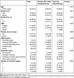 |
Table 1 Patient Clinicopathological Characteristics of the First-Line Therapy Groups |
Regarding the treatment response rate, the ORR was 27.6% and the DCR was 81.6% in the anti-PD-1-mAb combined group (n = 76), with 1 CR, 20 PR, 41 SD, and 14 PD. On the other hand, the ORR was 25.3% and the DCR was 67.5% in the chemotherapy group (n = 83), with 1 CR, 20 PR, 35 SD, and 27 PD. The DCR was higher in the PD-1-mAb combination group than in the chemotherapy group (p=0.047), while there was no significant difference in ORR between the two groups (p=0.857) (Table 2).
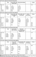 |
Table 2 Tumor Response for All the Five Analyzed Groups of the Research |
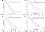
Regarding the survival data, the PD-1-mAb combination group achieved a mPFS of 7.3 months (HR: 0.59, 95% CI: 0.42, 0.84, p=0.003), while the chemotherapy group achieved a mPFS of 5.3 months (Figure 2A). The PD-1-mAb combination group yielded a median OS of 15.6 months (HR: 0.54, 95% CI: 0.36, 0.80, p = 0.002), while the chemotherapy group yielded a median OS of 11.4 months (Figure 2B).
For the univariable survival analysis of PFS, factors such as the first-line therapy regimen were identified as the significant predictive variables (Table 3). All these factors with p < 0.01 were included in the multivariable survival analysis. Only the first-line therapy regimen remained as an independent predictive factor for PFS in the multivariable analysis (HR: 0.63, 95% CI: 0.42, 0.93, p=0.021). For OS, ECOG PS status and the first-line therapy regimen were identified as the significant prognostic variables in the univariable survival analysis (Table 3); whereas only the first-line therapy regimen was demonstrated to be an independent prognostic factor for OS in the multivariable analysis (HR = 0.54, 95% CI: 0.36, 0.80, p=0.002) (Table 3).
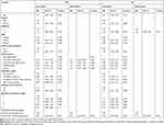 |
Table 3 Univariable and Multivariable Cox Proportional Hazards Regression Analyses for PFS and OS of the First-Line Therapy Group |
Subgroup Analysis of the First Line Groups
Further subgroup analysis was conducted in three groups, including the PD-1-mAb combined with TKI (n = 25), the PD-1-mAb combined with first-line chemotherapy (n = 29) and the first-line standard chemotherapy groups (n = 83). The regimens of the first-line chemotherapy were described as above.
Baseline clinical features of these subgroups are presented in Table 4. The patients had more metastatic organs in the PD-1-mAb combined with TKI group (p=0.018). No other significant differences were found between the three subgroups for gender, age, ECOG PS, HBV, tumor differentiation, primary tumor location, metastatic site, liver metastasis, and vascular tumor thrombus.
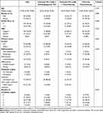 |
Table 4 Patient Clinicopathological Characteristics of the First-Line Therapy Subgroups |
Regarding the treatment response rate, the ORR was 28.0% and the DCR was 76.0% in the PD-1-mAb combined with TKI group (n = 25), with 7 PR, 12 SD, and 6 PD. Nevertheless, the ORR was 17.2% and the DCR was 79.3% in the PD-1-mAb combined with first-line chemotherapy group (n = 29), with 5 PR, 18 SD, and 6 PD. Finally, the ORR was 25.3% and the DCR was 67.5% in the chemotherapy group (n = 83), with 1 CR, 20 PR, 35 SD, and 27 PD. There was no significant difference in both of the response rates (ORR, DCR) between the PD-1-mAb combined with TKI group and chemotherapy group (p=0.787, p=0.417), and between the PD-1-mAb combined with first-line chemotherapy group and chemotherapy group, respectively (p=0.376, p=0.229) (Table 2).
Regarding the survival data, the PD-1-mAb combined with TKI group achieved a mPFS of 8.7 months (HR 0.468, 95% CI: 0.28, 0.79, p=0.005), and the PD-1-mAb combined with first-line chemotherapy achieved a mPFS of 6.2 months (HR 0.756, 95% CI: 0.47, 1.21, p=0.24), while the chemotherapy group achieved a mPFS of 5.3 months (Figure 2C). The PD-1-mAb combined with TKI group achieved a mOS of 15.6 months (HR 0.652, 95% CI: 0.36, 1.18, p=0.156), and the PD-1-mAb combined with first-line chemotherapy achieved a mOS of 15.9 months (HR 0.53, 95% CI 031, 0.92, p=0.023), while the chemotherapy group achieved a mOS of 11.4 months (Figure 2D).
For the univariable survival analysis of PFS, factors such as the regimen of first-line therapy were identified as the significant predictive variables (Supplement Table 1). All these factors with p < 0.01 were included in the multivariable survival analysis; however, no variable was demonstrated to be an independent predictive factor for PFS in the multivariable survival analysis. For OS, the first-line PD-1-mAb combined chemotherapy group (HR = 0.55, 95% CI: 0.32, 0.96, p=0.034) and ECOG PS status (HR = 1.64, 95% CI: 1.04, 2.57, p=0.033) were identified as the independent prognostic factors in the multivariable analysis (Supplement Table 1).
Second-Line Treatment of PD-1-mAb Combination Regimen vs Chemotherapy in BTC
The second-line treatment population included 50 patients who underwent PD-1-mAb combination therapy and 56 patients who received second-line chemotherapy or target therapy. Baseline clinical features of these patients are presented in Table 5.
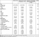 |
Table 5 Patient Clinicopathological Characteristics of the Second-Line Therapy Groups |
There were more patients with undifferentiated carcinoma in the second-line chemotherapy group. No significant differences were found between the groups of PD-1-mAb combination treatment and second-line chemotherapy for gender, age, ECOG PS, HBV, primary tumor location, metastatic site, number of metastatic organs, liver metastasis, and vascular tumor thrombus.
Regarding the treatment response rate, the ORR was 32.0%, and the DCR was 84.0% in the PD-1-mAb combination group (n = 50), with 2 CR, 14 PR, 26 SD, and 8 PD (Table 2). Subsequently, the ORR was 10.7% and the DCR was 46.4% in the second-line chemotherapy or target therapy group (n = 56), with 6 PR, 20 SD, and 30 PD. There was a significant difference in these two response rates between the two groups (p = 0.009, p < 0.001 for ORR and DCR respectively) (Table 2).
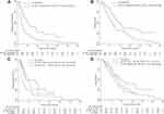
Regarding the survival data, the PD-1-mAb combination group yielded a mPFS of 6.1 months (HR 0.44, 95% CI: 0.29, 0.68, p<0.001), while the second-line chemotherapy or target therapy group yielded a mPFS of 2.6 months (Figure 3A). The PD-1-mAb combination group achieved a median OS of 11.7 months (HR 0.54, 95% CI: 0.34, 0.86, p=0.008), while the second-line chemotherapy or target therapy group achieved a mOS of 7.2 months (Figure 3B).
For the univariable survival analysis of PFS, factors such as the regimen of the second-line therapy were identified as the significant predictive variables (Table 6). All these factors with p < 0.01 were included in the multivariable survival analysis. The regimen of second-line therapy (HR = 0.44, 95% CI: 0.29, 0.68, p < 0.001) and the number of metastatic organs (HR = 14.84, 95% CI: 3.05, 72.14, p=0.001) were demonstrated as the independent predictive factors for PFS in the multivariable survival analysis. For OS, only the regimen of second-line therapy was identified as the significant prognostic variable in the univariable survival analysis. All other factors obtained p<0.1 in the univariable survival analysis. As a result, the multivariable survival analysis was not conducted (Table 6).
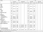 |
Table 6 Univariable and Multivariable Cox Proportional Hazards Regression Analyses for PFS and OS of the Second-Line Therapy Group |
Subgroup Analysis of the Second-Line Group
In order to further compare the efficacy of different treatments, we performed subgroup analyses in the following three subgroups: the PD-1-mAb combined with TKI group (n = 22), the PD-1-mAb combined with second-line chemotherapy group (n = 18), and the second-line chemotherapy group (n = 41). Baseline clinical features of the above 3 subgroups were balanced and presented in Supplement Table 2.
The ORR was 27.3% and the DCR was 86.4% in the PD-1-mAb combined with TKI group, with 1 CR, 5 PR, 13 SD, and 3 PD; whilst, the ORR was 22.2% and the DCR was 83.3% in the PD-1-mAb combined with second-line chemotherapy group, with 4 PR, 11 SD, and 3 PD. On the other hand, the ORR was 12.2% and the DCR was 51.2% in the second-line chemotherapy group, with 5 PR, 16 SD, and 20 PD. The DCR was higher in the PD-1-mAb combined with TKI group (p=0.006) and the PD-1-mAb combined with second-line chemotherapy group (p=0.02), than that in the second-line chemotherapy group. According to our data, there was no significant difference in ORR between the PD-1-mAb combined with TKI group and the second-line chemotherapy group (p=0.133), and between the PD-1-mAb combined with second-line chemotherapy group and the second-line chemotherapy group, respectively (p=0.324) (Table 2).
The PD-1-mAb combined with TKI group achieved a mPFS of 4.5 months (HR 0.45, 95% CI: 0.25, 0.82, p=0.009), and the PD-1-mAb combined with second-line chemotherapy group achieved a mPFS of 5.8 months (HR 0.54, 95% CI: 0.30, 0.98, p=0.044), while the second-line chemotherapy group achieved a mPFS of 2.6 months (Figure 3C). The PD-1-mAb combined with TKI group had a mOS of 11.7 months (HR 0.618, 95% CI: 0.33, 1.17, p=0.14), and the PD-1-mAb combined with second-line chemotherapy had a mOS of 10.0 months (HR 0.82, 95% CI: 0.42,1.60, p=0.561), while the second-line chemotherapy group had a mOS of 7.7 months (Figure 3D).
For the univariable survival analysis, factors such as the regimen of second-line therapy were identified as a significant predictive incidence for PFS. All these factors with p < 0.01 were included in the multivariable survival analysis. The Second-line PD-1-mAb combined anti-angiogenesis TKI group (HR = 0.32, 95% CI: 0.16,0.62, p=0.001), Second-line PD-1-mAb combined chemotherapy group (HR = 0.49, 95% CI: 0.25,0.92, p=0.028) and the number of metastatic organs (HR = 16.6, 95% CI: 2.8, 98.4, p=0.002) were demonstrated as the independent predictive factors for PFS in the multivariable survival analysis. For OS, no factor was identified as the significant prognostic incidence in the univariable and multivariable survival analysis (Supplement Table 3).
Discussion
Advanced BTC has a poor prognosis, and conventional chemotherapy has a limited efficacy.3,4,18 The collaborative immunization strategy has the potential to be a game-changer. A majority of immunotherapy trials in BTC are currently phase I-II back-line clinical investigations, with little indication of therapeutic data. There are very few reports on the first-line immunotherapy of BTC and its long-term outcomes. To the best of our knowledge, our study was the first to reveal that, no matter in the first or second-line treatment of advanced BTC, the PD-1-mAb combination therapy was more effective than the standard chemotherapy.
The first-line treatment data of an anti-PD-1 combined regimen in advanced BTC was rarely reported. The mPFS and mOS of the standard first-line regimens in our study are similar to those reported in the clinical trials.3,4 In the subgroup analysis, the PD-1-mAb combined with TKI was superior to the chemotherapy in terms of PFS through the first line to the second line, but had no significant difference in OS. The first-line PD-1-mAb combined with TKI group has more metastatic lesions, which may affect its efficacy in OS, resulting in its non-superiority to the chemotherapy group. On the other hand, the baseline characteristics of all groups were balanced in the second-line subgroup analysis, so it implied that the PD-1-mAb combined with TKI might be superior to the second-line chemotherapy in controlling the tumor progression for BTC.
As for PD-1 combined with TKI, there are only several phase I-II clinical trials of the single-arm posterior-line therapy. The reported ORR ranged from 32.3% to 41.7%, and the DCR ranged from 74.2% to 75.0%, while the mPFS was around 6 months and the OS was not reached.20–22 Our research found that the ORR and DCR were similar as reported, and the mPFS and mOS were better, with 8.7 months and 15.6 months, respectively. Therefore, our data suggest that the PD-1-mAb combined with TKI has a promising treatment efficacy in BTC. And this is rational because antiangiogenic therapy and immunotherapy are reported to be mutually synergistic in the mechanism of action.23,24 The anti-angiogenic targeted drugs enhanced the anti-tumor immunity by reversing the endothelial cell incapacitation.23 It reported that dual PD-1 and Vascular Endothelial Growth Factor Receptor-2 (VEGFR-2) blockade can promote vascular normalization and improve tumor microenvironment to enhance the antitumor immune responses.25–27
Recently, Zhou et al reported that lenvatinib combined with GEMOX chemotherapy and treprizumab achieved a great efficacy in unresectable advanced ICC. But the toxicity of 4 drug combination will limit its utility in advanced ICC patients.14 PD-1-mAb combined with TKI has shown a relatively low toxicity with acceptable safety profile in clinical practice. It is an acceptable scheme for most advanced-stage BTC patients and has a broader clinical value.
The phase III TOPAZ-1 trial reports that PD-L1 monoclonal antibody combined with GP chemotherapy (mOS 12.8m, mPFS 7.2m) is more effective than placebo combined with GP (mOS 11.5m, mPFS 5.7m) in the first-line therapy of BTC.17 However, there is no phase III clinical trial of PD-1 combined with chemotherapy in the first-line BTC. Previous phase I-II single-arm posterior line clinical studies revealed that the mPFS ranged from 4.2 to 6.7 months, the ORR ranged from 20.6% to 36.7%, and the OS was not reached.15,28 We found that the ORR and mPFS were similar to the previous study. However, our data showed that, no matter in the first or second line, the mPFS and mOS of PD-1-mAb combined with chemotherapy group were longer than the standard chemotherapy group. However, only the first-line mOS and the second-line mPFS of PD-1-mAb combined with chemotherapy obtain a statistically significant difference. This may be due to the small sample size. It has been reported that chemotherapy has a synergistic effect with immunotherapy, by increasing the release of tumor antigen and promoting immune activation.29,30 Therefore, the PD-1-mAb combined with chemotherapy was a promising regimen for advanced BTC patients.
Although our study has been evaluated by strict quality control and inter-group balancing, it still has inevitable bias due to its retrospective nature. Because this was a retrospective study, less than 5% of enrolled patients had the data of PD-L1 expression, so the prognostic difference between PD-L1+ and PD-L1- patients were not analyzed in this study. The sample size of some subgroups is small, and the evidence level of retrospective research is not strong enough, so large-scale studies such as the phase III clinical trials were required to verify our findings.
Conclusion
This study reveals that PD-1-mAb combination therapy is superior to the standard chemotherapy in advanced or unresectable BTC, whether as a first-line or second-line treatment. Further first- and second-line subgroup analyses revealed that the combination of PD-1-mAb combined with anti-angiogenesis TKI and PD-1-mAb combined with standard chemotherapy were the preferred regimen for advanced BTC patients.
Acknowledgments
This work was supported by funding from the National Natural Science Foundation of China (82173191), Guangdong Basic and Applied Basic Research Foundation (2019B151502009), the Guangdong Natural Science Foundation of Distinguished Youth Scholar (No. 2022B1515020060), the Guangdong Natural Science Foundation (No. 2021A1515010450), and Kelin Outstanding Young Scientist of the First Affiliated Hospital of Sun Yet‐sen University (Y12002, R08030).
Author Contributions
All authors contributed significantly to the conception and design, data acquisition, or data analysis and interpretation, participated in the drafting of the article or critically revising it for important intellectual content, agreed to submit to the current journal, gave final approval for the version to be published, and agreed to be accountable for all aspects of the work.
Disclosure
The authors have no actual or potential conflicts of interest related to this manuscript.
References
1. Bertuccio P, Malvezzi M, Carioli G, et al. Global trends in mortality from intrahepatic and extrahepatic cholangiocarcinoma. J Hepatol. 2019;71:104–114. doi:10.1016/j.jhep.2019.03.013
2. Endo I, Gonen M, Yopp AC, et al. Intrahepatic cholangiocarcinoma: rising frequency, improved survival, and determinants of outcome after resection. Ann Surg. 2008;248(1):84–96. doi:10.1097/SLA.0b013e318176c4d3
3. Morizane C, Okusaka T, Mizusawa J, et al. Combination gemcitabine plus S-1 versus gemcitabine plus cisplatin for advanced/recurrent biliary tract cancer: the FUGA-BT (JCOG1113) randomized phase III clinical trial. Ann Oncol. 2019;30(12):1950–1958. doi:10.1093/annonc/mdz402
4. Valle J, Wasan H, Palmer D, et al. Cisplatin plus gemcitabine versus gemcitabine for biliary tract cancer. N Engl J Med. 2010;362(14):1273–1281. doi:10.1056/NEJMoa0908721
5. de Groen PC, Gores GJ, LaRusso NF, Gunderson LL, Nagorney DM. Biliary tract cancers. N Engl J Med. 1999;341(18):1368–1378. doi:10.1056/nejm199910283411807
6. Spolverato G, Bagante F, Weiss M, et al. Comparative performances of the 7th and the 8th editions of the American Joint Committee on Cancer staging systems for intrahepatic cholangiocarcinoma. J Surg Oncol. 2017;115(6):696–703. doi:10.1002/jso.24569
7. Ebata T, Ercolani G, Alvaro D, Ribero D, Di Tommaso L, Valle JW. Current status on cholangiocarcinoma and gallbladder cancer. Liver Cancer. 2016;6(1):59–65. doi:10.1159/000449493
8. Reck M, Rodríguez-Abreu D, Robinson AG, et al. Pembrolizumab versus chemotherapy for PD-L1-positive non-small-cell lung cancer. N Engl J Med. 2016;375(19):1823–1833. doi:10.1056/NEJMoa1606774
9. Larkin J, Chiarion-Sileni V, Gonzalez R, et al. Combined nivolumab and ipilimumab or monotherapy in untreated melanoma. N Engl J Med. 2015;373(1):23–34. doi:10.1056/NEJMoa1504030
10. Kato K, Cho BC, Takahashi M, et al. Nivolumab versus chemotherapy in patients with advanced oesophageal squamous cell carcinoma refractory or intolerant to previous chemotherapy (ATTRACTION-3): a multicentre, randomised, open-label, Phase 3 trial. Lancet Oncol. 2019;20(11):1506–1517. doi:10.1016/s1470-2045(19)30626-6
11. Asaoka Y, Ijichi H, Koike KPD-1. Blockade in tumors with mismatch-repair deficiency. N Engl J Med. 2015;373(20):1979. doi:10.1056/NEJMc1510353
12. Mody K, Starr J, Saul M, et al. Patterns and genomic correlates of PD-L1 expression in patients with biliary tract cancers. J Gastrointest Oncol. 2019;10(6):1099–1109. doi:10.21037/jgo.2019.08.08
13. Piha-Paul SA, Oh DY, Ueno M, et al. Efficacy and safety of pembrolizumab for the treatment of advanced biliary cancer: results from the KEYNOTE-158 and KEYNOTE-028 studies. Int J Cancer. 2020;147(8):2190–2198. doi:10.1002/ijc.33013
14. Zhou J, Fan J, Shi G, et al. 56P Anti-PD1 antibody toripalimab, lenvatinib and gemox chemotherapy as first-line treatment of advanced and unresectable intrahepatic cholangiocarcinoma: a Phase II clinical trial. Ann Oncol. 2020;31:S262–S263. doi:10.1016/j.annonc.2020.08.034
15. Ueno M, Ikeda M, Morizane C, et al. Nivolumab alone or in combination with cisplatin plus gemcitabine in Japanese patients with unresectable or recurrent biliary tract cancer: a non-randomised, multicentre, open-label, Phase 1 study. Lancet Gastroenterol Hepatol. 2019;4(8):611–621. doi:10.1016/s2468-1253(19)30086-x
16. Jian Z, Fan J, Shi G-M, et al. Lenvatinib plus toripalimab as first-line treatment for advanced intrahepatic cholangiocarcinoma: a single-arm, Phase 2 trial. J Clin Oncol. 2021;39(15_suppl):4099. doi:10.1200/JCO.2021.39.15_suppl.4099
17. Do-Youn O, Aiwu Ruth HE, Qin S, et al. A Phase 3 Randomized, Double-Blind, Placebo-Controlled Study of Durvalumab in Combination with Gemcitabine Plus Cisplatin (Gemcis) in Patients (Pts) with Advanced Biliary Tract Cancer (BTC): TOPAZ-1. ASCO Gastrointestinal Cancers Symposium; 2022. doi:10.1200/JCO.2022.40.4_suppl.378
18. Neuzillet C. First-line chemotherapy plus immunotherapy in biliary tract cancer. Lancet Gastroenterol Hepatol. 2022;7(6):496–497.
19. Armato SG 3rd, Nowak AK. Revised modified response evaluation criteria in solid tumors for assessment of response in malignant pleural mesothelioma (Version 1.1). J Thorac Oncol. 2018;13(7):1012–1021. doi:10.1016/j.jtho.2018.04.034
20. Sun Y, Zhou A, Zhang W, Jiang Z, Qu W. A phase Ib study of anlotinib plus TQB2450 as second-line therapy for advanced biliary tract adenocarcinoma. J Clin Oncol. 2021;39:4075. doi:10.1200/JCO.2021.39.15_suppl.4075
21. Villanueva L, Lwin Z, Chung H, et al. Lenvatinib plus pembrolizumab for patients with previously treated biliary tract cancers in the multicohort phase 2 LEAP-005 study. J Clin Oncol. 2021;39:4080. doi:10.1200/JCO.2021.39.15_suppl.4080
22. Wang D, Yang X, Long J, et al. The efficacy and safety of apatinib plus camrelizumab in patients with previously treated advanced biliary tract cancer: a prospective clinical study. Front Oncol. 2021;11:646979. doi:10.3389/fonc.2021.646979
23. Huinen ZR, Huijbers EJM, van Beijnum JR, Nowak-Sliwinska P, Griffioen AW. Anti-angiogenic agents - overcoming tumour endothelial cell anergy and improving immunotherapy outcomes. Nat Rev Clin Oncol. 2021;18(8):527–540. doi:10.1038/s41571-021-00496-y
24. Rahma OE, Stephen Hodi F. The Intersection between Tumor Angiogenesis and Immune Suppression. Clin Cancer Res. 2019;25(18):5449–5457.
25. Shigeta K, Datta M, Hato T, et al. Dual programmed death receptor-1 and vascular endothelial growth factor receptor-2 blockade promotes vascular normalization and enhances antitumor immune responses in hepatocellular carcinoma. Hepatology. 2020;71(4):1247–1261. doi:10.1002/hep.30889
26. Tada Y, Togashi Y, Kotani D, et al. Targeting VEGFR2 with Ramucirumab strongly impacts effector/ activated regulatory T cells and CD8 + T cells in the tumor microenvironment. J Immunother Cancer. 2018;6(1):106. doi:10.1186/s40425-018-0403-1
27. Song Y, Yang F, Xie Q, et al. Anti-angiogenic agents in combination with immune checkpoint inhibitors: a promising strategy for cancer treatment. Front Immunol. 2020;25(11):1956. doi:10.3389/fimmu.2020.01956
28. Liu T, Li W, Yu Y, et al. 53P Toripalimab with chemotherapy as first-line treatment for advanced biliary tract tumors: a preliminary analysis of safety and efficacy of an open-label phase II clinical study. Ann Oncol. 2020;31:S261. doi:10.1016/j.annonc.2020.08.031
29. Wei-Di Y, Sun G, Li J, et al. Mechanisms and therapeutic potentials of cancer immunotherapy in combination with radiotherapy and/or chemotherapy. Cancer Lett. 2019;28(452):66–70.
30. Galon J, Bruni D. Approaches to treat immune hot, altered and cold tumours with combination immunotherapies. Nat Rev Drug Discov. 2019;18(3):197–218.
 © 2022 The Author(s). This work is published and licensed by Dove Medical Press Limited. The full terms of this license are available at https://www.dovepress.com/terms.php and incorporate the Creative Commons Attribution - Non Commercial (unported, v3.0) License.
By accessing the work you hereby accept the Terms. Non-commercial uses of the work are permitted without any further permission from Dove Medical Press Limited, provided the work is properly attributed. For permission for commercial use of this work, please see paragraphs 4.2 and 5 of our Terms.

