Back to Journals » International Journal of Chronic Obstructive Pulmonary Disease » Volume 17
Pathological Mechanism and Targeted Drugs of COPD
Authors Guo P, Li R, Piao TH, Wang CL, Wu XL, Cai HY
Received 12 March 2022
Accepted for publication 4 July 2022
Published 12 July 2022 Volume 2022:17 Pages 1565—1575
DOI https://doi.org/10.2147/COPD.S366126
Checked for plagiarism Yes
Review by Single anonymous peer review
Peer reviewer comments 2
Editor who approved publication: Prof. Dr. Richard Russell
Peng Guo,1 Rui Li,2 Tie Hua Piao,3 Chun Lan Wang,3 Xiao Lu Wu,3 Hong Yan Cai3
1College of Traditional Chinese Medicine, Changchun University of Chinese Medicine, Changchun, Changchun, 130000, People’s Republic of China; 2Graduate School, Beijing University of Chinese Medicine, Beijing, 100000, People’s Republic of China; 3Pulmonology Department, The First Clinical Hospital of Jilin Academy of Traditional Chinese Medicine, Changchun, 130000, People’s Republic of China
Correspondence: Hong Yan Cai, The First Clinical Hospital of Jilin Academy of Traditional Chinese Medicine, Changchun, 130000, People’s Republic of China, Email [email protected]
Abstract: Chronic obstructive pulmonary disease (COPD) includes chronic bronchitis, emphysema, and small airway obstruction. Incompletely reversible airflow limitation, inflammation, excessive mucus secretion and bronchial mucosal epithelial lesions are the main pathological basis of the disease. The prevalence of COPD is increasingly worldwide, which has caused the burden on individuals and society. This paper summarizes the pathogenesis of COPD and clarifies the effect and mechanism of the latest targeted drugs for COPD. Besides, we focus on NOD-like receptor thermal protein domain associated protein 3 inflammasome (NLRP3 inflammasome). NLRP3 can promote production of interleukin-1β (IL-1β) and interleukin-18 (IL-18). NLRP3 is an important factor in the migratory aggregation of macrophages and neutrophils and the generation of oxidative stress. Inhibition of NLRP3 inflammasome indirectly blocks the inflammatory effects of IL-1β and IL-18, which may be regarded as an ideal target for COPD treatment.
Keywords: chronic obstructive pulmonary disease, pathogenesis, targeted drugs, NLRP3
Introduction
COPD is an incurable chronic lung disease, which is also complicated by pulmonary heart disease and respiratory failure in some individuals with a tremendous burden on individuals and society. At present, it is not clear whether the pathological mechanisms of COPD are mainly thought to be the result of genetic and environmental interactions. Moreover, smoking is considered to be the main environmental factor to trigger COPD.1–3 Except for genetics, gender,4 occupation,4,5 airway hyperresponsiveness,6 lung growth and development,3,7 and infection8 also play an important role in the development of COPD. It is easily understood that gender may influence the history of smoking and the particulate environment in which the occupation is located. However, the status of lung growth and development determines the susceptibility to COPD, which seems to have a close relationship with genetics.9,10 We should focus on airway hyperreactivity, which is an independent predictor of COPD and can exist independently without asthma and bronchitis, suggesting that the inflammatory response in COPD is different from asthma.6,11 In addition to the above causes, inflammatory mechanisms, oxidative stress, and protease-antiprotease imbalance are also involved in the development of COPD. Various reasons for bronchial mucosal epithelial cell degeneration, necrosis, squamous metaplasia and recurrent injury-repair airway wall eventually lead to the occurrence of structural repeated remodeling of the airways and scar formation.12,13
Currently, there is still no specific treatment for COPD, and palliative regimens to improve airflow limitation are the mainstay methods. We generally do not advocate drug intervention for COPD in the stable period, but in the acute attack stage, antibiotics, inhaled corticosteroids, bronchodilators, and other medicines are widely used in clinical practice. However, the negative effects of these drugs should not be ignored. For example, frequent use of inhaled corticosteroids can cause side effects such as osteoporosis, immunosuppression and increased probability of infection, especially infection that promotes the recurrence of COPD,8,14 while bronchodilators, for example, the anticholinergic agents and β2 agonists commonly used in clinical practice have side effects such as heart rate disturbance, impact on vision, urinary retention, and metabolic disorders, which cannot be ignored.15,16 With the development of molecular biology, targeted drugs for treatment by blocking COPD development are gradually being developed. This paper not only reviews the pathogenesis of COPD and the pharmacological mechanisms of COPD-related targeted drugs but also elaborates the concerned contribution of NLRP3 to COPD and the effectiveness of NLRP3 inhibitors and related advances.
Pathogenesis of COPD
Oxidative Stress Response
Oxidative stress is involved in the development of several inflammatory conditions and is an important pathogenetic factor in COPD. Stimulation of patients with smoke or dust leads to lung cell damage. Excessive mucus secretion and accumulation of neutrophils produce a large amount of reactive oxygen species (ROS). Oxidative inactivation of antiproteases loses inactivation and the structure of lung tissue is destroyed due to ROS.17 The aggregation of neutrophils also leads to activation of a large number of inflammatory factors to produce more ROS, and aggravates the oxidative stress response.18,19 The oxidative system involves the secretion of the airway epithelial mucus, and the stimulation of noxious gases such as cigarettes20 generates oxidative stress, which causes a large accumulation of ROS and regulates the relevant mucus genes, such as Muc5b21 and Mu5ac.22 In addition, epidermal growth factor23 is also involved in the production of mucus. The signaling pathway of this kind of factor often resides in the oxidants’ activation of airway cells, and then involves in COPD. Regarding the structural destruction of lung tissue caused by protease-antiprotease imbalance, due to α1-antitrypsin24 is the most active one, its inactivation is the most critical one. And the large amount of oxidants released by noxious gases, oxidative stress also makes the antiprotease inactivated and finally the protease-antiprotease imbalance occurs. Oxidative stress also enhances the inflammatory response in the lung by regulating redox-sensitive transcription factors such as nuclear factor kappa-B (NF-κB) and activator protein 1 (AP-1), releasing amounts of cell factors such as IL-1β, tumor necrosis factor-α (TNF-α). In addition,25 the accumulation of ROS decreases the activity of histone deacetylases (HADC) and increases the activity of histone acetyltransferase, which can lead to a further aggregation of inflammatory cells, especially neutrophils.26 So, we always observe a large number of neutrophil infiltrates during COPD pathological sections. If COPD worsens, the excessive oxidative stress will cause inflammatory cells to generate large amounts of ROS after accumulating. Then, a systemic response will occur.27 Nuclear factor E2 (Nrf2)28 can regulate antioxidant genes. In oxidative stress response, Nrf2 will dissociate. It is then transported to the nucleus to activate the transcription of antioxidant genes. Patients with COPD go through a diminished self-protective mechanism due to reduction of Nrf2 in level, resulting in lower endogenous antioxidant production.29,30
Inflammatory Cells, Inflammatory Mediators Cell Factors
Neutrophils and macrophages play a significant role in the oxidative stress response in COPD and are involved in the remodeling of COPD’s airway. Neutrophils7 accumulate in large numbers in the airways of COPD patients under oxidative stress, and this cell can secrete serine proteases,31 including matrix metalloproteinase (MMP) and neutrophil elastase (NE). And MMP is significantly increased in patients with emphysema, and goes through the extracellular matrix of the lung destroyed by serine proteases, leading to the remodeling of the airway.32 Neutrophils are sensitive to the infection response. When COPD patients are stimulated by infection, neutrophils will leave the circulation to aggregate in the lungs and protect the cells and surrounding tissues by phagocytosing the infectious agent to form proteases and bactericidal proteins, and produce ROS, which is the necessary mechanism to protect the body from free radical damage and the inductor of oxidative stress. In addition, the accuracy of neutrophil migratory aggregation is affected by physical fitness. Therefore, COPD is more common in the elderly population, which is associated with the expression of phosphatidylinositol 3-kinase (PI3K).33 The accuracy of neutrophil migration can be improved by inhibiting type I PI3K-δ or PI3K-γ.34
Activation of macrophages35 can regulate the beginning and the end of multiple inflammation. For COPD patients, the combination of Interferon-γ (IFN-γ) secreted by Th1, CD8+ cells, and B cells with the IFN-γ receptor will trigger a series of signaling cascades that lead to the activation and differentiation of M1 macrophages, which then36 produce a large number of cytokines such as TNF-α, IL-1β, interleukin-6 (IL-6) depending on the tissue site. M2 macrophages, on the other hand, are activated by a variety of cell factors (interleukin-4 (IL-4), interleukin-10 (IL-10) and interleukin-13 (IL-13) et al), and help the remodeling of airway by remodeling and repairing damaged tissues.37 The inflammatory factor IL-6, produced by neutrophils and macrophages, can induce the production of elastase and oxygen radicals, which will increase the permeability of pulmonary vascular and aggravate the destruction of lung tissue.38,39 TNF-α, on the other hand, can modulate endothelial adhesion molecules, which will make polymorphonuclear leukocytes accumulate, and then release large amounts of elastase and ROS’s destroyed alveolar epithelium. And during the progression of COPD, TNF-α will generate an inflammatory cascade40 with IL-1β. In COPD patients, macrophages and neutrophils, entering the airways and upregulating chemokines such as monocyte chemotactic proteins (MCP-1, CCL-2),41 and releasing large amounts of inflammatory factors, all indicate their contribution to the development of COPD.
Access Mechanism
NF-κB Access
NF-κB plays a role in systemic inflammation, such as rheumatoid arthritis and bronchial asthma. Activation of NF-kB is achieved by activating protein inhibitor kappa B (IkB)42 to make ubiquitination of IkB. Because of IkB ubiquitination, NF-kB is released from the NF-kB/IkB complex, activates, exposes the nuclear localization domain, forms a p50/RelA dimer, and binds to target genes via the p50 subunit, thereby initiating the expression of target genes, such as TNF-α and IL-1, causing inflammatory responses.43 In addition, the inhalation of mixtures, such as ozone, cigarette smoke and so on, leads to the migration of inflammatory cells such as neutrophils into the lungs, and generates ROS that is also a kind of factor in the activation of (NF-κB).44 It will promote helper T cell type 1 (Th1)45 to produce cell factors such as TNF-α, IL-1, IL-6, and so on which promote the maturation of resting monocytes into mature dendritic cells, which then provides autoantigens to self-reactive T lymphocytes, causing them to move to target tissues and the destruction of inflammation and lung tissue. All of these suggest that activation of the NF-κB pathway drives the release of inflammatory factors, leads to further enhancement of the oxidative stress response and exacerbates lung injury in patients. In respiratory tests in COPD patients, it is confirmed that the NF-κB expression is higher than in normal people. And in the case of smoking patients, the NF-κB expression is even higher. In addition, factors such as MCP-1,46 IL-6, and CXCL-547 are released in large amounts during COPD exacerbations, and hypomethylation of NF-κB-mediated pathway gene DNA has also been observed to contribute to COPD exacerbation.48
MAPK
Mitogen-activated protein kinase (MAPK)49 is an important signaling pathway that transmits signals from the cell membrane to the nucleus. The pathway can be activated by the stimulation of cytokines, neurotransmitters, serine proteases, and oxidative stress to participate in stress adaptation and inflammatory responses.50 In COPD, mitogen-activated protein kinase (p38MAPK)51 plays an important role. The release of inflammatory factors caused by various environmental and genetic factors can lead to the activation of p38MAPK, while IL-8 and TNF-α, which are key factors associated with the development of COPD, are regulated by p38MAPK. The excessive release of these inflammatory factors eventually leads to aggregation of neutrophils, secretion of serine proteases, and destruction of lung structures. And all of p38MAPK isoforms (α, β, δ, γ)52 in COPD patients occur with high expression and mediate lung inflammation53 together. In addition, IL-8 and TNF-α, which are regulated by p38MAPK, appear to mediate glucocorticoid insensitivity in COPD patients. And these factors impair the function of glucocorticoid receptor (GR) by phosphorylating the GR, while the anti-inflammatory effects of glucocorticoids are exerted by GR. The endogenous p38MAPK antagonist MAPK-phosphatase-1 (MKP-1)54 may be central to the reversal of glucocorticoid insensitivity in COPD patients. And it is found that glucocorticoid insensitivity could be reversed by blocking p38MAPK-α, γ and thus upregulating MKP-1.55
PI3K/Akt
The PI3K/Akt56 pathway plays an important role in inhibiting cell proliferation and apoptosis. The activation of this pathway is mainly related to tyrosine kinase and G protein-coupled receptors.57 Upon receiving the signal, the p8558 regulatory subunit in PI3K aggregates to the plasma membrane site. The p110 and p8559 subunit converts the substrate phosphatidylinositol 2 phosphate (PIP2) to phosphatidylinositol 3 phosphate (PIP3). And PIP3 binds to the N-terminal end of protein kinase B (Akt) and translocates to the cell membrane for activation.60 It is an important mechanism of airway remodeling in COPD33 that regulates the PI3K/Akt pathways and promote apoptosis through the tumor suppressor gene encoded by chromosome 10 negatively. In COPD patients, low expression of Nrf2 is closely associated with the oxidative stress response and the release of inflammatory factors and Nrf2 is a PI3K/Akt downstream signaling target.61 In addition, when neutrophil migration is influenced by PI3K expression the persistent lung inflammation triggers neutrophil aggregation by activating the PI3K/Akt pathways. And the aggregation of associated cells generates large amounts of ROS to stimulate further exacerbation of oxidative stress. The activation of the PI3K/Akt pathways downregulates HADC activity, leading to the activation of inflammatory gene. Thus, inactivation of HADC is associated with glucocorticoid insensitivity.62
Molecularly Targeted Drugs for COPD
As the pathogenesis of COPD has become better understood, research on molecularly targeted drugs has increased. Here, we will describe most (we only list those that have been registered in clinical trials and published results) of the targeted drugs used in COPD treatment and introduce the mechanisms of these drugs.
Antioxidants
Oxidative stress is an important mechanism in the development of COPD. The application of antioxidants is feasible in COPD patients, because the significantly elevated level of airway oxidative stress markers (eg, H2O2) and has been validated in in vivo experiments.63 It is proved that N-acetylcysteine (NAC) can control COPD patients’ airway function in clinical trials.64,65 However, data from various studies are inconsistent, possibly due to oral administration affecting bioavailability. Other trials, such as superoxide dismutase, have a good anti-inflammatory effect achieved in animal studies, but still lack more clinical evidence.
Sulforaphane can attenuate oxidative stress by activating Nrf2 and regulating reactive nitrogen and ROS.66 However, in a 4-week clinical trial,67 no evidence was found to improve clinical outcomes in COPD (including Nrf2 expression, levels of relevant inflammatory markers). Therefore, the ability of turnip-sulfur as a therapeutic agent for COPD lacks the support of clinical evidence. The drugs are shown in Table 1.
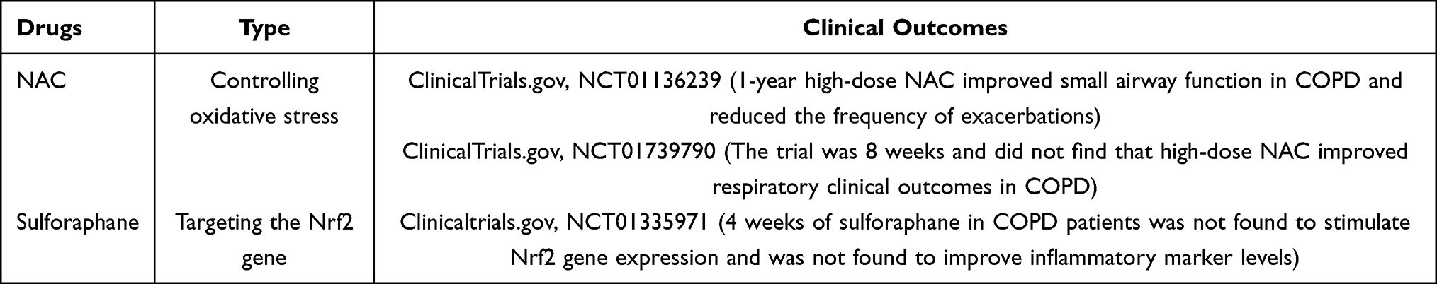 |
Table 1 Antioxidants |
Cytokine-Targeted Drugs
Numerous cytokines are involved in the development of COPD. High expression in COPD patients is also an important factor in activating various inflammatory signaling pathways, and oxidative stress responses. TNF-α plays an important role in interstitial lung disease. As a TNF-α targeting agent, Infliximab has been shown in animal studies68 to prevent smoke-induced emphysema in rats, and reduce the percentage of neutrophils and the level of IL-8 and TNF-α in rats. However, clinical trials69,70 had less favorable outcomes and did not find an improvement in clinical outcomes in COPD patients. It has even been reported that it is possible to increase in the incidence of lung malignancies in COPD patients. Also, as an antagonist of TNF-α, the outcomes reported by etanercept71 were slightly better, but still not as effective as those of inhaled corticosteroids. Due to the lack of clinical evidence, whether TNF-α antagonists can be used as a treatment for COPD remains to be discussed.
IL-5 is in high expression in COPD patients. It seems feasible to inhibit IL-5 to achieve suppression of inflammatory and oxidative stress responses. Although mepolizumab is a target drug for IL-5, clinical evidence points to COPD with increased eosinophilia, probably because of the close association between IL-5 and the aggregation and differentiation of eosinophil.72 Clinical trials have also demonstrated a reduction in COPD and exacerbation and a more pronounced reduction in eosinophil percentage than with placebo.73 However, the FDA has not approved mepolizumab for COPD and trials related to it still have to be planned. Benralizumab, also an IL-5 antagonist, is approved for asthma with eosinophilia. However, clinical trials reported that benralizumab did not reduce the frequency of exacerbations in moderate-to-severe COPD.74,75 So, there is still a lack of more evidence to support it.
MK-7123 is a CXCR2 inhibitor, which can decrease neutrophil chemotaxis to reduce inflammatory manifestations in COPD patients. And in a clinical trial of 616 patients,76 it was shown that MK-7123 at a 50 mg dose was effective in improving lung function and reducing lung inflammation in patients. Those drugs are shown in Table 2.
 |
Table 2 Cytokine-Targeted Drugs |
Enzyme Inhibitors
Protease-antiprotease imbalance is the main cause of lung damage in COPD patients. Protection against lung tissue damage and suppression of inflammation by inhibited NE seem feasible; however, clinical evidence77,78 seems unsatisfactory, AZD9668, an NE inhibitor, failed to improve the lung function and the airway structure in patients. MMP inhibitors are one of the targets of anticancer drugs, but the clinical value of MMP inhibition for COPD remains unknown. MMP-9 and MMP-12 are significantly associated with airway inflammatory damage, both enzymes have enhanced activity in COPD patients, and their inhibition has been found to achieve better anti-inflammatory efficacy in animal model species of COPD,79 however, no valuable outcomes have been reported in clinical trials.80
Phosphodiesterases-3 and phosphodiesterases-4 (PED3 and PED4) are involved in the development of COPD, and they regulate cellular activity by hydrolyzing intracellular cAMP and cGMP.81 PED3 is widely distributed in T lymphocytes, and lymphocyte function can be regulated by inhibition of PED3; inhibiting PED4 can reduce IL-4 and 5 gene expression in TH2 cells, decreases levels of inflammatory factors and has a synergistic effect with PED3 inhibitors in T cells.82 Clinical evidence83,84 also indicated that PED3/4 inhibitors (RPL-5 and roflumilast) both improve the lung function in COPD patients. However, when these patients simultaneously use standard bronchodilators, the function of RPL-554 and roflumilast benefits little. So, it did not justify the clinical efficacy in RPL-554 and roflumilast. Besides, roflumilast also reported side effects of severe gastrointestinal reactions (diarrhea and nausea)85 and headache. Those drugs are shown in Table 3.
 |
Table 3 Enzyme Inhibitors |
Signaling Pathway Inhibitors
In COPD patients, activation of related pathways can enhance oxidative stress and cytokines, chemokines were massive release, which can further COPD development. Vitro trials86 demonstrated that verproside can achieve inflammation suppression by blocking the TNF-α/NF-κB pathway, but no clinical trials related to verproside have been seen, and no clinical evidence has been found for other inhibitors related to the NF-κB pathway.
p38MAPK inhibitors have recently received wide attention and have shown beneficial anti-inflammatory effects in smoke-induced pneumonia models,87 in addition, inhibiting p38MAPK also reduces the production of associated cytokines by macrophages, whose aggregation is an important cell for oxidative stress and inflammatory factor release in COPD patients88 Currently, some clinical evidence89,90 also confirmed that p38MAPK inhibitors (PH-797804, SB-681323) improve lung function and inflammatory factor levels (TNF-α) in COPD patients and perform well in hormone-insensitive classes of patients.
Inhibited PI3K can activate Nrf2, improve HDAC activity, modulate oxidative stress and improve inhaled corticosteroids resistance.61,62 In clinical trials,91–94 the PI3K inhibitor GSK2269557 can improve lung function and related inflammatory factor levels (IL-8, IL-6) in COPD patients, but it has also been reported that it cannot change the clinical outcome. In addition, some studies95 pointed out that excessive PI3K inhibition may lead to immunosuppression. Nrf2, as a downstream target of PI3K, although there is no evidence of clinical effectiveness at this time. It remains to be validated and developed the medicine of downstream target of PI3K. Those drugs are shown in Table 4.
 |
Table 4 Signaling Pathway Inhibitors |
Relation of NLRP3 and Inflammatory Factors/Cells
Studies on NLRP3 in COPD, animal and in vitro experiments demonstrated that reducing NLRP3 expression can improve lung inflammation, inflammatory factor levels, and immune system function in COPD patients and so on, which also appears to improve glucocorticoid-insensitive classes of airway disease.
NLRP3 was demonstrated in mice experiments96 that Chlamydia and Haemophilus can increase NLR3, IL-1β responses and develop drug-resistant neutrophil inflammation. This experimental blockade of airway inflammation was made possible by NLRP3 inhibitor (MCC950), airway inflammation in asthma and the degree of glucocorticoid resistance were all related with IL-1β and NLRP3 expression. In in vitro models, NLRP3 expression is similarly elevated in models of COPD and exacerbated COPD, and IL-1β is similarly elevated.97 In addition, experiments by Yang98 et al verified that in a tobacco-made COPD mouse model, knockdown of NLRP3 caused mice to lose evidence of lung inflammation and did not show pathological damage, while in NLRP3 knockdown mice, IL-1β, IL-18, macrophage, neutrophil, and lymphocyte levels were significantly lower than in COPD model mice. Although also affected by tobacco, NLRP3 knockdown significantly alleviated lung inflammation in mice, providing us with evidence that NLRP3 is a target for COPD treatment.
Relation of NLRP3 and Oxidative Stress
Research pointed99 that NLRP3 deficiency can reduce oxidative stress and scavenge damaged mitochondria, and it seems to play an important role in neuroinflammation. After activation of NLRP3 inflammasome, the release of IL-1 β, IL-18 and other inflammatory factors will activate the polymorphonuclear neutrophils, produce a large number of ROS, and initiate the inflammatory response.100 Furthermore, studies101 suggest that in smog-induced responses, activation of NLRP3 can induce apoptosis through the p53-Bax mitochondrial pathway. These results demonstrate the importance of NLRP3 inflammasome in the cell injury and apoptosis.
Conclusions
The pathogenesis of COPD is complex, mainly related to oxidative stress, inflammatory factors and over-expression or activation of signaling pathways. We found that these factors often co-exist, and it is difficult for us to achieve treatment of COPD through a single target among these interacting factors. Although some targeted drugs have achieved therapeutic efficacy, there are unknown consequences for the interaction of inflammatory factors and signaling pathways that inhibit only a single pathway. Therefore, most targeted drugs are still at a hypothetical stage in clinical practice. We need to reexamine the mechanisms of COPD and study each targeted pathway in depth to assess the safety of new targets through extensive in vitro and animal experiments. NLRP3 is highly correlated with the development of COPD and achieves therapeutic effects in COPD by controlling inflammation to inhibit the production of inflammatory factors by blocking the activation of related pathways. It is also effective in hormonic tolerant patients. Inhibited NLRP3 has not been reported any adverse effects in animal studies. Therefore, we believe that the targeted drugs have implications for continued development in the treatment of COPD.
Disclosure
The authors report no conflicts of interest in this work.
References
1. Rao W, Wang S, Duleba M, et al. Regenerative metaplastic clones in COPD lung drive inflammation and fibrosis. Cell. 2020;181(4):848–864. doi:10.1016/j.cell.2020.03.047
2. Wang C, Zhou J, Wang J, et al. Progress in the mechanism and targeted drug therapy for COPD. Signal Transduct Target Ther. 2020;5(1):248. doi:10.1038/s41392-020-00345-x
3. Barnes PJ, Burney PG, Silverman EK, et al. Chronic obstructive pulmonary disease. Nat Rev Dis Primers. 2015;1:15076. doi:10.1038/nrdp.2015.76
4. Raherison C, Girodet PO. Epidemiology of COPD. Eur Respir Rev. 2009;18(114):213–221. doi:10.1183/09059180.00003609
5. Salvi SS, Barnes PJ. Chronic obstructive pulmonary disease in non-smokers. Lancet. 2009;374(9691):733–743. doi:10.1016/S0140-6736(09)61303-9
6. Kume H, Hojo M, Hashimoto N. Eosinophil inflammation and hyperresponsiveness in the airways as phenotypes of COPD, and usefulness of inhaled glucocorticosteroids. Front Pharmacol. 2019;10:765. doi:10.3389/fphar.2019.00765
7. Benjamin JT, Plosa EJ, Sucre JM, et al. Neutrophilic inflammation during lung development disrupts elastin assembly and predisposes adult mice to COPD. J Clin Invest. 2021;131(1):e139481. doi:10.1172/JCI139481
8. Linden D, Guo-Parke H, Coyle PV, et al. Respiratory viral infection: a potential “missing link” in the pathogenesis of COPD. Eur Respir Rev. 2019;28(151):180063. doi:10.1183/16000617.0063-2018
9. Huang X, Mu X, Deng L, et al. The etiologic origins for chronic obstructive pulmonary disease. Int J Chron Obstruct Pulmon Dis. 2019;14:1139–1158. doi:10.2147/COPD.S203215
10. Nasri A, Foisset F, Ahmed E, et al. Roles of mesenchymal cells in the lung: from lung development to chronic obstructive pulmonary disease. Cells. 2021;10(12):3467. doi:10.3390/cells10123467
11. Kim HT, Yin W, Nakamichi Y, et al. WNT/RYK signaling restricts goblet cell differentiation during lung development and repair. Proc Natl Acad Sci U S A. 2019;116(51):25697–25706. doi:10.1073/pnas.1911071116
12. Berg K, Wright JL. The pathology of chronic obstructive pulmonary disease: progress in the 20th and 21st centuries. Arch Pathol Lab Med. 2016;140(12):1423–1428. doi:10.5858/arpa.2015-0455-RS
13. Bagdonas E, Raudoniute J, Bruzauskaite I, Aldonyte R. Novel aspects of pathogenesis and regeneration mechanisms in COPD. Int J Chron Obstruct Pulmon Dis. 2015;10:995–1013. doi:10.2147/COPD.S82518
14. Agusti A, Fabbri LM, Singh D, et al. Inhaled corticosteroids in COPD: friend or foe? Eur Respir J. 2018;52(6):1801219. doi:10.1183/13993003.01219-2018
15. Gupta P, O’Mahony MS. Potential adverse effects of bronchodilators in the treatment of airways obstruction in older people: recommendations for prescribing. Drugs Aging. 2008;25(5):415–443. doi:10.2165/00002512-200825050-00005
16. Tanabe N, Sato S, Muro S, et al. Regional lung deflation with increased airway volume underlies the functional response to bronchodilators in chronic obstructive pulmonary disease. Physiol Rep. 2019;7(24):e14330. doi:10.14814/phy2.14330
17. Chang RM, Kauffman RJ, Kwon Y. Understanding the paradigm shift to computational social science in the presence of big data. Decis Support Syst. 2014;63:67–80. doi:10.1016/j.dss.2013.08.008
18. Dang X, He B, Ning Q, et al. Alantolactone suppresses inflammation, apoptosis and oxidative stress in cigarette smoke-induced human bronchial epithelial cells through activation of Nrf2/HO-1 and inhibition of the NF-κB pathways. Respir Res. 2020;21(1):95. doi:10.1186/s12931-020-01358-4
19. Zhang MY, Jiang YX, Yang YC, et al. Cigarette smoke extract induces pyroptosis in human bronchial epithelial cells through the ROS/NLRP3/caspase-1 pathway. Life Sci. 2021;269:119090. doi:10.1016/j.lfs.2021.119090
20. Shih YM, Chang YJ, Cooke MS, et al. Alkylating and oxidative stresses in smoking and non-smoking patients with COPD: implications for lung carcinogenesis. Free Radic Biol Med. 2021;164:99–106. doi:10.1016/j.freeradbiomed.2020.12.442
21. Roy MG, Livraghi-Butrico A, Fletcher AA, et al. Muc5b is required for airway defence. Nature. 2014;505(7483):412–416. doi:10.1038/nature12807
22. Samsuzzaman M, Uddin MS, Shah MA, Mathew B. Natural inhibitors on airway mucin: molecular insight into the therapeutic potential targeting MUC5AC expression and production. Life Sci. 2019;231:116485. doi:10.1016/j.lfs.2019.05.041
23. Wu YF, Li ZY, Dong LL, et al. Inactivation of MTOR promotes autophagy-mediated epithelial injury in particulate matter-induced airway inflammation. Autophagy. 2020;16(3):435–450. doi:10.1080/15548627.2019.1628536
24. Geramizadeh B, Jowkar Z, Karami L, Masoumpour M, Mehrabi S, Ghayoumi MA. Alpha-1 antitrypsin deficiency in Iranian patients with chronic obstructive pulmonary disease. Iran Red Crescent Med J. 2013;15(11):e7508. doi:10.5812/ircmj.7508
25. Rajendrasozhan S, Yao H, Rahman I. Current perspectives on role of chromatin modifications and deacetylases in lung inflammation in COPD. COPD. 2009;6(4):291-297..
26. Sahakian E, Chen J, Powers JJ, et al. Essential role for histone deacetylase 11 (HDAC11) in neutrophil biology. J Leukoc Biol. 2017;102(2):475–486. doi:10.1189/jlb.1A0415-176RRR
27. Zuo L, Wijegunawardana D. Redox role of ROS and inflammation in pulmonary diseases. Adv Exp Med Biol. 2021;1304:187–204.
28. Cui W, Zhang Z, Zhang P, et al. Nrf2 attenuates inflammatory response in COPD/emphysema: crosstalk with Wnt3a/β-catenin and AMPK pathways. J Cell Mol Med. 2018;22(7):3514–3525. doi:10.1111/jcmm.13628
29. Mizumura K, Maruoka S, Shimizu T, Gon Y. Role of Nrf2 in the pathogenesis of respiratory diseases. Respir Investig. 2020;58(1):28–35. doi:10.1016/j.resinv.2019.10.003
30. Fratta PA, Stranieri C, Ferrari M, et al. Oxidative stress and Nrf2 expression in peripheral blood mononuclear cells derived from COPD patients: an observational longitudinal study. Respir Res. 2020;21(1):37. doi:10.1186/s12931-020-1292-7
31. Genschmer KR, Russell DW, Lal C, et al. Activated PMN exosomes: pathogenic entities causing matrix destruction and disease in the lung. Cell. 2019;176(1–2):113–126. doi:10.1016/j.cell.2018.12.002
32. Mahor D, Kumari V, Vashisht K, et al. Elevated serum matrix metalloprotease (MMP-2) as a candidate biomarker for stable COPD. BMC Pulm Med. 2020;20(1):302. doi:10.1186/s12890-020-01323-3
33. Lu J, Xie L, Liu C, Zhang Q, Sun S. PTEN/PI3k/AKT regulates macrophage polarization in emphysematous mice. Scand J Immunol. 2017;85(6):395–405. doi:10.1111/sji.12545
34. Sun X, Chen L, He Z. PI3K/Akt-Nrf2 and anti-inflammation effect of macrolides in chronic obstructive pulmonary disease. Curr Drug Metab. 2019;20(4):301–304. doi:10.2174/1389200220666190227224748
35. Li N, Liu Y, Cai J. LncRNA MIR155HG regulates M1/M2 macrophage polarization in chronic obstructive pulmonary disease. Biomed Pharmacother. 2019;117:109015. doi:10.1016/j.biopha.2019.109015
36. Huang H, Feng H, Zhuge D. M1 macrophage activated by notch signal pathway contributed to ventilator-induced lung injury in chronic obstructive pulmonary disease model. J Surg Res. 2019;244:358–367. doi:10.1016/j.jss.2019.06.060
37. Takiguchi H, Yang CX, Yang C, et al. Macrophages with reduced expressions of classical M1 and M2 surface markers in human bronchoalveolar lavage fluid exhibit pro-inflammatory gene signatures. Sci Rep. 2021;11(1):8282. doi:10.1038/s41598-021-87720-y
38. El-Gazzar AG, Kamel MH, Elbahnasy O, El-Naggar ME. Prognostic value of platelet and neutrophil to lymphocyte ratio in COPD patients. Expert Rev Respir Med. 2020;14(1):111–116. doi:10.1080/17476348.2019.1675517
39. Shyam PSB, Chaya SK, Kumar VS, et al. Inflammatory biomarkers interleukin 1 beta (IL-1β) and tumour necrosis factor alpha (TNF-α) are differentially elevated in tobacco smoke associated COPD and biomass smoke associated COPD. Toxics. 2021;9(4):72. doi:10.3390/toxics9040072
40. Wang Z, Locantore N, Haldar K, et al. Inflammatory endotype-associated airway microbiome in chronic obstructive pulmonary disease clinical stability and exacerbations: a multicohort longitudinal analysis. Am J Respir Crit Care Med. 2021;203(12):1488–1502. doi:10.1164/rccm.202009-3448OC
41. Di Stefano A, Coccini T, Roda E, et al. Blood MCP-1 levels are increased in chronic obstructive pulmonary disease patients with prevalent emphysema. Int J Chron Obstruct Pulmon Dis. 2018;13:1691–1700. doi:10.2147/COPD.S159915
42. Hoffmann A, Levchenko A, Scott ML, Baltimore D. The IkappaB-NF-kappaB signaling module: temporal control and selective gene activation. Science. 2002;298(5596):1241–1245. doi:10.1126/science.1071914
43. Lee UJ, Choung SR, Prakash KV, et al. Dual knockdown of p65 and p50 subunits of NF-kappaB by siRNA inhibits the induction of inflammatory cytokines and significantly enhance apoptosis in human primary synoviocytes treated with tumor necrosis factor-alpha. Mol Biol Rep. 2008;35(3):291–298. doi:10.1007/s11033-007-9084-4
44. Zhuan B, Yu Y, Yang Z, Li P, Li P. Mechanisms of oxidative stress effects of the NADPH oxidase-ROS-NF-κB transduction pathway and VPO1 on patients with chronic obstructive pulmonary disease combined with pulmonary hypertension. Eur Rev Med Pharmacol Sci. 2017;21(15):3459–3464.
45. Mojiri-Forushani H, Hemmati AA, Khodadadi A, et al. Valsartan attenuates bleomycin-induced pulmonary fibrosis by inhibition of NF-κB expression and regulation of Th1/Th2 cytokines. Immunopharmacol Immunotoxicol. 2018;40(3):225–231. doi:10.1080/08923973.2018.1431924
46. Balamayooran G, Batra S, Cai S, et al. Role of CXCL5 in leukocyte recruitment to the lungs during secondhand smoke exposure. Am J Respir Cell Mol Biol. 2012;47(1):104–111. doi:10.1165/rcmb.2011-0260OC
47. Starrett W, Blake DJ. Sulforaphane inhibits de novo synthesis of IL-8 and MCP-1 in human epithelial cells generated by cigarette smoke extract. J Immunotoxicol. 2011;8(2):150–158. doi:10.3109/1547691X.2011.558529
48. Kaur G, Batra S. Regulation of DNA methylation signatures on NF-κB and STAT3 pathway genes and TET activity in cigarette smoke extract-challenged cells/COPD exacerbation model in vitro. Cell Biol Toxicol. 2020;36(5):459–480. doi:10.1007/s10565-020-09522-8
49. Drosten M, Barbacid M. Targeting the MAPK pathway in KRAS-driven tumors. Cancer Cell. 2020;37(4):543–550. doi:10.1016/j.ccell.2020.03.013
50. Yang G, Chang CC, Yang Y, et al. Resveratrol alleviates rheumatoid arthritis via reducing ROS and inflammation, inhibiting MAPK signaling pathways, and suppressing angiogenesis. J Agric Food Chem. 2018;66(49):12953–12960. doi:10.1021/acs.jafc.8b05047
51. Knobloch J, Jungck D, Kronsbein J, Stoelben E, Ito K, Koch A. LABAs and p38MAPK inhibitors reverse the corticosteroid-insensitivity of IL-8 in airway smooth muscle cells of COPD. J Clin Med. 2019;8(12):2058. doi:10.3390/jcm8122058
52. Banerjee A, Koziol-White C, Panettieri RJ. p38 MAPK inhibitors, IKK2 inhibitors, and TNFα inhibitors in COPD. Curr Opin Pharmacol. 2012;12(3):287–292. doi:10.1016/j.coph.2012.01.016
53. Pelaia C, Vatrella A, Sciacqua A, Terracciano R, Pelaia G. Role of p38-mitogen-activated protein kinase in COPD: pathobiological implications and therapeutic perspectives. Expert Rev Respir Med. 2020;14(5):485–491. doi:10.1080/17476348.2020.1732821
54. Keränen T, Moilanen E, Korhonen R. Suppression of cytokine production by glucocorticoids is mediated by MKP-1 in human lung epithelial cells. Inflamm Res. 2017;66(5):441–449. doi:10.1007/s00011-017-1028-4
55. Pinart M, Hussain F, Shirali S, et al. Role of mitogen-activated protein kinase phosphatase-1 in corticosteroid insensitivity of chronic oxidant lung injury. Eur J Pharmacol. 2014;744:108–114. doi:10.1016/j.ejphar.2014.10.003
56. Fruman DA, Chiu H, Hopkins BD, Bagrodia S, Cantley LC, Abraham RT. The PI3K pathway in human disease. Cell. 2017;170(4):605–635. doi:10.1016/j.cell.2017.07.029
57. Desale SE, Chidambaram H, Chinnathambi S. G-protein coupled receptor, PI3K and Rho signaling pathways regulate the cascades of Tau and amyloid-βin Alzheimer’s disease. Mol Biomed. 2021;2(1):17. doi:10.1186/s43556-021-00036-1
58. De la Cruz-Herrera CF, Baz-Martínez M, Lang V, et al. Conjugation of SUMO to p85 leads to a novel mechanism of PI3K regulation. Oncogene. 2016;35(22):2873–2880. doi:10.1038/onc.2015.356
59. Wang Q, Zhang P, Zhang W, et al. PI3K activation is enhanced by FOXM1D binding to p110 and p85 subunits. Signal Transduct Target Ther. 2020;5(1):105. doi:10.1038/s41392-020-00218-3
60. An X, Wei Z, Ran B, et al. Histone deacetylase inhibitor trichostatin A suppresses cell proliferation and induces apoptosis by regulating the PI3K/AKT signalling pathway in gastric cancer cells. Anticancer Agents Med Chem. 2020;20(17):2114–2124. doi:10.2174/1871520620666200627204857
61. Mercado N, Thimmulappa R, Thomas CM, et al. Decreased histone deacetylase 2 impairs Nrf2 activation by oxidative stress. Biochem Biophys Res Commun. 2011;406(2):292–298. doi:10.1016/j.bbrc.2011.02.035
62. Malhotra D, Thimmulappa RK, Mercado N, et al. Denitrosylation of HDAC2 by targeting Nrf2 restores glucocorticosteroid sensitivity in macrophages from COPD patients. J Clin Invest. 2011;121(11):4289–4302. doi:10.1172/JCI45144
63. Aydemir Y, Aydemir Ö, Şengül A, et al. Comparison of oxidant/antioxidant balance in COPD and non-COPD smokers. Heart Lung. 2019;48(6):566–569. doi:10.1016/j.hrtlng.2019.07.005
64. Tse HN, Raiteri L, Wong KY, et al. High-dose N-acetylcysteine in stable COPD: the 1-year, double-blind, randomized, placebo-controlled HIACE study. Chest. 2013;144(1):106–118. doi:10.1378/chest.12-2357
65. Johnson K, Mcevoy CE, Naqvi S, et al. High-dose oral N-acetylcysteine fails to improve respiratory health status in patients with chronic obstructive pulmonary disease and chronic bronchitis: a randomized, placebo-controlled trial. Int J Chron Obstruct Pulmon Dis. 2016;11:799–807. doi:10.2147/COPD.S102375
66. Li D, Shao R, Wang N, et al. Sulforaphane activates a lysosome-dependent transcriptional program to mitigate oxidative stress. Autophagy. 2021;17(4):872–887. doi:10.1080/15548627.2020.1739442
67. Wise RA, Holbrook JT, Criner G, et al. Lack of effect of oral sulforaphane administration on Nrf2 expression in COPD: a randomized, double-blind, placebo controlled trial. PLoS One. 2016;11(11):e163716. doi:10.1371/journal.pone.0163716
68. Zhang XY, Zhang C, Sun QY, et al. Infliximab protects against pulmonary emphysema in smoking rats. Chin Med J. 2011;124(16):2502–2506.
69. Rennard SI, Flavin SK, Agarwal PK, Lo KH, Barnathan ES. Long-term safety study of infliximab in moderate-to-severe chronic obstructive pulmonary disease. Respir Med. 2013;107(3):424–432. doi:10.1016/j.rmed.2012.11.008
70. van der Vaart H, Koëter GH, Postma DS, Kauffman HF, ten Hacken NH. First study of infliximab treatment in patients with chronic obstructive pulmonary disease. Am J Respir Crit Care Med. 2005;172(4):465–469. doi:10.1164/rccm.200501-147OC
71. Aaron SD, Vandemheen KL, Maltais F, et al. TNFα antagonists for acute exacerbations of COPD: a randomised double-blind controlled trial. Thorax. 2013;68(2):142–148. doi:10.1136/thoraxjnl-2012-202432
72. Nagase H, Ueki S, Fujieda S. The roles of IL-5 and anti-IL-5 treatment in eosinophilic diseases: asthma, eosinophilic granulomatosis with polyangiitis, and eosinophilic chronic rhinosinusitis. Allergol Int. 2020;69(2):178–186. doi:10.1016/j.alit.2020.02.002
73. Pavord ID, Chanez P, Criner GJ, et al. Mepolizumab for eosinophilic chronic obstructive pulmonary disease. N Engl J Med. 2017;377(17):1613–1629. doi:10.1056/NEJMoa1708208
74. Criner GJ, Celli BR, Singh D, et al. Predicting response to benralizumab in chronic obstructive pulmonary disease: analyses of GALATHEA and TERRANOVA studies. Lancet Respir Med. 2020;8(2):158–170. doi:10.1016/S2213-2600(19)30338-8
75. Criner GJ, Celli BR, Brightling CE, et al. Benralizumab for the prevention of COPD exacerbations. N Engl J Med. 2019;381(11):1023–1034. doi:10.1056/NEJMoa1905248
76. Rennard SI, Dale DC, Donohue JF, et al. CXCR2 antagonist MK-7123. A phase 2 proof-of-concept trial for chronic obstructive pulmonary disease. Am J Respir Crit Care Med. 2015;191(9):1001–1011. doi:10.1164/rccm.201405-0992OC
77. Kuna P, Jenkins M, O’Brien CD, Ahy WA. AZD9668, a neutrophil elastase inhibitor, plus ongoing budesonide/formoterol in patients with COPD. Respir Med. 2012;106(4):531–539. doi:10.1016/j.rmed.2011.10.020
78. Nordenmark LH, Taylor R, Jorup C. Feasibility of computed tomography in a multicenter COPD trial: a study of the effect of AZD9668 on structural airway changes. Adv Ther. 2015;32(6):548–566. doi:10.1007/s12325-015-0215-3
79. Navratilova Z, Kolek V, Petrek M. Matrix metalloproteinases and their inhibitors in chronic obstructive pulmonary disease. Arch Immunol Ther Exp. 2016;64(3):177–193. doi:10.1007/s00005-015-0375-5
80. Dahl R, Titlestad I, Lindqvist A, et al. Effects of an oral MMP-9 and −12 inhibitor, AZD1236, on biomarkers in moderate/severe COPD: a randomised controlled trial. Pulm Pharmacol Ther. 2012;25(2):169–177. doi:10.1016/j.pupt.2011.12.011
81. Ntontsi P, Detta A, Bakakos P, Loukides S, Hillas G. Experimental and investigational phosphodiesterase inhibitors in development for asthma. Expert Opin Investig Drugs. 2019;28(3):261–266. doi:10.1080/13543784.2019.1571582
82. Martin C, Burgel PR, Roche N. Inhaled dual phosphodiesterase 3/4 inhibitors for the treatment of patients with COPD: a short review. Int J Chron Obstruct Pulmon Dis. 2021;16:2363–2373. doi:10.2147/COPD.S226688
83. Singh D, Abbott-Banner K, Bengtsson T, Newman K. The short-term bronchodilator effects of the dual phosphodiesterase 3 and 4 inhibitor RPL554 in COPD. Eur Respir J. 2018;52(5):1801074. doi:10.1183/13993003.01074-2018
84. Martinez FJ, Calverley PM, Goehring UM, Brose M, Fabbri LM, Rabe KF. Effect of roflumilast on exacerbations in patients with severe chronic obstructive pulmonary disease uncontrolled by combination therapy (REACT): a multicentre randomised controlled trial. Lancet. 2015;385(9971):857–866. doi:10.1016/S0140-6736(14)62410-7
85. Wedzicha JA, Calverley PM, Rabe KF. Roflumilast: a review of its use in the treatment of COPD. Int J Chron Obstruct Pulmon Dis. 2016;11:81–90. doi:10.2147/COPD.S89849
86. Lee SU, Sung MH, Ryu HW, et al. Verproside inhibits TNF-α-induced MUC5AC expression through suppression of the TNF-α/NF-κB pathway in human airway epithelial cells. Cytokine. 2016;77:168–175. doi:10.1016/j.cyto.2015.08.262
87. Li T, Wu YN, Wang H, Ma JY, Zhai SS, Duan J. Dapk1 improves inflammation, oxidative stress and autophagy in LPS-induced acute lung injury via p38MAPK/NF-κB signaling pathway. Mol Immunol. 2020;120:13–22. doi:10.1016/j.molimm.2020.01.014
88. Arora S, Dev K, Agarwal B, Das P, Syed MA. Macrophages: their role, activation and polarization in pulmonary diseases. Immunobiology. 2018;223(4–5):383–396. doi:10.1016/j.imbio.2017.11.001
89. Macnee W, Allan RJ, Jones I, Das P, Syed MA. Efficacy and safety of the oral p38 inhibitor PH-797804 in chronic obstructive pulmonary disease: a randomised clinical trial. Thorax. 2013;68(8):738–745. doi:10.1136/thoraxjnl-2012-202744
90. Singh D, Smyth L, Borrill Z, Sweeney L, Tal-Singer R. A randomized, placebo-controlled study of the effects of the p38 MAPK inhibitor SB-681323 on blood biomarkers of inflammation in COPD patients. J Clin Pharmacol. 2010;50(1):94–100. doi:10.1177/0091270009347873
91. Cahn A, Hamblin JN, Begg M, et al. Safety, pharmacokinetics and dose-response characteristics of GSK2269557, an inhaled PI3Kδ inhibitor under development for the treatment of COPD. Pulm Pharmacol Ther. 2017;46:69–77. doi:10.1016/j.pupt.2017.08.008
92. Begg M, Hamblin JN, Jarvis E, et al. Exploring PI3Kδ molecular pathways in stable COPD and following an acute exacerbation, two randomized controlled trials. Int J Chron Obstruct Pulmon Dis. 2021;16:1621–1636. doi:10.2147/COPD.S309303
93. Cahn A, Hamblin JN, Robertson J, et al. An inhaled PI3Kδ inhibitor improves recovery in acutely exacerbating COPD patients: a randomized trial. Int J Chron Obstruct Pulmon Dis. 2021;16:1607–1619. doi:10.2147/COPD.S309129
94. Fahy WA, Homayoun-Valiani F, Cahn A, et al. Nemiralisib in patients with an acute exacerbation of COPD: placebo-controlled, dose-ranging study. Int J Chron Obstruct Pulmon Dis. 2021;16:1637–1646. doi:10.2147/COPD.S309320
95. Rohrbacher L, Brauchle B, Ogrinc WA, von Bergwelt-Baildon M, Bücklein VL, Subklewe M. The PI3K∂-selective inhibitor idelalisib induces T- and NK-cell dysfunction independently of B-cell malignancy-associated immunosuppression. Front Immunol. 2021;12:608625. doi:10.3389/fimmu.2021.608625
96. Kim RY, Pinkerton JW, Essilfie AT, et al. Role for NLRP3 inflammasome-mediated, IL-1β-dependent responses in severe, steroid-resistant asthma. Am J Respir Crit Care Med. 2017;196(3):283–297. doi:10.1164/rccm.201609-1830OC
97. Nachmias N, Langier S, Brzezinski RY, et al. NLRP3 inflammasome activity is upregulated in an in-vitro model of COPD exacerbation. PLoS One. 2019;14(5):e214622. doi:10.1371/journal.pone.0214622
98. Yang W, Ni H, Wang H, Gu H. NLRP3 inflammasome is essential for the development of chronic obstructive pulmonary disease. Int J Clin Exp Pathol. 2015;8(10):13209–13216.
99. Wu X, Gong L, Xie L, et al. NLRP3 deficiency protects against intermittent hypoxia-induced neuroinflammation and mitochondrial ROS by promoting the PINK1-parkin pathway of mitophagy in a murine model of sleep apnea. Front Immunol. 2021;12:628168. doi:10.3389/fimmu.2021.628168
100. Yang H, Lv H, Li H, et al. Oridonin protects LPS-induced acute lung injury by modulating Nrf2-mediated oxidative stress and Nrf2-independent NLRP3 and NF-κB pathways. Cell Commun Signal. 2019;17(1):62. doi:10.1186/s12964-019-0366-y
101. Li C, Zhihong H, Wenlong L, et al. The nucleotide-binding oligomerization domain-like receptor family pyrin domain-containing 3 inflammasome regulates bronchial epithelial cell injury and proapoptosis after exposure to biomass fuel smoke. Am J Respir Cell Mol Biol. 2016;55(6):815–824. doi:10.1165/rcmb.2016-0051OC
 © 2022 The Author(s). This work is published and licensed by Dove Medical Press Limited. The
full terms of this license are available at https://www.dovepress.com/terms
and incorporate the Creative Commons Attribution
- Non Commercial (unported, 3.0) License.
By accessing the work you hereby accept the Terms. Non-commercial uses of the work are permitted
without any further permission from Dove Medical Press Limited, provided the work is properly
attributed. For permission for commercial use of this work, please see paragraphs 4.2 and 5 of our Terms.
© 2022 The Author(s). This work is published and licensed by Dove Medical Press Limited. The
full terms of this license are available at https://www.dovepress.com/terms
and incorporate the Creative Commons Attribution
- Non Commercial (unported, 3.0) License.
By accessing the work you hereby accept the Terms. Non-commercial uses of the work are permitted
without any further permission from Dove Medical Press Limited, provided the work is properly
attributed. For permission for commercial use of this work, please see paragraphs 4.2 and 5 of our Terms.
Recommended articles
GSTP1 rs4147581 C>G and NLRP3 rs3806265 T>C as Risk Factors for Chronic Obstructive Pulmonary Disease: A Case-Control Study
Li H, Wang R, Wei X, Zhang C, Pei W, Zhang X, Yang Z, Li Z, Zhang Y, Shi Y, Wang Y, Wang X
International Journal of Chronic Obstructive Pulmonary Disease 2024, 19:489-500
Published Date: 22 February 2024