Back to Journals » Infection and Drug Resistance » Volume 16
Pathogen Burden Among ICU Patients in a Tertiary Care Hospital in Hail Saudi Arabia with Particular Reference to β-Lactamases Profile
Authors Saleem M , Syed Khaja AS, Hossain A, Alenazi F, Said KB , Moursi SA , Almalaq HA, Mohamed H, Rakha E, Alharbi MS, Babiker SAA, Usman K
Received 8 November 2022
Accepted for publication 16 January 2023
Published 5 February 2023 Volume 2023:16 Pages 769—778
DOI https://doi.org/10.2147/IDR.S394777
Checked for plagiarism Yes
Review by Single anonymous peer review
Peer reviewer comments 4
Editor who approved publication: Professor Suresh Antony
Mohd Saleem,1 Azharuddin Sajid Syed Khaja,1 Ashfaque Hossain,2 Fahaad Alenazi,3 Kamaleldin B Said,1 Soha Abdallah Moursi,1 Homoud Abdulmohsin Almalaq,4 Hamza Mohamed,5 Ehab Rakha,6,7 Mohammed Salem Alharbi,8 Salma Ahmed Ali Babiker,9 Kauser Usman10
1Department of Pathology, College of Medicine, University of Hail, Hail, Kingdom of Saudi Arabia; 2Department of Medical Microbiology and Immunology, RAK Medical and Health Sciences University, Ras Al Khaimah, United Arab Emirates; 3Department of Pharmacology, College of Medicine, University of Hail, Hail, Kingdom of Saudi Arabia; 4Hail Health Cluster, King Khalid Hospital, College of Pharmacy, King Saud University, Riyadh, Kingdom of Saudi Arabia; 5Anatomy Department, Faculty of Medicine, Northern Border University, Arar, Kingdom of Saudi Arabia; 6Laboratory Department, King Khalid Hospital, Hail, Kingdom of Saudi Arabia; 7Clinical Pathology Department, Faculty of Medicine, Mansoura University, Mansoura, Egypt; 8Department of Internal Medicine, College of Medicine, University of Hail, Hail, Kingdom of Saudi Arabia; 9Department of Family Medicine, Hail University Medical Clinics, University of Hail, Hail, Kingdom of Saudi Arabia; 10Department of Internal Medicine, King George’s Medical University, Lucknow, India
Correspondence: Azharuddin Sajid Syed Khaja, Department of Pathology, College of Medicine, University of Hail, Hail, Kingdom of Saudi Arabia, Tel +966 59 184 9573, Email [email protected]
Purpose: Ventilator-associated pneumonia (VAP) is associated with a higher mortality risk for critical patients in the intensive care unit (ICU). Several strategies, including using β-lactam antibiotics, have been employed to prevent VAP in the ICU. However, the lack of a gold-standard method for VAP diagnosis and a rise in antibiotic-resistant microorganisms have posed challenges in managing VAP. The present study is designed to identify, characterize, and perform antimicrobial susceptibility of the microorganisms from different clinical types of infections in ICU patients with emphasis on VAP patients to understand the frequency of the latter, among others.
Patients and Methods: A 1-year prospective study was carried out on patients in the ICU unit at a tertiary care hospital, Hail, Saudi Arabia.
Results: A total of 591 clinically suspected hospital-acquired infections (HAI) were investigated, and a total of 163 bacterial isolates were obtained from different clinical specimens with a high proportion of bacteria found associated with VAP (70, 43%), followed by CAUTI (39, 24%), CLABSI (25, 15%), and SSI (14, 8.6%). Klebsiella pneumoniae was the most common isolate 39 (24%), followed by Acinetobacter baumannii 35 (21.5%), Pseudomonas aeruginosa 25 (15.3%), and Proteus spp 23 (14%). Among the highly prevalent bacterial isolates, extended-spectrum beta-lactamase was predominant 42 (42.4%).
Conclusion: Proper use of antibiotics, continuous monitoring of drug sensitivity patterns, and taking all precautionary measures to prevent beta-lactamase-producing organisms in clinical settings are crucial and significant factors in fending off life-threatening infections for a better outcome.
Keywords: ICU, VAP, ESBL, Klebsiella pneumoniae, Acinetobacter baumannii
Introduction
Hospital-associated infections (HAI) pose severe challenges in treating patients admitted to ICU. The frequencies of these infections are high for several reasons, including but not limited to the increased age of the patients, invasive procedures and devices used for the treatment, and associated comorbidities. During the last few decades, there has been a significant drop in HAI due to the better handling of devices and the implementation of rules and regulations related to aseptic conditions. However, infections, particularly with resistant pathogens associated with ICU settings, remain high. Artificial ventilation clinches the proper maintenance of gas exchange vital for the body, which is considered therapeutic support for patients with respiratory and metabolic disturbance in ICU. However, it divulges the compromised patients towards acquiring ventilator-associated pneumonia (VAP). It is perceived that prolonged intubation in ICU patients is prone to trigger VAP by forming a microbial biofilm on the device surface, subscribing to the pathogenesis of the infection.1 Also, it plays a vital role in deteriorating the effectiveness of antimicrobial agents as biofilm act as a diffusional barrier against the antibiotics making those less effective, stretching the morbidity and mortality rates.2
In all mechanically ventilated patients, the chance of developing VAP ranges from 9–27%, with the highest risk being early during hospitalization.3 During the first five days of mechanical ventilation, the risk is 3%, with 3.3 days of the mean duration. However, the risk factor dropped to 2% per day between days 5 to 10 of ventilation and 1% per day after ten days.4 In another study by Skrupky et al, based on the criteria by the American College of Chest Physicians and the National Healthcare Safety Network, rates of VAP ranged from 1.2 to 8.5 per 1000 ventilator days.5 The community-originated infectious agents are mainly responsible for the early onset of VAP. These comprise pathogens like community-acquired Methicillin-resistant Staphylococcus aureus (CA-MRSA), Haemophilus influenzae, Streptococcus pneumoniae, Moraxella catarrhalis, or Enterobacteria. The occurrence of opportunistic pathogens is seen with late-onset VAP, such as Klebsiella pneumoniae, Pseudomonas aeruginosa, Acinetobacter baumannii, and Acinetobacter spp.6
The early diagnosis of VAP is quite complex. Signs and symptoms, chest radiography, and diagnostic tests are the main parameters for VAP diagnosis.7 Still, there is no gold-standard method for diagnosing pneumonia, and the claim used to define this has the least sensitivity and specificity to establish such a diagnosis.5,8 Hence the support provided by microbiological data to refine diagnostic accuracy is very important.9 The present study identified and characterized clinical isolates while considering their antibiotic susceptibility.
Materials and Methods
Sample Collection
A prospective study over one year, from January 2019 to December 2019, was conducted at King Khalid hospital, Hail, Kingdom of Saudi Arabia (KSA). The study population comprised 591 clinically suspected hospital-associated infections (HAI) cases. Informed consent was obtained from the patients before the start of the study.
Institutional Review Board Statement
The study was conducted after obtaining ethical approval from the Ethics Committee, Research Deanship, University of Hail (H-2020-236, letter number 23561/5/42; IRB Registration Number with KACS: H-08-L-074). Patients were informed about the research, and informed consent was taken from them. During the study, the Helsinki Declaration of Human Rights was strictly observed.
Clinical Sampling
Respiratory secretions were collected in a sterile universal container for early morning sputum samples, endotracheal aspirates, and bronchial aspirates. Ten mL of venous blood was collected from adult patients following strict aseptic precautions. 10mL of urine sample was collected from the catheter tube using a sterile disposable syringe in a sterile universal container from each patient with aseptic precaution.
Pus samples were collected from the infected wound using a sterile disposable syringe. If the sample amount was inadequate, the sample was collected by using a transportable cotton swab.
Fluid samples like pleural fluid were collected using a sterile syringe under aseptic conditions. Blood samples were collected in anticoagulants containing tubes to avoid clotting.10
Phenotypic Identification
Standard conventional biochemical and microbiological tests were performed for phenotypic identification of the isolates. Further confirmation was done using the BD Phoenix M50 system (BD Diagnostic Systems, Oxford, UK), in which identification was based on conventional, chromogenic, and fluorogenic reactions.
BD Phoenix M50 Test Procedure
Gram staining was done to select the appropriate Phoenix panel for inoculation, and then colonies were suspended in the Phoenix ID broth (4.5mL). A nephelometer was used to adjust the turbidity of the inoculum to 0.5–0.6 of the McFarland turbidity standards. The panels were loaded into the Phoenix and incubated at 35°C. Data were automatically generated and collected by the system every 20 minutes. After 16 hours of incubation, the Epicenter data management software version 6.61A (BD Diagnostic Systems) was used to analyze the result.
Antibiotic Susceptibility Testing
The Kirby-Bauer disk diffusion method was used to test the susceptibility of the isolates against various antibiotics, Amikacin (30 µg), Amoxicillin/clavulanic acid (20/10 µg), Ampicillin (10 µg), Aztreonam (30 µg), Cefepime (30 µg), Cefoxitin (30 µg), Ceftazidime (30 µg), Ceftriaxone (30 µg), Cefuroxime (30 µg), Cephalothin (30 µg), Ciprofloxacin (5 µg), Colistin (10 µg), Ertapenem (10 µg), Erythromycin (15 µg), Gentamicin (10 µg), Imipenem (10 µg), Levofloxacin (5 µg), Meropenem (10 µg), Nitrofurantoin (300 µg), Oxacillin (5 µg), Piperacillin/tazobactam (100/10 µg), and Tigecycline (15 µg), following Clinical and laboratory standards institute guidelines 2020.11 The diameters of the inhibition zones were recorded and interpreted as sensitive and resistant, according to the CLSI guidelines 2020.11 According to Jones et al, the interpretation for tigecycline was ≥16 mm as sensitive and ≤12 as resistant.12
Broth microdilution (BMD) is recommended for polymyxin (colistin) susceptibility testing by both CLSI and EUCAST. In our study, we used BMD on Phoenix M50 to test colistin susceptibility. Isolates with MIC < 1 ug/mL are tested with another confirmatory method: Colistin broth disk elution using cation-adjusted MH (Mueller Hinton) broth. In this method, 0, 1, 2, and 4 10-g colistin disks are placed in four different 10-mL cation-adjusted Mueller-Hinton broth tubes, respectively. These tubes are then incubated at room temperature to allow the colistin to elute from the disks, leading to presumed colistin concentrations of 0, 1, 2, and 4 g/mL in these tubes, respectively. The organisms are then inoculated into these tubes and incubated overnight, and the MIC is read as the lowest concentration where turbidity is not observed.13,14
β-Lactamases Profiling of Clinical Isolates
Common Steps Used Before Performing Phenotypic Methods
a) Few bacterial colonies were picked by the inoculating loop and mixed in nutrient broth, incubated at 37°C for 15–20 minutes. Then the turbidity of the broth was matched with 0.5 McFarland standards. b) Excess inoculum was drained by squeezing the swab on the mouth of the tube. c) Carpet culture was done on Mueller Hinton Agar (MHA) plate with a sterile cotton swab with a regular rotation of the swab stick for uniform distribution. d) The inoculated plate was allowed to dry for 5–10 minutes before placing the antibiotic disks.
Detection of Antibiotic Resistance
Extended-Spectrum β-Lactamases (ESBL) Detection
For ESBL detection, a disk of ceftazidime (30µg) and ceftazidime+clavulanic acid (30/10µg) was incorporated on MHA plates and incubated at 37°C for 16–18 hrs. A ≥5mm increase in zone diameter for ceftazidime+clavulanic acid (30/10µg) is considered a positive beta-lactamase enzyme.
AmpC Detection
Clinical isolates which came up with a synergistic effect with cefepime only in a modified double-disk synergy test (MDDST) were further tested for the AmpC enzyme production by AmpC disk test after an initial screening with a Cefoxitin (30 µg) disk.
Determination of Carbapenemase Production (Metallo-Beta Lactamase)
Isolates resistant to one or more carbapenems, ie, imipenem and/or meropenem, were subjected to the Modified Hodge test (MHT).11
Statistical Analysis
All statistical analysis was performed using Microsoft Excel and Statistical Package for Social Sciences software (SPSS, version 23, Chicago, IL, USA). Qualitative variables were expressed as percentages (%). Chi-square tests were performed to find out the differences between different variables. A value of P <0.05 was considered statistically significant.
Results
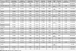
A total of 163 bacterial isolates were identified and isolated from the different clinical samples from the patients admitted to ICU during the study. As shown in Table 1, the prevalence of VAP was the highest (70, 43%), followed by catheter-associated urinary tract infection (CAUTI; 39, 24%), central line-associated bloodstream infection (CLABSI; 25, 15%), surgical site infection (SSI; 14, 8.6%), other low-level non-surgical infections (such as otitis media and wound, non-SSI; 7, 4.3%), bloodstream infection (BSI; 5, 3%), and the lowest prevalence was for respiratory tract infection (RTI; 3, 1.8%). In the case of VAP, the most predominant pathogen reported was Acinetobacter baumannii (18, 51%), while Klebsiella pneumoniae was the predominant pathogen for CAUTI (11, 28%) and CLABSI cases (6, 24%). As shown in Table 1, A. baumannii was also the most common pathogen isolated from SSI cases (9, 64%). We also examined the distribution of pathogens associated with ICU infections (Table 1) and found that K. pneumoniae was the predominant microorganism in the ICU infections, as it was found in nearly 24% of the samples (39, 24%). It was followed by A. baumannii (35, 21%), P. aeruginosa (25, 15%), and Proteus spp. (23, 14%). Other bacterial pathogens were also isolated from the clinical samples, but their prevalence rate was low (Table 1). We also investigated the month-wise distribution of infections associated with ICU patients (Table 2). The highest healthcare-associated ICU infections were observed in September (19, 11.7%), followed by the month of December and January (17, 10.4%), May (16, 9.8%), April (15, 9.2%), with the least in March (9, 5.5%).
 |
Table 1 Pathogen Burden Among Different Types of Infections in ICU Patients |
 |
Table 2 Month-Wise Distribution of Infections Associated with ICU Patients |
Next, we performed antibiotic resistant pattern for these bacteria (Table 3) and found that A. baumannii was resistant to most of the antibiotics, as it was 100% resistant towards amoxicillin/clavulanic acid, ciprofloxacin, and meropenem, followed by cefepime, ceftazidime, and nitrofurantoin (34, 97.1%). A. baumannii also showed nearly 90% resistance against the other carbapenems (such as ertapenem and imipenem; 88.6%). In fact, A. baumannii showed more than 80% resistance even against other antibiotics (such as amikacin, ampicillin, aztreonam, cefoxitin, cefuroxime, cephalothin, gentamicin, and levofloxacin). Only against tigecycline it showed 60% resistance. K. pneumoniae showed nearly 90% resistance to ampicillin (36/39), levofloxacin (36/39), amoxicillin/clavulanic acid (35/39), and ciprofloxacin (35/39). K. pneumoniae was the least resistant to tetracycline (8, 20.5%). In the case of P. aeruginosa, nearly 80% of the isolates were resistant to amoxicillin/clavulanic acid (21/25), cefuroxime (21/25), ceftazidime (20/25), ertapenem (20/25), and nitrofurantoin (20/25). However, P. aeruginosa isolates were least resistant to tigecycline (1/23, 4%).
 |
Table 3 Antibiotic Resistant Pattern of Isolated Bacterial Isolates Against Various Antibiotics |
Proteus spp. bacterial isolates were 100% resistant only against ampicillin and more than 90% resistant against cephalothin and ciprofloxacin (22/23; Table 3). Proteus spp. was the least resistant against erythromycin (1, 4.3%). Providencia stuartii showed more resistance than the above-mentioned gram-negative bacteria as it was 100% resistant towards amoxicillin/clavulanic acid, ampicillin, cefuroxime, gentamicin, levofloxacin, and nitrofurantoin, followed by cephalothin (11/12, 91.7%), aztreonam (10/12, 83.3%). In contrast, only two isolates of Providencia stuartii were resistant against imipenem (2/12, 16.7%).
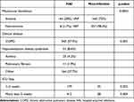
The beta-lactamases profile of the most prevalent gram-negative bacteria isolated in ICU patients was also examined, as shown in Table 4. ESBL production was the highest in K. pneumoniae (22, 56%), followed by A. baumannii (12, 34.2%), and the lowest was in P. aeruginosa (8, 32%). In the same manner, AmpC production was highest in K. pneumonia (7, 17.9%), followed by P. aeruginosa (4, 16%), and least for A. baumannii (5, 14.2%). In contrast to ESBL and AmpC, MBL production was not seen in K. pneumoniae. Only A. baumannii (8, 22.8%) and P. aeruginosa (6, 24%) isolates were producing MBL. Few bacterial isolates produced ESBL and AmpC, as shown in Table 4. In our study, we also observed that endotracheal intubation (p<0.0001) was the dominant risk factor significantly found to be associated with VAP in ICU (Table 5). The other significant risk factors associated with ICU infections observed in our study were chronic obstructive pulmonary disease (COPD) (p=0.001), prolonged ICU stay that is 1–2 weeks (P=0.025) and >2 weeks (P= 0.004).
 |
Table 4 Various Beta-Lactamases Among Highly Prevalent Bacterial Isolates |
 |
Table 5 Risk Factors Associated with Hospital-Acquired Infections (HAI) |
Discussion
In our study, we found that the gram-negative bacterial (GNB) infections in ICU patients are extremely high (156/163, 95%), and this may be due to the adoption of inadequate preventive measures. However, in this study, the association of GNB with ICU infection is higher than in most of the studies performed around the globe; in Bosnia (65.2%), India (62%), and Nigeria (50.9%).15–17 We found that the prevalence of VAP was highest (70, 43%), followed by CAUTI (39, 24%), CLABSI (25, 15%), SSI (14, 8.6%), non-SSI (7, 4.3%), BSI (5, 3%), and the lowest prevalence was for RTI (3, 1.8%). Similar findings were also reported by Parajul et al18 with VAP (53%), but in his study, the second most common infection is the bloodstream infection (18.8%) which differs from this study. Other reports also describe VAP as the most common healthcare-associated infection in the ICU.19,20.
However, in a previous study, the authors pointed to urinary tract infection as the predominant infection, followed by pneumonia and SSI.21 A study by Datta et al22 reported that CLABSI (13.50%) was the most common healthcare-associated infection, followed by UTI (10.75%) and VAP (6.15%). In our study, one isolate of S. epidermidis was reported from CLABSI infection, while the study of Datta et al did not report any S. epidermidis isolates from CLABSI. This variation in clinical types might be due to the differences in the ICU precaution measures and patient population. The most common bacterial isolates reported from our study were K. pneumoniae (39, 24%), followed by A. baumannii (35, 21.5%) and P. aeruginosa (25, 15.3%); other authors reported similar findings.23 In another study by Agarwal et al, A. baumannii was the most common isolate, followed by P. aeruginosa in VAP patients.24
In our study, the prevalence of non-fermenters was high at 60 (36.8%, 60/163). Of the non-fermenters, the A. baumannii shows the highest occurrence in VAP (18/35, 51.4%) and SSI (9/35, 25.7%) and the least in non-SSI and BSI (1/35, 2.8%) respectively. P. aeruginosa shows an equal distribution in VAP and CAUTI (9/25, 36%), with the least in non-SSI (1/25, 4%), and similar findings were reported in other studies.18,25,26 A single isolate of Burkholderia cepacia was reported in VAP in our ICU setting.
Several studies have shown that seasonal variations and climatic changes affect the frequencies of infections in hospitals,27–30 which was also observed in our study, as the highest infection cases were observed during September. Blot et al (2022) have reported an increase in the incidence of gram-negative bacteria with increasing temperature.27 In KSA, summer usually peaks from July to September. The second highest cases of infections were observed during the months of December and January, which are winter months. Reduced temperature and low humidity also caused an increase in the RTIs, as reported by Mäkinen et al (2009).30
In the ICU setting of our study, multi-drug resistance (MDR) pathogens such as Acinetobacter baumannii, Klebsiella pneumoniae, Pseudomonas aeruginosa, Proteus spp., and Providencia stuartii showed strong resistance against several antibiotics, a result which has been shown in several other studies.31–33 The emergence of multi-drug resistance strains of pathogens has been a major public health problem faced by healthcare institutions around the globe during the past few decades, because of which the efficacies of several antibiotics have decreased. These MDR pathogens are increasing the burden on the healthcare systems due to nosocomial infections in the ICU settings.34–36 Our study showed that P. aeruginosa and A. baumannii produced three types of beta-lactamases, including MBL, ESBL, and AmpC. In contrast, K. pneumoniae isolates did not produce MBL. Interestingly, a study reported the opposite, while K. pneumoniae produced MBL and P. aeruginosa isolates did not produce MBL and AmpC.37 However, in this study, 42 (42.4%, 42/99) of the ICU patients had infections with ESBL-producing gram-negative bacilli, of which the most common ESBL producer is K. pneumoniae (22/42, 56%), followed by A. baumannii (12/44, 34.2%) and P. aeruginosa (8/44, 32%). The prevalence of ESBL in this study was higher than those reported from different parts of the world, like India (35.2%), Nepal (28.2%), Qatar (26%), and France (25%).38–40 The highly significant associated risk factors related to our study were endotracheal tube (ET) intubation, prolonged mechanical ventilation, and a comorbid condition such as chronic obstructive pulmonary disease (COPD). Duration of ICU stay was also a significant risk factor associated with ICU infection, which is in agreement with the study by Confalonieri et al.41 But there is another disagreement with the finding that COPD is an associated risk factor for ICU patients in developing healthcare-associated infection.23
Gram-negative bacteria were found to be more common with HAIs than gram-positive bacteria. Post-operative state and prior hospitalization have not been shown to significantly influence HAIs in ICU. Public health problems can be triggered by hospital strains spreading into the community.42,43 The isolation of antibiotic-resistant organisms in ICU is particularly high.41,44 The wide prevalence of nosocomial infections is due to poor hygiene in hospitals, negligence on the part of medical personnel, and non-compliance with antibiotic stewardship guidelines. Hence infections caused by multi-drug resistant microorganisms continue to be one of the leading causes of hospital deaths worldwide.39,45
Study Limitations
One of the main limitations of the present study is that this was a single-center study based on the results from only one tertiary-care hospital. Large-scale sampling from different hospitals in different cities and regions would reveal much more insight into the antibiotic-resistant pattern of the microorganisms responsible for hospital-acquired infections. Also, sampling other hospital units for the prevalence of circulating ecotype strain profiles would indicate what to expect in the ICU. The other limitation of our present study is the missing or incomplete data on some of the microorganisms’ resistant patterns against various antibiotics, because of which some of the clinical specimens were excluded from our study.
Conclusion
This study presented a high prevalence of K. pneumoniae in ICU patients, followed by A. baumannii, with VAP as the most common clinical type, followed by CAUTI. ESBL production was the highest in comparison to AmpC and MBL. Hence, proper use of antibiotics, continuous monitoring of drug sensitivity patterns, and taking all precautions to prevent infections from beta-lactamase-producing microorganisms in clinical settings are crucial and significant factors in fending off life-threatening infections and for a better outcome.
Abbreviations
BSI, bloodstream infection; CAUTI, catheter-associated urinary tract infection; CLABSI, central line-associated bloodstream infection; COPD, chronic obstructive pulmonary disease; ESBL, Extended-spectrum β-lactamases; HAI, hospital-acquired infections; ICU, Intensive care unit; MBL, metallo β- lactamase; MDR, multi-drug resistant, MRSA, Methicillin-resistant Staphylococcus aureus; RTI, respiratory tract infection; SSI, surgical site infection; VAP, Ventilator-associated pneumonia.
Acknowledgments
The authors acknowledge all the participants for their involvement in the study. This research has been funded by the Scientific Research Deanship at the University of Ha’il, Saudi Arabia through the project number RG-20143.
Disclosure
The authors report no conflicts of interest in this work.
References
1. Lima J, Alves LR, Paz J, Rabelo MA, Maciel MAV, Morais MMC. Analysis of biofilm production by clinical isolates of Pseudomonas aeruginosa from patients with ventilator-associated pneumonia. [Analise da producao de biofilme por isolados clinicos de Pseudomonas aeruginosa de pacientes com pneumonia associada a ventilacao mecanica]. Rev Bras Ter Intensiva. 2017;29(3):310–316. doi:10.5935/0103-507X.20170039
2. Rodrigues ME, Lopes SP, Pereira CR, et al. Polymicrobial ventilator-associated pneumonia: fighting in vitro candida albicans-pseudomonas aeruginosa biofilms with antifungal-antibacterial combination therapy. PLoS One. 2017;12(1):e0170433. doi:10.1371/journal.pone.0170433
3. Chastre J, Fagon JY. Ventilator-associated pneumonia. Am J Respir Crit Care Med. 2002;165(7):867–903. doi:10.1164/ajrccm.165.7.2105078
4. Kalanuria AA, Ziai W, Mirski M. Ventilator-associated pneumonia in the ICU. Crit Care. 2014;18(2):208. doi:10.1186/cc13775
5. Skrupky LP, McConnell K, Dallas J, Kollef MH. A comparison of ventilator-associated pneumonia rates as identified according to the national healthcare safety network and American college of chest physicians criteria. Crit Care Med. 2012;40(1):281–284. doi:10.1097/CCM.0b013e31822d7913
6. Amaral SM, Cortes Ade Q, Pires FR. Nosocomial pneumonia: importance of the oral environment. J Bras Pneumol. 2009;35(11):1116–1124. doi:10.1590/s1806-37132009001100010
7. Torres A, Niederman MS, Chastre J, et al. International ERS/ESICM/ESCMID/ALAT guidelines for the management of hospital-acquired pneumonia and ventilator-associated pneumonia: guidelines for the management of hospital-acquired pneumonia (HAP)/ventilator-associated pneumonia (VAP) of the European Respiratory Society (ERS), European Society of Intensive Care Medicine (ESICM), European Society of Clinical Microbiology and Infectious Diseases (ESCMID) and Asociacion Latinoamericana del Torax (ALAT). Eur Respir J. 2017;50(3). doi:10.1183/13993003.00582-2017
8. Ling L, Wong WT, Lipman J, Joynt GM. A narrative review on the approach to antimicrobial use in ventilated patients with multidrug resistant organisms in respiratory samples-to treat or not to treat? that is the question. Antibiotics. 2022;11(4). doi:10.3390/antibiotics11040452
9. Rello J, Ollendorf DA, Oster G, et al. Epidemiology and outcomes of ventilator-associated pneumonia in a large US database. Chest. 2002;122(6):2115–2121. doi:10.1378/chest.122.6.2115
10. Tille PM. Bailey & Scott’s diagnostic microbiology; 2022.
11. CLSI. Performance Standards for Antimicrobial Susceptibility Testing. In: CLSI Supplement M100.
12. Jones RN, Ferraro MJ, Reller LB, Schreckenberger PC, Swenson JM, Sader HS. Multicenter studies of tigecycline disk diffusion susceptibility results for Acinetobacter spp. J Clin Microbiol. 2007;45(1):227–230. doi:10.1128/JCM.01588-06
13. Matuschek E, Ahman J, Webster C, Kahlmeter G. Antimicrobial susceptibility testing of colistin - evaluation of seven commercial MIC products against standard broth microdilution for Escherichia coli, Klebsiella pneumoniae, Pseudomonas aeruginosa, and Acinetobacter spp. Clin Microbiol Infect. 2018;24(8):865–870. doi:10.1016/j.cmi.2017.11.020
14. Satlin MJ. The search for a practical method for colistin susceptibility testing: have we found it by going back to the future? J Clin Microbiol. 2019;57(2). doi:10.1128/JCM.01608-18
15. Kovacevic P, Zlojutro B, Kovacevic T, Baric G, Dragic S, Momcicevic D. Microorganisms profile and antibiotics sensitivity patterns in the only medical intensive care unit in Bosnia and Herzegovina. Microb Drug Resist. 2019;25(8):1176–1181. doi:10.1089/mdr.2018.0458
16. Siddique SG, Bhalchandra MH, Wyawahare AS, Bansal VP, Mishra JK, Naik SD. Prevalence of MRSA, ESBL and carbapenemase producing isolates obtained from endotracheal and tracheal tubes secretions of ICU patient at tertiary care centre. Int J Curr Microbiol Appl Sci. 2017;6(4):288–299. doi:10.20546/ijcmas.2017.604.032
17. Yusuf I, Rabiu AT, Haruna M, Abdullahi SA. Carbapenem-Resistant Enterobacteriaceae (CRE) in intensive care units and surgical wards of hospitals with no history of carbapenem usage In Kano, North West Nigeria. Nigerian J Microbiol. 2015;27(1):7.
18. Parajuli NP, Acharya SP, Mishra SK, Parajuli K, Rijal BP, Pokhrel BM. High burden of antimicrobial resistance among gram negative bacteria causing healthcare associated infections in a critical care unit of Nepal. Antimicrob Resist Infect Control. 2017;6(1). doi:10.1186/s13756-017-0222-z
19. Cosic G, Djekic J, Rajcevic S, Ristic M, Ikonic N. Nosocomial infections and microbiological agents in an intensive care unit. Arch Biol Sci. 2012;64(4):1357–1362. doi:10.2298/abs1204357c
20. Dasgupta S, Das S, Chawan NS, Hazra A. Nosocomial infections in the intensive care unit: incidence, risk factors, outcome and associated pathogens in a public tertiary teaching hospital of Eastern India. Indian J Crit Care Med. 2015;19(1):14–20. doi:10.4103/0972-5229.148633
21. Mythri H, Kashinath KR. Nosocomial infections in patients admitted in intensive care unit of a Tertiary Health Center, India. Ann Med Health Sci Res. 2014;4(5):738. doi:10.4103/2141-9248.141540
22. Datta P, Rani H, Chauhan R, Gombar S, Chander J. Health-care-associated infections: risk factors and epidemiology from an intensive care unit in Northern India. Indian J Anaesth. 2014;58(1):30. doi:10.4103/0019-5049.126785
23. Rynga D, Shariff M, Deb M. Phenotypic and molecular characterization of clinical isolates of Acinetobacter baumannii isolated from Delhi, India. Ann Clin Microbiol Antimicrob. 2015;14(1). doi:10.1186/s12941-015-0101-5
24. Agarwal R, Gupta D, Ray P, Aggarwal AN, Jindal SK. Epidemiology, risk factors and outcome of nosocomial infections in a respiratory intensive care unit in North India. J Infect. 2006;53(2):98–105. doi:10.1016/j.jinf.2005.10.021
25. Habibi S, Wig N, Agarwal S, et al. Epidemiology of nosocomial infections in medicine intensive care unit at a tertiary care hospital in northern India. Trop Doct. 2008;38(4):233–235. doi:10.1258/td.2008.070395
26. Sah M, Mishra S, Ohara H, Kirikae T, Rijal B, Pokhrel B. Nosocomial bacterial infection and antimicrobial resistant pattern in a tertiary care hospital in Nepal. J Inst Med. 2014;36:38–48.
27. Blot K, Hammami N, Blot S, Vogelaers D, Lambert ML. Seasonal variation of hospital-acquired bloodstream infections: a national cohort study. Infect Control Hosp Epidemiol. 2022;43(2):205–211. doi:10.1017/ice.2021.85
28. Clark JM, Durrani H, Hagan JD, et al. Statewide Seasonal variations of infections within the intensive care unit among the trauma population. Am Surg. 2021;87(4):623–630. doi:10.1177/0003134820951496
29. Herrera-Lara S, Fernández-Fabrellas E, Cervera-Juan Á, Blanquer-Olivas R. Do seasonal changes and climate influence the etiology of community acquired pneumonia? Arch Bronconeumol. 2013;49(4):140–145. doi:10.1016/j.arbr.2013.02.004
30. Mäkinen TM, Juvonen R, Jokelainen J, et al. Cold temperature and low humidity are associated with increased occurrence of respiratory tract infections. Respir Med. 2009;103(3):456–462. doi:10.1016/j.rmed.2008.09.011
31. Prestinaci F, Pezzotti P, Pantosti A. Antimicrobial resistance: a global multifaceted phenomenon. Pathog Glob Health. 2015;109(7):309–318. doi:10.1179/2047773215y.0000000030
32. Wani FA, Bandy A, Alenzi MJS, et al. Resistance patterns of gram-negative bacteria recovered from clinical specimens of intensive care patients. Microorganisms. 2021;9(11):2246. doi:10.3390/microorganisms9112246
33. Ibrahim ME. High antimicrobial resistant rates among Gram-negative pathogens in intensive care units. Saudi Med J. 2018;39(10):1035–1043. doi:10.15537/smj.2018.10.22944
34. Saleem M, Syed Khaja AS, Hossain A, et al. Molecular characterization and antibiogram of acinetobacter baumannii clinical isolates recovered from the patients with ventilator-associated pneumonia. Healthcare. 2022;10(11):2210. doi:10.3390/healthcare10112210
35. Saleem M, Syed Khaja AS, Hossain A, et al. Catheter-associated urinary tract infection in intensive care unit patients at a tertiary care hospital, hail, kingdom of Saudi Arabia. Diagnostics. 2022;12(7):1695. doi:10.3390/diagnostics12071695
36. Pérez-Rodríguez F, Mercanoglu Taban B. A state-of-art review on multi-drug resistant pathogens in foods of animal origin: risk factors and mitigation strategies. Front Microbiol. 2019;10. doi:10.3389/fmicb.2019.02091
37. Deshmukh DG, Damle AS, Bajaj JK, Bhakre JB, Patwardhan NS. Metallo-β-lactamase-producing clinical isolates from patients of a tertiary care hospital. J Lab Physicians. 2020;3(02):093–097. doi:10.4103/0974-2727.86841
38. Kayastha K, Dhungel B, Karki S, et al. Extended-spectrum beta-lactamase-producing Escherichia coli and Klebsiella species in pediatric patients visiting international friendship children’s hospital, Kathmandu, Nepal. Infect Dis. 2020;13:1178633720909798. doi:10.1177/1178633720909798
39. Oberoi L, Singh N, Sharma P, Aggarwal A. ESBL, MBL and ampc beta lactamases producing superbugs - havoc in the intensive care units of Punjab India. J Clin Diagn Res. 2013;7(1):70–73. doi:10.7860/JCDR/2012/5016.2673
40. Pilmis B, Zahar J-R. Ventilator-associated pneumonia related to ESBL-producing gram negative bacilli. Ann Transl Med. 2018;6(20):424. doi:10.21037/atm.2018.09.34
41. Confalonieri M, Gorini M, Ambrosino N, Mollica C, Corrado A. Scientific group on respiratory intensive care of the Italian association of hospital P. Respiratory intensive care units in Italy: a national census and prospective cohort study. Thorax. 2001;56(5):373–378. doi:10.1136/thorax.56.5.373
42. Al-Tawfiq JA, Tambyah PA. Healthcare associated infections (HAI) perspectives. J Infect Public Health. 2014;7(4):339–344. doi:10.1016/j.jiph.2014.04.003
43. Klevens RM, Edwards JR, Richards CL
44. Phillips I, Shannon K. Class I ??-Lactamases. Drugs. 1989;37(4):402–407. doi:10.2165/00003495-198937040-00002
45. Giacometti A, Siquini FM, Cirioni O, Petroni S, Scalise G. Imipenem and meropenem induced resistance to beta-lactam antibiotics inPseudomonas aeruginosa. Eur J Clin Microbiol Infect Dis. 1994;13(4):315–318. doi:10.1007/bf01974609
 © 2023 The Author(s). This work is published and licensed by Dove Medical Press Limited. The full terms of this license are available at https://www.dovepress.com/terms.php and incorporate the Creative Commons Attribution - Non Commercial (unported, v3.0) License.
By accessing the work you hereby accept the Terms. Non-commercial uses of the work are permitted without any further permission from Dove Medical Press Limited, provided the work is properly attributed. For permission for commercial use of this work, please see paragraphs 4.2 and 5 of our Terms.
