Back to Journals » Nanotechnology, Science and Applications » Volume 19
Parenteral Berberine vs Cisplatin- Loaded Lipid Nanoparticles – Development, Characterisation, Comparative Safety Profiling and Cytotoxicity in Cholangiocarcinoma Cell Models
Authors Manov M, Stefanova D, Tzankova V, Tzankov B, Lozano E, Delgado-Calvo K ![]() , Macias RIR
, Macias RIR ![]() , Marin JJG, Spassova I, Kovacheva D, Slavkova M
, Marin JJG, Spassova I, Kovacheva D, Slavkova M ![]()
Received 14 January 2026
Accepted for publication 7 May 2026
Published 20 May 2026 Volume 2026:19 596038
DOI https://doi.org/10.2147/NSA.S596038
Checked for plagiarism Yes
Review by Single anonymous peer review
Peer reviewer comments 5
Editor who approved publication: Professor Kattesh Katti
Martin Manov,1 Denitsa Stefanova,1 Virginia Tzankova,1 Borislav Tzankov,2 Elisa Lozano,3 Kevin Delgado-Calvo,3 Rocio IR Macias,3 Jose JG Marin,3 Ivanka Spassova,4 Daniela Kovacheva,4 Marta Slavkova2
1Department of Pharmacology, Pharmacotherapy and Toxicology, Faculty of Pharmacy, Medical University of Sofia, Sofia, Bulgaria; 2Department of Pharmaceutical Technology and Biopharmaceutics, Faculty of Pharmacy, Medical University of Sofia, Sofia, Bulgaria; 3National Institute for the Study of Liver and Gastrointestinal Diseases (CIBEREHD), Institute of Biomedical Research (IBSAL), University of Salamanca, Salamanca, Spain; 4Institute of General and Inorganic Chemistry, Bulgarian Academy of Sciences, Sofia, Bulgaria
Correspondence: Marta Slavkova, Department of Pharmaceutical Technology and Biopharmaceutics, Faculty of Pharmacy, Medical University of Sofia, 2 Dunav str., 1000, Sofia, Bulgaria, Tel +359 2 923 65 27, Email [email protected]
Introduction: Cholangiocarcinoma (CCA) is an aggressive and heterogeneous malignancy of the biliary tract with a poor prognosis. Berberine can be a therapeutic option with low toxicity, but its delivery remains challenging. Encapsulation in lipid nanoparticles offers a promising biocompatible strategy to improve the delivery and safety of berberine.
Purpose: This study aimed to develop, characterize berberine-loaded nanostructured lipid carriers (N-Ber) and evaluate their cytotoxicity in CCA cell models in comparison to a standard chemotherapeutic, cisplatin, in both free and nanoparticle-loaded form (N-Cis). The in vitro biocompatibility for potential parenteral delivery of the nanocarriers was a secondary goal of the work.
Methods: Nanoparticles were prepared by solvent evaporation with Precirol 5 ATO and oleic acid as lipids and Tween 20 as a stabilizer and were characterized by DLS, morphology, encapsulation efficiency, and in vitro drug release at physiological (pH 7.4) and tumor-mimicking (pH 5.5) conditions. Cytotoxicity was evaluated in three CCA cell lines (TFK-1, EGI-1, and HuCCT1) using the MTT assay, while endothelial cells (Ea.hy926) and the hemolysis test in human erythrocytes were employed to evaluate safety.
Results: N-Ber and N-Cis displayed mean sizes 159.5 nm and 146.7 nm, respectively, negative surface charge (− 27.95 mV and − 50.75 mV), and high encapsulation efficiencies (berberine: 88.8%; cisplatin: 95.8%). Both nanoformulations showed significantly altered dissolution profiles compared to the free drugs. Berberine showed potent cytotoxicity against all CCA cell lines (IC50 0.4– 5.2 μM), comparable or superior to cisplatin (IC50 ≈ 10 μM) and these effects were preserved after nanoencapsulation. In contrast, N-Ber exhibited low toxicity towards endothelial cells and no relevant hemolytic activity (< 5% hemolysis) at therapeutic concentrations, while N-Cis induced marked hemolysis at all tested doses.
Discussion: N-Ber combines strong cytotoxicity in CCA cells with a favorable biocompatibility compared to N-Cis. The promising in vitro data warrant further in vivo evaluation.
Keywords: cholangiocarcinoma, berberine, cisplatin, nanostructured lipid carriers, cytotoxicity
Introduction
Cholangiocarcinoma (CCA) is a malignancy originating from the epithelial cells of the bile ducts and is broadly classified into intrahepatic and extrahepatic subtypes.1 CCA’s aggressive nature is associated with increasing morbidity and mortality in Europe.2,3 CCA accounts for about 3% of all gastrointestinal cancers and represents 10 to 15% of all primary liver tumors.3 Their asymptomatic course usually leads to late diagnosis, which makes treatment difficult and results in a very bad prognosis of less than 5% survival in 1–5 years.3,4 In biliary tract cancer (BTC), standard first-line chemotherapy has traditionally relied on gemcitabine/cisplatin-based chemotherapy, with other cytotoxic agents such as paclitaxel and capecitabine often used as adjuvant or neoadjuvant therapy.5 More recently, the addition of immune checkpoint inhibitors (anti-PD/PD-L1 antibodies) to gemcitabine/cisplatin has been introduced as a new first-line option, improving overall survival.6 Although cisplatin-containing regimes can reduce tumor burden and prolong survival, their clinical use is limited by significant limitations. Cisplatin (Cis) treatment is frequently accompanied by adverse drug reactions, including nephrotoxicity, ototoxicity and neurotoxicity, which often lead to dose reduction and markedly impair patients’ quality of life.7 In addition, tumor cells can acquire resistance to cisplatin through different mechanisms, resulting in treatment failure and disease progression.8 In addition, there is limited overall survival achieved by standard available therapies.9 These limitations underscore the need to develop alternative or complementary therapeutic strategies for CCA.
Among the emerging strategies, two complementary trends stand out: the application of nanotechnology-based drug delivery systems and the use of bioactive compounds of natural origin. Nanotechnology offers the potential to improve the bioavailability of anticancer agents and to modulate their release profile over time or at specific target sites.10 In parallel, phytochemicals are attractive candidates in oncology because they may mitigate toxicity in normal cells or enhance cytotoxic effects in tumor cells through diverse mechanisms.11 Natural compounds often display multi-target modes of action and a favorable safety profile, making them promising scaffolds for anticancer drug development.
Berberine (Ber) is an isoquinoline alkaloid present in several medicinal plants (Berberis sp., Coptis sp.) with documented antitumor activity. It can modulate multiple signaling pathways involved in cell proliferation, apoptosis and metastasis, and has therefore been proposed as an anticancer agent.12 Its antiproliferative effects are associated with multi-kinase inhibitory activity, including suppression of the EGF/EGFR signaling pathway and its downstream effectors Erk, STAT3 and Akt, as well as inhibition of EMT-related mediators such as VEGFA and Slug, thereby regulating cell proliferation, migration and apoptosis.12 Several studies demonstrated its applicability in gastrointestinal-related tumors, eg colorectal, gastric, hepatocellular, gallbladder cancer, and cholangiocarcinoma.13–18 In addition, there is evidence for berberine’s liver tropism regardless of the administration route.19,20 Within nanoparticulate formulations, berberine ameliorates liver injury of different origins.21–23 Different studies showed that berberine in the free state or loaded within nanoparticles is superior or at least comparable to the effects of cisplatin in damaged liver, non-small cell lung cancer cell lines (A549), breast cancer cells (MCF-7), human hepatocellular carcinoma cell line (HepG2) and others.22,24–26 Together with these anticancer effects, the off-site toxicity to normal cells is lower than that of standard chemotherapeutics.24,27,28 Further advantages can be expected from the enhancement of cisplatin sensitivity in different tumor cells when combined.29–31 Taken together, all these properties make berberine a suitable natural compound for studying its potential applicability in cholangiocarcinoma.
However, the clinical translation of berberine is limited by its physicochemical properties. It has low aqueous solubility (4.51 mmol/l) and a low partition coefficient (logP = −0.91), indicating that the molecule is difficult to be absorbed in the gastrointestinal tract.32 Hence, berberine’s oral bioavailability is low. This observation is further enhanced by extensive intestinal metabolism and efflux mediated by P-glycoprotein pumps.33 Consequently, a suitable approach is necessary to enhance berberine exposure at the tumor site while maintaining an acceptable safety profile. One possibility is the parenteral application, which can be further enhanced by the formulation of a suitable nanocarrier system.
Nanostructured lipid carriers (NLCs) are a relatively recent class of lipid-based nanoparticles, introduced in 1999/2000 to overcome several drawbacks associated with conventional lipid carriers such as liposomes and solid lipid nanoparticles (SLNs).34 NLCs offer important advantages, including increased drug-loading capacity, improved long-term stability, the ability to encapsulate both hydrophilic and hydrophobic molecules, better control over drug release, and suitability for large-scale production.34–37 They are typically composed of solid and liquid lipids together with an appropriate stabilizer, which can be tailored in different ratios to optimize their properties.35 Due to the inherent biodegradability and biocompatibility, NLCs are suitable for various routes of administration and have been widely explored as carriers for chemotherapeutic drugs.35,37 In this context, NLCs can help mitigate systemic toxicity, enable combinatorial loading of different active agents and potentially overcome multidrug resistance.37 Moreover, lipid-based nanocarriers tend to accumulate preferentially in the liver following parenteral administration,38,39 which represents an additional advantage for targeting hepatobiliary malignancies such as CCA. Furthermore, typically, NLC preparation is more cost-effective in comparison to lipid nanoparticles that use ionizable lipids and microfluidic preparation techniques because of the more common lipids used and simpler techniques.40
Several nanoparticles have already been investigated as carrier systems for berberine. These include polymeric (albumin, chitosan, gelatin) and inorganic nanocarriers (copper oxide, zinc oxide, silver, mesoporous silica) as well as dendrimers.22,24,26,41–45 Different lipid-based nanoformulations were also studied.18,46–49 Our previous work showed that a nanostructured lipid carrier system with berberine has the potential to ameliorate gastrointestinal-related cancers in vitro (colorectal adenocarcinoma (CaCo-2) and cholangiocarcinoma (HuCC-T1)).18 Even though all these data are promising, the potential of nanoformulated berberine in cholangiocarcinoma still needs evaluation. There is a significant molecular heterogeneity between BTC with different localizations or even in the same localisation.50 This variability is substantially challenging for active targeting and, together with the common development of multidrug resistance, turns the chemotherapy of BTC into a very difficult task. Thus, investigating berberine’s effectiveness within lipid-based nanoformulation in different cholangiocarcinoma cell lines and comparing it to a standard chemotherapeutic such as cisplatin may answer whether this natural alkaloid can be a suitable candidate for a new therapeutic strategy. Other studies also used cisplatin as a reference when preparing innovative nanoparticles for breast cancer treatment, for example. However, cisplatin is not loaded in a similar formulation but is used in its free state.51
Based on this rationale, this study aimed to test the proof-of-concept development and characterization of berberine-loaded lipid nanoparticles (N-Ber) and to evaluate their cytotoxic effects in different CCA cell lines. Cisplatin was used as a comparator to provide a benchmark for the cytotoxicity measurement. For this purpose, we also prepared cisplatin-loaded NLCs (N-Cis), used as a reference nanoformulation, and with the secondary task to investigate whether the nanoparticles affect the cytotoxicity performance. Endothelial cells Ea.hy926 and hemolysis assay in human erythrocytes were used to assess the safety and biocompatibility profiles of empty and drug-loaded lipid nanoparticles for the intended parenteral delivery. These investigations are preliminary but still can bridge the gap between the nanotechnological approach and the potential clinical translation of novel natural drugs in cholangiocarcinoma treatment.
Materials and Methods
Chemicals
RPMI medium and DMEM/F12 were obtained from GIBCO (Thermo Fisher, Madrid, Spain); DMEM low glucose and non-essential amino acids solutions were purchased from Sigma-Aldrich (Madrid). Precirol 5 ATO (5ATO) was kindly gifted by Gattefosse, France. Berberine hydrochloride (Ber) and 1,2-phenylene diamine (OPDA) were obtained from Fluorochem (Hadfield, UK). Cisplatin (Cis), oleic acid (OA), dichloromethane and Tween 20 were purchased from Thermo Fischer Scientific (Loughborough, UK). Methanol was obtained from Merck KGaA (Darmstadt, Germany). All other reagents and chemicals were of analytical grade.
Cell Lines
Three CCA cell lines were used: TFK-1 and EGI-1 (extrahepatic CCA origin), obtained from the German Collection of Microorganisms and Cell Cultures (DSMZ, Braunschweig, Germany), and HuCCT1 (intrahepatic CCA origin), from the Japanese Collection of Research Bioresources (JCRB, Cell Bank Osaka, Japan). TFK-1 cells were cultured in RPMI 1640, EGI-1 cells with DMEM low glucose and HuCCT1 cells with DMEM/F12. All media were supplemented with 10% heat-inactivated foetal bovine serum (FBS) and 1% penicillin/ streptomycin (Gibco, Thermo Fisher). A non-essential amino acid solution was also added to EGI-1 media. Cells were periodically tested to confirm that they were mycoplasma-free (Biotools B&M Labs, Madrid). Ea.hy926 cells (CRL-2922) were acquired from the American Type Culture Collection (ATCC, Manassas, Virginia, USA) and were cultured in DMEM low glucose with 10% FBS, 2 mM L-glutamine and 1 x HAT (5 mM hypoxanthine, 20 lM aminopterin, and 0.8 mM thymidine; 21060–017; Gibco). All the cell lines were maintained in 5% CO2 and 37 °C.
NLC Preparation and Drug Loading
NLCs were prepared by the solvent evaporation method with minor alterations.52 The parameters were previously selected based on trial and error without systematic optimization. In brief, the lipids (5ATO: OA; 2:1 w/w) were dissolved in a mixture of dichloromethane and methanol (4:1 v/v). The aqueous phase contained Tween 20 (0.3% w/v) and was heated to 70°C. The organic phase was then added dropwise to the aqueous phase and subjected to magnetic stirring (1000 rpm) for 15 min. The resulting coarse emulsion was subjected to vapor evaporation using Büchi Rotavapor R-124 (Büchi Labortechnik AG, Postfach, Switzerland) at 40°C for 30 min to remove the organic phase. The final nanoparticles were obtained after pulse ultrasonication at 50% amplitude (20 s/10 s on/off) for 3 min with a probe sonicator (Bandelin Sonopuls HD3100, Bandelin Electronics, Berlin, Germany). For drug-loaded formulations, berberine and cisplatin were introduced in the organic phase in 15.6% w/w and 3.6% w/w relative to the total lipid content, respectively. The resulting nanocarriers were designated as NE for the empty nanoparticles (drug-free NLCs), N-Ber for berberine-loaded NLCs and N-Cis for cisplatin-loaded NLCs.
Encapsulation Efficiency and Loading Capacity
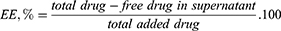
Encapsulation efficiency and loading capacity were determined indirectly by the amount in the filtered dispersion with the help of Vivaspin 100 KDa MWCO centrifugal separator (Sartorius Stedim Lab Ltd., UK) at 4000 rpm, 15 °C for 30 min in a cooling centrifuge (Micro 220R, Hettich, Tuttlingen, Germany). The concentration of free berberine was measured spectrophotometrically at 430 nm (Thermo Scientific Evolution 300, Madison, WI, USA) based on previous calibration. For cisplatin, a derivatizing reaction based on complexation with 1,2-phenylene diamine in phosphate buffer with pH 6.8 (PBS) was applied.53,54 Briefly, an aliquot of the filtrate (after centrifugation) was mixed with 1 mL of freshly prepared OPDA solution, heated at 90 °C for 30 min, cooled to room temperature, and the final volume was adjusted to 10 mL with dimethyl formamide: water (7:3 v/v, pH 6.2 adjusted with 0.1 M HCl). The resulting cisplatin-OPDA complex was quantified by UV-vis spectrophotometry at 705 nm against a blank. The amount was measured based on a previously prepared calibration curve with a series of samples with a known concentration of cisplatin. The encapsulation efficiency (EE) and the loading capacity (LC) were calculated according to the following formulas:
Particle Size, Polydispersity and Surface Charge
Zetasizer (Malvern Instruments, Malvern, UK) was utilized to determine the physicochemical properties by dynamic light scattering. Freshly prepared dispersions of the empty nanoparticles and drug-loaded N-Ber and N-Cis formulations were diluted 1:10 (v:v) with distilled water prior to analysis. The measurements were carried out at 25°C and a scattering angle of 173°. Each sample was analyzed in triplicate, and results are expressed as mean ± standard deviation (SD).
FTIR-ATR
Attenuated Total Reflectance-Fourier-transform infrared (FTIR-ATR) spectroscopy was used to investigate possible drug-excipient interactions and the structural organization of the lipid matrix. The freshly prepared NLC suspensions (NE, N-Ber, N-Cis) were lyophilized using an Alpha 344 LSC basic semi-industrial freeze-dryer (Martin Christ, Gefriertrocknunganlagen GmbH, Osterode, Germany). Samples were first frozen at −40 °C for 24 h and then freeze-dried in two stages: first at −110 °C and 0.125 mbar for 72 h, followed by a second drying at −110 °C and 0.05 mbar for 6 h.
The dried samples and individual components were then analyzed on a Thermo-Nicolet FTIR instrument equipped with an attenuated total reflectance (ATR) device (Thermo Scientific Nicolet). FTIR-ATR spectra were recorded in the range of 4000–400 cm−1 and with a resolution of 4 cm−1.
P-XRD
Powder X-ray diffraction (XRD) was used to assess the crystalline state of the drugs and the lipid matrix. Measurements were performed on a Bruker D8 Advance diffractometer equipped with Cu Kα radiation (λ = 1.5406 Å) and a LynxEye position-sensitive detector. Data were collected in Bragg-Brentano geometry using a step size of 0.02° in 2θ and an angular range spanning 5.3–80° 2θ. For small-angle XRD measurements in the angle region (0.4–5° 2θ), the instrument was configured with a manually adjustable knife-edge to reduce parasitic scattering from the direct beam. The small-angle scans were acquired using the same step increment of 0.02° 2θ, ensuring consistent angular resolution across the full measurement range.
Shape and Surface Morphology
The morphology of NE, N-Ber, and N-Cis was evaluated by scanning electron microscopy using a Hitachi TM 4000. The samples were prepared following the procedure proposed by Saghafi et al.55 The freshly prepared samples were diluted with ethanol, and glass coverslips were submerged in the dispersion for 5 min. The glass slides were then dried in a desiccator over silica, sputter-coated with gold (JEOL SC7620) and observed at different magnifications.
In vitro Drug Release
In vitro release of berberine and cisplatin from N-Ber and N-Cis was evaluated using a dialysis method. Release studies were carried out in PBS pH 7.4, simulating physiological blood conditions, and phosphate buffer pH 5.5, simulating the acidic tumor microenvironment.54 Regenerated cellulose dialysis bags (MWCO 12–14 kDa) were soaked overnight in release medium before use. 3 mL of N-Ber or N-Cis dispersion, containing approximately 5 mg berberine and 1 mg cisplatin, were placed inside the dialysis bag, which was sealed and immersed in 100 mL release medium. Samples were incubated in a shaking water bath at 37 ± 0.5 °C and 100 rpm.
Samples containing aqueous dispersions of the investigated drugs were used as a control for the unrestricted diffusion through the membrane. At different time points (1 h, 2 h, 3 h, 4 h, 5 h, 6 h, 12 h and 24 h), samples were withdrawn to determine the released amount based on spectrophotometric evaluation explained earlier. At each interval, fresh medium was added to maintain a constant volume of 100 mL. All tests were performed in triplicate, and the results are given as means with standard deviations.
To compare the release profiles, two-way ANOVA statistical analysis followed by Tukey’s post hoc comparison was applied with a significance level set to p<0.05 to compare samples and time points. In addition, the similarity factor (f2) was calculated based on the following equation:
The comparison was made between the free and the nanoencapsulated drugs’ mean cumulative released amounts at the sampling points within each pH medium. If the f2 value is above 50, it demonstrates similarity between the dissolution profiles.56
The dissolution data was also fitted to various release kinetics models (Zero order, First order, Higuchi and Korsmeyer-Peppas) with the help of the DDSolver add-in in Excel. The best-fitted model is the one with the highest R2 value.57
Cell Viability Assay
Cell viability was measured using the MTT assay.58 After the treatment with the test substances – Ber 3.125–200 μM, Cis 1.56–100 μM, N-Ber 3.125–200 μM, N-Cis 1.56–100 μM and NE 18.75–1200 μM, MTT solution (0.5 mg/mL) was added to each well. After 4–5 h incubation at 37 °C the MTT solution was aspirated and 200 μL DMSO were used to solubilize the formazan crystals, and absorbance was measured at 540 nm, using Synergy 2 microplate reader (BioTek Instruments, Inc., Highland Park, Winooski, VT, USA). Values are expressed as a percentage of the viability of untreated cells (Control) and are the mean ± SD of at least 6 independent wells from three separate experiments.
Cytostatic Effect
To evaluate the cytostatic effect of free and nanoencapsulated drugs, cells were seeded in 96-well plates at a density of 6.5 × 10^3 cells/100 µL for TFK-1 and HuCCT1, and 7.5 × 10^3 cells/100 µL for EGI-1 and 100 µL cell suspension was added to each well. After 24 h to allow cell attachment, the culture medium was replaced with fresh medium containing increasing concentrations of the test compounds (1.56, 3.125, 6.25, 12.5, 25, 50 and 100 μM for Cis and 3.125, 6.25, 12.5, 25, 50, 100 and 200 μM for Ber). Cells were exposed for 72 h, after which cell viability was determined by the MTT assay as described. Untreated cells were used as negative controls in all cytotoxicity assays and served as a reference condition for normalization (100% cell viability). Cytotoxicity of N-Ber, N-Cis, and NE was compared with that of the corresponding free drugs. All experiments were performed using independent nanoparticle batches.
Safety Evaluation in Endothelial Cells
The endothelial cell line Ea.hy926 was used as a model to assess the safety of the nanoparticles. The cells were seeded in a 96-well plate at a density of 1 × 10^4 cells/well. They were exposed to different concentrations (the same as in the test for cytotoxicity) of NE, N-Ber and N-Cis for 24 h, and cell viability was determined using the MTT assay. Untreated cells were used as negative controls and defined as 100% cell viability for normalization.
Haemolysis Assay
The hemolytic potential of loaded and non-loaded nanoparticles is a standard test for biocompatibility. Human blood was acquired from a certified clinical laboratory, according to the rules of the Institutional Ethics Committee (KENIMUS) at the Medical University, Sofia, Bulgaria (link at: https://mu-sofia.bg/nauka/nauka/etika-nauchni-izsledvania/). The assay was performed according to a previously described protocol.59 Whole blood samples were centrifuged to separate erythrocytes from plasma. The erythrocytes were resuspended in PBS (pH 7.4). 1 µL of packed erythrocytes was diluted in 49 mL PBS. Aliquots of 190 μL of the erythrocyte suspension and 10 µL of NE, N-Ber, or N-Cis formulations were mixed and added to 96-well plates. PBS was used as a negative control (0% haemolysis), while Triton X-100 (1% v/v) served as a positive control (100% haemolysis). These controls were used to normalise the haemolysis values of the tested formulations and to ensure assay validity. After incubation at 37 °C for 1 h, the plates were centrifuged, and 100 μL of the supernatant of each well was transferred to a new plate. The amount of released hemoglobin, a marker for hemolysis, was determined spectrophotometrically at 430 nm with a Synergy 2 microplate reader (BioTek Instruments, Inc., Highland Park, Winooski, VT, USA).
Statistical Analysis
Experimental data were statistically analysed using GraphPad Prism8. All in vitro experiments were performed in at least three independent biological replicates. For each concentration, six wells were used per condition within each experiment. Data are expressed as mean ± standard deviation (SD), reflecting variability between wells. Reproducibility between independent experiments was confirmed, showing consistent trends across datasets. Statistical comparisons between multiple groups were carried out using one-way analysis of variance (ANOVA). When a significant overall effect was detected, Dunnett’s post hoc test was applied to compare each treatment group against the corresponding untreated control. This approach was selected due to the study design focusing on comparisons versus a single control group. A p-value < 0.05 was considered statistically significant.
Results
Physicochemical Characterization of Lipid Nanoparticles
Particle Size, Polydispersity and Surface Change
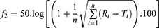
Dynamic light scattering (DLS) analysis revealed that all formulations consisted of relatively small nanoparticles ranging from 128 nm to 159 nm (Table 1). Drug loading resulted in a moderate increase in the size of the nanocarriers compared with NE. It could be explained by the additional space needed for the drug. Encapsulation efficiency was high for both drugs (Table 1).
 |
Table 1 Nanoparticles z-Average Size, Polydispersity Index (PDI), and Surface Charge (Zeta Potential) Determined in Water at 25°C and 173° Scattering Angle; Encapsulation Efficiency (EE) and Loading Capacity (LC) (Mean ± SD, n=3) |
All formulations exhibited relatively narrow size distributions (Figure 1A), with polydispersity index (PDI) values in the range 0.38–0.43, indicative of acceptable homogeneity for colloidal dispersions. However, some additional populations with particle sizes different from the z-average reported (Table 1). It is worth noting that the presented formulations are not systematically optimised as the aim of the study is proof-of-concept. The inclusion of berberine and cisplatin was successful, as can be seen from the change in the zeta potential (Figure 1b). The molecule of berberine possesses a quaternary ammonium atom in its structure, which leads to some compensation of the nanocarriers’ negative charge. Similar values were reported by other researchers.54,60 In the case of cisplatin, our results correspond to already published data.61
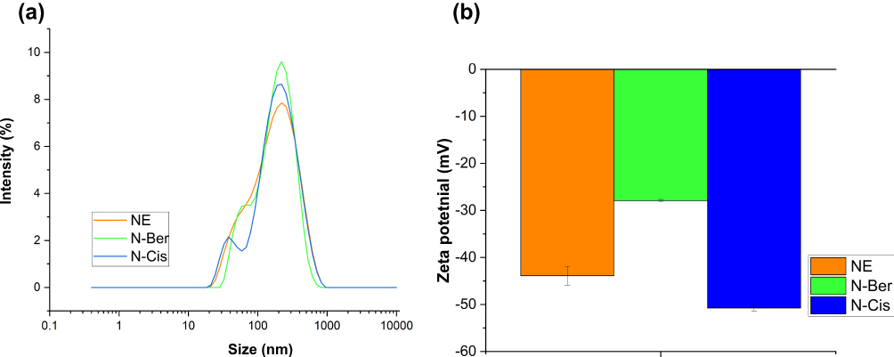 |
Figure 1 Size distribution by intensity (a) and zeta potential (b) of the empty (NE), berberine- (N-Ber) and cisplatin- (N-Cis) loaded nanostructured lipid carriers measured in distilled water at 25°C. |
Morphology
Scanning electron microscopy (SEM) images confirmed that NE, N-Ber and N-Cis consisted mainly of spherical particles with smooth surfaces (Figure 2). All formulations have uniform and relatively small sizes. Drug-loaded particles increased their size, although this tendency is limited. Both DLS and SEM results provided clear evidence that all three formulations resulted in nanometer-sized particles. However, SEM provides information about the morphology of the particles as they exist in a dried state, whereas DLS measures the hydrodynamic diameter of the particles in a liquid medium. Therefore, some minor discrepancies can occur when comparing results from the two different techniques.
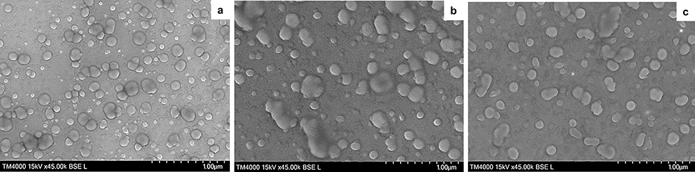 |
Figure 2 Morphology of the of the empty (a), berberine- (b) and cisplatin- (c) loaded nanostructured lipid carriers. |
Fourier-Transform Infrared Analysis
FTIR-ATR spectra of the individual components (Precirol ATO 5, oleic acid, Tween 20) and the corresponding nanoparticles are presented in Figure 3.
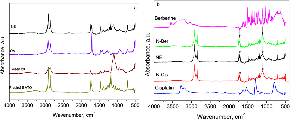 |
Figure 3 FTIR-ATR spectra of (a): Precirol 5 ATO, Tween 20, oleic acid and empty lipid nanoparticles NE; (b): free drugs berberine and cisplatin and the corresponding loaded nanoparticles N-Ber and N-Cis. |
The lipid matrix was characterized by peaks at ~2916 cm−1 and ~2849 cm−1 (Figure 3A), attributed to asymmetric and symmetric C–H stretching vibrations of –CH2 groups, as well as the carbonyl stretching vibration around ~1730 cm−1 for Precirol 5 ATO and ~1710 cm−1 for oleic acid, which are typical for solid and liquid lipids used in NLC formulations.62
The successful formation of the lipid nanoparticles is based mainly on physical interactions, practically without chemical modifications of the lipid backbone. In NE (Figure 3a), the bands were preserved, including a carbonyl doublet at 1732 and 1714 cm−1, confirming the coexistence of both Precirol 5 ATO and oleic acid. The retention of individual bands confirms that lipids are partially immiscible and form microphase-separated domains without chemical interaction.63 Bands in the 1100–1000 cm−1 region, corresponding to C–O–C stretching vibrations, reflected the presence of Tween 20 PEG chains on the nanoparticle surface.64
In Figure 3b, one can see the spectra of initial drugs (Berberine and Cisplatin) along with the spectra of the respective loaded lipid nanoparticles (marked as N-Ber, N-Cis).
The spectrum of Berberine is consistent with the literature.65 The bands in the region from 3570 to 3050 cm−1 originate from the vibrations of loosely and strongly hydrogen-bonded water molecules with different environments. The spectrum also contains a well-resolved peak at 1632 cm−1 due to C=N stretching of quaternary bonded nitrogen as well as C–O stretching vibrations of the methoxy group appearing in the 1200–1000 cm−1 region.
In the spectrum of cisplatin, N–H stretching vibrations at 3280 cm−1 and 3200 cm−1 are lower than those in free ammonia due to coordination with platinum. The N–H bending vibrations appear in the region 1630–1540 cm−1. The 1293 cm−1 and 798 cm−1 peaks arise from the N–H vibrations of the coordinated –NH3 ligands in cisplatin.61
In N-Ber and N-Cis, the characteristic bands of berberine and cisplatin were largely overlapped by the lipid signals, but their spectral features remained compatible with physical entrapment rather than covalent interaction, indicating that drug loading did not induce major structural changes in the lipid matrix.
Powder X-Ray Diffraction
(P-XRD) Patterns provided further insight into the solid-state organisation of the components. The wide- and small-angle parts of powder diffraction patterns of the empty lipid nanoparticles NE, pure berberine and cisplatin, as well as the drug-loaded nanoparticles N-Ber and N-Cis, are shown in Figure 4.
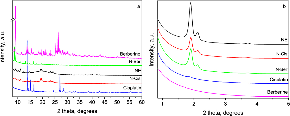 |
Figure 4 Wide-angle X-Ray diffraction patterns (a) and small-angle X-Ray diffraction patterns (b) of empty lipid nanoparticles NE, pure berberine and cisplatin, as well as the drug-loaded nanoparticles N-Ber and N-Cis, respectively. |
The wide-angle diffraction pattern of berberine (Figure 4a) displays sharp, intense peaks, indicating a high degree of crystallinity. The pattern is consistent with the ICDD PDF file #00-048-2342 corresponding to the berberine chloride tetrahydrate. The refined unit cell parameters within the space group P-1 are: a = 7.010(1) Å, b = 11.501(4) Å, c = 13.165(3) Å, α = 76.07(2)°, β = 88.99(2)°, and γ = 85.14(3)°, which are close to previously reported values.66,67
The wide-angle diffraction pattern of cisplatin (Figure 4A) also shows sharp peaks with high intensities typical of a substance with high crystallinity. The observed set of interplanar distances corresponds to the ICDD PDF file #00-050-0643. The crystal modification is the triclinic (P-1) room-temperature β-form, and the refined unit cell parameters were: a = 6.2829(4) Å, b = 13.5602(9) Å, c = 6.6984(3) Å, α = 69.194(6)°, β = 86.27(1)°, and γ = 83.140(6)°, close to previous observations.65
The wide-angle diffraction pattern of lipid nanoparticles (NE) (Figure 4A) is a complex one and shows the results of the interaction between the solid (Precirol ATO 5) and liquid (OA) lipid components in the presence of surfactant (Tween 20). In the present case, a splitting of characteristic lines of Precirol ATO 5 in the region 19–20° and 23–24° 2θ is observed, combined with the appearance of the additional lines at 6.2°, 7.5°, 9.2°, 11.7°, and 14.8° 2θ.
The small-angle part of this diffraction pattern (Figure 4B, NE) consists of three peaks at 1.9°, 2.1° and 3.7° 2θ corresponding to the interplanar distances of 46.3, 41.7, and 23.8 Å, respectively. Having in mind that Precirol ATO 5 is a mixture of mono-, di-, and triglycerides of C16 and C18 fatty acids, these long interplanar distances are supposed to be due to the mixture of different crystal modifications of the triglycerides and the different chain length of the fatty acids.68
The XRD pattern of N-Cis contains all major reflections of the lipid carrier together with the strongest peak of cisplatin (around 14° 2θ), confirming successful loading of the cisplatin on the lipid nanoparticles. In contrast, N-Ber reveals that during the incorporation, the berberine drug transforms from tetrahydrate to dihydrate form, since the characteristic peaks of the dihydrate form are clearly seen together with the peaks of the carrier.67 The small-angle patterns of the loaded samples contain only peaks of the carrier, as can be seen in Figure 4.
In Vitro Drug Release
The in vitro release studies showed sustained berberine and cisplatin release from N-Ber and N-Cis under both physiological (pH 7.4) and acidic (pH 5.5) conditions (Figure 5). The quantitative comparison of the dissolution profiles between the free berberine and its corresponding nanoparticulate formulation at pH 7.4 and 5.5 resulted in f2 values of 26.89 and 28.04, respectively. In the case of cisplatin, the same assessment also indicated that the two profiles are not similar (f2 = 27.07 and f2 = 27.69 for pH 7.4 and 5.5, respectively). For berberine, nanoencapsulation increased the amount of drug available over time compared with the free compound, particularly at pH 7.4, where N-Ber provided a more controlled release profile. In the case of cisplatin, the free drug showed no marked pH dependence, whereas N-Cis displayed a clearly sustained release pattern, more pronounced at pH 7.4.
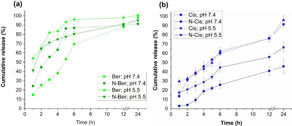 |
Figure 5 In vitro dissolution profiles of (a) free berberine - Ber, single-loaded berberine – N-Ber, and (b) free cisplatin (Cis) and the single-loaded cisplatin – N-Cis. Release media phosphate buffer pH 5.5 and 7.4, temperature 37 ± 0.5°C, 100 rpm, three replicates (mean ± SD). Statistical significance was determined by two-way ANOVA followed by Tukey’s multiple comparisons test. All two sets of profiles are significantly different (p<0.001). |
The assessment of dissolution kinetics (Table 2) demonstrated that the free cisplatin followed first order regardless of the pH. Similar data were reported earlier.69 Upon loading in the NLCs, the release was diffusion-controlled from the insoluble matrix system of the nanocarrier without any impact of the pH medium (highest linearity for the Higuchi model). Such a behaviour for insoluble drugs was demonstrated before.70 In the case of berberine, there was a change to the Higuchi model only for the nanoencapsulated drug at pH 7.4. At this pH, the solubility of berberine is lower than at pH 5.5, which can explain the observations. In other samples, the highest linearity is observed for the first order, which is supported by earlier studies.71
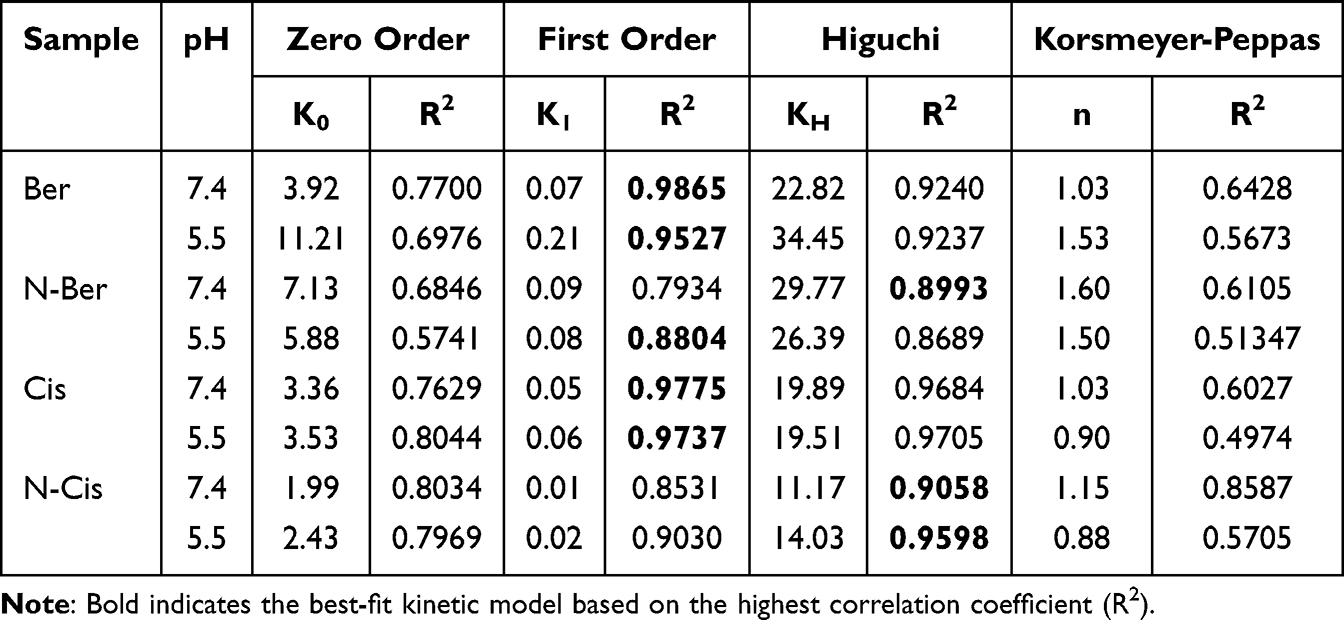 |
Table 2 Dissolution Kinetic Parameters for the Free and Encapsulated Berberine and Cisplatin at pH 7.4 and pH 5.5 Obtained from Zero-Order, First-Order, Higuchi and Korsmeyer-Peppas Models |
Biological Characterization
Cytotoxic Potential in CCA Cells
The cytotoxic effects of free berberine, free cisplatin, N-Ber, N-Cis and NE were evaluated in TFK-1 (Figure 6), EGI-1 (Figure 7) and HuCCT1 (Figure 8) CCA cell lines after 72 h exposure using the MTT assay.
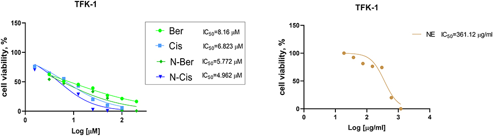 |
Figure 6 Effects of free berberine (Ber; 3.125 μM – 200 μM), free cisplatin (Cis; 1.56 μM – 100 μM), single-loaded berberine (N-Ber; 3.125 μM – 200 μM), single-loaded cisplatin (N-Cis; 1.56 μM – 100 μM) and non-loaded lipid nanoparticles (NE; 18.75 μg/mL – 1200 μg/mL) on TFK-1 cell viability after 72 h treatment. The results are expressed as means ± SD of triplicate assays (n=6). All groups were compared statistically vs untreated controls by one-way ANOVA with Dunnett’s post-test. |
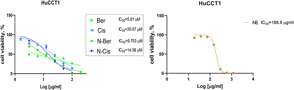 |
Figure 7 Effects of free berberine (Ber; 3.125 μM – 200 μM), free cisplatin (Cis; 1.56 μM – 100 μM), single-loaded berberine (N-Ber; 3.125 μM – 200 μM), single-loaded cisplatin (N-Cis; 1.56 μM – 100 μM) and non-loaded lipid nanoparticles (NE; 18.75 μg/mL – 1200 μg/mL) on HuCCT1 cell viability after 72 h treatment. The results are expressed as means ± SD of triplicate assays (n=6). All groups were compared statistically vs untreated controls by one-way ANOVA with Dunnett’s post-test. |
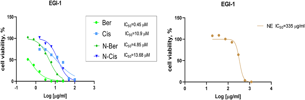 |
Figure 8 Effects of free berberine (Ber; 0.39 μM – 25 μM), free cisplatin (Cis; 1.56 μM – 100 μM), single-loaded berberine (N-Ber; 0.39 μM – 25 μM), single-loaded cisplatin (N-Cis; 1.56 μM – 100 μM) and non-loaded lipid nanoparticles (NE; 18.75 μg/mL – 1200 μg/mL) on EGI-1 cell viability after 72 h treatment. The results are expressed as means ± SD of triplicate assays (n=6). All groups were compared statistically vs untreated controls by one-way ANOVA with Dunnett’s post-test. |
Both free berberine and N-Ber induced a pronounced, concentration-dependent reduction in cell viability in all three cell lines. Berberine demonstrated strong cytotoxicity, with IC50 values ranging from 0.4 to 5.2 µM, which was comparable to or more effective than cisplatin, whose IC50 values were approximately 10 µM across all models. Notably, the highest sensitivity to berberine was observed in EGI-1 cells, indicating cell line–dependent variability in drug responsiveness.
Importantly, nanoencapsulation of both berberine and cisplatin preserved their cytotoxic effects. N-Ber showed a modest reduction in cytotoxic potency compared with free berberine at equivalent concentrations, which may be attributed to a sustained release profile from the lipid matrix, resulting in delayed but prolonged intracellular drug availability. In contrast, N-Cis exhibited equal or slightly enhanced cytotoxic effects relative to free cisplatin in selected concentration ranges, which may be explained by possible improved cellular uptake and enhanced intracellular accumulation facilitated by the nanostructured lipid carriers.
Both N-Ber and N-Cis induced statistically significant, dose-dependent inhibition of cell viability in all tested CCA cell lines, with dose–response profiles closely paralleling those of the corresponding free drugs. These findings indicate that lipid-based nanoencapsulation does not compromise drug bioactivity and may modulate pharmacodynamic behaviour without loss of efficacy, supporting the suitability of nanostructured lipid carriers as effective delivery systems.
The blank nanocarrier (NE) exhibited negligible cytotoxicity over most concentrations tested, with only minimal effects observed at the highest doses (300–1200 µg/mL), confirming that the observed cytotoxic activity is primarily attributable to the encapsulated active compounds rather than the lipid matrix itself.
Safety Evaluation in Endothelial Cells Ea.hy926
The safety profile of the formulations was further investigated in Ea.hy926 endothelial cells (Figure 9). Overall, the cytotoxicity of N-Ber, N-Cis and NE was markedly lower in endothelial cells than in CCA cells. A reduction in cell viability to approximately 73% was observed after treatment with N-Ber at a concentration of 50 µM, while at concentrations above 100 µM, cell viability decreased further to approximately 11%. N-Cis induced a statistically significant reduction in cell viability at concentrations above 12.5 µM, up to 40% at 12.5 µM and to 12% at concentrations above 25 µM. Specifically, significant reductions in endothelial cell viability were detected only at concentrations of 25–100 µM for N-Cis and 100–200 µM for N-Ber, whereas the IC50 values in tumour cells were in the low micromolar range (0.4–5.2 µM for berberine-based treatments and approximately 10 µM for cisplatin).
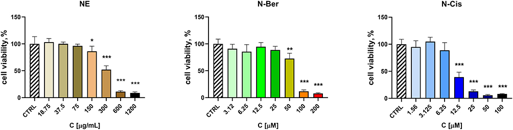 |
Figure 9 Effects of free berberine (Ber; 3.125 μM – 200 μM), free cisplatin (Cis; 1.56 μM – 100 μM), single-loaded berberine (N-Ber; 3.125 μM – 200 μM), single-loaded cisplatin (N-Cis; 1.56 μM – 100 μM) and non-loaded lipid nanoparticles (NE; 18.75 μg/mL – 1200 μg/mL) on Ea.hy926 cell viability after 72 h treatment. The results are expressed as means ± SD of triplicate assays (n=6). All groups were compared statistically vs untreated controls by one-way Anova with Dunnett’s post-test * p < 0.05, ** p < 0.01; *** p < 0.001. |
NE showed detectable toxicity only in the 300–1200 µg/mL range, similar to the pattern observed in tumour cells. These findings support the existence of a therapeutic window in which berberine-loaded nanoparticles can exert cytotoxic effects on cancer cells while being comparatively less harmful to endothelial cells, suggesting a favourable selectivity profile for N-Ber.
Hemolysis Assay
Hemocompatibility was assessed by measuring the haemolytic activity of NE, N-Ber and N-Cis on human erythrocytes (Figure 10). According to ISO 10993–5, formulations are considered non-haemolytic when haemolysis remains below 5% (ISO 10993–4).
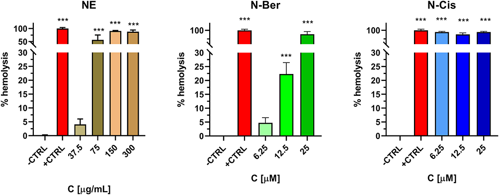 |
Figure 10 Haemolytic effects on human erythrocytes of non-loaded lipid nanoparticles (NE; 37.5 μg/mL – 300 μg/mL), nanoparticles, loaded with berberine (N-Ber; 6.25 μM – 25 μM) and nanoparticles, loaded with cisplatin (N-Cis; 6.25 μM – 25 μM). The results are expressed as means ± SD of triplicate assays (n=6). All groups were compared statistically vs untreated controls by one-way ANOVA with Dunnett’s post-test *** p < 0.001. Abbreviations: ATR, attenuated total reflection; BTC, biliary tract cancer; CCA, cholangiocarcinoma; DLS, dynamic light scattering; EE, encapsulation efficiency; FTIR-ATR, Attenuated Total Reflectance-Fourier-transform infrared spectroscopy; LC, loading capacity; NLC, nanostructured lipid carriers; SD, standard deviation; SEM, scanning electron microscopy; SLN, solid lipid nanocarriers; P-XRD, powder x-ray diffraction; PDI, polydispersity index. |
NE induced 4.1% haemolysis at the lowest concentration tested (37.5 µg/mL), therefore complying with the safety threshold at this dose. N-Cis caused significant haemolysis at all tested concentrations, indicating a relevant risk of erythrocyte membrane damage even in its nanoform. In contrast, N-Ber met the safety criterion at 6.25 µM, with haemolysis of 4.7%, while higher concentrations exceeded the 5% limit.
Taken together, these results demonstrate that berberine-loaded lipid nanoparticles combine strong in vitro antitumor activity in cholangiocarcinoma cells with a relatively favourable safety and hemocompatibility profile compared with cisplatin-loaded formulations.
Discussion
This work explores nanostructured lipid carriers as vehicles for berberine delivery in CCA and positions N-Ber as a potential alternative or complement to cisplatin-based approaches. Here, the cytotoxicity of berberine, either in free or encapsulated form, is investigated in three different cholangiocarcinoma cell lines. The novelty of the study is further enhanced by the use of cisplatin as a comparator, both free and encapsulated within nanoparticles with the same composition and preparation method. This strategy allows for the proper interpretation of the nanoparticle’s role in the cytotoxicity effects. Rather than focusing on incremental formulation optimization, our data collectively support the concept that a natural, pleiotropic agent can be formulated in a potentially clinically relevant nanocarrier with preserved antitumor activity and an improved in vitro safety profile.
The selected NLC composition, solid/liquid lipid mixture stabilized with a non-ionic surfactant, aligns with current nanomedicine design principles for parenteral delivery. The choice of components in the lipid nanoparticles preparation varies based on the purpose, route of administration, drug solubility and compatibility, among others. In the present study, we have selected oleic acid as a liquid lipid due to its known pharmacological effects, especially its antiproliferative effects in gastrointestinal tract-related neoplasms.47,72,73 Furthermore, in the case of cholangiocarcinoma, it was demonstrated that the tumor cells have increased intake of fatty acids, specifically oleic and palmitic acid, as opposed to healthy cells.72 We consider this as a potential additional benefit for the specific chemotherapeutic delivery to the target site. In addition, it was shown that mono and di-unsaturated liquid lipids enhance the cell internalization in comparison to saturated ones.74 Additionally, our previous study demonstrated an IC50 of 11.86 µM in HuCC-T1 cells for berberine loaded in NLC with medium chain triglycerides.18 We assumed that modifying the lipid composition may alter the cytotoxicity results. Indeed, the results here demonstrate improved cytotoxicity in the same cell line (IC50 9.75 µM in HUCC-T1). On the other hand, glyceryl palmitostearate (Precirol 5 ATO) is one of the most commonly used solid lipids for the development of NLCs.62 The berberine solubility in this solid lipid is also high, which is a prerequisite for high encapsulation efficiency.18 The nanoparticles were stabilized with the help of Tween 20. Its structure possesses polyethylene glycol units that are expected to shield the nanoparticles and prolong their blood circulation.64 These structural features are particularly relevant for CCA, a disease in which repeated systemic administration is required and cumulative toxicity is a major limitation. A carrier that combines high loading with physicochemical robustness is a prerequisite for any realistic translational pathway.
The successful preparation of the nanostructured lipid carriers was demonstrated by the proposed method. In this study, we chose the solvent evaporation method over the melt emulsification in order to produce relatively smaller nanoparticles. Indeed, the particle size observed in this work is smaller than that obtained by melt emulsification in our previous work.18 The observed encapsulation efficiency is relatively high, although studies show the possibilities for higher percentages.75 It should be noted that in this work, we intended to test the hypothesis of lipid-based nanoparticles for berberine’s effects in cholangiocarcinoma. Optimization of the formulation was not intended and can be the aim of future work. Nanoparticle size is of paramount importance for in vitro and in vivo cell interactions. It is evident that nanoparticles smaller than 200 nm have improved cell membrane penetration.76 The nanostructured lipid carriers developed in this work fall in the lower limit of this range. Their negative charge suggests good physicochemical stability and is considered more biocompatible with respect to hemolysis.9
The release profiles of N-Ber and N-Cis point to an interplay between the nanocarrier and the tumor microenvironment. The more sustained release at physiological pH and the relatively higher availability under acidic conditions are in line with the design goal of limiting systemic exposure while favoring drug liberation in tumor tissue. This type of “passive” pH-responsiveness, even if modest, is attractive in hepatobiliary malignancies, where local acidosis, altered vasculature and biliary obstruction coexist.
The coexistence of different lipid domains and the mixed crystalline structure are typical of NLCs and are usually associated with high loading capacity and reduced risk of drug expulsion over time. In this context, the solid-state transition of berberine from tetrahydrate to dihydrate within the carrier is notable: rather than indicating instability, it likely reflects the adaptation of the drug to the lipid environment and may contribute to its sustained release behavior. For cisplatin, the persistence of crystalline domains suggests a different mode of incorporation that may influence release kinetics and interaction with biological milieu.
The berberine in vitro dissolution showed its improved release at pH 7.4 in comparison to the free drug. At pH 5.5, the N-Ber was characterized with some burst release and a sustained pattern over time. A similar trend of higher available berberine amount at pH 5.5 was shown in other studies.77 At the same time, the cisplatin in vitro dissolution exhibited no pH-dependent effect for the free drug. A sustained pattern was also noticeable in the case of the NLC formulation, and it was more pronounced at pH 7.4. Such behavior is considered favorable since, after parenteral administration and during blood circulation (pH 7.4), a smaller amount of cisplatin will be released, and hence the unwanted side effects would be mitigated. Meanwhile, at the tumor-simulating pH (5.5), the available cisplatin will be higher and better efficacy could be expected. A similar trend has been described in earlier studies.61,76
A key observation is that nanoencapsulation preserves the strong cytotoxic effect of berberine in CCA cell lines, yielding cytotoxicity at least comparable to cisplatin. Importantly, this effect was preserved after nanoencapsulation, indicating that the lipid matrix did not impede the biological activity of the loaded agents in vitro. The slightly lower cytotoxicity observed for N-Ber compared with free berberine in equal concentrations may be attributed to the sustained release behavior of the nanocarrier, which limits the immediate availability of the drug and results in a more gradual cellular exposure. In addition, nanoencapsulation may modify cellular uptake pathways and reduce efflux, leading to prolonged intracellular retention. This controlled delivery profile may be advantageous, as it could reduce acute toxicity while maintaining overall antitumor effect. N-Cis exhibited comparable or minimally enhanced cytotoxicity relative to free cisplatin. This effect may be explained by improved cellular uptake via nanoparticle-mediated endocytosis and prolonged intracellular exposure due to sustained drug release. From a nanomedicine perspective, this indicates that the lipid matrix does not hinder cellular uptake or intracellular access to targets, which is not trivial given the cationic, amphiphilic nature of berberine. Importantly, comparison between free and nanoencapsulated formulations showed that N-Ber and N-Cis retained cytotoxic activity with dose–response profiles comparable to their respective free drugs. N-Ber displayed a slight reduction in potency relative to free berberine, while N-Cis showed comparable or slightly improved activity compared with free cisplatin in selected concentration ranges. Overall, these findings indicate that nanoencapsulation preserves the biological activity of both compounds, and it is justified to further assess the modulation of pharmacodynamics. It also raises the possibility that N-Ber could exploit transport proteins and membrane dynamics differently from free drug, potentially modulating subcellular distribution in ways that merit mechanistic investigation. These findings align with previous research on nanoparticle-mediated delivery of plant-derived compounds, which often enhances cellular uptake and stability.
The cytotoxic activity observed in this study aligns with existing literature on berberine-based nanoformulations, which frequently demonstrate enhanced or comparable antiproliferative effects relative to the free compound. Previous reports established IC50 values in the low micromolar range for berberine loaded into different lipid nanoparticles across various solid tumor lines, including cholangiocarcinoma.18,78 These improved outcomes are generally attributed to enhanced cellular uptake and superior metabolic stability provided by the nanocarrier. Consequently, the potency of N-Ber observed here confirms that encapsulation preserves the inherent anticancer efficacy of berberine while optimizing its in vitro release profile and possibly intracellular delivery.
Equally important is the selectivity pattern. Even at borderline hemolytic concentrations, the advantages of the N-Ber are noticeable. The lower toxicity of N-Ber towards endothelial cells, together with its more favorable hemocompatibility compared with N-Cis, suggests a window in which tumor cells are more affected than normal vascular or blood cells. For a disease treated with regimens already constrained by systemic toxicity, even modest improvements in therapeutic index can be clinically meaningful. In contrast, the marked hemolytic effect of N-Cis underscores a recurrent challenge in formulating platinum drugs and argues against simple “nanonisation” as a universal solution for classic chemotherapeutics.
The cytotoxic effects of N-Ber were particularly potent in EGI-1 cells (IC50 = 0.4 μM), suggesting differential sensitivity of various cholangiocarcinoma subtypes. This could be attributed to distinct molecular signatures or variations in drug transport and metabolism among the lines. N-Ber showed a favourable toxicity profile in endothelial cells, with IC50 values far above those observed in cancer cells, supporting the potential therapeutic selectivity of this formulation.
Hemocompatibility of N-Ber is an important issue, especially for parenteral drug administration. The hemolysis assay confirmed that N-Ber, unlike N-Cis, meets international safety standards (<5% hemolysis at therapeutic concentrations). This result underscores a major limitation of cisplatin, even in its nanoform, as it induced membrane lysis at all concentrations tested, raising concerns about systemic toxicity upon intravenous administration.
The current therapeutic landscape of CCA is dominated by gemcitabine–cisplatin combinations, now frequently partnered with immune checkpoint inhibitors as mentioned before. Within this context, N-Ber shows significant in vitro potential for future cholangiocarcinoma-related studies, but it could be strategically integrated in several ways: as a partner to existing regimens to enhance tumor cell killing; as a tool to explore dose-reduction strategies for cisplatin; or as a platform to exploit transporter-mediated uptake in CCA cells. The latter is particularly relevant for berberine, whose interaction with membrane transporters and efflux systems might be deliberately harnessed or modulated through nanoparticle design.
Study Limitations
This study has several limitations that should be acknowledged. First, the biological evaluation was performed exclusively in vitro over a limited time frame, which does not fully reflect the complexity of in vivo tumor environments. Therefore, further in vivo studies are required to confirm the therapeutic potential and safety profile of the investigated formulations. Second, pharmacokinetic and biodistribution analyses were not performed, which limits the understanding of systemic behavior, tissue accumulation, and circulation profile of the nanostructured lipid carriers. Finally, mechanistic studies investigating pathways such as apoptosis induction, oxidative stress, and cellular uptake were not included, and thus, the detailed molecular mechanisms underlying the observed cytotoxic effects remain to be further elucidated.
Conclusion
The in vitro experimental data of the current work demonstrated the possibility of modifying berberine and cisplatin dissolution from NLCs. The physicochemical properties of the carriers are adequate for provisional parenteral delivery. The cytotoxicity tests in various cell cultures elucidate the potential effectiveness of free and encapsulated berberine in cholangiocarcinoma cell lines, which are superior or comparable to cisplatin. Preliminary results also demonstrated the biocompatibility and safety of the prepared nanoparticles in the case of parenteral delivery. These data provide solid foundational insights for more comprehensive future studies. In vivo experiments of the biodistribution, efficacy and safety, as well as mechanistic investigations, are necessary to bring berberine closer to the clinical context. Nevertheless, the potential is clearly demonstrated in vitro in the present work and may help optimize therapeutic strategies in cholangiocarcinoma.
Funding
This publication is based upon work from COST Action Precision-BTC Network, supported by COST (European Cooperation in Science and Technology). This research was supported by the National Science Fund of Bulgaria under grant number KP-06-COST/21/19.12.2023 to MM, DS, VT, BT, MS. Martin Manov is gratefully acknowledged for funding the in vitro cytotoxicity tests, sponsored by the Medical Science Council at the Medical University of Sofia under the project “Young Researcher 2025”, D-220/4.06.2025. The sponsor had no involvement in the study design, manuscript preparation and decision for submission of the manuscript for publication in Nanotechnology, Science and Applications.
Disclosure
The authors report no conflicts of interest in this work.
References
1. Brindley PJ, Bachini M, Ilyas SI, et al. Cholangiocarcinoma. Nat Rev Dis Primers. 2021;7(1):1–19. doi:10.1038/s41572-021-00300-2
2. Izquierdo-Sanchez L, Lamarca A, La Casta A, et al. Cholangiocarcinoma landscape in Europe: diagnostic, prognostic and therapeutic insights from the ENSCCA Registry. J. Hepatol. 2022;76(5):1109–1121. doi:10.1016/j.jhep.2021.12.010
3. Banales JM, Rodrigues PM, Affò S, et al. Cholangiocarcinoma 2026: status quo, unmet needs and priorities. Nat Rev Gastroenterol Hepatol. 2026;23(1):65–96. doi:10.1038/s41575-025-01153-w
4. Yuan ZG, Zeng TM, Tao CJ. Current and emerging immunotherapeutic approaches for biliary tract cancers. Hepatobiliary Pancreat Dis Int. 2022;21(5):440–449. doi:10.1016/j.hbpd.2022.08.015
5. Wheless M, Agarwal R, Goff L, Lockney N, Padmanabhan C, Heumann T. Current standards, multidisciplinary approaches, and future directions in the management of extrahepatic cholangiocarcinoma. Curr Treat Options in Oncol. 2024;25(1):127–160. doi:10.1007/s11864-023-01153-5
6. Beri N. Immune Checkpoint Inhibitors in Cholangiocarcinoma. Immunotherapy. 2023;15(7):541–551. doi:10.2217/imt-2022-0288
7. Oun R, Moussa YE, Wheate NJ. The side effects of platinum-based chemotherapy drugs: a review for chemists. Dalton Trans. 2018;47(19):6645–6653. doi:10.1039/C8DT00838H
8. Galluzzi L, Senovilla L, Vitale I, et al. Molecular mechanisms of cisplatin resistance. Oncogene. 2012;31(15):1869–1883. doi:10.1038/onc.2011.384
9. Tang J, Yang Y, He Z, et al. Construction of dual-targeted liposomes loaded with celastrol and their application in treating intrahepatic cholangiocarcinoma. Materials Today Bio. 2025;31:101581. doi:10.1016/j.mtbio.2025.101581
10. Yadav P, Ambudkar SV, Rajendra Prasad N. Emerging nanotechnology-based therapeutics to combat multidrug-resistant cancer. J Nanobiotechnol. 2022;20(1):423. doi:10.1186/s12951-022-01626-z
11. Huang M, Lu JJ, Ding J. Natural products in cancer therapy: past, present and future. Nat Prod Bioprospect. 2021;11(1):5–13. doi:10.1007/s13659-020-00293-7
12. Obchoei S, Detarya M, Boonnate P, et al. Low dose berberine suppresses cholangiocarcinoma cell progression as a multi-kinase inhibitor. Asian Pac J Cancer Prev. 2022;23(10):3379–3386. doi:10.31557/APJCP.2022.23.10.3379
13. Zhang H, Li G, Ni L, et al. Berberine hydrochloride inhibits the proliferation and metastasis of p53 mutant gallbladder Cancer cells by regulating the IL6/STAT3 pathway. Sci Rep. 2025;15(1):26808. doi:10.1038/s41598-025-11480-2
14. Skonieczna M, Adamiec-Organisciok M, Hudy D, et al. Hepatocellular cancer cell lines, Hep-3B and Hep-G2, display the pleiotropic response to resveratrol and berberine. Adv. Med. Sci. 2022;67(2):379–385. doi:10.1016/j.advms.2022.09.003
15. ying LS, Jian SC, ming FW, Zhang J. Berberine inhibits tumour growth in vivo and in vitro through suppressing the lincROR-Wnt/β-catenin regulatory axis in colorectal cancer. J Pharm Pharmacol. 2023;75(1):129–138. doi:10.1093/jpp/rgac067.
16. Palaniyandi T, Sivaji A, Natarajan S, Ravi M, Viswanathan S. Chemotherapeutic effect of berberine, oxidative stress, and genotoxic studies in methylnitronitrosoguanidine-induced gastric carcinoma in experimental rats. Pharm Chem J. 2024;58(8):1226–1236. doi:10.1007/s11094-024-03262-3
17. Rai D, George S. Computational analysis of ligands from natural products on the cellular targets of combined hepatocellular carcinoma and cholangiocarcinoma. Nat Prod Res. 2025;39(22):6459–6467. doi:10.1080/14786419.2024.2373960
18. Stefanova D, Yordanov Y, Bogdanova R, et al. In vitro evaluation of the safety and antineoplastic effects in gastrointestinal tumors of nanostructured lipid carriers loaded with berberine. Pharmaceutics. 2025;17(3):331. doi:10.3390/pharmaceutics17030331
19. Qi L, Qian S, Wang L, et al. Properties of approved antitumour chinese herbal medicines: integrating evidence and tradition. Drug Des Devel Ther. 2026;20:575608. doi:10.2147/DDDT.S575608
20. Attia HG, Elmataeeshy ME, Aleraky M, et al. The assessment of pharmacokinetics and neuroprotective effect of berberine hydrochloride-embedded albumin nanoparticles via various administration routes: comparative in-vivo studies in rats. J. Microencapsul. 2024;41(7):576–600. doi:10.1080/02652048.2024.2395976
21. Farag AA, Kamal M, Youssef HS, et al. The neonicotinoid imidacloprid provokes ferritinophagy/ferroptosis axis disruption in rats’ liver: the attenuation impact of berberine chloride-loaded nano-liposomes. Toxicol Appl Pharmacol. 2026;506:117622. doi:10.1016/j.taap.2025.117622
22. Zaied H, Ashmawy MI, Abdel Karim AE, Ghareeb DA, El Wakil A. Berberine-loaded albumin nanoparticles alleviate liver damage in rats by modulating mitochondrial biogenesis and mitochondria-endoplasmic reticulum interactions. Biochem Biophys Res Commun. 2025;754:151555. doi:10.1016/j.bbrc.2025.151555
23. Suman I, Klepac D, Vragović M, et al. Targeted delivery of berberine via ROS-sensitive polymersomes enhances its hepatoprotective activity in CCl 4 -intoxicated mice. Nanoscale Adv. 2026;8(2):595–611. doi:10.1039/D5NA00706B
24. Sakthivel MB, Dass PDN, Munusamy T, et al. Berberine-enriched copper oxide nano formulation synthesized using Solanum torvum: a strategic advancement in lung cancer therapy and wound healing. J App Pharm Sci. 2025;15(6):096–114. doi:10.7324/JAPS.2025.237203
25. Zhao Y, Jing Z, Li Y, Mao W. Berberine in combination with cisplatin suppresses breast cancer cell growth through induction of DNA breaks and caspase-3-dependent apoptosis. Oncol. Rep. 2016;36(1):567–572. doi:10.3892/or.2016.4785
26. Khaled AM, Othman MS, Obeidat ST, et al. Green-synthesized silver and selenium nanoparticles using berberine: a comparative assessment of in vitro anticancer potential on human hepatocellular carcinoma cell line (HepG2). Cells. 2024;13(3):287. doi:10.3390/cells13030287
27. Shi Y, Cao L, Zhao W, et al. Enhancing chemo-immunotherapy in triple-negative breast cancer: co-delivery of doxorubicin and berberine using nanoparticles to downregulate PD-L1 and eliminate cancer stem cells. International Journal of Pharmaceutics. 2025;670:125134. doi:10.1016/j.ijpharm.2024.125134
28. Sun Q, Tu K, Xu Q, et al. Berberine suppresses colorectal cancer progression by inducing ferroptosis-mediated energy metabolism disorders. J Adv Res. doi:10.1016/j.jare.2025.10.025
29. Laishram S, Laishram B, Moirangthem DS. Berberine enhances cisplatin sensitivity in non-small cell lung cancer H1299 cells via modulation of the p38-MAPK signaling pathway. J. Biol. Active Prod. Nature. 2025;15(3):222–233. doi:10.1080/22311866.2025.2518163
30. Xu B, Luo X, Xiong J, et al. Berplatin: a conjugate of berberine and cisplatin serving as a potent antitumor agent with distinctive tumor cell uptake and minimal side effects. Adv Ther. 2023;6(3):2200160. doi:10.1002/adtp.202200160
31. Bhattacharjee K, Nath M, Choudhury Y, Choudhury Y. Berberine mitigates betel-nut induced hepatocarcinogenesis, enhances chemosensitivity to cisplatin and reduces cisplatin- induced nephrotoxicity in mice exposed to an aqueous extract of betel nut. Pharmacogn. Mag. 2024;16(5):1021–1028. doi:10.5530/pj.2024.16.165
32. Lu H, Yan Z, Sun M. Studies on synthesis, physicochemical properties, biological activities of two novel berberine-based salts with DL-mandelate and cinnamate anions. J. Mol. Struct. 2023;1294:136359. doi:10.1016/j.molstruc.2023.136359
33. Imenshahidi M, Hosseinzadeh H. Berberis vulgaris and berberine: an update review. Phytother. Res. 2016;30(11):1745–1764. doi:10.1002/ptr.5693
34. Salvi VR, Pawar P. Nanostructured lipid carriers (NLC) system: a novel drug targeting carrier. J. Drug Deliv. Sci. Technol. 2019;51:255–267. doi:10.1016/j.jddst.2019.02.017
35. Mall J, Naseem N, Haider MF, Rahman MA, Khan S, Siddiqui SN. Nanostructured lipid carriers as a drug delivery system: a comprehensive review with therapeutic applications. Intelligent Pharm. doi:10.1016/j.ipha.2024.09.005
36. Khosa A, Reddi S, Saha RN. Nanostructured lipid carriers for site-specific drug delivery. Biomed. Pharmacother. 2018;103:598–613. doi:10.1016/j.biopha.2018.04.055
37. Haider M, Abdin SM, Kamal L, Orive G. Nanostructured lipid carriers for delivery of chemotherapeutics: a review. Pharmaceutics. 2020;12(3):3. doi:10.3390/pharmaceutics12030288
38. Nakamura T, Sato Y, Yamada Y, et al. Extrahepatic targeting of lipid nanoparticles in vivo with intracellular targeting for future nanomedicines. Adv. Drug Deliv. Rev. 2022;188:114417. doi:10.1016/j.addr.2022.114417
39. Jia L, Zhang D, Li Z, et al. Nanostructured lipid carriers for parenteral delivery of silybin: biodistribution and pharmacokinetic studies. Colloids and Surfaces B: Biointerfaces. 2010;80(2):213–218. doi:10.1016/j.colsurfb.2010.06.008
40. Kambanis J, Lowe LA, Yao J, et al. Lipid nanoparticles in drug delivery: overcoming challenges and unlocking new frontiers. Small Structures. 2026;7(3):e202500583. doi:10.1002/sstr.202500583
41. Esnaashari F, Zamani H, Zahmatkesh H, Soleimani M, Dashtaki GA, Rasti B. Berberine decorated zinc oxide loaded chitosan nanoparticles a potent anti cancer agent against breast cancer. Sci Rep. 2025;15(1):3185. doi:10.1038/s41598-025-87445-2
42. Xia X, Zhang Z, He Z, et al. Alginate/sodium carboxymethyl cellulose nanocomposite hydrogels loaded with berberine-rosmarinic acid nanoparticles: pH-responsive release for enhanced acid stability and colon-targeted delivery. Int. J. Biol. Macromol. 2025;328:147629. doi:10.1016/j.ijbiomac.2025.147629
43. Sharaf HA, Saied MAA, Ghareeb DA, Kandil SH, El-Fattah AA. Enhanced controlled drug delivery of berberine-loaded gelatin nanoparticles: characterization and in vitro assessment. RSC Adv. 2026;16(7):6119–6131. doi:10.1039/D5RA08567E
44. Yadav D, Semwal BC, Dewangan HK. Grafting, characterization and enhancement of therapeutic activity of berberine loaded PEGylated PAMAM dendrimer for cancerous cell. Biomater. Sci. Polym. Ed. 2023;34(8):1053–1066. doi:10.1080/09205063.2022.2155782
45. Cao X, Pang S, Li Y, et al. Berberine-loaded mesoporous silica nanomaterials inhibit pancreatic cancer targeting cancer stem cells. J Pharm Pharmacol. 2025;77(11):1534–1541. doi:10.1093/jpp/rgaf058
46. Al rashid MH, Mishra S, Pattnaik S, Mohanty C. Berberine loaded glyceryl monooleate nanoparticles exhibited potent intrinsic anticancer activity against pancreatic cancer therapy: in vitro and in silico studies. Nano Trends. 2025;9:100092. doi:10.1016/j.nwnano.2025.100092
47. El-Fakharany EM, Ashry M, Abu‑Serie,Marwa M, Abdel-Wahhab KG, El-Sahra DG, El‑Gendi H. In vitro and in vivo synergistic antitumor activity of albumin-coated oleic acid-loaded liposomes toward hepatocellular carcinoma. Cancer Investigation. 2023;41(7):621–639. doi:10.1080/07357907.2023.2241083
48. Liu Y, Hussain SA, Yue H. Protective effects of berberine-loaded chitosan/solid lipid nanoparticles in streptozotocin-induced gestational diabetes mellitus rats. Exp Biol Med. 2025;250:10749. doi:10.3389/ebm.2025.10749
49. Skorokhyd N, Panchuk R, Zaichenko O, et al. Modulating the biological effect of berberine via its immobilization on different polymer nanocarriers. Cytol Genet. 2025;59(3):270–280. doi:10.3103/S0095452725030077
50. Filippi R, Lombardi P, Quarà V, et al. Pharmacotherapeutic options for biliary tract cancer: current standard of care and new perspectives. Expert Opinion on Pharmacotherapy. 2019;20(17):2121–2137. doi:10.1080/14656566.2019.1667335
51. Yari E, Sari S, Kelidari H, Asare-Addo K, Nokhodchi A. Effect of rosa damascena essential oil loaded in nanostructured lipid carriers on the proliferation of human breast cancer cell line mda-mb-231 in comparison with cisplatin. J Pharm Innov. 2024;19(1):4. doi:10.1007/s12247-024-09809-x
52. Jia B, He J, Zhang Y, et al. Pulmonary delivery of magnolol-loaded nanostructured lipid carriers for COPD treatment. Int. J. Pharm. 2024;662:124495. doi:10.1016/j.ijpharm.2024.124495
53. Basotra M, Singh SK, Gulati M. Development and validation of a simple and sensitive spectrometric method for estimation of cisplatin hydrochloride in tablet dosage forms: application to dissolution studies. Int Sch Res Notices. 2013;2013(1):936254. doi:10.1155/2013/936254
54. Zhang Y, Zhang P, Zhu T. Ovarian carcinoma biological nanotherapy: comparison of the advantages and drawbacks of lipid, polymeric, and hybrid nanoparticles for cisplatin delivery. Biomed. Pharmacother. 2019;109:475–483. doi:10.1016/j.biopha.2018.10.158
55. Saghafi Z, Mohammadi M, Mahboobian MM, Derakhshandeh K. Preparation, characterization, and in vivo evaluation of perphenazine-loaded nanostructured lipid carriers for oral bioavailability improvement. Drug Development and Industrial Pharmacy. 2021;47(3):509–520. doi:10.1080/03639045.2021.1892745
56. Nurohman I, Chaerunisaa AY, Wilar G, Jafar G, Suhandi C, Sriwidodo S. Sacha inchi oil–based nanostructured lipid carriers for curcumin delivery: development and physicochemical characterization. Nanotechnol. Sci. Appl. 2026;19:589429. doi:10.2147/NSA.S589429
57. Zuo J, Gao Y, Bou-Chacra N, Löbenberg R. Evaluation of the ddsolver software applications. Biomed Res. Int. 2014;2014(1):204925. doi:10.1155/2014/204925
58. Kumar P, Nagarajan A, Uchil PD. Analysis of Cell Viability by the MTT Assay. Cold Spring Harb Protoc. 2018;2018(6):
59. Evans BC, Nelson CE, Yu SS, et al. Ex vivo red blood cell hemolysis assay for the evaluation of ph-responsive endosomolytic agents for cytosolic delivery of biomacromolecular drugs. J Vis Exp. 2013;(73):50166. doi:10.3791/50166
60. Raju M, Kunde SS, Auti ST, Kulkarni YA, Wairkar S. Berberine loaded nanostructured lipid carrier for Alzheimer’s disease: design, statistical optimization and enhanced in vivo performance. Life Sci. 2021;285:119990. doi:10.1016/j.lfs.2021.119990
61. Mittal D, Singh A, Kohli K, Verma AK. Engineering biosafe cisplatin loaded nanostructured lipid carrier: optimisation, synthesis, pharmacokinetics and biodistribution. J. Microencapsul. 2022;39(6):522–538. doi:10.1080/02652048.2022.2131919
62. Apostolou M, Assi S, Fatokun AA, Khan I. The effects of solid and liquid lipids on the physicochemical properties of nanostructured lipid carriers. J. Pharm. Sci. 2021;110(8):2859–2872. doi:10.1016/j.xphs.2021.04.012
63. Fidorra M, Heimburg T, Seeger HM. Melting of individual lipid components in binary lipid mixtures studied by FTIR spectroscopy, DSC and Monte Carlo simulations. Biochimica et Biophysica Acta (BBA) - Biomembranes. 2009;1788(3):600–607. doi:10.1016/j.bbamem.2008.12.003
64. Göppert TM, Müller RH. Plasma protein adsorption of tween 80- and poloxamer 188-stabilized solid lipid nanoparticles. J Drug Target. 2003;11(4):225. doi:10.1080/10611860310001615956
65. Georgieva I, Trendafilova N, Dodoff N, Kovacheva D. DFT study of the molecular and crystal structure and vibrational analysis of cisplatin. Spectrochim Acta A Mol Biomol Spectrosc. 2017;176:58–66. doi:10.1016/j.saa.2017.01.008
66. Zhang Y, Zhang D, Zhang Y, et al. Improving solubility and avoiding hygroscopicity of tetrahydroberberine by forming hydrochloride salts by introducing solvents: [HTHB]Cl, [HTHB]Cl·CH3OH and [HTHB]Cl·CH3COOH. New J Chem. 2017;41(22):13268–13275. doi:10.1039/C7NJ02423A
67. Singh M, Bhandary S, Bhowal R, Chopra D. Observation of bending, cracking and jumping phenomena on cooling and heating of tetrahydrate berberine chloride crystals. CrystEngComm. 2018;20(16):2253–2257. doi:10.1039/C8CE00114F
68. Caselli L, Conti L, De Santis I, Berti D. Small-angle X-ray and neutron scattering applied to lipid-based nanoparticles: recent advancements across different length scales. Adv. Colloid Interface Sci. 2024;327:103156. doi:10.1016/j.cis.2024.103156
69. Demirbolat GM, Cakirli E, Saglam AB, Sarigul-Ozbek S, Abas BI, Cevik O. pH-triggered liposomal strategy for cisplatin in lung cancer therapy. Sci Rep. 2025;15(1):36349. doi:10.1038/s41598-025-20144-0
70. Chaudhuri A, Kumar DN, Srivastava SK, et al. Combinatorial delivery of docetaxel- and erlotinib-loaded functionalized nanostructured lipid carriers for the treatment of triple-negative breast cancer using quality-by-design approach. Pharmaceutics. 2024;16(7):926. doi:10.3390/pharmaceutics16070926
71. Zhang S, Zhao Y, Tan L, et al. A novel berberine–glycyrrhizic acid complex formulation enhanced the prevention effect to doxorubicin-induced cardiotoxicity by pharmacokinetic modulation of berberine in rats. Front Pharmacol. 2022:13. doi:10.3389/fphar.2022.891829.
72. de Gauna M R, Biancaniello F, González-Romero F, et al. Cholangiocarcinoma progression depends on the uptake and metabolization of extracellular lipids. Hepatology. 2022;76(6):1617–1633. doi:10.1002/hep.32344
73. Giulitti F, Petrungaro S, Mandatori S, et al. Anti-tumor effect of oleic acid in hepatocellular carcinoma cell lines via autophagy reduction. Front Cell Dev Biol. 2021:9. doi:10.3389/fcell.2021.629182.
74. Jeitler R, Glader C, König G, et al. On the structure, stability, and cell uptake of nanostructured lipid carriers for drug delivery. Mol Pharmaceutics. 2024;21(7):3674–3683. doi:10.1021/acs.molpharmaceut.4c00392
75. Gendy AM, Elnagar MR, Allam MM, et al. Berberine-loaded nanostructured lipid carriers mitigate warm hepatic ischemia/reperfusion-induced lesion through modulation of HMGB1/TLR4/NF-κB signaling and autophagy. Biomed. Pharmacother. 2022;145:112122. doi:10.1016/j.biopha.2021.112122
76. Vhora I, Khatri N, Desai J, Thakkar HP. Caprylate-conjugated cisplatin for the development of novel liposomal formulation. AAPS PharmSciTech. 2014;15(4):845–857. doi:10.1208/s12249-014-0106-y
77. Abolhasanzadeh N, Dehghan G, Abbaspour-Ravasjani S. Enhancement of the stability and cytotoxicity of berberine by liposomal nanocarriers for gastric cancer treatment and its application in gummy candy. Front Gastroenterol. 2024;3. doi:10.3389/fgstr.2024.1387343
78. Paudel KR, Mehta M, Yin GHS, et al. Berberine-loaded liquid crystalline nanoparticles inhibit non-small cell lung cancer proliferation and migration in vitro. Environ Sci Pollut Res Int. 2022;29(31):46830–46847. doi:10.1007/s11356-022-19158-2
 © 2026 The Author(s). This work is published and licensed by Dove Medical Press Limited. The
full terms of this license are available at https://www.dovepress.com/terms
and incorporate the Creative Commons Attribution
- Non Commercial (unported, 4.0) License.
By accessing the work you hereby accept the Terms. Non-commercial uses of the work are permitted
without any further permission from Dove Medical Press Limited, provided the work is properly
attributed. For permission for commercial use of this work, please see paragraphs 4.2 and 5 of our Terms.
© 2026 The Author(s). This work is published and licensed by Dove Medical Press Limited. The
full terms of this license are available at https://www.dovepress.com/terms
and incorporate the Creative Commons Attribution
- Non Commercial (unported, 4.0) License.
By accessing the work you hereby accept the Terms. Non-commercial uses of the work are permitted
without any further permission from Dove Medical Press Limited, provided the work is properly
attributed. For permission for commercial use of this work, please see paragraphs 4.2 and 5 of our Terms.
Recommended articles
The Chemotherapy Medication of Evodia lepta (Spreng). Merr. on the Viability of Tongue Cancer Cells Through the PD-L1/MMP14/HSPA5 Pathway

Chen J, Zheng X, Wang X, Weng CF
Cancer Management and Research 2025, 17:1613-1623
Published Date: 12 August 2025