Back to Journals » International Journal of General Medicine » Volume 15
Paraclinical Aspects in Systemic Sclerosis
Authors Bobeica C ![]() , Niculet E
, Niculet E ![]() , Musat CL, Craescu M, Stefanescu BI
, Musat CL, Craescu M, Stefanescu BI ![]() , Dinu C
, Dinu C ![]() , Chiscop I, Chirobocea S
, Chiscop I, Chirobocea S ![]() , Nechita L, Iancu AV
, Nechita L, Iancu AV ![]() , Stefanescu V
, Stefanescu V ![]() , Balan G, Stefanopol IA
, Balan G, Stefanopol IA ![]() , Pelin AM
, Pelin AM ![]() , Tatu AL
, Tatu AL ![]()
Received 24 December 2021
Accepted for publication 5 April 2022
Published 26 April 2022 Volume 2022:15 Pages 4391—4398
DOI https://doi.org/10.2147/IJGM.S355662
Checked for plagiarism Yes
Review by Single anonymous peer review
Peer reviewer comments 3
Editor who approved publication: Dr Scott Fraser
Carmen Bobeica,1,* Elena Niculet,1,2 Carmina Liana Musat,1 Mihaela Craescu,1,* Bogdan Ioan Stefanescu,3,* Ciprian Dinu,4,* Iulia Chiscop,3,* Silvia Chirobocea,5,* Luiza Nechita,6,* Alina Viorica Iancu,1,* Victorita Stefanescu,7,* Gabriela Balan,6,8,9,* Ioana Anca Stefanopol,1,10,* Ana Maria Pelin,11,* Alin Laurentiu Tatu2,6,12
1Department of Morphological and Functional Sciences, Faculty of Medicine and Pharmacy, “Dunărea de Jos” University of Galați, Galati, 800008, Romania; 2Multidisciplinary Integrated Center of Dermatological Interface Research MIC-DIR (Centrul Integrat Multidisciplinar de Cercetare de Interfata Dermatologica - CIM-CID), “Dunărea de Jos” University, Galați, 800008, Romania; 3Clinical Surgical Department, Faculty of Medicine and Pharmacy, “Dunărea de Jos” University, Galați, Romania; 4Dental Department, Faculty of Medicine and Pharmacy, Dunărea de Jos University, Galați, Romania; 5Department of Neurology, Municipal Emergency Hospital, Moinești, Romania; 6Clinical Medical Department, Faculty of Medicine and Pharmacy, Dunărea de Jos University, Galați, Romania; 7Medical Department, Faculty of Medicine and Pharmacy, Dunărea de Jos University, Galați, Romania; 8Department of Gastroenterology, “Sf. Apostol Andrei” County Emergency Clinical Hospital, Galați, Romania; 9Research Center in the Field of Medical and Pharmaceutical Sciences, “Dunărea de Jos” University, Galați, Romania; 10Department of Pediatrics, Clinical Emergency Hospital for Children “Sf. Ioan”, Galati, Romania; 11Department of Pharmaceutical Sciences, Faculty of Medicine and Pharmacy, “Dunărea de Jos” University, Galați, Romania; 12Dermatology Department, “Sf. Cuvioasa Parascheva” Clinical Hospital of Infectious Diseases, Galați, Romania
*These authors contributed equally to this work
Correspondence: Elena Niculet; Carmina Liana Musat, Department of Morphological and Functional Sciences, Faculty of Medicine and Pharmacy, “Dunărea de Jos” University of Galați, 35 Alexandru Ioan Cuza Street, Galați, 800008, Romania, Tel +40741398895 ; +40723338438, Email [email protected]; [email protected]
Abstract: Systemic sclerosis (SSc) is a chronic inflammatory disease with an autoimmune substrate that affects the skin and a large number of internal organs. The chronic inflammatory process is sustained by a wide range of cytokines and chemokines, which are discharged by inflammatory cells, with fibrosis and nail bed vascular changes (disorganized vasculature architecture with microhemorrhages, megacapillaries and areas without capillaries). Confocal microscopy contributes to the understanding of the molecular mechanism involved in chronic inflammation and mainly targets the field of research. Coherent optical tomography, capillaroscopy, and skin biopsy are useful for the differential diagnosis of SSc with other sclerodermoid syndromes. The immunological profile is a classification criterion for SSc and directs the diagnosis to the two subsets of the disease. Multisystemic damage requires evaluation with the help of a set of investigations specific to each affected organ, such as: diffusing capacity for carbon monoxide, forced vital capacity, 6-minute walk test, high-resolution computed tomography standard and reduced sequential, cardiac ultrasound and right cardiac catheterization. The current possibilities of diagnosis, treatment and monitoring are permanently adapting to new medical discoveries.
Keywords: systemic sclerosis, antinuclear antibodies, capillaroscopy, confocal microscopy, coherent optical tomography
Introduction
Systemic sclerosis (SSc) is an autoimmune disease with an inflammatory substrate evolving into extensive multiorgan fibrosis, with microvasculature changes that reflect the stage of the disease. Inflammation is maintained by the marked expression of cytokines: interleukin (IL)-17A, IL-4, IL-25, transforming growth factor-β1 (TGF-β1). Recent research has shown that the IL-9 molecule is deeply involved in the autoimmune pathogenic process in ScS and other autoimmune diseases: psoriatic arthropathy, rheumatoid arthritis, and giant cell arthritis. The IL-9 molecule is secreted mainly by Th9 lymphocytes (Ly) and to a lesser extent by LyTh17. IL-9 appears to mediate cutaneous and visceral fibrosis in SSc. LyTh9 maintain the inflammatory process and are activated by IL-33, IL-25 and stromal thymic lymphopoietin (TSLP). The analysis of the hardened skin also showed that the accumulated inflammatory cells, endothelial cells and fibroblasts in the dermis, can be a source of IL-9. Confocal microscopy revealed excessive production of IL-9 in the skin of patients belonging to both subsets of ScS, limited and diffuse. IL-9 stimulates the infiltration with LyT, LyB and (through competition with IL-17) maintains the inflammatory process by neutrophils accumulation.1,2
A 2017 study by Sawamura et al revealed another cytokine’s involvement in SSc, namely IL-22, which was found to be overexpressed in the lymphocytes infiltrating the affected skin, but not in the patients’ serum (not being a useful marker for disease activity, as opposed to IL-6, which is elevated in the blood of these patients). IL-22 overexpression in lymphocytes leads to the accumulation of type I collagen in the dermis of SSc-suffering patients.3,4
The multiple systemic disturbances in SSc require a wide and complex range of imaging studies and laboratory investigations.5 This manuscript aims at evaluating SSc from the perspective of its immunological profile, microvessel changes (with available techniques), confocal laser microscopy and coherent optical tomography evaluation, skin biopsy results and other investigations specific to the organ damage that SSc inflicts, making use of established and updated information.
Immunological Profile
High percentages of patients (90% to 95%) have an elevated serum antinuclear antibody (ANA) titer.6,7 SSc-specific autoantibodies are useful for diagnosis. The type of autoantibodies that are present indicates a certain subset of SSc and foreshadows the risk of developing certain complications. Haustein appreciates good autoantibody predictability for disease subsets and reveals their usefulness for early diagnosis. Anti-Scl70, ACA and anti-RNA polymerase III antibodies have the highest specificity for SSc. Therefore, elevated autoantibody titers direct the therapeutic attitude towards the type of impairment characteristic for each subset of SSc.8,9
Anti-Scl70 (anti-topoisomerase I) antibodies are highly specific for diffuse SSc and the patient has a high risk of developing interstitial pulmonary fibrosis;8,9 the risk for renal dysfunction is equal to that found in other disease models. These autoantibodies are present more frequently in Thai and Japanese people and to a lesser extent in African Americans.9 Like anti-Scl 70 antibodies, high titers of RNA polymerase III are characteristic for diffuse SSc and indicate a worse prognosis with a higher risk for internal organ damage and especially for scleroderma development.8,10
Compared to the diffuse SSc subset, ACA have specificity for limited SSc and CREST syndrome, and they also indicate a better prognosis, higher survival rates, with a lower risk for impaired pulmonary and renal functions.9 Jacobsen noted that ACA are often present in patients with calcinosis, telangiectasia, and digital ulceration.11
Interstitial pulmonary fibrosis and decreased carbon monoxide diffusion capacity (DLCO) have been less observed in patients with elevated ACA. Patients with elevated anti-Scl70 antibody titers were associated with pulmonary fibrosis, digital joint deformities, and acroosteolysis, but calcinosis was less frequent in these cases.11
SSc-specific autoantibodies are not found in association.9 The serological profile is positive in most patients, as noted by several authors during their studies done on patient groups originating from European countries. Thus, Jacobsen identified a ratio of 86% of patients with high ANA titers,11 and Fabri and Hunzelmann identified more than 90% of patients with such antibody values.10
Haustein recorded a percentage of 85% of patients with positive ANA, and, in dynamics, the percentage rose to 98%.9 While Haustein observed ANA positivity developing during the course of the disease,9 other studies revealed that autoantibody detection takes place many years before the onset of SSc.10
Although some authors classify Ac anti-Scl70 as having a 100% specificity for diffuse SSc,9 numerous studies have found an atypical immune profile for this specific subset of SSc. As such, Steen identified high titers of anti-Scl70 Ac in limited SSc and ACA in diffuse SSc.12
Research has also identified other serological abnormalities specific to SSc. Thus, antibodies anti-U3-RNP (fibrillarin) targets 3 enzymes involved in the synthesis of ribosomal proteins; this suggests the presence of an intense process of skin fibrosis and a low survival rate. These autoantibodies are associated with diffuse SSc; they indicate early disease onset, the presence of telangiectasias, and an increased risk of progression to pulmonary arterial hypertension and to sclerodermal renal crisis.9
SSc-specific autoantibodies may be accompanied by other autoantibodies - anti-PM/Scl, anti-PL7 (anti-threonyl-tRNA synthetase), anti-Jo1 antibodies, anti-Ku antibodies and anti-U1-RNP antibodies (in the context of Overlap syndrome). Anti-Ro (SSA) antibodies present in 9% of SSc cases indicate a rapid and severe evolution of renal failure and pulmonary arterial hypertension.9
Monfort et al highlight the fact that SSc is associated with the risk of developing neoplasms, especially when anti-RNA polymerase III antibodies are elevated. As early as 2010, Monfort described the phenomena of paraneoplastic SSc as being an association between SSc with negative ANA and the existence of a neoplasm. Paraneoplastic SSc has a negative immune profile and a normal capillary appearance.13,14 Although autoantibodies characteristic of other concomitant autoimmune diseases are not always present, they may occasionally be clinically associated with systemic scleroderma. Some medications used for comorbidities or experimental/off label ones, such as statins, may have benefits in lowering the level of cytokines involved in SS, this fact making the patient possibly subjected to potential known side effects of these drugs.15–20
Capillaroscopy
Peripheral microvasculopathy can be assessed qualitatively and quantitatively at nail bed level by using video capillaroscopy. This has the advantage of being a non-invasive investigation with high specificity and sensitivity, all at low costs.21 The specific microvascular changes that take place in SSc (in early, active and late stages) are recognized and can be used as a tool for disease monitoring.2 Capillaroscopy establishes the differential diagnosis between the secondary Raynaud’s phenomenon SSc and the primary Raynaud’s phenomenon.22
The normal videocapillaroscopic appearance is represented by parallel capillaries with the skin surface, lack of dilated vessels and hemorrhages. The elderly and children may have atypical nail bed capillary appearance. It has been observed that dilated capillary loops represent a normal aspect in the elderly, and the presence of inhomogeneous images and varied capillary aspects in children are not representative of pathology. Several parameters are evaluated: dilations and capillary ramifications, giant capillaries and capillary density.23
Characteristics of ScS are megacapillaries and areas without capillaries (Figure 1).10 The capillary appearance can quantify the degree of microvascular damage from early to active and late.24 Dilated capillaries of giant capillaries and microhemorrhages describe an early pattern of microangiopathy, and the active disease pattern is identified by disorganization of the capillary architecture and capillary loss.23,25 In the late model, areas without vascularization alternate with focal neoangiogenesis,26,27 while megacapillaries are missing.23 The hemorrhages have a lower specificity for SSc, being more limited both in surface and depth, as compared to those found in other collagenoses. Some authors consider the presence of a megacapillary, in the background of Raynaud’s phenomenon, as being highly suggestive of SSc.23 The degree of microvessel anomalies increases gradually in severity as SSc progresses and might be useful in stratifying patients according to their risk of developing organ damage.27
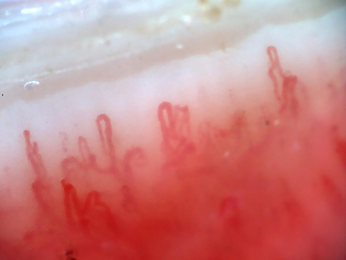 |
Figure 1 SSc capillaroscopy image – nail bed with dilated capillaries and areas without vascularization. |
In a study of a group of 204 patients with SSc, Barsotti found that there were obstructions in the ulnar artery in 37.3% of the evaluated patients and 24% of them had bilateral occlusion. As a result, the microscopic irregularities of the digital arteries’ vascular wall may be accompanied by unilateral or bilateral ulnar artery occlusion, which is revealed by Doppler ultrasonography.28
Nail bed videocapillaroscopic evaluation reveals capillary changes, which cannot always be classified into one of the SSc’s stages, and are thus labeled as a scleroderma-like pattern; there are a number of other connective tissue diseases, which can present with nail bed capillary changes, such as: systemic lupus erythematosus, mixed connective tissue disease, dermatomyositis.2
Laser speckle contrast analysis (LSCA) is a newer technique that dynamically evaluates peripheral blood perfusion in SSc patients, in vivo, in real-time, with a higher spatial resolution. A study from 2018, by Cutolo et al, found that this laser tool, LSCA, had excellent intra- and inter-observer reliability in the study of blood flow in SSc, but more studies need to be done in order to evaluate results in a parallel manner.29 High-resolution laser Doppler imaging (laser Doppler flowmetry – LDF) can be also used as an objective investigation tool for evaluating microvessel changes in SSc (in the right temperature and setting, with the patient’s hand away from its body, on a mat), by analyzing the dorsum of the hands, which reveals a blood flow map, more precisely revealing blood perfusion (which is altered at the finger level in SSc patients). For LSCA and LDF more studies are needed due to the lack of a gold standard for measuring, but there have been found links between the structural altering of microvessels (capillaroscopy) and the functional vessel activity (LDF or LSCA).30,31
Confocal Laser Microscopy
Lung tissue samples collected from patients with SSc with secondary pulmonary fibrosis are analyzed using confocal laser microscopy images. Confocal microscopy (CM) identified the presence of cells that express surface molecular markers that are specific to endothelial cells located in the lung parenchyma and in the perivascular and subendothelial space. Also, by using confocal microscopy, the presence of endothelial cells with molecular markers characteristic of myofibroblasts in the endothelial and in the subendothelial space of small and medium-sized arteries in the lungs was highlighted. These observations support the theory of the transition of endothelial cells into mesenchymal cells – the so-called EndoMT mechanism. EndoMT has long been thought to be present only during embryonic development of cardiovascular structures. Recent studies have shown that EndoMT-type phenotypic modification is also present in experimental models of excessive tissue fibrosis. There is also ample evidence of the involvement of EndoMT in the pathogenesis of SSc-associated pulmonary hypertension and even in idiopathic pulmonary hypertension. In other words, endothelial cells in the lungs can be converted into activated myofibroblasts by the EndoMT mechanism, which induces progressive fibrosis of the intima of the vessels, obstruction of the pulmonary vessels, and interstitial pulmonary fibrosis. This phenotypic change could be a useful new therapeutic target in the prevention of pulmonary complications in SSc.32 Other in vitro studies have shown the possibility of bone remodeling by CM, a phenomenon that may have implications and utility in the treatment of calcinosis in some cases of SSc.33
Cascio et al used a confocal microscope to analyze the confocal images of blood samples and hardened skin. They identified the molecular mechanism by which LyT CD8+ produce IL-13. The thickened skin samples were pre-frozen, then stained with immunofluorescence techniques. Subsequently, LyT CD8+ were isolated from both fibrosed skin and blood samples. It appears that in the early stages of SSc, fibrosed skin contains high levels of IL-13-producing Ly T CD8+. LyT CD4+, while also producing lower levels of IL-13. Therefore, they found that IL-13-generating LyT CD8+ play a key role in excessive tissue fibrosis. Cascio and his collaborators also made an in-depth analysis of the molecular process which takes place in the diffuse subset of SSc. They noted that LyT CD8+ overexpress GATA-3 transcription factor and induce a high level of T-bet transcription factor.34
Coherent Optical Tomography
Classical studies have shown that the intima of the pulmonary vessels suffers from a process of excessive fibrosis, accompanied by arteriopathy with lower plexogenic potential than in idiopathic pulmonary hypertension. Coherent optical tomography, which can quantify the intima-media index in cases of SSc during right cardiac catheterization, has been shown to be particularly useful. It was observed that patients with secondary pulmonary arterial hypertension SSc had a significantly greater thickness of the intima and an average one in the pulmonary vascular bed with a caliber of less than 2 mm, as compared to those without pulmonary hypertension. Coherent optical tomography showed a significant increase in pulmonary artery branches smaller than 300 μm in the course of treating pulmonary hypertension.35
Skin Biopsy
Although the histological examination of the hardened skin is not part of the classification criteria for SSc, skin biopsy is useful when there is diagnostic uncertainty. SSc histological examination is different from that found in other sclerodermoid diseases, such as Bursche’s scleredema or scleromyxedema. Skin biopsy has proven to be particularly useful in research and less so in SSc management. Studies aimed at identifying microvascular lesions and chronic inflammatory infiltrates in the early stages of SSc, at identifying the type of cytokines involved in the chronic inflammatory process and the excessive collagen deposition in advanced disease stages.36
Tokumura et al observed that patients with SSc have elevated serum lysophosphatidic acid levels.37,38 Of particular interest is the observation of several researchers who noted that skin inflammation appears to be a constant source of lysophosphatidic acid,38,39 and on the surface of fibroblasts, in the dermis of patients with SSc, there are overexpressed, high levels of lysophosphatidic acid receptors.37,38 Under the action of lysophosphatidic acid, fibroblasts in the hardened dermis overexpress fibronectin and smooth muscle alpha-actin αSMA. These two components direct the differentiation of activated fibroblasts from myofibroblasts, a process known to be an important link in the pathogenic process of SSc. At the same time, the markers involved in the Wnt pathway are increasingly expressed under the action of lysophosphatidic acid.38,40
Experimental stimulation with lysophosphatidic acid in animal models induced a proinflammatory effect by accumulating chemokines and cytokines. Of these, chemokine CCL2 appears to have maximum potential in the process of inflammation and fibrosis in SSc. Similarly, an elevated level of IL6 was observed.38,41 Skin biopsies in patients with SSc revealed high levels of CCL2 in both serum and epidermis. Lysophosphatidic acid induces a strong chemotactism of neutrophils and monocytes in endothelial cells,38,42 followed by the differentiation of smooth muscle fibers from the walls of blood vessels.38,43 This proves the involvement of lysophosphatidic acid in microangiopathy and chronic inflammation in SSc.38,44
Investigations Specific to Organ Damage
Pulmonary fibrosis and pulmonary arterial hypertension are common complications of SSc that can sometimes be associated.5,45 A decrease in DLCO below 60% suggests the presence of an increased risk of developing pulmonary hypertension and requires regular monitoring accompanied by ultrasound quantification of the degree of hypertension and even right cardiac catheterization.46
The gold standard in identifying and quantifying the degree of lung damage remains High-resolution computed tomography (HRCT). The extension of pulmonary fibrosis to over 33% of the lung parenchyma, accompanied by a reduction in the forced vital capacity (FVC) in an early-stage SSc patient suggests a disease type with an unfavorable prognosis and a high risk of progression.45,47,48 The features associated with SSc affecting the lung are as follows: ground-glass opacities with fibrosis inside (upper and lower lobes) and bilateral, symmetrical lower-lobe reticulations (associated or not with fibrosis).48 Sequentially reduced HRCT has been shown to be almost as effective as standard HRCT in determining the extent of pulmonary fibrosis. Only the identification of bronchiectasis was limited to sequentially reduced HRCT.45,49
Studies show that disease progression can be anticipated using the 6-minute walking test applied in dynamics to individual patient. Two evaluations of the 3-month gait test done on a group of 56 patients demonstrated good predictability of patient prognosis, although the test was limited by some pathological aspects of SSc. The 6-minute gait test was consistent with a number of global parameters: DLCO, FVC, left ventricular ejection fraction, Rodnan score, tendon friction and arthralgia.45,50–52
Conclusion
This paper brings together the most widely used investigations in SSc management and study, which are available so far in the medical literature and practice. The multisystemic involvement and complex pathogenesis of SSc are still unknown, but they open up the opportunity for extensive studies on various aspects of the disease, all the while making useful different investigative techniques for each affected organ.53 There is an important burden of systemic sclerosis complications which stem from the progressive and intractable fibrotic and microvessel damages (non-lethal ones) that characterize this disease, making it a very challenging one to evaluate and treat. As such, the current possibilities of diagnosis, treatment and monitoring are in a permanent adaptation to the new medical discoveries.
Abbreviations
SSc, systemic sclerosis; IL, interleukin; TGF-β1, transforming growth factor-β1; Ly, lymphocyte; TSLP, thymic stromal lymphopoietin; ANA, antinuclear antibodies; ACA, anti-centromere antibodies; RNA, ribonucleic acid; DLCO, diffusing capacity for carbon monoxide; LSCA, laser speckle contrast analysis; LDF, laser Doppler flowmetry; αSMA, smooth muscle alpha-actin; HRCT, high-resolution computed tomography; FVC, forced vital capacity; CM, confocal microscopy.
Acknowledgments
The authors wish to acknowledge that the present study was supported by the ‘Dunărea de Jos’ University of Galați, Romania, through the research center – Multidisciplinary Integrated Center of Dermatological Interface Research MIC-DIR (Centrul Integrat Multidisciplinar de Cercetare de Interfata Dermatologica – CIM-CID). We would also like to acknowledge the contribution made by Professor Codrina Ancuta, MD, PhD, the Head of Rheumatology 2 Department, part of the Clinical Rehabilitation Hospital and Professor of Rheumatology at the University of Medicine and Pharmacy “Grigore T. Popa” Iasi.
Author Contributions
All authors made a significant contribution to the work reported, whether that is in the conception, study design, execution, acquisition of data, analysis and interpretation, or in all these areas; took part in drafting, revising or critically reviewing the article; gave final approval of the version to be published; have agreed on the journal to which the article has been submitted; and agree to be accountable for all aspects of the work.
Funding
The article publishing charge was paid by the “Dunarea de Jos” University of Galati, Romania.
Disclosure
The authors report no conflicts of interest in this work.
References
1. Guggino G, Lo Pizzo M, Di Liberto D, et al. Interleukin-9 over-expression and T helper 9 polarization in systemic sclerosis patients. Clin Exp Immunol. 2017;190(2):208–216. doi:10.1111/cei.13009
2. Bernero E, Sulli A, Ferrari G, et al. Prospective capillaroscopy-based study on transition from primary to secondary Raynaud’s phenomenon: preliminary results. Reumatismo. 2013;65(4):186–191. doi:10.4081/reumatismo.2013.186
3. Sawamura S, Jinnin M, Inoue K, et al. Regulatory mechanisms of collagen expression by interleukin-22 signaling in scleroderma fibroblasts. J Dermatol Sci. 2018;90(1):52–59. doi:10.1016/j.jdermsci.2017.12.017
4. Ruaro B, Soldano S, Smith V, et al. Correlation between circulating fibrocytes and dermal thickness in limited cutaneous systemic sclerosis patients: a pilot study. Rheumatol Int. 2019;39(8):1369–1376. doi:10.1007/s00296-019-04315-7
5. Barsotti S, Bruni C, Orlandi M, et al. One year in review 2017: systemic sclerosis. Clin Exp Rheumatol. 2017;35Suppl 106(4):3–20.
6. Stochmal A, Czuwara J, Trojanowska M, Rudnicka L. Antinuclear antibodies in systemic sclerosis: an update. Clin Rev Allergy Immunol. 2020;58(1):40–51. doi:10.1007/s12016-018-8718-8
7. Bobeica C, Niculet E, Halip AI, et al. Predictive value of immunological markers in systemic sclerosis. Exp Ther Med. 2021;22(3):1–5. doi:10.3892/etm.2021.10426
8. Denton CP. Advances in pathogenesis and treatment of systemic sclerosis. Clin Med. 2016;16(1):55–60. doi:10.7861/clinmedicine.16-1-55
9. Haustein UF. Systemic sclerosis-scleroderma. Dermatol Online J. 2002;8(1):3. doi:10.5070/D30VD8P0XW
10. Fabri M, Hunzelmann N. Differential diagnosis of scleroderma and pseudoscleroderma. J Dtsch Dermatol Ges. 2007;5(11):977–984. doi:10.1111/j.1610-0387.2007.06311.x
11. Jacobsen S, Halberg P, Ullman S, et al. Clinical features and antinuclear serum in 230 Danish patients with systemic sclerosis. Br J Rheumatol. 1998;37(1):39–45. doi:10.1093/rheumatology/37.1.39
12. Steen VD, Medsger JTA. The value of the health assessment questionnaire and special patient-generated scales to demonstrate change in systemic sclerosis patients over time. Arthritis Rheum. 1997;40(11):1984–1991. doi:10.1002/art.1780401110
13. Monfort JB, Mathian A, Amoura Z, Francès C, Barbaud A, Senet P. Cancers associated with systemic sclerosis involving anti-RNA polymerase III antibodies. Ann Dermatol Venereol. 2018;145(1):33–36. doi:10.1016/j.annder.2017.08.005
14. Monfort JB, Lazareth I, Priollet P. Paraneoplastic systemic sclerosis: about 3 cases and literature review. J Mal Vasc. 2016;41(6):365–370. doi:10.1016/j.jmv.2016.07.001
15. Tatu AL, Ionescu MA. Multiple autoimmune syndrome type III-thyroiditis, vitiligo and alopecia areata. Acta Endocrinol. 2017;13(1):124–125. doi:10.4183/aeb.2017.124
16. Goussot R, Francès C, Cury K, et al. Prospective evaluation of the frequency of genital lichen sclerosus in 79 patients with systemic sclerosis. Br J Dermatol. 2018;179(4):999–1000. doi:10.1111/bjd.16898
17. Tatu AL, Nwabudike LC The treatment options of male genital lichen sclerosus et atrophicus. Short Title for a Running Head: treatments of genital lichen sclerosus.
18. Gonçalves RSG, Dantas AT, Pereira MC, et al. Statins inhibit cytokines in a dose-dependent response in patients with systemic sclerosis. Inflammation. 2019;42(2):407–411. doi:10.1007/s10753-018-0907-3
19. Tatu AL, Nwabudike LC. Male genital lichen sclerosus — a permanent therapeutic challenge. J Am Acad Dermatol. 2018;79(3Suppl1):AB185.
20. Nwabudike LC, Elisei AM, Buzia OD, Miulescu M, Tatu AL. Statins. A review on structural perspectives, adverse reactions and relations with non-melanoma skin cancer. RevChim. 2018;69(9):2557–2562.
21. Cutolo M, Melsens K, Herrick AL, et al. Reliability of simple capillaroscopic definitions in describing capillary morphology in rheumatic diseases. Rheumatology. 2018;7(4):757–759. doi:10.1093/rheumatology/kex460
22. Cutolo M, Sulli A, Secchi M, Paolini S, Pizzorni C. Nailfold capillaroscopy is useful for the diagnosis and follow-up of autoimmune rheumatic diseases. A future tool for the analysis of microvascular heart involvement? Rheumatology. 2006;45Suppl4:iv43–46. doi:10.1093/rheumatology/kel310
23. Ioniţescu RC, Mihai C, Danciu O, et al. Capilaroscopia periunghială în bolile colagen vasculare. Rev Med Interna. 2009;4:1.
24. Ciaffi J, van Leeuwen NM, Huizinga TWJ. Cumulative endogenous estrogen exposure is not associated with severity of peripheral microangiopathy in patients with systemic sclerosis. Clin Exp Rheumatol. 2019;37Suppl 119(4):82–87.
25. Cutolo M, Sulli A, Pizzorni C, Accardo S. Nailfold videocapillaroscopy assessment of microvascular damage in systemic sclerosis. J Rheumatol. 2000;27(1):155–160.
26. Cutolo M, Pizzorni C, Secchi ME, Sulli A. Capillaroscopy. Best Pract Res Clin Rheumatol. 2008;22(6):1093–1108. doi:10.1016/j.berh.2008.09.001
27. Trombetta AC, Smith V, Pizzorni C, et al. Quantitative alterations of capillary diameter have a predictive value for development of the capillaroscopic systemic sclerosis pattern. J Rheumatol. 2016;43(3):599–606. doi:10.3899/jrheum.150900
28. Barsotti S, Orlandi M, Codullo V, et al. One year in review 2019: systemic sclerosis. Clin Exp Rheumatol. 2019;37 Suppl 119(4):3–14.
29. Cutolo M, Vanhaecke A, Ruaro B, et al. Is laser speckle contrast analysis (LASCA) the new kid on the block in systemic sclerosis? A systematic literature review and pilot study to evaluate reliability of LASCA to measure peripheral blood perfusion in scleroderma patients. Autoimmun Rev. 2018;17(8):775–780. doi:10.1016/j.autrev.2018.01.023
30. Britton J. Repeatability of high resolution laser Doppler images of the hands in patients with systemic sclerosis and secondary Raynaud’s phenomenon. Photodermatol Photoimmunol Photomed. 2020. PMID: 32181935. doi:10.1111/phpp.12549
31. Melsens K, Van Impe S, Paolino S, Vanhaecke A, Cutolo M, Smith V. The preliminary validation of laser Doppler flowmetry in systemic sclerosis in accordance with the OMERACT filter: a systematic review. Semin Arthritis Rheum. 2020;50(2):321–328. doi:10.1016/j.semarthrit.2019.08.007
32. Mendoza FA, Piera-Velazquez S, Farber JL, Feghali-Bostwick C, Jiménez SA. Endothelial cells expressing endothelial and mesenchymal cell gene products in lung tissue from patients with systemic sclerosis-associated interstitial lung disease. Arthritis Rheumatol. 2016;68(1):210–217. doi:10.1002/art.39421
33. Coman MG, Hîncu M. Study of bone cells by confocal microscopy in fractures stimulated by ultrasound. Rom J Morphol Embryol. 2013;54(2):357–360.
34. Cascio S, Medsger JTA, Hawse WF, et al. 14-3-3z cytosolic T-bet sequesters, upregulating IL-13 levels in TC 2 and CD8 + lymphocytes from patients with scleroderma. J Allergy Clin Immunol. 2017;14(1):109–119.
35. Schwaiger JP, Loder CD, Dobarro D, et al. Optical coherence tomography evaluation of pulmonary arterial vasculopathy in systemic sclerosis. Sci Rep. 2017;7(1):43304. doi:10.1038/srep43304
36. Rongioletti F, Ferreli C, Atzori L, Bottoni U, Soda G. Scleroderma with an update about clinico-pathological correlation. G Ital Dermatol Venereol. 2018;153(2):208–215. doi:10.23736/S0392-0488.18.05922-9
37. Tokumura A, Carbone LD, Yoshioka Y, et al. Elevated serum levels of arachidonoyl - lysophosphatidic acid and sphingosine 1 ‐ phosphate in systemic sclerosis. Int J Med Sci. 2009;6(4):168–176. doi:10.7150/ijms.6.168
38. Ledein L, Léger B, Dees C, et al. Translational engagement of lysophosphatidic acid receptor 1 in skin fibrosis: from dermal fibroblasts of patients with scleroderma to tight skin 1 mouse. Br J Pharmacol. 2020;177(18):4296–4309. doi:10.1111/bph.15190
39. Mazereeuw‐Hautier J, Gres S, Fanguin M, et al. Production of lysophosphatidic acid in blister fluid: involvement of a lysophospholipase D activity. J Invest Dermatol. 2005;125(3):421–427. doi:10.1111/j.0022-202X.2005.23855.x
40. Wei J, Fang F, Lam AP, et al. Wnt/β ‐ catenin signaling is hyperactivated in systemic sclerosis and induces Smad ‐ dependent fibrotic responses in mesenchymal cells. Arthritis Rheum. 2012;64(8):2734–2745. doi:10.1002/art.34424
41. Denton CP, Shi ‐ Wen X, Sutton A, Abraham DJ, Black CM, Pearson JD. Scleroderma fibroblasts promote migration of mononuclear leukocytes across endothelial cell monolayers. Clin Exp Immunol. 1998;114(2):293–300. doi:10.1046/j.1365-2249.1998.00721.x
42. Idzko M, Laut M, Panther E, et al. Lysophosphatidic acid induces chemotaxis, oxygen radical production, CD11b up-regulation, Ca2 + mobilization, and actin reorganization in human eosinophils via toxin-sensitive pertussis G proteins. J Immunol. 2004;172(7):4480–4485. doi:10.4049/jimmunol.172.7.4480
43. Zhou ZB, Niu JP, Zhang ZJ. Receptor ‐ mediated vascular smooth muscle migration induced by LPA involves p38 mitogen ‐ activated protein kinase pathway activation. Int J Mol Sci. 2009;10(7):3194–3208. doi:10.3390/ijms10073194
44. Tigyi G, Parrill AL. Molecular mechanisms of lysophosphatidic acid action. Prog Lipid Res. 2003;42(6):498–526. doi:10.1016/S0163-7827(03)00035-3
45. Orlandi M, Barsotti S, Lepri G, et al. One year in review 2018: systemic sclerosis. Clin Exp Rheumatol. 2018;36Suppl113(4):3–23.
46. Coghlan JG, Wolf M, Distler O, et al. Incidence of pulmonary hypertension and determining factors in patients with systemic sclerosis. Eur Respir J. 2018;51(4):1701197. doi:10.1183/13993003.01197-2017
47. Barsotti S, Stagnaro C, d’Ascanio A, Della RA. One year in review 2016: systemic sclerosis. Clin Exp Rheumatol. 2016;34Suppl100(5):3–13.
48. Orlandi M, Landini N, Sambataro G, et al. The role of chest CT in deciphering interstitial lung involvement: systemic sclerosis versus COVID-19. Rheumatology 2021;61(4):1600–1609.
49. Nguyen-Kim TDL, Maurer B, Suliman YA, Morsbach F, Distler O, Frauenfelder T. The impact of slice-reduced computed tomography on histogram-based densitometry assessment of lung fibrosis in patients with systemic sclerosis. J Thorac Dis. 2018;10(4):2142–2152. doi:10.21037/jtd.2018.04.39
50. Sanges S, Giovannelli J, Sobanski V, et al. Factors associated with the 6-minute walk distance in patients with systemic sclerosis. Arthritis Res Ther. 2017;19(1):279. doi:10.1186/s13075-017-1489-4
51. Vandecasteele E, Thevissen K, Melsens K, et al. Six-minute walk test in or out in evaluation of systemic sclerosis patients? Clin Exp Rheumatol. 2017;35Suppl106(4):122–129.
52. Pugnet G, Marjanovic Z, Deligny C, et al. Reproducibility and utility of the 6-minute walk test in systemic sclerosis. J Rheumatol. 2018;45(9):1273–1280. doi:10.3899/jrheum.170994
53. Bobeica C, Tatu AL, Craescu M, Solovastru-Gheuca L. Dynamics of digital ulcers in systemic sclerosis. Exp Ther Med. 2020;20(1):):61–67. doi:10.3892/etm.2020.8572
 © 2022 The Author(s). This work is published and licensed by Dove Medical Press Limited. The
full terms of this license are available at https://www.dovepress.com/terms
and incorporate the Creative Commons Attribution
- Non Commercial (unported, 3.0) License.
By accessing the work you hereby accept the Terms. Non-commercial uses of the work are permitted
without any further permission from Dove Medical Press Limited, provided the work is properly
attributed. For permission for commercial use of this work, please see paragraphs 4.2 and 5 of our Terms.
© 2022 The Author(s). This work is published and licensed by Dove Medical Press Limited. The
full terms of this license are available at https://www.dovepress.com/terms
and incorporate the Creative Commons Attribution
- Non Commercial (unported, 3.0) License.
By accessing the work you hereby accept the Terms. Non-commercial uses of the work are permitted
without any further permission from Dove Medical Press Limited, provided the work is properly
attributed. For permission for commercial use of this work, please see paragraphs 4.2 and 5 of our Terms.