Back to Journals » Journal of Pain Research » Volume 17
Optimizing Temporal Summation of Heat Pain Using a Constant Contact Heat Stimulator
Authors Kell PA, Vore CN, Hahn BJ, Payne MF, Rhudy JL
Received 12 September 2023
Accepted for publication 30 January 2024
Published 8 February 2024 Volume 2024:17 Pages 583—598
DOI https://doi.org/10.2147/JPR.S439862
Checked for plagiarism Yes
Review by Single anonymous peer review
Peer reviewer comments 2
Editor who approved publication: Dr Dawood Sayed
Parker A Kell,1 Claudia N Vore,1 Burkhart J Hahn,1,2 Michael F Payne,1,3 Jamie L Rhudy1
1Department of Psychology, The University of Tulsa, Tulsa, OK, USA; 2Department of Psychology, Oklahoma State University, Stillwater, OK, USA; 3Department of Pediatrics, Division of Child & Adolescent Psychiatry, Cincinnati Children’s Hospital Medical Center, Cincinnati, OH, USA
Correspondence: Jamie L Rhudy, University of Oklahoma Health Sciences Center, TSET Health Promotion Research Center, Department of Health Promotion Sciences, 4502 E. 41st Street, Tulsa, OK, 74104, USA, Tel +1 918-660-3050, Email [email protected]
Purpose: Temporal summation (TS) of pain occurs when pain increases over repeated presentations of identical noxious stimuli. TS paradigms can model central sensitization, a state of hyperexcitability in nociceptive pathways that promotes chronic pain onset and maintenance. Many experimenters use painful heat stimuli to measure TS (TS-heat); yet, TS-heat research faces unresolved challenges, including difficulty evoking summation in up to 30– 50% of participants. Moreover, substantial variability exists between laboratories regarding the methods for evoking and calculating TS-heat.
Patients and Methods: To address these limitations, this study sought to identify optimal parameters for evoking TS-heat in healthy participants with a commercially available constant contact heat stimulator, the Medoc TSA-II. Working within constraints of the TSA-II, stimulus trains with varying parameters (eg, stimulus frequency, baseline temp, peak temp, peak duration, testing site) were tested in a sample of 32 healthy, chronic pain-free participants to determine which combination best evoked TS-heat. To determine whether TS scoring method altered results, TS-heat was scored using three common methods.
Results: Across all methods, only two trains successfully evoked group-level TS-heat. These trains shared the following parameters: site (palmar hand), baseline and peak temperatures (44°C and 50°C, respectively), and peak duration (0.5 s). Both produced summation that peaked at moderate pain (~50 out of 100 rating).
Conclusion: Future TS-heat investigations using constant contact thermodes and fixed protocols may benefit from adopting stimulus parameters that include testing on the palmar hand, using 44°C baseline and 50°C peak temperatures, at ≥ 0.33 Hz stimulus frequency, and peak pulse durations of at least 0.5 seconds.
Keywords: temporal summation, heat pain, second pain, wind-up, Medoc TSA-II
Introduction
Temporal summation (TS) of pain is a C fiber-mediated phenomenon in which pain is amplified over repeated presentations of identical noxious stimuli delivered at a frequency of at least 0.33Hz.1 It is the psychophysical correlate of windup, a state of temporary hyperexcitability in spinal neurons,2 and is used by investigators as a model for central sensitization, a prolonged state of spinal neuronal hyperexcitability that can cause hyperalgesia and allodynia.3,4 That is, TS is used as a measure of pain and nociceptive amplification which can be augmented in chronic pain states.4–6 Physiologically, all healthy individuals should experience TS as a result of windup of dorsal horn neurons, but greater TS reflects enhanced central pain processing and is observed in people with chronic pain.4,7
TS has been successfully evoked using multiple noxious stimulus modalities, including electric, mechanical pressure, and heat (TS-heat). When assessing TS-heat, stimuli are delivered using either intermittent contact thermodes (ie, a preheated thermode is applied and removed to the test site for each stimulus) or constant contact thermodes (ie, an attached thermode rapidly heats and cools to deliver each stimulus).8 The constant contact thermode approach is appealing due to the ease of standardizing duration of stimulus presentations and pulse intervals.
Despite this, TS-heat paradigms face unresolved challenges, including difficulty evoking TS-heat in a large subset of participants. Indeed, previous studies suggest that 30–50% of participants show no evidence of TS-heat.9–11 Whereas some TS-heat investigations have employed fixed protocols,9,12 in which all participants received identical noxious stimuli, other investigators have developed protocols that individually tailor heat stimuli for participants in an attempt to elicit TS-heat for a greater proportion of participants by accounting for individual differences in heat sensitivity.13–15 Yet, the use of individually calibrated protocols limits comparisons across groups, requires participants to receive more painful stimulations, and still may not yield sufficient summation. In fact, one recent study using individually calibrated stimuli and state-of-The-art procedures/equipment found no summation in over 300 healthy participants.16 Thus, it would be beneficial to identify and design fixed protocols that better evoke TS-heat.
Additionally, there is a lack of procedural standardization in TS-heat research that decrease the ability to make generalized conclusions from TS-heat research across laboratories.8 For instance, there is no consensus on an optimal body site to evoke TS-heat, and there is considerable variability in the equipment and stimulus parameters used to evoke TS-heat, further limiting generalizability.12,17 Methods of scoring summation also vary across laboratories and studies,9,11,12,18 further precluding conclusions about the effectiveness of TS-heat paradigms.
To address these limitations, this study was conducted to identify optimal stimulus parameters for evoking TS-heat employing a widely used, commercially available constant-contact heat stimulator in healthy, chronic pain-free participants.
Materials and Methods
Participants
Participants were 32 (mean age = 21.44 years, SD = 4.57) healthy, chronic pain-free cisgender men (n=10), and women (n = 22). Participants reported their race as predominantly White (n = 25, 78.1%) or Asian (n = 6, 18.8%), and most participants reported their ethnicity as non-Hispanic (n = 30, 93.8%). Participants had a mean body mass index (BMI) of 24.32 (SD = 4.41). Of these 32 participants, 30 completed all testing in this study. The two participants who did not complete the study withdrew during the last of three testing blocks (see procedure description below), but their available data were used in analyses, nonetheless. Participants were undergraduate students at a small university in the United States who received course credit for their participation. Enrollment began in 2017 and ended in 2019. Exclusion criteria were as follows: <18 years of age; currently ill with infectious disease (eg, cold, flu); current symptoms of psychosis; diagnosis of a chronic pain condition; an inability to speak or read English; neurological, cardiovascular, or circulatory problems; or recent use of analgesic, anxiolytic, antihypertensive, or antidepressant medication. The study protocol was approved by the university of Tulsa’s Institutional Review Board and all participants provided written and verbal consent to participate. All study procedures complied with the Declaration of Helsinki.
To determine whether the current study’s sample size of 32 (30 of whom completed the study) would provide adequate statistical power for the multilevel modeling analyses, ANOVA-based sensitivity analyses were conducted using G*Power 319 to estimate the minimum effect sizes that could be detected given a Type 1 Error probability of 0.05 for each of the statistical models used in the study. ANOVA-based sensitivity analyses were conducted due to difficulties in conducting power analyses for multilevel models. For the first two statistical models, described fully in the Data Analysis section, the parameters entered for the sensitivity analysis (ANOVA, repeated measures, within factor) were as follows: total sample size = 30, number of groups = 1, and number of measurements = 72. Even when entering extremely low correlations among repeated measures (ie, 0.01), the G*Power output found that we could detect effect sizes as low as f = 0.1522, which is considered a small-to-medium effect size.
For the third statistical model, a nearly identical sensitivity analysis was conducted, although there were 720 measurements per participant instead of 72. When entering extremely low correlations among repeated measures (ie, 0.01), the G*Power output found that we could detect effect sizes as low as f = 0.0795, which is also considered a small effect size.
Overview of Procedures and Testing Environment
After providing informed consent, participants were brought into an electrically shielded and sound-attenuated testing room by a male experimenter. Participants were seated in a reclining chair (Perfect Chair Zero Gravity Recliner, Human Touch, Long Beach, CA) and were asked to turn off all electronic devices and remove any jewelry on their arms and hands. Then, participants completed a demographics questionnaire and were screened for exclusion criteria. Next, they completed a computer-presented pre-test questionnaire (not presented) and received instructions for rating their pain in response to heat pulses, which were administered in trains (ie, groups) of 10 stimuli. A total of 72 trains of stimulations were delivered to each participant.
During the instructions for pain ratings, participants were presented with a 0–100 numerical rating scale (NRS) previously used in TS-heat paradigms, which had increments every 5 points and verbal descriptions every 10 points.20 The intervals were: 0 = no sensation, 10 = warm, 20 = a barely painful sensation, 30 = very weak pain, 40 = weak pain, 50 = moderate pain, 60 = slightly strong pain, 70 = strong pain, 80 = very strong pain, 90 = nearly intolerable pain, and 100 = intolerable pain.20 The NRS remained visible to the participant on a computer screen during testing to help them anchor their ratings. Participants could choose any whole number between 0 and 100 on the NRS. They were reminded that they would receive 10 painful heat pulses in a row and were told to provide verbal pain ratings after each pulse. If a participant rated their pain in response to a heat pulse as a 100 on the VAS, the experimenter asked the participant if they would like to discontinue the study and reminded them that they could withdraw at any time. To facilitate participants’ rating of pain related to TS and not the initial (first) pain of the heat stimuli, participants were trained to differentiate between first and second pain using instructions similar to those used in previous TS-heat protocols.13,21 During these instructions, they were told that they would feel two painful sensations after each pulse, with the first sensation being a sharp pain occurring immediately after the pulse (ie, first pain) and the second pain sensation beginning approximately one second after the pulse (ie, second pain).13,21 Since the slower second pain reflects C fiber activation,22,23 which is responsible for triggering states of wind-up in the spinal cord,3 participants were instructed to only make pain ratings based on second pain. To ensure that participants could accurately distinguish between first and second pain, they received at least two trains of heat pulses as practice until they could confidently distinguish between the two.
For the first practice train, the experimenter attached the heat probe to the participant’s left volar forearm and delivered practice heat pulses so that the participant could become familiar with the rating procedure. After the train, participants were asked if they could successfully distinguish between first and second pain; if they could differentiate between the two, they continued to the second practice train. If not, experimenters provided verbal feedback about the sensations before continuing to the second practice train. If a participant did not provide pain ratings for all 10 stimuli, the experimenter reminded the participant to make all 10 ratings before moving to the second practice train. For the second practice train, the heat probe was attached to the thenar eminence (palm) of the participant’s left hand. Once participants were comfortable with identifying second pain and making verbal pain ratings for both sites, they began the experiment, which consisted of three testing blocks. In each block, there were 24 trains of 10 heat pulses delivered; as described in the next section, each of the 24 trains had different stimulus parameters and/or stimulation site. In total, 72 trains (ie, 720 stimulations) were delivered. The order of the trains was pseudo-randomized within blocks and across participants. That is, trains alternated between stimulation sites (volar forearm and palmar hand) to minimize habituation or sensitization, while the order of the other train types (see description below) was fully randomized. Thus, there was approximately 45–60 s between trains at each stimulation site. During testing, the experimenter remained in the testing room to initiate stimulation trains, record pain ratings, and hold the thermode on the stimulation sites. Stimulation trains alternated between sites (palmar hand and volar forearm.) To minimize peripheral sensitization, the thermode was moved slightly between trains for both sites.24,25 There were 5-minute breaks between blocks.
Temporal Summation of Heat Pain Parameters
Heat stimuli were delivered using the Medoc TSA-II Advanced Thermosensory Stimulator (Haifa, Israel) with a 30x30mm thermode. Based on previously published manuscripts describing TS-heat methods using continuous contact thermodes as well as technological limitations of the TSA-II,14,21 five variables relevant to evoking TS-heat were manipulated in this study. First, baseline and peak pulse temperatures were paired together to create three possible conditions (42–48, 43–49, or 44–50°C). These pairs were created based on ramp speed limitations of the TSA-II to ensure that peak temperatures of heat pulses and stimulation frequencies ≥0.33 Hz could be consistently achieved. Similar peak temperatures have been employed in previous TS-heat protocols.7,8,20 Based on constraints of the TSA-II, two conditions for pulse ramp speed to and from the peak temperature (6°C or 8°C/s) were created. Given that TS-heat requires stimulation frequencies of at least 0.33Hz,1 two conditions for pulse frequency were created (0.33 or 0.4Hz). Since second pain occurs approximately 1–1.5 seconds after a noxious stimulus,10 these intervals gave participants sufficient time (1–1.5 seconds) to experience and report their sensation of second pain.8,26 Next, two conditions were created for both peak temperature duration (0.25 or 0.5s) and stimulation site (left volar forearm or left thenar eminence.) Together, 24 trains of 10 identical stimuli were developed using all possible permutations of these variables. Table 1 depicts the parameters for each train and the percentage of participants showing summation during the trains, and Figure 1 depicts the time course of an example heat pulse within a train with definitions of the parameters manipulated. Although all stimuli were delivered at a frequency of either 0.33 or 0.4Hz, the amount of time at the baseline temperature differed depending on the values of the other stimulus parameters. At one extreme (ie, the slowest ramp speed [6°C/s], highest peak pulse duration [0.5s], and fastest frequency [0.4Hz]), the next stimulus within a train would begin as soon the previous stimulus ended). At the other extreme (ie, the fastest ramp speed [8°C/s], lowest peak pulse duration [0.25s], and slowest frequency [0.33Hz), the next stimulus within a train would return to and remain at the baseline temperature for 1.25s before the next stimulus would begin. Each of the three testing blocks contained all 24 trains, and participants provided ratings for each train three times across the whole experiment. Thus, participants completed 72 trains during testing, which consisted of 720 individual heat stimulations.
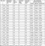 |
Table 1 Parameters for Trains of Noxious Heat Stimuli Used to Evoke Temporal Summation (TS) |
Methods to Score Summation
Investigators have used varying methods to score TS, which can limit generalizability of results across studies.12,17 For instance, researchers have frequently calculated TS as a change score by subtracting either the first pain rating within a stimulus train from either the final pain rating within the train9 or the maximum pain rating within the train.18 Additionally, researchers have used more sophisticated regression methods to assess the slope in pain ratings over the stimulus train.11 Thus, all three methods were used in this study to calculate TS to determine whether calculation method affected results. That is, the first two analyses calculated TS using change scores: the first analysis calculated TS as the last rating minus the first rating within a train, and the second analysis calculated TS as the maximum rating of stimulus 2 to 10 minus the first rating within a train. The third analysis used a growth curve analysis to generate the slope (ie, predicted summation in pain ratings within a train at the individual level), as well as the intercept (ie, the predicted pain rating at the first stimulus in a train at the individual level) of a train.
Data Analysis
All multilevel model analyses were conducted in SPSS 27 using the SPSS MIXED command. All data obtained during the study were used in the analyses, including partial data from the two participants who withdrew during testing. Missing data were handled using maximum likelihood estimations. In the SPSS datasets, there were two primary nominal independent variables: site, which was coded as 1 (palmar hand) or 2 (volar forearm), and stimulus code, which ranged from 1 to 12 and reflected all combinations of stimulus parameters excluding site (see Table 1).
For models 1 and 2, first change scores were calculated for each train that represented the degree of summation. Next, 2 multilevel models (ie, random effects ANOVA models) were used to analyze each change score. The fixed effects were site, stimulus code, and the Site x Stimulus Code interaction term. For these models, data were organized into long form such that each participant had 72 rows of data corresponding to the change score for each of the trains (24 trains x 3 repetitions = 72). This represents the level 1 unit in the multilevel models. The intercept for each participant was entered as a random effect, and both the testing block number (1–3) and order of trains within a block (1–24) were entered as repeated measures. To account for autocorrelations across repeated measures, a first-order autoregressive matrix (AR1) was used as the within-subject variance covariance structure. To facilitate comparisons of group-level TS for each stimulus train in Bonferroni-corrected post-hoc analyses, a second multilevel ANOVA model was conducted, but this time collapsing the two nominal independent variables (site, stimulus code) into a single nominal independent variable called “train” with 24 levels. Bonferroni comparisons were then made to reduce the probability of Type I Error due to multiple comparisons.
In model 3, the multilevel growth curve model (ie, random effects regression model), data in SPSS were organized in long-form; that is, each row contained a single pain rating in response to a heat pulse (level 1 unit). Since all 24 stimulus trains were presented three times, participants with no missing data each had 720 rows of pain ratings (24 trains × 10 heat pulses × 3 repetitions = 720). Like the change score models, the site, stimulus code, and Site x Stimulus Code interaction term were entered as fixed effects. In addition, the stimulus number of each heat pulse within each train (ranging from 1 to 10) was entered as a fixed effect to model the regression slope of summation, as were the Site x Stimulus Number, Stimulus Code x Stimulus Number, and Site x Stimulus Code x Stimulus Number interaction terms. Stimulus number (1–10), the order of trains within each testing block (1–24), and each testing block (1–3) were entered as repeated measures. To account for autocorrelations across repeated measures, a first-order autoregressive matrix (AR1) was used as the within-subject variance covariance structure. Stimulus code and site were entered as categorical variables and stimulus number was entered as a continuous variable. To determine simple slopes and intercepts for each train with pain rating as the outcome variable, a final model was built using this long-form dataset; in this model, each stimulus train (1–24) was added as a fixed effect, as were the stimulus number (1–10) and every Stimulus Train x Stimulus Number interaction term. Simple slopes and intercepts were then calculated for each train using an online tool for testing two-way interactions in hierarchical linear models.27 Ordinary least squares regression slopes were calculated for each train for each participant to determine the percentage of participants showing positive summation in each train.
Results
Group Level Pain Ratings for TS Trains
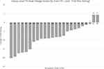
Figure 2A presents findings for trains delivered to the left volar forearm. Figure 2B presents findings for the left palmar hand. To briefly summarize the findings, trains 17 (base temp = 44°C, peak temp = 50°C, 0.5s peak duration, ramp = 6°C/s, 0.33Hz, at hand) and 21 (base temp = 44°C, peak temp = 50°C, 0.5s peak duration, ramp = 8°C/s, 0.40Hz, at hand) showed a positive TS slope at the group level across all three methods of calculating TS (denoted with asterisks on figures/tables). The group-level TS for each stimulus train and TS calculation method are shown in Table 2.
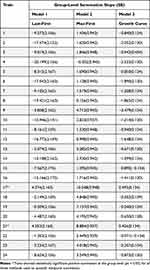 |
Table 2 Average Group-Level Temporal Summation Slopes and Standard Errors for Each Stimulation Train and Calculation Method |
Model 1: Predicting Temporal Summation Last Minus First Change Score
Table 3 presents results from the first analysis. Significant main effects of stimulus code and stimulation site were found. The interaction term between stimulation code and stimulus site was marginally significant (p = 0.088), thus the simple effects were probed to avoid a Type II error (Figure 3), but Bonferroni adjustments were applied to reduce the probability of Type I Error. Trains 17 and 21 showed a positive TS slope and were significantly different from all other trains except Train 19. As shown in Table 1, approximately 59% of participants showed summation during Train 17, 71% showed summation during Train 21, and 46% for Train 19.
 |
Table 3 Results of the Temporal Summation Slope Calculated by Subtracting the Last Pain Rating Minus the First Pain Rating of Each Train |
Model 2: Predicting Temporal Summation Change Score 2 (Maximum Minus First)
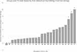
Table 4 and Figure 4 present results from the second analysis. Like the first model, the main effects of stimulus code and site were significant, as was the interaction between stimulation code and stimulus site. According to Bonferroni multiple comparisons, Train 17 showed the greatest group level summation and was significantly greater than all other trains (ps < 0.05), except trains 19 and 21 (ps > 0.05). Train 21 showed the second highest summation and was significantly greater than all other trains, except for trains 22, 19, and 17. Train 19, which showed the third highest summation, was significantly greater than all trains except for trains 23, 20, 9, 18, 22, 21, and 17. When quantifying TS as the difference between the maximum rating and the first rating, approximately 91% of participants showed positive summation during Train 17, 84% showed positive summation during Train 21, and 69% during Train 19 (Table 1).
 |
Table 4 Results of the Temporal Summation Slope Calculated by Subtracting the Maximum Pain Rating Minus the First Pain Rating of Each Train |
Model 3: Predicting Temporal Summation Slopes Using Growth Curve Analysis
The results of this analysis are presented in Table 5 and Figure 5. The variable called “stimulus number” codes for the degree of summation (ie, the linear trend slope) across the 10 pulse trains. The main effects of stimulus code, site, and stimulus number were all significant, as were the 2-way interactions of Stimulus Code x Site, Stimulus Code x Stimulus Number, and Site x Stimulus Number. But all these effects were qualified by the Stimulus Code x Site x Stimulus Number interaction, which indicates that the degree of summation depended on the stimulus site and stimulus code. Post-hoc analyses were conducted to test which slopes were significantly positive, thus showing summation. The simple slopes and standard errors for each train are shown in Table 2. Only the slopes for two trains, 17 and 21, were positive (ps < 0.002), suggesting only those parameters produced significant TS. When calculating TS using this method, about 66% of participants showed positive summation during Train 17, and 72% showed summation during Train 21 (Table 1). Person-level plots of the slopes across all stimulus codes and sites are depicted in Supplemental Figure 1. Additionally, we re-ran the third model using an unstructured covariance structure, which allowed us to examine the covariance between the random intercept (pain rating at the first stimulation) and random slope (degrees of summation) (Table 5). The covariance estimate was non-significant, indicating that the degree of summation was not determined by the pain evoked by the first stimulus in the train. It also ruled out ceiling and floor effects that could be caused by high or low pain ratings to the first stimulus in the train, respectively.
 |
Table 5 Results of the Growth Curve Analysis Predicting Temporal Summation Slope |
Exploratory Analyses: Assessing for Peripheral Sensitization Across the Study
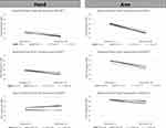
This study was designed to test several train parameters and sites to determine which of them best evoked TS-heat in healthy participants. To do so, it was necessary to present multiple painful trains to two test sites which might have evoked long-term sensitization unrelated to TS thus confounding our findings (although the location of the thermode was moved slightly between trains to minimize sensitization).24,25 Yet, is worth noting that this might not be a problem since peripheral heat injuries are not related to TS magnitude (at least for electric and heat stimuli), although they are related to reduced pain thresholds and hyperalgesia.28,29 To test for possible sensitization due to exposure to multiple stimulation trains during the testing session, additional analyses were conducted (using multilevel modeling) to determine if pain ratings to the first stimulus in each train increased across trains which would suggest peripheral sensitization (Figure 6). Two analyses were conducted, one for each testing site.
The independent variables for each model were testing block (1–3) and trial (ie, order of trains within each testing block [1–24]). For the volar forearm, there was not a significant main effect of trial, F (23, 1101.391) = 1.313, p = 0.148, such that initial pain ratings did not significantly change across the 24 trains within a testing block. But, there was a significant main effect of testing block, F (2, 1102.117) = 5.170, p = 0.006. According to Bonferroni comparisons, pain ratings tended to increase slightly as testing progressed, such that pain ratings were significantly higher in block 3 than in block 1 (p = 0.004) (Figure 6A). The block x trial interaction was not significant, F (46, 1101.242) = 0.655, p = 0.964.
For the palmar hand, there was a significant main effect of trial, F (23, 596.077) = 1.934, p = 0.006), such that initial pain ratings decreased across the 24 trains within each testing block (Figure 6B). There was also a significant main effect of testing block, F (2, 287.744) = 3.437, p = 0.033, with pain ratings on the palmar hand decreasing as testing progressed. Yet, differences between testing blocks were not significantly different using post-hoc Bonferroni comparisons (ps >0.05). Like the volar forearm, the Block x Trial interaction was not significant, F (46, 610.523) = 1.310, p = 0.087) for the palmar hand.
Overall, these analyses demonstrate that sensitization did not confound our results. In fact, the palmar hand showed significant habituation across testing, not sensitization. Moreover, the graphs clearly depict that pain ratings were generally higher on the volar forearm than the palmar hand, suggesting that stimulations to the palmar hand should be more tolerable even for stimulations that peak at 50°C.
Discussion
This study sought to identify optimal parameters for assessing TS-heat with a commonly used, commercially available stimulator, the Medoc TSA-II. In addition to determining the stimulus parameters and stimulation site, three methods for calculating TS-heat used in prior studies were compared to determine if calculation method altered results.9,11,12,18 Across all three calculation methods, two stimulus trains evoked significant TS. Both trains shared many features, including the same site (ie, left thenar eminence of palmar hand), a baseline pulse temperature of 44°C, a peak pulse temperature of 50°C, and a peak stimulus duration of 0.5s. The trains varied in ramp speed and stimulation frequency; one train ramped from the baseline pulse temperature to the peak pulse temperature at 6°C/s with pulses delivered at 0.33 Hz intervals, and the other ramped from baseline to peak temperature up at 8°C/s at 0.40 Hz intervals.
Implications for Assessing TS-Heat Using a Constant Contact Stimulator
Since the two stimulus trains that evoked group-level TS-heat had many similarities, it is plausible that these variables could be important for evoking TS. First, both trains were delivered to the thenar eminence of the palmar hand. While previous studies have successfully evoked TS at both the thenar eminence of the palmar hand and the volar forearm,8 our findings indicate that the palmar hand may be a preferable site for reliably eliciting TS-heat when using the TSA-II. In part, this finding may be related to differences in the skin thickness between these sites, which consequently alters subcutaneous nociceptive input detected by A-delta and C fibers. Whereas the thinner, hairy skin of the volar forearm allows for greater subcutaneous nociceptive input from surface stimuli, the thicker, glabrous skin of the palmar hand somewhat dampens input to subcutaneous nociceptors.30 This is consistent with our finding of greater pain in response to stimuli on the volar forearm relative to the palmar hand. Moreover, this finding may be related to differences in the distribution and/or density of afferent nociceptive fibers present in the glabrous skin of the palmar hand versus the non-glabrous skin of the volar forearm. Relative to non-glabrous skin, glabrous skin contains much fewer low threshold mechano- and heat-sensitive A-delta fiber nociceptors (Type II AMHs),31,32 which are responsible for detection of first pain in response to temperatures around 43°C.31,33,34 Given that A-delta fiber activation can evoke central inhibition of C fibers,31,35 assessment of TS-heat on glabrous skin may be preferable due to the presence of fewer Type II AMHs, which may otherwise inhibit the C fiber activation that causes TS. Finally, hairy and glabrous skin differ in their intra-epidermal density of afferent nerve fibers, which may also contribute to the observed differences in TS-heat slopes between the palm and forearm.36,37
Additionally, both trains that evoked group-level TS had baseline/peak pulse temperatures of 44/50°C, which is congruent with evidence that higher stimulation intensity is more likely to elicit TS.38 That is, greater stimulation intensity is associated with increased afferent nociceptive signaling and nociceptive output, which could in turn linger following the termination of the noxious stimulus, causing TS.39
Finally, both trains had peak stimulus durations of 0.5s, suggesting that a longer stimulus duration may be important for evoking TS-heat; this may be related to increased nociceptive signaling that occurs in response to longer presentation of the noxious stimulus, and consequently greater C fiber activation. By contrast, the stimulus frequency and ramp speeds tested in this study were not as instrumental in eliciting TS, indicating that there may be a range of frequencies and ramp speeds that are within the optimal parameters for eliciting TS.
Notably, despite the high peak temperature and longer stimulus duration, both trains summated to an average peak pain around 50 (moderate pain) on the NRS. This suggests these TS trains should be tolerable for most participants and still have significant room for higher summation peaks in patient populations with central sensitization.
Additional Methodological Considerations for TS-Heat Research
Beyond manipulating stimulus parameters, this study also calculated TS-heat using three methods common to the literature: 1) pain change score = last pain rating minus first pain rating, 2) max pain change score = maximum pain rating of stimuli 2 through 10 minus first pain rating, and 3) regression slope calculated from multilevel growth curve. Results were almost wholly consistent across all three methods, indicating that scoring method may have less of an impact on TS than the stimulus train parameters and site. Yet, the percentage of participants showing positive summation for each stimulus train somewhat differed by calculation method (Table 1). For methods 1 and 3, about 50–70% of participants showed positive summation during Trains 17 and 21, consistent with previous findings for TS-heat.12,13 When using the max pain change score calculation method, however, a larger proportion of participants showed positive TS, with about 85–90% showing summation during Trains 17 and 21.
Given the aim of the present study was to inform a standardized protocol that may be used across laboratories, all conditions used a constant contact thermode. While TS can be elicited using an intermittent contact approach,8 this was not explored in the present study due to the challenges of standardization without custom-built equipment. However, all pain ratings were made based on the sensation of second pain, therefore these findings may not be applicable to TS protocols that do not distinguish between first and second pain.
Strengths and Limitations
The current study assessed a range of stimulus train parameters that could be employed within the equipment and safety limits of the Medoc TSA-II. Thus, results can be generalized to most pain laboratories, as the TSA-II remains a widely used device for assessing for heat pain and TS. Since newer devices (eg, CHEPS, TSA-2) can deliver stimuli with the same parameters as the TSA-II, these findings may also be applicable to investigations assessing TS-heat with other devices as well. Finally, demonstrating that results were consistent across all three TS scoring methods helps ensure replicability across laboratories.
Despite its strengths, there are limitations in our study. First, participants were healthy and chronic pain-free, such that results cannot be generalized to populations with chronic pain. Moreover, participants were predominately White, college-aged students, further limiting generalizability to other populations, such as older adults and/or minoritized individuals. Next, the study had a relatively low sample size (n = 32), and the sample consisted mainly of cisgender women (n = 22), such that the study was not powered to examine sex differences in TS-heat. Further, the experimenter in this study was a cisgender men, which may have influenced participant responses and ratings. Also, stimulation trains were developed based on constraints of the TSA-II and therefore could not assess a full range of parameters, like pulse ramp speed. Finally, this study was designed to assess the optimal train parameters and scoring methods to observe TS at the individual and group levels. However, it was not able to determine which methods best correlate with chronic pain risk (external criterion validity). Future research is needed to address that issue.
Future Directions
Future research is warranted to address the limitations of the present study. The replication of these findings in larger and more diverse samples may provide additional support for the use of these parameters across laboratories. Given the improved capabilities of newer constant contact stimulators (eg, Medoc TSA2) to deliver heat pulses with quicker ramp speeds, additional parametric testing is needed. Identifying ways to optimize these parameters for TS-heat protocols may produce more consistent TS in future investigations. Finally, TS has been successfully elicited at other glabrous skin sites, like the plantar surface of the foot,20 face,11 and finger;40 therefore, future research may clarify which site is most reliable for eliciting TS.
Summary
This study sought to identify optimal stimulus parameters for evoking temporal summation of heat pain (TS-heat) using a commercially available heat stimulator, the Medoc TSA-II, to provide methodical recommendations for future investigations. Based on our findings, future protocols should deliver stimulations to the palmar hand (as opposed to the volar forearm), utilize the highest possible 44°C baseline and 50°C peak pulse temperatures, and use a peak pulse duration of 0.5 seconds. Pulse intervals of 0.33 and 0.40Hz were successful for evoking TS.
Disclosure
The authors have no conflicts of interest to report in this work.
References
1. Price DD. Characteristics of second pain and flexion reflexes indicative of prolonged central summation. Exp Neurol. 1972;37(2):371–387. doi:10.1016/0014-4886(72)90081-7
2. Woolf CJ. Windup and central sensitization are not equivalent. Pain. 1996;66(2–3):105–108. doi:10.1097/00006396-199608000-00001
3. Eide PK. Wind-up and the NMDA receptor complex from a clinical perspective. Eur J Pain. 2000;4(1):5–15. doi:10.1053/eujp.1999.0154
4. Staud R, Robinson ME, Price DD. Temporal summation of second pain and its maintenance are useful for characterizing widespread central sensitization of fibromyalgia patients. J Pain. 2007;8(11):893–901. doi:10.1016/j.jpain.2007.06.006
5. Li J, Simone DA, Larson AA. Windup leads to characteristics of central sensitization. Pain. 1999;79(1):75. doi:10.1016/S0304-3959(98)00154-7
6. Woolf CJ. Evidence for a central component of post-injury pain hypersensitivity. Nature. 1983;306(5944):686–688. doi:10.1038/306686a0
7. Staud R, Bovee CE, Robinson ME, Price DD. Cutaneous C-fiber pain abnormalities of fibromyalgia patients are specifically related to temporal summation. Pain. 2008;139(2):315–323. doi:10.1016/j.pain.2008.04.024
8. Eckert NR, Vierck CJ, Simon CB, et al. Methodological considerations for the temporal summation of second pain. J Pain. 2017;18(12):1488–1495. doi:10.1016/j.jpain.2017.07.009
9. Granot M, Granovsky Y, Sprecher E, Nir RR, Yarnitsky D. Contact heat-evoked temporal summation: tonic versus repetitive-phasic stimulation. Pain. 2006;122(3):295–305. doi:10.1016/j.pain.2006.02.003
10. Price DD, Staud R, Robinson ME, Mauderli AP, Cannon R, Vierck CJ. Enhanced temporal summation of second pain and its central modulation in fibromyalgia patients. Pain. 2002;99(1–2):49–59. doi:10.1016/S0304-3959(02)00053-2
11. Raphael KG, Janal MN, Anathan S, Cook DB, Staud R. Temporal summation of heat pain in temporomandibular disorder patients. J Orofac Pain. 2009;23(1):54–64.
12. Anderson RJ, Craggs JG, Bialosky JE, et al. Temporal summation of second pain: variability in responses to a fixed protocol. Eur J Pain. 2013;17(1):67–74. doi:10.1002/j.1532-2149.2012.00190.x
13. Kong JT, Johnson KA, Balise RR, Mackey S. Test-retest reliability of thermal temporal summation using an individualized protocol. J Pain. 2013;14(1):79–88. doi:10.1016/j.jpain.2012.10.010
14. Mackey IG, Dixon EA, Johnson K, Kong JT. Dynamic quantitative sensory testing to characterize central pain processing. J Visualized Exp. 2017;120:e54452.
15. Suzan E, Aviram J, Treister R, Eisenberg E, Pud D. Individually based measurement of temporal summation evoked by a noxious tonic heat paradigm. J Pain Res. 2015;8:409. doi:10.2147/JPR.S83352
16. Rhudy JL, Lannon EW, Kuhn BL, et al. Assessing peripheral fibers, pain sensitivity, central sensitization, and descending inhibition in Native Americans: main findings from the Oklahoma Study of Native American Pain Risk. Pain. 2020;161(2):388–404. doi:10.1097/j.pain.0000000000001715
17. Kong JT, Bagarinao E, Olshen RA, Mackey S. Novel characterization of thermal temporal summation response by analysis of continuous pain vs time curves and exploratory modeling. J Pain Res. 2019;12:3231. doi:10.2147/JPR.S212137
18. Hastie BA, Riley JL, Robinson ME, et al. Cluster analysis of multiple experimental pain modalities. Pain. 2005;116(3):227–237. doi:10.1016/j.pain.2005.04.016
19. Faul F, Erdfelder E, Lang AG, Buchner A. G* Power 3: a flexible statistical power analysis program for the social, behavioral, and biomedical sciences. Behavior Res Method. 2007;39(2):175–191. doi:10.3758/BF03193146
20. Staud R, Craggs JG, Robinson ME, Perlstein WM, Price DD. Brain activity related to temporal summation of C-fiber evoked pain. Pain. 2007;129(1–2):130–142. doi:10.1016/j.pain.2006.10.010
21. Staud R, Price DD, Fillingim RB. Advanced continuous-contact heat pulse design for efficient temporal summation of second pain (windup). J Pain. 2006;7(8):575–582. doi:10.1016/j.jpain.2006.02.005
22. Ploner M, Gross J, Timmermann L, Schnitzler A. Cortical representation of first and second pain sensation in humans. Proc Natl Acad Sci. 2002;99(19):12444–12448. doi:10.1073/pnas.182272899
23. Price DD, Dubner R. Mechanisms of first and second pain in the peripheral and central nervous systems. J Invest Dermatol. 1977;69(1 (Print)):167. doi:10.1111/1523-1747.ep12497942
24. Fillingim RB, Maixner W, Kincaid S, Silva S. Sex differences in temporal summation but not sensory-discriminative processing of thermal pain. Pain. 1998;75(1):121. doi:10.1016/S0304-3959(97)00214-5
25. Morris MC, Walker L, Bruehl S, Hellman N, Sherman AL, Rao U. Race effects on temporal summation to heat pain in youth. Pain. 2015;156(5):917. doi:10.1097/j.pain.0000000000000129
26. Moriguchi D, Ishigaki S, Lin X, et al. Clinical identification of the stimulus intensity to measure temporal summation of second pain. Sci Rep. 2022;12(1):12915. doi:10.1038/s41598-022-17171-6
27. Preacher KJ, Curran PJ, Bauer DJ. Computational tools for probing interactions in multiple linear regression, multilevel modeling, and latent curve analysis. J Educ Behav Stat. 2006;31(4):437–448. doi:10.3102/10769986031004437
28. Pedersen JL, Andersen OK, Arendt-Nielsen L, Kehlet H. Hyperalgesia and temporal summation of pain after heat injury in man. Pain. 1998;74(2–3):189–197. doi:10.1016/S0304-3959(97)00162-0
29. Yucel A, Miyazawa A, Andersen OK, Arendt-Nielsen L. Comparison of hyperalgesia induced by capsaicin injection and controlled heat injury: effect on temporal summation. Somatosens Mot Res. 2004;21(1):15–24. doi:10.1080/0899022042000201263
30. Iannetti G, Zambreanu L, Tracey I. Similar nociceptive afferents mediate psychophysical and electrophysiological responses to heat stimulation of glabrous and hairy skin in humans. J Physiol. 2006;577(1):235–248. doi:10.1113/jphysiol.2006.115675
31. Granovsky Y, Matre D, Sokolik A, Lorenz J, Casey KL. Thermoreceptive innervation of human glabrous and hairy skin: a contact heat evoked potential analysis. Pain. 2005;115(3):238–247. doi:10.1016/j.pain.2005.02.017
32. Vierck CJ, Cannon RL, Fry G, Maixner W, Whitsel BL. Characteristics of temporal summation of second pain sensations elicited by brief contact of glabrous skin by a preheated thermode. J Neurophysiol. 1997;78(2):992. doi:10.1152/jn.1997.78.2.992
33. Cain DM, Khasabov SG, Simone DA. Response properties of mechanoreceptors and nociceptors in mouse glabrous skin: an in vivo study. J Neurophysiol. 2001;85(4):1561–1574. doi:10.1152/jn.2001.85.4.1561
34. Raja SN, Meyer RA, Campbell JN. Peripheral mechanisms of somatic pain. Anesthesiology. 1988;68(4):571–590. doi:10.1097/00000542-198804000-00016
35. Chung J, Lee K, Hori Y, Endo K, Willis W. Factors influencing peripheral nerve stimulation produced inhibition of primate spinothalamic tract cells. Pain. 1984;19(3):277–293. doi:10.1016/0304-3959(84)90005-8
36. Provitera V, Nolano M, Pagano A, Caporaso G, Stancanelli A, Santoro L. Myelinated nerve endings in human skin. Muscle and Nerve. 2007;35(6):767–775. doi:10.1002/mus.20771
37. Thomsen N, Englund E, Thrainsdottir S, Rosén I, Dahlin L. Intraepidermal nerve fibre density at wrist level in diabetic and non‐diabetic patients. Diabetic Med. 2009;26(11):1120–1126. doi:10.1111/j.1464-5491.2009.02823.x
38. Nielsen J, Arendt-Nielsen L. The importance of stimulus configuration for temporal summation of first and second pain to repeated heat stimuli. Eur J Pain. 1998;2(4):329–341. doi:10.1016/s1090-3801(98)90031-3
39. Arendt‐Nielsen L, Morlion B, Perrot S, et al. Assessment and manifestation of central sensitisation across different chronic pain conditions. Eur J Pain. 2018;22(2):216–241. doi:10.1002/ejp.1140
40. Maixner W, Fillingim R, Sigurdsson A, Kincaid S, Silva S. Sensitivity of patients with painful temporomandibular disorders to experimentally evoked pain: evidence for altered temporal summation of pain. PAIN. 1998;76(1):1. doi:10.1016/S0304-3959(98)00028-1
 © 2024 The Author(s). This work is published and licensed by Dove Medical Press Limited. The full terms of this license are available at https://www.dovepress.com/terms.php and incorporate the Creative Commons Attribution - Non Commercial (unported, v3.0) License.
By accessing the work you hereby accept the Terms. Non-commercial uses of the work are permitted without any further permission from Dove Medical Press Limited, provided the work is properly attributed. For permission for commercial use of this work, please see paragraphs 4.2 and 5 of our Terms.


