Back to Journals » International Journal of Women's Health » Volume 15
Once We Find Grade III Meconium Stained Amniotic Fluid, Must We Act as Early as Possible?
Authors Zhu X, Huang S, Tang Y, Wu Z, Sun Y, Ren H, Lu H, Yin T, Zuo Q, Ge Z, Jiang Z
Received 7 August 2022
Accepted for publication 6 December 2022
Published 5 January 2023 Volume 2023:15 Pages 7—23
DOI https://doi.org/10.2147/IJWH.S385356
Checked for plagiarism Yes
Review by Single anonymous peer review
Peer reviewer comments 2
Editor who approved publication: Professor Elie Al-Chaer
Xinxin Zhu,1,* Shiyun Huang,1,* Yuxuan Tang,1,* Zhonglan Wu,2,* Yue Sun,1 Huiyan Ren,1 Hongmei Lu,1 Tingting Yin,1 Qing Zuo,1,* Zhiping Ge,1,* Ziyan Jiang1,*
1Department of Obstetrics and Gynecology, First Affiliated Hospital of Nanjing Medical University, Nanjing, People’s Republic of China; 2Department of Obstetrics and Gynecology, Women’s Hospital of Nanjing Medical University (Nanjing Maternity and Child Health Care Hospital), Nanjing, People’s Republic of China
*These authors contributed equally to this work
Correspondence: Ziyan Jiang, Department of Obstetrics, the First Affiliated Hospital of Nanjing Medical University, 300 Guangzhou Road, Nanjing, 210029, People’s Republic of China, Tel +86-13512534017, Email [email protected]
Background: Grade III meconium stained amniotic fluid (MSAF) is a common obstetric disease, and has the greatest impact on poor maternal and neonatal outcomes.
Question or Hypothesis or Aim: There is no consensus on treatment, especially on the timing of delivery.
Methods: We collected the medical records of 345 women who gave birth with grade III MSAF and analyzed the difference in baseline characteristics and maternal and neonatal outcomes relative to different labor stage, observation times in the first stage of labor, and the presence or absence of abnormal fetal heart rate (FHR) or thick amniotic fluid.
Findings: Higher rate of cesarean section was observed when grade III MSAF was found in active labor. Intervention occurred at an observation time of 90– 120 min, but there were no significant differences in maternal or neonatal outcomes shown when the observation time was greater than 3 or 4 hours. However, a higher rate of admission to the neonatal intensive care unit was demonstrated in cases with grade III MSAF with abnormal FHR either in the first or second stage of labor or in cases with thick MSAF in the second stage of labor.
Discussion: Higher rate of composite adverse neonatal outcomes was found when secondary MSAF (a transition from clear AF to MSAF) was diagnosed > 3 h before delivery.
Conclusion: In the first stage of labor, an observation time of greater than 4 hours might be possible after grade III MSAF is found if the labor has progressed and is without abnormal FHR.
Keywords: MSAF, grade III MSAF, observation time, abnormal FHR, thick AF
Corrigendum for this paper has been published.
Statement of Significance
Problem or Issue.
Grade III MSAF has the greatest impact on poor maternal and neonatal outcomes, like higher caesarean section and operative vaginal delivery rates in women, and respiratory complications and higher neonatal intensive care unit admission rates in neonates.
What is Already Known.
No consensus about the treatment algorithms when grade III MSAF was observed before delivery, especially the timing of active intervention.
What this Paper Adds.
It might be feasible that waiting for up to 4 hours once finding grade III MSAF in the first stage of labor if the labor was progressed and without abnormal FHR or without thick AF.
Introduction
Meconium is a thick, dark-green, germ-free, odorless stool, found in the fetal gut from the age of 10 weeks, but it normally passes within the first 24 to 48 h after birth. If meconium is passed into amniotic fluid during pregnancy, it stains the fluid, leading to meconium stained amniotic fluid (MSAF).1–3 MSAF is a common obstetric disease, prevalent in approximately 8–38% of pregnancies; the large span in prevalence depends on sample size. Two large studies reported that the incidence was approximately 14.7% (35,897/243,725; 35,609/242,342).4,5 Before 37 weeks, MSAF was less common, occurring in approximately 4.3–6.9% of pregnancies, but with increasing gestation week, a higher incidence of MSAF was observed, in approximately 10.0–14.6% of pregnancies (listed in Table S1),6 which was in accordance with previous studies showing that MSAF most commonly occurred from 38–41 weeks of gestation.7
According to the standard grading system for meconium, MSAF can be classified into 3 grades: I (translucent, light green or yellow flesh), II (opalescent, deep green and light yellow, brown), and III (opaque and deep green), and it can also be classified as thin or thick based on the different solid contents.8 The proportion of grade III MSAF in all MSAF populations is approximately 7.8–10.6% (listed in Table S1).9,10 Grade III MSAF, which results from especially thick meconium, is regarded as posing a higher risk of poor neonatal outcomes than grade I or II MSAF. For example, sepsis, respiratory complications (such as meconium aspiration syndrome, MAS), hypoglycemia, seizures, lower Apgar scores and higher neonatal intensive care unit (NICU) admission rates may occur. For this reason, a higher risk of vaginal operation and cesarean section was also observed.11 There is still no consensus or guidelines on the management of grade III amniotic fluid, a dilemma regarding the timing of active treatment like operative vaginal delivery or cesarean section for either midwife or doctor.
The aims of this study were to collect recent two-year data and analyze the observation times, and maternal and neonatal outcomes in pregnancies with grade III MSAF.
Methods
Data Collection
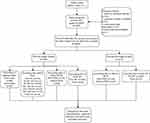
This retrospective study was conducted in the obstetrics department, of Jiangsu Province Hospital, the First Affiliated Hospital of Nanjing Medical University. Clinical data were collected from January 2020 to December 2021. All pregnant women with grade III MSAF (opaque and deep green AF) were included except for cases with elective cesarean section, induction after a stillbirth, incomplete data, and fetal renal abnormalities. Baseline information includes age, body mass index (BMI), gestation, primipara rate, the rate of application of assisted reproductive technology (ART), and complications (including diabetes mellitus (DM), preeclampsia, cord or placental abnormalities, positive Group B Streptococcus (GBS), and premature rupture of membrane (PROM)); Maternal outcomes include observation time (from the discovery of grade III amniotic fluid to the birth of the fetus), thin and thick amniotic fluid (AF) percentages (“thin” or “thick” meconium were defined, respectively, as fluid that was normal except for a greenish or yellowish color, or fluid that was viscous, tenacious and opaque12), AF volume (estimated by a midwife or doctor), artificial rupture of membrane rate, the mode of labor, intrapartum regional analgesia rate, intrapartum fever rate (>37.5°C), abnormal fetal heart rate (FHR) (category II and III FHR pattern according to the 3-tier classification system defined by the 2008 National Institute of Child Health and Human Development workshop13), delivery mode, vaginal bleeding over 24 hours (estimated by gravimetry; the volume of blood in milliliters is the difference in wet and dry weight in grams approximates), and a routine blood test 24 hours after delivery; Neonatal outcomes include: gender, birth weight, abnormal fetal development rate, Apgar scores at 1 and 5 min, and NICU admission rate. Data analysis is shown in Figure 1. Data acquired from our hospital was approved by the Ethical Board of the First Affiliated Hospital of Nanjing Medical University and were kept anonymized and in confidentiality and complied with the Declaration of Helsinki (IRB-GL1-AF06).
 |
Figure 1 The flow chart of the present study design. |
Statistical Analysis
Statistical analysis was performed using the SPSS statistical package (version 26.0, SPSS Inc., Chicago, IL, USA). The normality of continuous variables was analyzed by a Shapiro–Wilk test. The standard normally distributed data are described as the mean ± standard deviation (SD) and were compared by a Student’s t-test. Non normally distributed variables are expressed as the median (interquartile range) and were compared by a Mann–Whitney U-test. Categorical variables were described as concrete cases (percentages) and compared by a chi-square test or Fisher’s exact test. A value of p < 0.05 was considered statistically significant.
Results
The Higher the Thick Amniotic Fluid Ratio is in the First Stage of Labor, the Higher the Cesarean Delivery Rate
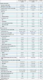
A total of 345 cases were selected in the present retrospective study. Although the age of participants between the first or second stage of labor seemed to be different, when comparing those over 35 years (20 cases vs 22 cases, respectively, in the first or second stage of labor), the P-value was 0.150 (data was not shown). Other baseline characteristics were not significantly different in different stages of labor. There was a significant difference in the cord or placental abnormality ratio between the first and second stages of labor. In the first stage of labor, 1 case had battledore placenta, and 1 case had the cord around the neck. In the second stage of labor, 9 cases of the cord around the neck, 1 case of a true knot of cord, 2 cases of placental adherence, 3 cases of battledore placenta, and 1 case of velamentous placenta were observed. A longer observation time was found in the first stage, and 75% of cases had less than 3 hours of observation time. More cases with thick amniotic fluid were observed in the first stage of labor than in the second stage, and higher rates of cesarean section rates (41.5%) but lower rates of vaginal delivery (25.4%) were observed in the first stage of labor than in the second stage (6.5%, 67.0%). Other maternal and neonatal outcomes were not found to be significantly different between the first and second stages of labor (Table 1).
 |
Table 1 The Differences of Grade III MSAF Between the First and Second Stage of Labor |
When Grade III MSAF was Found in Active Labor, a Lower Cesarean Delivery Rate, but a Higher NICU Admission Rate Occurred
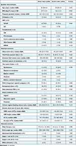
When we compared the difference between grade III AF found in early labor and active labor, more women had higher BMI and PROM in early labor, and more primipara were in active labor. With the progress of labor, intrapartum regional analgesia rates gradually increased. Higher intrapartum regional analgesia rates were found in active labor. Higher cesarean delivery rates occurred in early labor as well as higher operative vaginal delivery rates (vacuum extraction was used in 2 cases). Higher NICU admission rates were observed in active labor (21.9%) than in early labor (9.1%). Other baseline characteristics, and maternal and neonatal outcomes did not demonstrate a significant difference between early and active labor (Table 2).
 |
Table 2 The Differences of Grade III MSAF Between Early and Active Labor |
If Grade III MSAF was Found in the First Stage of Labor, Waiting for Vaginal Delivery Not Less Than 4 Hours was Possible if Clinically Indicated
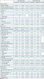
When we grouped comparisons according to observation time from the discovery of grade III MSAF to the final delivery, with a wait of more than 30 or 60 min, a higher WBC count and neutrophil count were observed. In addition, over 60 min, the C-reactive protein expression level rose, but the neutrophil percentage and intrapartum fever did not show a significant difference. If a wait of more than 60 min occurred, the 24-hour vaginal bleeding volume showed a slight increase. For waits of more than 90 and 120 min, significantly lower cesarean rates but higher operative vaginal delivery rates occurred than for waits of less than 90 or 120 min. For waits of more than 180 or 240 min, a nonstatistical difference in either baseline characteristics or maternal and neonatal outcomes was observed (Table 3).
 |  |  |
Table 3 The Maternal and Neonatal Differences of Grade III MSAF Among Different Observation Times |
In Both the First and Second Stages of Labor, NICU Admission Rates Increased with the Occurrence of Abnormal FHR
In the first stage of labor, the artificial rupture of membrane ratio was higher in pregnancies with an abnormal FHR than in those without. A higher cesarean rate (55.4%) occurred in pregnancies with an abnormal FHR than in those without (31.1%), and the vaginal delivery rate (16.1%) among those with an abnormal FHR was lower than that among those without (32.4%). In the second stage of labor, the thick MSAF ratio was higher in pregnancies with an abnormal FHR (21.3%) than in pregnancies without (3.0%). In either the first or second stage of labor, the morbidity of pregnancies with an intrapartum fever was higher in pregnancies with an abnormal FHR (44.7%, 40.4%) than in those without (21.6%, 20.8%). In the first stage of labor, only the WBC count showed a significant difference between pregnancies with and without an abnormal FHR. In the second stage of labor, neutrophil percentage and count were higher in pregnancies with an abnormal FHR in addition to WBC count. In addition, NICU admission rates were higher in pregnancies with an abnormal FHR, whether women were the first stage of labor or the second stage of labor. However, only in the second stage of labor, was the percentage of 1-minute Apgar scores of 4 to 7 was higher in pregnancies with FHR abnormalities than in those without (Table 4).
 |
Table 4 The Differences Between Grade III MSAF with and without Abnormal FHR |
In the Second Stage of Labor, NICU Admission Rates Increased with the Occurrence of Thick Grade III MSAF
In the first stage of labor, a longer gestation week and a larger AF volume were observed in the thick AF group than in the thin AF group. In addition, thick MSAF was found more frequently in early labor than in active labor. Vaginal bleeding volume was greater in women with thick MSAF than in those with thin AF. The rate of cesarean section was significantly higher in women with thick MSAF than in women with thin AF (73.1% vs 33.7%), but there was a lower vaginal operation rate (7.7% vs 39.4%). A higher incidence of intrapartum fever occurred in women with thick AF, in either the first or second stage of labor. Only in the second stage of labor, the neutrophil percentage was higher in thick AF than in thin AF, and more neonates with Apgar scores of 4~7 at 1 or 5 min were in the thick AF group. Similarly, a higher NICU admission rate was shown in women with thick AF (60.0%) than in thin AF (19.5%) (Table 5).
 |
Table 5 The Differences Between Thin and Thick MSAF |
Discussion
The etiology of MSAF is unknown, although studies of the difference between women with clear AF and MSAF have reported that some risks associated with the occurrence of MSAF include advanced maternal age, prolonged labor, postterm pregnancy, maternal complications (such as DM, hypertensive disorders of pregnancy, and intrahepatic cholestasis of pregnancy), and oligohydramnios.3,14 In our study, only women with grade III MSAF were selected, but in women with thick grade III MSAF, a longer gestational week, a lower AF volume, and a higher incidence of intrapartum fever were observed compared with women with thin AF. In another study on the difference in the degree of MSAF (yellow, green and thick groups), more women with intrapartum fever were in the thick AF group, and the authors explained that the occurrence of MSAF, especially thick MSAF, might be a potential alert of maternal infection.15 However, it cannot explain why not all women with MSAF have an intrapartum fever, or why this acute response is not shown by a change in routine blood tests, for WBC, CRP and neutrophil percentage. The causal association between intrapartum fever and MSAF or more severe MSAF (thick AF) remains unknown. Maternal intrapartum fever might result in the occurrence of thick MSAF, and in turn, MSAF might be a risk factor for uterine infection or a stress-induced maternal response via of intrapartum fever.
Unignorably, some maternal and perinatal outcomes might increase as the staining and consistency of amniotic fluid increases,12 which might affect a doctor’s decision regarding intervention in cases of MSAF. Some studies reported the difference between MSAF and clear AF, and their results showed increasing rates of cesarean section, puerperal infection, postpartum hemorrhage (PPH) and even mortality (such as labor dystocia) among women with MSAF.16,17 It is easy to understand the increasing rates of cesarean section or operative vaginal delivery with the occurrence of MSAF; these shorten the delivery time, especially when accompanied by thick AF or abnormal FHR in the first stage of labor, in cases of poor neonatal outcome. In addition, our results showed that in women with thick MSAF in the first stage of labor, the 24-hour vaginal bleeding volume was greater than that in women with thin MSAF, but the rate of PPH was not significantly different (data not shown). Previously published literature revealed that MSAF is significantly associated with a higher risk of moderate (1000–2000 mL) and severe (>2000 mL) PPH than clear AF,18 but another study reported the risk is higher for minor (500–1000 mL) and moderate but not for severe PPH,19 a finding which may need further studies. Regarding puerperal infections, our results showed only a slight change in WBC count, CRP and neutrophil percentage or count, and no cases of puerperal infection or death existed in our data, which may be due to the use of antibiotics.
To neonates, MAS, a neonatal complication of MSAF, can develop into a serious condition and even cause mortality.20 MAS is mainly characterized by the early onset of respiratory distress with poor lung compliance and hypoxemia and is diagnosed by a chest X‑ray. Treatment for MAS may even require mechanical ventilation and intubation.20 Most importantly, the incidence of MAS increased with gestational age, from 1.3% at 38 weeks to 4.8% at 42 weeks.10 Prolonged gestation might be an independent risk factor for the occurrence of neonatal MAS in nulliparous women.21 Other associated neonatal short-term adverse effects of MSAF include acidosis, NICU admission, respiratory distress, hypoglycemia, and seizures.22 Our results showed a higher NICU admission rate in pregnancies with grade III MSAF and an abnormal FHR either in the first or second stage of labor. FHR reflects the fetal condition in the uterus, and abnormal FHR indicates intrauterine fetal hypoxia. Normal FHR is between 110–160 bpm, as defined by cardiotocography monitoring. However, one recent study demonstrated that a baseline FHR between 150 and 160 bpm might be a signal of MSAF, which was approximately 2.6 times more likely to occur than in 110~149 bpm group.23 A previously published report viewed MSAF as a predictor of delayed closure among neonates with gastroschisis.24 In our data, only 5 abnormal fetal developments (3 cases of congenital heart disease, 1 case of anotia, and 1 case of agenesis of the corpus callosum), were likely not directly associated with the occurrence of MSAF. Among pregnancies with thick MSAF in the second stage of labor, a higher rate of NICU admission and more Apgar scores of 4~7 (indicating fetal distress) at 1 and 5 min occurred than in pregnancies with thin AF. Similar research was published in which thick MSAF was associated with a low APGAR score, a high rate of emergency cesarean section, and MAS.11 A possible explanation is that an exposure under thick MSAF, where there is a lack of oxygen compared with “light”-stained MSAF, resulted in the intestinal ischemia. Then, the fetal anal sphincter relaxed and gastrointestinal peristalsis increased, leading to the passage of meconium when umbilical vein oxygen saturations was below 30%.25 In addition to the abovementioned short-term neonatal outcomes, recent studies have followed up on the long-term outcome in newborns. Fetal exposure to MSAF during labor and delivery may result in lower rates of long-term infectious-related hospitalizations, lower incidence of long-term infectious morbidity in the offspring,4 lower rates of long‐term respiratory-related hospitalizations,5 and an independent protective factor for dermatitis and skin rash-related hospitalizations throughout childhood and adolescence.22,26
It seems that MSAF may affect newborns more than women, but newborns also benefit from the long-term effects of MSAF. Researchers have even put forward the view that “MSAF is more like a symptom rather than a syndrome”,27 but no clinical practice guidelines have been developed by any international organization such as the world health organization or gynecological and obstetrical society. Although it is well-known that intensive delivery room and continuous fetal heart monitoring by the midwife regarding the intrapartum management,15 and non-necessary routine endotracheal suctioning at birth in vigorous term meconium-stained babies28 and other routine postnatal care. However, the observation time once MSAF is found remains unknown. Our results showed non significantly different maternal or neonatal outcomes even after a wait of up to 4 hours, regardless of abnormal FHR or thick MSAF, but a watershed of significantly different rates of operative vaginal delivery time or cesarean section with a wait of 90–120 min, which was associated with our doctor’s decision. One study found a higher rate of composite adverse neonatal outcomes when secondary MSAF (a transition from clear AF to MSAF) was diagnosed >3 h before delivery.29
In conclusion, it might be plausible that waiting for up to 4 hours after finding grade III MSAF in the first stage of labor if labor was progressed and without abnormal FHR. However, the limitations of our study should also be noted. The present study was a single-center retrospective study with a small population size sample. More cases, and more centers, even in different countries, would be needed to design a prospective study. The differences between early and active labor and the observation time in the two labor stages respectively must be investigated. It is unclear whether 4 hours is that the endpoint for observation in the first stage of labor, or whether it is 5 or more hours; more studies are needed. Due to a lack of objectivity in the definition and quantitative diagnostic criteria for MSAF, many scholars questioned the timing of intervention for MSAF, especially grade III MSAF, but have not investigated it. In the future, we hope there will be quantitative diagnostic methods and further investigation to dilute the subjective bias of MSAF diagnosis.
Abbreviations
MSAF, meconium stained amniotic fluid; AF, amniotic fluid; BMI, body mass index; ART, assisted reproductive technology; IVF-ET, in vitro fertilization-embryo implantation; IUI, intrauterine insemination; DM, diabetes mellitus; ICP, intrahepatic cholestasis; PROM, premature rupture of membrane; GBS, group B Streptococcus; FHR, fetal heart rate; WBC, white blood cell; CRP, C-reactive protein; NICU, neonatal intensive care unit; MAS, meconium aspiration syndrome; PPH, postpartum hemorrhage.
Acknowledgment
Xinxin Zhu, Shiyun Huang, Yuxuan Tang, Zhonglan Wu contributed equally to this work (co-first authors). Qing Zuo, Zhiping Ge, Ziyan Jiang contributed equally to this work (co-corresponding authors).
Disclosure
The authors report no conflicts of interest in this work.
References
1. Liabsuetrakul T, Meher S. Intrapartum care algorithms for liquor abnormalities: oligohydramnios, meconium, blood and purulent discharge. BJOG. 2022. doi:10.1111/1471-0528.16728
2. Abraham K, Thomas E, Lionel J. New evidence to support antibiotic prophylaxis in meconium-stained amniotic fluid in low-risk women in labor a prospective cohort study. J Obstet Gynaecol India. 2018;68(5):360–365. doi:10.1007/s13224-017-1043-y
3. Addisu D, Asres A, Gedefaw G, Asmer S. Prevalence of meconium stained amniotic fluid and its associated factors among women who gave birth at term in Felege Hiwot comprehensive specialized referral hospital, North West Ethiopia: a facility based cross-sectional study. BMC Pregnancy Childbirth. 2018;18(1):429. doi:10.1186/s12884-018-2056-y
4. Paz Levy D, Walfisch A, Wainstock T, et al. Meconium-stained amniotic fluid exposure is associated with a lower incidence of offspring long-term infectious morbidity. Am J Reprod Immunol. 2019;81(6):e13108. doi:10.1111/aji.13108
5. Rodavsky G, Sheiner E, Walfisch A, et al. Meconium stained amniotic fluid exposure and long-term respiratory morbidity in the offspring. Pediatr Pulmonol. 2021;56(7):2328–2334. doi:10.1002/ppul.25357
6. Hiersch L, Krispin E, Aviram A, Wiznitzer A, Yogev Y, Ashwal E. Effect of meconium-stained amniotic fluid on perinatal complications in low-risk pregnancies at term. Am J Perinatol. 2016;33(4):378–384. doi:10.1055/s-0035-1565989
7. Oyelese Y, Culin A, Ananth CV, Kaminsky LM, Vintzileos A, Smulian JC. Meconium-stained amniotic fluid across gestation and neonatal acid-base status. Obstet Gynecol. 2006;108(2):345–349. doi:10.1097/01.AOG.0000226853.85609.8d
8. Unsworth J, Vause SJO. Meconium in labour. Gynaecol Med R. 2010;20(10):289–294. doi:10.1016/j.ogrm.2010.06.005
9. Itzhaki-Bachar L, Meyer R, Levin G, Weissmann-Brenner A. Incidental finding of meconium-stained amniotic fluid in elective cesarean deliveries: features and perils. Int J Gynecol Obstet. 2021;158:418.
10. Ward C, Caughey AB. The risk of meconium aspiration syndrome (MAS) increases with gestational age at term. J Matern Fetal Neonatal Med. 2022;35(1):155–160. doi:10.1080/14767058.2020.1713744
11. Mohammad N, Jamal T, Sohaila A, Ali SR. Meconium stained liquor and its neonatal outcome. Pak J Med Sci. 2018;34(6):1392–1396. doi:10.12669/pjms.346.15349
12. Sheiner E, Hadar A, Shoham-Vardi I, et al. The effect of meconium on perinatal outcome: a prospective analysis. J Matern Fetal. 2002;11(1):54–59. doi:10.1080/jmf.11.1.54.59
13. Macones GA, Hankins GD, Spong CY, Hauth J, Moore TJJ. The 2008 national institute of child health and human development workshop report on electronic fetal monitoring: update on definitions, interpretation, and research guidelines. Gynecol Nurs. 2008;37(5):510–515.
14. Levin G, Tsur A, Shai D, Cahan T, Shapira M, Meyer R. Prediction of adverse neonatal outcome among newborns born through meconium-stained amniotic fluid. Int J Gynecol Obstet. 2021;154(3):515–520. doi:10.1002/ijgo.13592
15. Rodríguez Fernández V, López RY, Cajal C, Marín OE. Intrapartum and perinatal results associated with different degrees of staining of meconium stained amniotic fluid. Eur J Obstet Gynecol Reprod Biol. 2018;228:65–70. doi:10.1016/j.ejogrb.2018.03.035
16. Ma’ayeh M, Snyder A, Oliver EA, Gee SE. Meconium-stained amniotic fluid and the risk of postcesarean surgical site infection. Medicine N. 2021;34(9):1361–1367.
17. Attali E, Lavie M, Lavie I, et al. Prolonged exposure to meconium in cases of spontaneous premature rupture of membranes at term and pregnancy outcome. J Matern Fetal Neonatal Med. 2021;2021:1–6.
18. Bouchè C, Wiesenfeld U, Ronfani L, et al. Meconium-stained amniotic fluid: a risk factor for postpartum hemorrhage. Ther Clin Risk Manag. 2018;14:1671–1675. doi:10.2147/TCRM.S150049
19. Fang ZJ, Liu HF, Zhang YL, Yu L, Yan JY. Relation of meconium-stained amniotic fluid and postpartum hemorrhage: a retrospective cohort study. Eur Rev Med Pharmacol Sci. 2020;24(20):10352–10358. doi:10.26355/eurrev_202010_23384
20. Mehar V, Agarwal N, Agarwal A, Agarwal S, Dubey N, Kumawat H. Meconium-stained amniotic fluid as a potential risk factor for perinatal asphyxia: a single-center experience. J Clin Neonatol. 2016;5(3):157.
21. Lindegren L, Stuart A, Herbst A, Källén K. Improved neonatal outcome after active management of prolonged pregnancies beyond 41(+2) weeks in nulliparous, but not among multiparous women. Acta Obstet Gynecol Scand. 2017;96(12):1467–1474. doi:10.1111/aogs.13237
22. Krieger Y, Horev A, Wainstock T, Sheiner E, Walfisch AJJ. Meconium‐stained amniotic fluid as a protective factor against childhood dermatitis and skin rash‐related hospitalization in the offspring–a population‐based cohort analysis. Venereology. 2020;34(2):319–324.
23. Ghi T, Di Pasquo E. Intrapartum fetal heart rate between 150 and 160 bpm at or after 40 weeks and labor outcome. Acta Obstet Gynecol Scand. 2021;100(3):548–554. doi:10.1111/aogs.14024
24. Koehler SM, Loichinger M, Peterson E, Christensen M, Szabo A, Wagner AJ. Meconium-stained amniotic fluid as a predictor of poor outcomes in gastroschisis. J Pediatr Surg. 2018;53(9):1665–1668. doi:10.1016/j.jpedsurg.2018.04.023
25. Lakshmanan J, Ross M. Mechanism (s) of in utero meconium passage. J Perinatol. 2008;28(3):S8–S13. doi:10.1038/jp.2008.144
26. Matalon R, Wainstock T, Walfisch A, Sheiner E. Exposure to meconium-stained amniotic fluid and long-term neurological-related hospitalizations throughout childhood. Am J Perinatol. 2021;38(14):1513–1518. doi:10.1055/s-0040-1713863
27. Monen L, Hasaart T, Kuppens SJ. The aetiology of meconium-stained amniotic fluid: pathologic hypoxia or physiologic foetal ripening? Early Hum Dev. 2014;90(7):325–328. doi:10.1016/j.earlhumdev.2014.04.003
28. Wei Q, Chen W, Liang Q, Song S, Li J. Effect of endotracheal suctioning on infants born through meconium-stained amniotic fluid: a meta-analysis. Am J Perinatol. 2022. doi:10.1055/s-0041-1741034
29. Tairy D, Gluck O, Tal O, et al. Amniotic fluid transitioning from clear to meconium stained during labor—prevalence and association with adverse maternal and neonatal outcomes. J Perinatol. 2019;39(10):1349–1355. doi:10.1038/s41372-019-0436-4
 © 2023 The Author(s). This work is published and licensed by Dove Medical Press Limited. The full terms of this license are available at https://www.dovepress.com/terms.php and incorporate the Creative Commons Attribution - Non Commercial (unported, v3.0) License.
By accessing the work you hereby accept the Terms. Non-commercial uses of the work are permitted without any further permission from Dove Medical Press Limited, provided the work is properly attributed. For permission for commercial use of this work, please see paragraphs 4.2 and 5 of our Terms.
