
Back to Journals » International Journal of Nanomedicine » Volume 21
Next Generation Calcium Nanomaterials: Disrupting Tumor Ca2⁺ Homeostasis for Enhanced Cancer Therapy
Authors Gangadhar L, Sana SS  , Venkatesan R , Bhandare RR, Kim SC, Yasin HKA
Received 2 August 2025
Accepted for publication 29 December 2025
Published 11 February 2026 Volume 2026:21 557935
DOI https://doi.org/10.2147/IJN.S557935
Checked for plagiarism Yes
Review by Single anonymous peer review
Peer reviewer comments 2
Editor who approved publication: Dr Krishna Nune
Lekshmi Gangadhar,1,* Siva Sankar Sana,2,* Raja Venkatesan,2,3 Richie R Bhandare,4,5 Seong-Cheol Kim,2 Haya Khader Ahmad Yasin4,5
1Department of Nanotechnology, Nanodot Research Private Limited, Nagercoil, Tamil Nadu, India; 2School of Chemical Engineering, Yeungnam University, Gyeongsan, Republic of Korea; 3Department of Biomaterials, Saveetha Dental College and Hospitals, SIMATS, Saveetha University, Chennai, Tamil Nadu, India; 4College of Pharmacy & Health Sciences, Ajman University, Ajman, United Arab Emirates; 5Center of Medical and Bio-Allied Health Sciences Research, Ajman University, Ajman, United Arab Emirates
*These authors contributed equally to this work
Correspondence: Seong-Cheol Kim, Email [email protected] Haya Khader Ahmad Yasin, Email [email protected]
Abstract: Calcium-based nanotechnology is a rapidly growing field that leverages the unique properties of calcium compounds at the nanoscale to develop innovative materials and applications. This field centers around the manipulation and utilization of calcium compounds, such as calcium carbonate, calcium phosphate, and calcium oxide (CaO), when their dimensions are reduced to the nanoscale. Calcium compounds are generally biocompatible, biodegradable, cost-effective, and versatile, making them suitable for biomedical applications. Calcium homeostasis plays a critical role in cancer development and its progression. Understanding the mechanisms underlying cancer and calcium homeostasis disruption is crucial for developing effective strategies in cancer therapy. This review explores the therapeutic implications of intracellular calcium homeostasis, its crucial cancer development, and advances in therapy. The link between calcium homeostasis and tumor therapy is examined in this review, with particular attention paid to common calcium-based nanomaterials and their multifunctionality with other metals and compounds. Potential treatment options for disrupting calcium homeostasis include calcification therapy for tumor diagnosis, indirect prevention of tumor growth by altering the tumor microenvironment, and direct killing of tumor cells by inducing calcium overload. This review also examines the progress made in creating and using multifunctional calcium-based nanomaterials to treat cancer. Nanomaterials, which are categorized according to their main constituents (hydroxyapatite, carbonate, peroxide, and calcium phosphate), show great promise in ion interference, gene delivery, protein delivery, and drug delivery. Despite the challenges in this field, calcium-based nanotechnology holds immense potential for a wide range of applications, from healthcare to environmental remediation to energy. Continued research and development in this field will undoubtedly lead to more innovative and impactful technologies in the future.
Keywords: calcium, homeostasis, cancer therapy, diagnosis, nanomedicine
Introduction
Currently, 10 million new cases of cancer are diagnosed worldwide each year, making it the second leading cause of death. Unless the number rises to 20 million in 17 years, the existing situation of an increase in cancer incidence necessitates an efficient prevention plan.1 Ions have been extensively employed for tumor therapy. Ions such as zinc (Zn2+), barium (Ba2+), potassium (K+), sodium (Na+), calcium (Ca2+) and so forth are essential for many biological processes, such as immune cell activation, yet they also disrupt intracellular communication, blood pH, intracellular osmotic pressure, and the inflammatory response.2 Ca2+ is an integral part of bone, which is composed of 5–10% water, 20–40% organic matrix, 50–70% minerals, and less than 3% lipids.3–5 About 90–95% of the bone organic matrix is made up of type I collagen fibers, with trace levels of other proteins, including alkaline phosphatase (ALP) and osteocalcin (OC).6 In order to give bone its mechanical rigidity and strength, tiny crystals of hydroxyapatite make up the majority of the mineral composition of bone. Additionally, tiny deposits of acid phosphate, magnesium, and carbonate accumulate on collagen fibers. Because they are more soluble than geologic hydroxyapatite crystals, these tiny, weakly crystalline, carbonate-substituted crystals can aid in mineral metabolism.7,8 As the primary extracellular signaling molecule Ca2+ serves as a universal secondary messenger used widely in signaling systems.9 It helps multiple living systems work through its participation in neural signaling and muscle activation. Ca2+ supports many cell tasks at the Deoxyribonucleic acid (DNA) level and assists in making proteins and controlling enzymes. Multiple body functions depend on the careful control of calcium levels. The body maintains a proper calcium balance through the action of calciotropic hormones. Disruptions in calcium homeostasis can have significant health consequences, as evidenced by the use of Ca2+ channel inhibitors in the treatment of conditions like pain and hypertension.10–12
Nanotechnology holds immense promise for revolutionizing cancer treatment.13 Continued research and development in this area are crucial for unlocking its full potential and improving the lives of cancer patients. The schematic diagram of cancer therapy strategies based on Ca2+ homeostasis, Figure 1. Recent developments in nanotechnology have led to the development of a wide spectrum of medicinal and diagnostic agents, which has increased interest in their intracellular administration. Most first-line cancer medications have dose-limiting adverse effects, are untargeted, and are water insoluble. Furthermore, several of these therapeutic agents, such as DNA or small interfering Ribonucleic acid (siRNA), are large, charged molecules that have difficulty passing through cell membranes to exert their intended effects. This is in addition to advancements in colloid technology. Therefore, in order to facilitate their trafficking and cellular uptake, a suitable delivery method is needed.14 Despite the fact that recombinant viral vectors have shown great effectiveness in treating human diseases, their significant disadvantages and eventual clinical failure have caused a rapid transition to synthetic nonviral carriers. Numerous nanoparticle compositions, such as calcium phosphate-based mineral systems, silica, dendrimers, biodegradable polymers, gold, iron oxide, and liposomes, have been proposed to overcome the drawbacks of drug delivery methods. Potential bioactive substances should be physically or chemically incorporated into an ideal drug carrier, which would then shield them from the bloodstream. To improve therapeutic efficacy, the carrier complex should gradually deconstruct and offer continuous drug release over an extended period. To lessen their off-target effects and increase the on-site drug concentration, it should also offer a workable mechanism for selectively binding to target cells or tissues.15
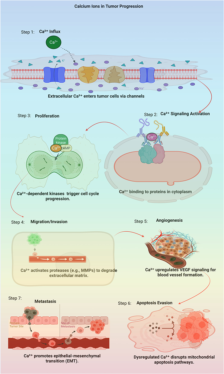 |
Figure 1 The Schematic diagram of cancer therapy strategies based on Ca2+ homeostasis. [Image is created by Biorender.com]. |
Calcium nanocomposites combine nano-scale compounds of calcium (CaCO3 or CaPO4) with other substances for unique material development. Combining calcium nanocomposites creates stronger materials than their separate parts. Research has indicated that calcium nanocomposites have numerous benefits, including being very durable, rigid, nontoxic, and biodegradable. These materials hold promise for biomedical applications as well as potential environmental remediation and energy storage.16 When cells maintain appropriate levels of calcium, they can live multiply, and die appropriately. Cancer cells disrupt the good relationship between the calcium levels and tissues.17 This review is focused on examining the therapeutic implications of intracellular calcium hemostasis in addition to its significant role in cancer development, and therapy improvement. This review discusses the connection between calcium hemostasis and tumor therapy, and gives specific interest to the typical calcium-based nanomaterials and their multifunctionality with other metals and compounds. Possible treatment approaches to disorient calcium homeostasis include the diagnosis of the tumor by calcification therapy, indirect by prevention of tumor growth by manipulating the microenvironment and direct killing of tumor cells by inducing calcium overload. This review also examines the developments in the application of multifunctional calcium-based nanomaterials for cancer treatment.
Altered Calcium Homeostasis in Tumorigenesis
Since cancer still poses a serious threat to global health, there is a constant need to find new and effective therapeutic options.18,19 Although the role of altered Ca2+ regulation in cancer has long been known, this field of study has only recently acquired traction. Figure 2 demonstrates the calcium signaling and associated pathways in cancer. In Figure 2, section A represents the area near the cell membrane and cytosol, where extracellular growth receptors activate phosphoinositide signaling, leading to IP3 generation and initial signal transduction for cell cycle regulation. B is related to ATPase-mediated Ca2⁺ translocation and protein trafficking involving the Golgi complex and endoplasmic reticulum (ER). Calcium signaling influences protein modification and export. C indicates the mitochondrion, focusing on reactive oxygen species (ROS) generation, electron transport chain activity, and protein damage or proteolysis under stress conditions. D is centered at the ER, highlighting processes such as protein folding, import/export, calumenin involvement, and Ca2⁺ signaling. Human malignancies often exhibit altered expression and activity of Ca2+ pumps, channels, and exchangers. Increased cell motility, proliferation, apoptosis resistance, and immune evasion are the results of this imbalance.20–22 Ca2+ balance within cells is crucial and is tightly controlled by various mechanisms. However, disruptions to this balance, often triggered by external factors, can overwhelm the cell’s regulatory systems, leading to cell damage or death. Cell death due to Ca2+ imbalances is not solely caused by changes in Ca2+ movement but also by upstream signaling pathways, such as those involving G-protein-coupled receptors (GPCRs), receptor tyrosine kinase (RTK), ion channels, and nicotinic acid dinucleotide phosphate (NAADP), which significantly influence intracellular Ca2+ levels and ultimately determine cell fate.23 Cancer cells, due to their uncontrolled growth, are particularly vulnerable to Ca2+ imbalances. This makes Ca2+ signaling a promising target for cancer treatment.24,25 Therapeutic approaches can directly target tumor cells or indirectly influence tumor behavior by interacting with the tumor microenvironment (TME). To target cancer cells in therapy, Ca2+ overload treatment is used, which damages their cellular structure, resulting in metabolic breakdown followed by cell death. The behavior of TME cells depends on calcium homeostasis, which helps or hinders tumor development by managing immune responses and new blood vessel development.26 Ca2+ homeostasis influences TME behaviors, such as immune responses and blood vessel formation, indirectly impacting tumor development.27 Manipulating Ca2+-related organelle coordination and controlling Ca2+ entry and exit routes creates effective cancer treatments by altering Ca2+ overload and disequilibrium.28 Cancer cell death mechanisms involving Ca2+ are shown in Table 1.
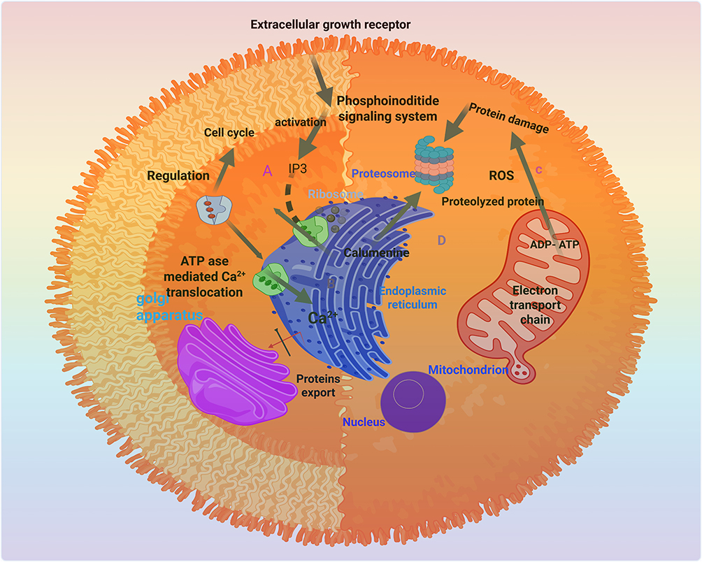 |
Figure 2 Ca2+ signaling involved in calcium homeostasis [Image is created by Biorender.com]. |
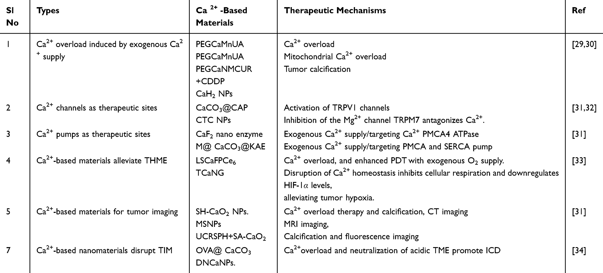 |
Table 1 Summary of Research on Ca2+-Based Nanomaterials for Manipulating Calcium Homeostasis in Cancer |
Aberrated Regulation of Intraorganellar Communication in Cancer
The survival and proliferation of cancer cells depend on the balanced functioning of their internal compartments, or organelles. These organelles play key roles in signaling pathways that promote cancer, such as increased energy production and protection from oxidative stress. Current cancer treatments, including chemotherapy, gene therapy, and therapies targeting ROS, often affect these organelles, as their intended targets reside within them. However, such therapies often interfere with the normal activity of healthy cells because they are not cell within the cell. There is still a strong demand for personalized approaches that are capable of precisely controlling subcellular organelles of cancer cells but causing minimal damage to healthy cells. Cell and tissue functions depend on the effective signaling of Ca2+ between cellular compartments. The proper functioning of cells and tissues depends on the efficient communication of Ca2+ between different cellular compartments. Disruptions in this intricate Ca2+ signaling network can have serious consequences, including the development of diseases such as cancer.35 Cancer cells exhibit altered cellular behavior, including uncontrolled growth, resistance to growth-inhibiting signals, and the ability to evade cell death. These alterations are driven by changes in communication between cellular organelles. Importantly, cellular metabolism, particularly the function of mitochondria, plays a critical role in supporting the growth and survival of cancer cells, interacting with various pathways that promote cancer development.36,37
Interplay of Endoplasmic Reticulum Stress and Calcium Dysregulation in Cancer
Endoplasmic Reticulum (ER) calcium depletion triggers protein misfolding, leading to chronic mitochondrial calcium overload and apoptosis through the B-cell leukemia/lymphoma 2- (Bcl-2) dependent pathway. ER stress induces the localization and oligomerization of pro-apoptotic Bcl-2 proteins (Bax and Bak), promoting calcium release from the ER into the cytosol via inositol-3 phosphate receptors (IP3Rs) and ryanodine receptors (RyRs).38 Increased cytosolic calcium activates muscle calpain, which cleaves and triggers ER-resident procaspase-12, initiating the ER stress-induced cell death pathway. Cytosolic calcium also enters the mitochondrial matrix, causing mitochondrial membrane depolarization, cytochrome release, and apoptosome activation, leading to apoptosis. C/EBP Homologous Protein (CHOP), a key regulator of ER stress-induced apoptosis, suppresses Bcl-2 expression and triggers pro-apoptotic gene transcription. The association of inositol requiring enzyme/ tumor necrosis factor receptor-associated factor 2 (IRE1/TRAF2) during ER stress triggers the release and activation of procaspase-12, ultimately leading to apoptosis.39
Misfolded proteins accumulating in ER membranes, which disturb homeostasis, lead to ER stress. ER stress develops from various sources, including disturbed Ca2+ concentrations, nutrient deficiencies, and oxidative damage. Significant Ca2+ deposits exist within the ER, and cell death occurs when ER stress leads to excessive cytoplasmic and mitochondrial Ca2+ accumulation.40 The uptake of Ca2+ ions into the ER occurs mostly through Sarco/Endoplasmic Reticulum Calcium ATPase (SERCAs). The IP3R and RyRs pathways control Ca2+ movement through ion channels during efflux events. Store-operated calcium entry (SOCEs) plays an essential role in sustaining the endoplasmic reticulum’s Ca2+ reserves. When ER stress affects ER Ca2+ homeostasis, it enables cancer development because high Ca2+ levels kill cells while simultaneously disrupting metabolic processes and energy production. The maintenance of Ca2+ homeostasis in cells is heavily influenced by ER stress. Research indicates that targeting calcium-regulatory mechanisms in the ER holds promise for the treatment of cancer.
Unravelling the Secrets of Endoplasmic Reticulum-Mitochondria Communication
Ca2+ serves as a signaling molecule that controls the communication dynamics between the ER and mitochondria.40 The interaction between these organelles facilitates various cellular processes, including lipid production and calcium transfer, and functions in promoting mitochondrial growth and division. Such organelle-to-organelle communication helps cells stay alive while determining the metabolic pathways and mechanisms to fight against dying cells. Improper calcium management results in cancer development. Three mechanisms contribute to cancer development: blocking excessive calcium entry into the mitochondria, blocking cellular death pathways, and inducing mitochondrial dysfunction. Tumor development depends on calcium signaling, which regulates the expression of cancer-promoting and tumor-blocking genes, while the protein Bcl-2 regulates these signaling mechanisms for tumor growth.41 Different forms of cancer such as lung cancer, myeloma, and lymphomas require Bcl-2 dysregulation for their development. Doctors use BIRD-2 as an inhibitor to treat B-cell malignancies. The anti-apoptotic protein Bcl-XL frequently shows increased expression levels in tumors.42 The protein acts as a survival protector by maintaining the regulation of cytochrome-C release and controlling mitochondrial membrane permeability. The IP3R channel functions as a direct target of Bcl-XL, which influences calcium flow.43 Research has directed investigators to examine the potential of the Bcl-XL inhibitor WEHI-539 for diverse solid tumors and lymphoma treatment. Phosphorylated K-Ras binds to IP3R1 to suppress channel sensitization as a mechanism to prevent mitochondrial calcium uptake alongside autophagy enhancement. Oncogenic Rat Sarcoma (Ras) variants bind with IP3Rs to control ER-calcium ion release, creating conditions favorable for cancer growth and cell survival. Tumors are more likely to survive due to the AK-strain transforming (Akt) pathway that drives phosphorylation events on IP3Rs, thus disrupting normal Ca2+ release mechanics.44 The pro-cancerous H-Ras and K-Ras strains modify calcium signaling pathways to block cellular apoptotic responses. Through Mitochondrial Associated Membranes (MAMs), H-Ras controls the movement of Ca2+ ions from the ER into the mitochondria but K-Ras reduces Ca2+ transfer to the mitochondria by altering the expression of IP3Rs in colorectal cancer cells. Cancer often shows deregulated Akt pathway activity, which contributes to improper calcium signaling in cells. The protein phosphorylation activity of Akt regulates both IP3 receptors and mitochondrial calcium uptake1 protein (MICU1), which controls ER calcium release and mitochondrial calcium uptake.
This results in abnormal levels of calcium inside the mitochondria, enhanced ROS production, and the enhancement of tumor growth. In turn, the mechanistic target of rapamycin C2 (mTORC2), another Akt-activating signaling pathway located at the interface between the ER and mitochondria, collaborates with Akt to ensure proper calcium homeostasis and regulate cell growth. These results highlight the key role of calcium signaling dysregulation in the development and progression of cancer. Tumor Suppressor proteins, such as phosphatase and tensin homolog (PTEN) and Promyelocytic Leukemia (PML), work at the ER-mitochondria interface, which inhibits the survival signaling pathways, such as phosphoinositide 3-kinase (PI3K)-Akt, and promotes the apoptotic response.45 Mitochondrial Dynamics also play a role in cancer development and progression. Abnormalities in mitochondrial morphology and function, influenced by proteins like mitofusin2 (MFN2) and protein tyrosine phosphatase interacting protein-51 (PTPIP51), have implications for tumorigenesis, affecting energy metabolism and apoptosis sensitivity. Orchestrating proteins, including voltage-dependent ion channels (VDAC), mitochondrial uniporter (MCU), and various oxysterol-binding proteins (ORPs), are highlighted for their roles in regulating the coupling between ER and mitochondria, influencing metabolic communication and cell signaling, with alterations linked to cancer progression.46 Interestingly, melatonin, a hormone that controls the sleep-wake cycle, has anticancer properties. This is achieved by modifying the Akt phosphorylation, which inhibits the growth of cancer cells. Therefore, it is a promising adjuvant therapy for the treatment of cancer because of its safety, nontoxicity, and efficacy. It has been demonstrated that MAMs are the location of the tumor suppressors PML and PTEN. Many primary human tumors have mutations in the deleted PTEN on chromosome 10, which causes PTEN function to be lost and constitutively activates the PI3K-Akt pathway. Changes in the physical connections between the ER and mitochondria can disrupt the Ca2+ flow, affecting cellular function. Changes in the expression of a protein called phosphofurin acidic cluster sorting protein 2 (PACS-2) have been associated with a type of lung cancer called non-small cell lung cancer (NSCLC). Furthermore, alterations in the shape and structure of mitochondria, such as fragmentation, can limit the excessive accumulation of calcium within mitochondria, preventing cell death. This disruption of calcium signaling contributes to the “Warburg effect”, a metabolic shift observed in cancer cells where they rely heavily on glycolysis for energy production instead of efficient cellular respiration.47,48
Plasma Membrane-Endoplasmic Reticulum Junctions: Regulators of Calcium Dynamics and Cancer Progression
PM-ER junctions are crucial for regulating intracellular Ca2+ levels by controlling Ca2+ channels in both the plasma membrane (PM) and ER. Dysregulation of SOCE components (like STIM1 and Orai1) is common in various cancers. Increased stromal interaction module protein 1 (STIM1) expression is associated with poor prognosis in several cancers.49,50 SOCE plays a role in cell cycle progression, migration, and invasion of cancer cells. Caveolin-1 (Cav1), a protein found in caveolae, has both tumor-suppressing and tumor-promoting roles. These are small membrane invaginations found in the plasma membrane. They regulate calcium flux, and their structure and function are often altered in cancer cells. It influences Ca2+ homeostasis and contributes to cancer development in various tissues, including the lungs. Oncogenic Ras can alter intracellular Ca2+ levels through Cav1. Cav1 antagonists (like nitrendipine) could be used for cancer therapy. However, through controlling Ca2+ homeostasis and migration, it has been shown that Cav1 affect the development of solid tumors.51 It is interesting to note that the oncogene Ras causes Cav1-mediated changes in intracellular Ca2+ levels, which result in changes to the mitochondria.52 Cav1 antagonists, including verapamil, nitrendipine, and nifedipine, are frequently used in treatments for various illnesses, suggesting that they may be used to treat cancer.53,54 Ca2+ plays a crucial role in regulating sphingosine kinase 1 (SK1) activity and localization within caveolae. Increased Ca2+ levels in caveolae, driven by oncogenic SK1 expression, disrupt focal adhesions and cytoskeletal organization, thereby affecting cell migration. Extended-synaptotagmin (E-Syt) proteins, which modulate ER/plasma membrane contact sites and calcium exchange, are also implicated in cancer.55 The oncogenic lung cancer fusion kinase CD74-ROS targets E-Syt1, leading to increased tumor invasiveness. This phosphorylation event, conserved in other E-Syt proteins, has been observed in human NSCLC biopsies and other cancer cell lines. These findings suggest that E-Syt proteins and their post-translational modifications represent potential targets for cancer therapy.
Targeting Calcium Signaling at the Endoplasmic Reticulum-Endo Lysosome Interface for Cancer Therapy
The cellular space between the ER and end-lysosomes represents a crucial site for communication between these organelles. The interface serves a significant function in cellular operations, including lipid regulation, and calcium signaling, autophagy, and immune array activation.56 The interface operates as a crucial component that substantially initiates cancer onset and disease progression. The abnormal lipid metabolism of lipids at this interface activates cancer cell growth mechanisms, whereas disruptions in calcium signaling promote cellular survival and therapeutic resistance. The interface between endo-lysosomes and the ER plays an essential role in cellular processes, including endocytosis, autophagy, and lysosomal function. Dysregulated biomolecular interactions between these compartments are detected as leading to major alterations in cellular energy utilization and determining cell type development particularly, during tumorigenic processes. Cancer cells depend on lysosomes because these cellular compartments serve both to provide an energy supply and produce the necessary building blocks for their growth.57 Extracellular matrix remodeling allows lysosomes to facilitate tumor spreading with cancer cell movement alongside new blood vessel development. Drug resistance in cancer cells appears to be controlled by functional lysosomes. Studies indicate that elevated levels of two-pore channels TPC1 and TPC2 correlate with enhanced cancer cell migration, metastasis, and their expression affects Ca2+ channels.58 Transient Receptor Potential channels (TRPMLs) have been identified as a tumor progression mechanism that particularly affects glioma cells.59 Cells experience enhanced survival and show increased resistance to cell death processes when TRPML2 protein expression levels rise in the cell. Toward novel cancer treatments, researchers propose focusing on Ca2+ signaling regulation of ER-endo lysosome interfaces specific to targets like TPCs and TRPMLs.31
Mitochondrial Calcium Ions Overload: A Therapeutic Target in Cancer
Modulation of calcium levels within mitochondria, along with the achievement of elevated cytoplasmic calcium signals, is an emerging approach in cancer research. Additional scientific investigations are essential for developing both safe and effective therapeutic approaches that exploit Ca2+ overload for cancer treatment. The cellular substance mitochondria control fundamental processes involving energy creation. When mitochondria contain excessive Ca2+, cells stop producing ATP and eventually die. The MCU functions as a regulator to allow entry of Ca2+ into the mitochondria. Cancer treatment appears promising when researchers focus on manipulating Ca2+ regulatory dynamics between mitochondria and endoplasmic reticulum organelles through MAMs pathways. A malfunctioning cytoplasm calcium balance results from the abnormal accumulation of Ca2+ ions. The abnormal buildup of Ca2+ across multiple cell organelles creates concerning levels of cellular stress, increasing the risk of apoptosis. The compatibility between Ca2+ channel activators, including bepridil and DIM, serves as an additional treatment method specifically targeting cancer-causing cellular Ca2+ overload. Tumor calcification happens as a result of the process that makes cell functions more severe.54
Calcium-Based Nanotherapeutics: Exploiting Calcium Ions Overload for Cancer Therapy
Nanomaterials offer a new approach for the precise management of intracellular signaling pathways in the treatment of cancer. Research scientists can attain efficient cancer therapy outcomes through precise nanoparticle design, which controls cellular activities.55 Researchers have developed new innovative calcium phosphate (CaP) nanomaterials that utilize calcium overload. Scientists create nanostructures by combining CaP-based materials with other substances to trigger the ion release of Ca2+ through acidic tumor cell conditions. These nanostructures are designed to release Ca2+ in response to external exposure to near infra-red (NIR) light or the addition of therapeutic drugs. The delivery of cancer drugs through CaP nanoparticles represents an alternative therapeutic approach. Various cell death mechanisms emerge when drug-loaded CaP nanoparticles affect intracellular calcium homeostasis and create cellular stress patterns. Experimental findings display the broad applicability of CaP-based nanomaterials for cancer therapy and establish exciting routes for better targeted therapeutic development.56 Calcium sulphate nanoparticles are effective in the control of cancerous cell intracellular calcium amounts. The acidic breakdown of calcium sulphate nanoparticles generates cellular signaling molecules comprising Ca2+ and H2S, which shape various cellular processes. Scientific research teams have developed Calcium sulphate-based nanomaterials to deliver both Ca2+ and H2S directly to cancer cells. By entering cancer cells, the nanoparticles initiate an internal reaction that destroys tumor cells while creating defensive immune responses against tumors. Experimental studies have shown that cancer therapy benefits from the use of calcium fluoride (CaF2) nanocrystals. Such materials show compatibility with biological systems because they function like enzymes that generate ROS. Scientists have confirmed that the combined use of ultrasound technology considerably boosts ROS-generation. These findings indicate that CaF2 nanocrystals, in synergy with ultrasound, are a good approach to target and kill the cancer cells with minimal damage to the healthy tissue.57 All the above-mentioned compounds ultimately increase the Ca2+ levels within mitochondria, and thereby inducing apoptosis. Figure 3a reveals that the size range is small with an average diameter of approximately 3.89 nm, indicating that the synthesis is well controlled and can be used in biomedical applications.46 Pb2+ removal mechanism is shown in Figure 3b.46 The CaCO3 NPs can be taken up into red blood cells and chelate lead ions, which is their role in the systemic elimination of heavy-metals. SEM pictures of plate-like and flower-like CaCO3 morphologies are presented in Figure 3c, and the particles provide a high surface area and favourable architecture to load nucleic acids and presents their application as mRNA delivery carriers in glioma.47 Figure 3d illustrates that the delivery of mRNA using CaCO3 in glioma immunotherapy induces the maturation and secretion of cytokines by the dendritic cells and this stimulates the production of potent anti-tumor immune responses.60 Figure 3e shows in vivo images of bioluminescence of RGD-modified CaCO3 NPs accumulating specifically in tumors and having therapeutic efficacy in tumor-bearing mice, which is backed up by flux data.60 These show the role of CaCO3 NPs as versatile platforms with an effective combination of metal ion scavenging, accurate tumor targeting, and effective immunomodulatory delivery of mRNA.
 |
Figure 3 (a) TEM image with the histogram of the nanoparticles size distribution of synthesized CaCO3, with an average value of ~3.89 nm (Reproduced with permission from ref.46), (b) Schematic diagram of mechanism of Pb2+ removal by CaCO3 NPs, with having the particles in red blood cells and chelating Pb (Reproduced with permission from ref.46), (c) SEM images of plate-like and flower-like CaCO3 particles and mRNA delivery based on CaCO3 nanoparticle used in glioma to activate immune responses (Reproduced with permission from ref.47), (d) Diagram showing applications of nanoparticle-based mRNA delivery of glioma immunotherapy, which triggers immune responses by maturation of dendritic cells and secretion of cytokines (Reproduced with permission from ref.60), (e) In vivo mouse bioluminescence to show targeted delivery and therapeutic effects of RGD-modified CaCO3 NPs with help of data on flux. (*P< 0.05) (Reproduced with permission from ref.60). |
Calcium Ion: A Key Regulator of the Tumor Microenvironment
Ca2+ homeostasis is a critical regulator of the tumor microenvironment (TME). Targeting Ca2+ signaling in immune and tumor cells can enhance anti-tumor immunity.61 Manipulating Ca2+ homeostasis to inhibit angiogenesis is a promising cancer treatment strategy. The TME is a complex that influences tumor growth. Ca2+ signaling plays a crucial role in regulating immune cell function in the TME. Ca2+ homeostasis influences immune cell functions, such as T cell activation, NK cell activity, and DC function. It regulates immune checkpoint molecules (such as CTLA-4 and PD-1), which help tumor cells evade immune surveillance. Ca2+ also affects the metabolism of immune cells. In addition, Ca2+ signaling is crucial for tumor angiogenesis. Various Ca2+ channels (TRPs and Orai1) and proteins promote angiogenesis. Ca2+ can modulate the release of pro-angiogenic factors. Understanding and manipulating Ca2+ homeostasis offers exciting new avenues for cancer therapy by improving immune responses and inhibiting tumor growth.62
Calcium Ion Signaling: A Key Player in the Hypoxic Tumor Microenvironment
Calcium signaling plays a critical role in tumor cell adaptation to hypoxic conditions. Targeting the pathways that control calcium signaling within the hypoxic tumor environment could be a promising approach to inhibit tumor growth, metastasis, and the development of resistance to cancer treatments.63 The tumor hypoxic microenvironment (THME), characterized by low oxygen levels, drives cancer progression. HIF-1α, a key regulator of cellular responses to hypoxia, plays a critical role in adapting to this environment. Figure 4 shows the Mechanisms of Ca2+ homeostasis disorders in cancer, including THME. Ca2+ signaling modulates HIF-1α activity. For instance, transient receptor potential melastatin (TRPM8), a Ca2+-related channel, enhances HIF-1α expression in prostate cancer, thereby promoting tumor growth. In hypoxic tumors, Ca2+ influx into endothelial cells induces angiogenesis to support tumor growth and metastasis. Dysregulated Ca2+signaling in THME disrupts mitochondrial function, thereby driving metabolic reprogramming, favoring tumor growth, and resistance to treatment.64 Ca2+ signaling is responsible for the advancement of cancer due to its ability to influence HIF-1α activity, induce angiogenesis, and drive metabolic reprogramming.65 A multifunctional microreactor by encapsulating catalase includes an enzyme that breaks down hydrogen peroxide, within calcium carbonate (CaCO3) nanoparticles. These microreactors, when injected into tumors, effectively neutralize the acidic environment, reduce hypoxia, and suppress the production of lactate, all of which are detrimental to the function of immune cells. This improved tumor microenvironment significantly enhanced the effectiveness of both immune checkpoint blockade therapy (using anti-PD-1 antibodies) and CAR-T cell therapy against various tumor models in mice. These findings demonstrate that by modifying the tumor microenvironment to be more favorable for immune cells, it is possible to significantly improve the success of immunotherapy in treating solid tumors.
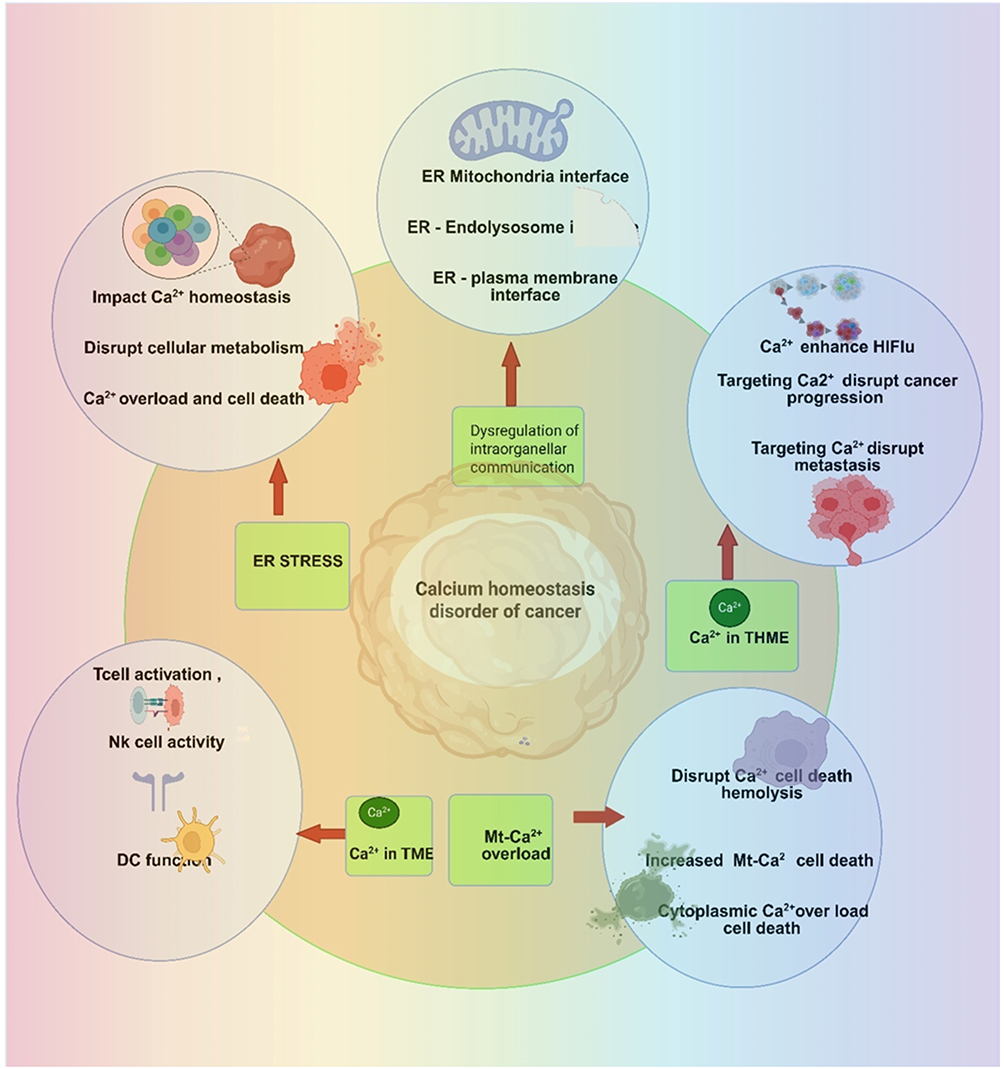 |
Figure 4 Schematic illustration of mechanisms of Ca2+ homeostasis disorders of cancer [Image is created by Biorender.com]. |
Calcium-Based Nano Materials in Tumor Immunotherapy and Immunogenic Cell Death
Tumor immunotherapy is frequently hampered by the development of a complex immunosuppressive TME that prevents immune cell activation and shows low tumor immunogenicity. Ca2+ controls the charge characteristics of T cell lipids, resulting in T cell activation and proliferation in response to immunostimulants, whereas Ca-NMs directly trigger immunogenic cell death in tumors. Furthermore, Ca2+ can stimulate dendritic cell (DC) maturation and macrophage polarization, offering potential cancer therapeutic alternatives. Defensive mitochondrial autophagy attenuates cellular pyroptosis, while damaging the mitochondria can temporarily increase it.65 A nano system that uses a hybrid membrane consisting of an equivalent mitochondrial membrane and a homologous cell membrane, to reduce the adverse feedback of compensatory induction of mitochondrial autophagy in cellular death.66 Through the delivery of calcium-supported glucose oxidase (Ca@GOx) to mitochondria, the nano system caused cellular death through a large amount of ROS production and induction of mitochondrial Ca2+ excess. For even more promotion of cellular death, chloroquine, which prevents mitochondrial autophagy, was also added to the nanoparticles. This procedure combines targeted mitochondrial damage and mitochondrial autophagy blocking to achieve cell death-based successful treatment. Tumor immunogenic cell death (ICD) has gained considerable interest over the years and is critical in tumor immunotherapy. It is crucial to the tumor immune cycle and initiates the expression of many tumor-associated antigens. Control of ROS generation by Ca2+ overload is a significant mechanism of ICD, which triggers the release of damage-associated molecular patterns (DAMPs). As a consequence, it initiates an immune response that is protective against the tumor.67 Ca2+ are innovative, promising, and physiologically safe inducers of ICD in light of these findings. They are appealing for further research because of their capacity to alter the tumor immune microenvironment (TIM) and elicit anticancer immune responses. Ca2+ plays multiple roles in ICD induction. Ca2+ acts as a nanomaterial’s gatekeepers, preventing other ingredients from premature release before they reach the tumor site and induce ICD.42
The pH-sensitive calcium phosphate-coated lanthanide-doped nanoparticles proved to be successful in protecting the ICD inducers from Adriamycin and Mn2+ ions, allowing their action only at the tumor site. This invention enables the prevention of lung metastases and distant tumors by overcoming photo dynamic therapy (PDT) limitations such as low potency, shallow tissue penetration, and phototoxicity. The CaCO3 @Pt-TiO2 nanocomposite releases a significant amount of Ca2+ in response to the TME.67 It led to the induction of high ICD activation and oxidative stress. To overcome hypoxia within the tumor, platinum nanoparticles were added to increase the production of ROS and catalyze the degradation of hydrogen peroxide.68 The combined action of Ca2+ and platinum nanoparticles effectively shifted the tumor microenvironment from “cold” to “hot”, significantly increasing the infiltration of inflammatory cells.69 By combining combretastatin A4 phosphate with calcium nanoparticles to improve T cell infiltration and the ability of doxorubicin to induce ICD.70 Beyond the direct effects of calcium ion overload, other mechanisms contribute to cancer cell death.71 Nanoparticles that utilize Ca2+ to generate carbon dioxide bubbles and upon ultrasound activation, these bubbles rupture, damaging cancer cell membranes and triggering ICD.72 Furthermore, manganese and Ca2+ can enhance immune responses and improve the effectiveness of photothermal therapy combined with immunotherapy.
Calcium-Based Nano Materials in Modulating the Tumor Immune Microenvironment
Numerous immune-related cells, including T cells, DCs, and macrophages, are drawn into the microenvironment surrounding the tumor cells over the course of tumor formation.73 The TIM is formed by the interaction of these cells with vascular endothelial cells, immune cell-secreted substances, the extracellular matrix (ECM), and other components.60 To control the TIM, several Ca2+- based NM’s mainly target immune-related cells. Calcium-based nanomaterials can influence macrophage polarization, shifting them from the tumor-promoting M2 phenotype to the anti-tumor M1 phenotype. These nanomaterials can enhance DC maturation, improve antigen presentation and activate T cell responses. Ca2+- based NM’ s can promote T cell activation and infiltration into tumors. They can help overcome the immunosuppressive mechanisms within the TME, such as those mediated by M2 macrophages.
Some Ca2+-dependent NM’s may increase the levels of oxygen inside the tumor to increase the suitability for the functioning of immune cells. They enhance the delivery of chemotherapy drugs and other therapeutic drugs into the tumors. Calcium-based nanomaterials can initiate ICD in tumor cells, liberating DAMPs and initiating an antitumor immune response. Ca2+-based NM can be used in combination with other therapeutic modalities, such as PDT and chemotherapy, to provide synergistic antitumor effects. Nanocomposites include calcium ion nanogenerators, which enhance DC maturation and improve antitumor immunity. CaCO3-based nanoparticles induce ICD, regulate macrophage polarization, and enhance antigen presentation. Calcium-based nanoparticles with Toll-like receptor agonists activate innate and acquired immune responses. Ca2+-based NM’s can affect macrophage polarization, switching them from the tumor-promoting M2 phenotype to the anti-tumor M1 phenotype. These nanomaterials can enhance DC maturation, thereby improving antigen presentation and activating T cell response. Calcium-based nanomaterials can promote T cell activation and infiltration into tumors. Calcium-based nanomaterials can help overcome the immunosuppressive mechanisms within the TME, such as those mediated by M2 macrophages. Some calcium-based materials enhance oxygen levels within the tumor, making the environment conducive to the proper functioning of immune cells.
Chemoimmunotherapy activates T cell immune responses that can produce targeted anticancer effects. However, the poor antigen presentation of DCs in the TIM reduces the efficacy of chemoimmunotherapy. The nanogenerator that can disrupt Ca2+ levels within dendritic cells (DCs) disrupts the immunosuppressive environment inside DCs and enhances their maturation and ability to recognize and attack tumor cells. This is attained through both the release of Ca2+ inside DCs and the concurrent promotion of the deleterious effects of calcium-derived molecular products from tumor cells. The most important immune cells in the TME are tumor-associated macrophages. They polarize to the M2 subtype, which induces immune suppression and supports tumor development by affecting lymphocyte infiltration. Calcium-based nanomaterial affects the intracellular calcium ion levels and, as a result, can modify the macrophage polarization. Studies have shown that increased CaP concentrations implicate the expression of genes related to M1 macrophages. More significantly, the immunomodulatory function of CaP materials may be dramatically affected by their crystalline form. To regulate macrophage polarization towards the M1 type, an upconverting nanocarrier that uses NIR light to alter intracellular calcium levels was used. The function of ECM in solid tumors has been studied.42 To shield tumor cells from cytotoxic T lymphocytes (CTL) and therapeutic medications, the ECM serves as a natural barrier. It is also necessary for create a hypoxic tumor microenvironment, which inhibits the immune system. Photodynamic therapy and calcium ion overburden may enhance the production of DAMPs, thereby facilitating CTL activation. CaP can alleviate tumor hypoxia by enhancing the oxygen supply capacity. ROS and the digesting enzyme chymotrypsin effectively enhance CTL entry into tumors. These factors downregulate immunosuppressive factors and enhance the efficacy of photodynamic treatment. A better approach would be multimodal therapy applied at different stages of the tumor immune process, given the complexity of the TIM. Salmonella (Sal) was biomineralized with CaCO3 to modulate an immunosuppressive tumor microenvironment (ITM) and perform in situ cancer vaccination.74 In the acidic tumor microenvironment, the shell of CaCO3 dissolves, lowering the acidity of the surrounding environment while simultaneously releasing the calcium ionophore A23187, Ca2+, and Sal. Sal enhances antigen presentation through ICD in cancer cells, thereby aiding the interaction between tumor cells and dendritic cells via gap junctions.
The calcium ionophore A23187 facilitates the uptake of Ca2+ by various immune cells and works synergistically with Sal to effectively regulate the immune system. This involves encouraging T cell activation, DC maturation, and macrophage polarization. In particular, in the acidic TME, multifunctional immunomodulatory calcium nanoparticles produce Ca2+ and DOX.75 The concentration of Ca2+ inside the tumor cells increases synergistically as a result of this special characteristic. These nanoparticles control the IFN-1 signaling system and aid in the ICD of tumor cells. Thus, they boost CD8+ T cell infiltration by promoting DC maturation and inducing M1-like macrophage polarization.76–78 These successfully rewire the ITM. To achieve synergistic effects, CaCO3 and curcumin were co-loaded into nanoparticles coated with cancer cell membranes.79 This impact entails raising Ca2+ levels and encouraging the production of ROS in the ER and mitochondria, which results in ICD.80 Additionally, by antigen cross-presentation, this mechanism can trigger DC maturation and macrophage polarization. As a result, ionic-crosslinking poly aspartic acid was used to reprogram the TME and deliver antitumor treatment. In addition to cross-linking poly aspartic acid, calcium-based nanomaterials can enhance both innate and acquired immune responses within the tumor microenvironment.81 When combined with Toll-like receptor 7/8 agonists or pathogen-associated molecular patterns (PAMPs), these nanomaterials can activate the innate immune system. This approach leads to tumor warming, remodeling the tumor microenvironment. Ca-NMs can induce ICD by interacting with and cyclically amplifying the ROS generated by photothermal therapy (PTT) or chemo-dynamic therapy (CDT) and the resulting calcium overload. This activation enhances anti-cancer immunity in vivo by stimulating the development of dendritic cells, activating helper and cytotoxic T cells, and releasing pro-inflammatory cytokines.82
Calcium‑based Nanomaterials in Calcification Therapy
Mineral buildup and calcification are basic biological processes observed in living organisms. An increase in intracellular Ca2+ levels during calcium overload causes metabolic problems and dysfunction, influences tumor cell metabolism and proliferation, and encourages tumor calcification.82 These factors contribute to the demise of tumor cells. A favorable prognostic sign is physiological calcification in the treated tumor region. Low Ca2+ concentrations in and around cancer tissues, high Ca2+ concentrations in the microenvironment, and the gradual and unpredictable nature of physiological calcification all affect the calcification process. Conversely, excessive systemic administration of calcium can lead to hypercalcemia, a potentially life-threatening condition characterized by elevated blood calcium levels, which can cause serious adverse effects such as cardiac arrest, organ failure, or even death.83 Consequently, a safe way to deliver significant exogenous Ca2+ into tumors is by using calcium-based nanomaterials. By disturbing calcium homeostasis and generating mitochondrial metabolic abnormalities, calcium-based nanomaterials are said to encourage tumor calcification. CaHPO4 covalently doped with L-ascorbic acid creates a biologically sensitive calcification initiator.84 This initiator promotes calcification by increasing ROS levels and causing mitochondrial malfunction. Furthermore, because of its pro-oxidative properties, exogenous L-ascorbic acid indirectly disrupts the metabolic balance. By upsetting mitochondrial homeostasis, this sensitive calcium source also caused intratumoral calcification. Liu developed a sodium hyaluronate-modified hollow calcium peroxide sphere capable of encapsulating L-buthionine sulfoximine and delivering chloroperoxidase to enhance CDT. Excessive calcium ion loading from these nanospheres disrupts calcium transport within cells, damages mitochondria, and increases oxidative stress. This ultimately leads to the deposition of calcium within tissues (calcification) and accelerates the death of cancer cells. In addition, directly increasing intracellular calcium levels, other strategies can induce calcification. Sialic acid, a calcium chelator, can be incorporated into nanomaterials to bind excess Ca2+ within tumor mitochondria. This Ca2+ aggregation initiates mitochondrial calcification, enhancing treatment efficacy. Most research has focused on examining the precise processes by which calcium-based nanomaterials cause tumor calcification; yet, a crucial avenue for treating tumor calcification is the connection to clinical prognosis. Fluorapatite nanoparticle surface modification of nanofibrous polyether sulfone scaffolds yielded advantageous results in osteosarcoma-related research.
Calcium Based Nano Composites in Cancer Therapy
Compared to other nanodrugs, Ca-NM’s offers a number of benefits. When it comes to production expenses, they are less expensive than those of expensive metals and polymer nanoparticles. Unreactive monomers or additives used in polymeric materials may have harmful side effects. Certain polymeric nanodrugs may accumulate in the body and have limited biodegradability, which could eventually result in negative side effects. Ca-NM drug delivery systems outperform liposomal nanodrugs because they effectively hold loaded medicine and maintain their structure and stability under heat. Their strong storage power, combined with sustained gene-protein-drug release makes NM’Ca an excellent choice for biological applications due to its biodegradability and compatibility with living tissue. The demonstrated nanomaterials show promising features to overcome material limitations from regular silica, carbon, and gold inorganic biomaterials. Traditional materials perform poorly when it comes to biochemical decomposition and can pose safety risks over time. Calcium-based nanomaterials dissolve into harmless ions during body metabolism, allowing them to be processed. Tests have shown that calcium-based nanomaterials work well to boost cancer cell’s calcium intake. These nanomaterials support multiple therapeutic options that work together with RT, PDT, gene therapy, chemotherapy, and hyperthermia treatments. Calcium nanomaterials show promise for cancer diagnostic imaging. Nano-composite compounds can combine with different imaging contrast agents to support multiple surgical imaging methods. Calcium-based nanomaterials are promising as a safer treatment option for medical professionals to detect and monitor illnesses during clinical management. Nanomaterials advance medical treatments and boost their effectiveness in delivering precise care to patients. Consequently, researchers are interested in creating multifunctional calcium-based nanomaterials.85,86 Given the progress made in personalized medicine, translating research on calcium-based biochemical processes into the development of new calcium-dependent clinical therapies would be highly advantageous for patients. An increasing amount of data highlights the possibility and efficacy of artificially regulating calcium as a cancer treatment technique. Table 2 shows the Ca2+ based nano constructs and their applications in therapy.
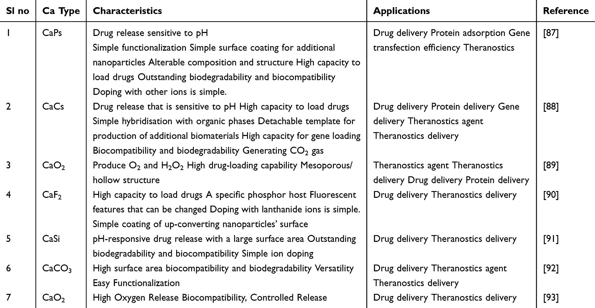 |
Table 2 Types of Ca2+ Based Nano Constructs and Their Features and Application Therapy |
Calcium Carbonate Based Nano Constructs
CaCO3 is an important inorganic mineral found in nature that has been widely used in various industrial applications. In the field of cancer treatment, CaCO3 nanoparticles have attracted significant attention as drug carriers due to their tumor-targeting ability and acid-responsive degradation properties.94 On the one hand, CaCO3 nanomaterials have the potential to increase intracellular Ca2+ levels in tumor cells, leading to mitochondrial damage and eventually causing tumor cell death. Conversely, the high drug loading capacity and slow biodegradation of these calcium- based materials make them highly significant in the field of controlled release therapy. Recently, surface modification methods have been developed to endow CaCO3 nanoparticles with different structures, such as porous, hollow, or core-shell structures, through template-induced biomineralization, layer-by-layer assembly, and etching techniques. When combined with other contrast agents, polymers, photosensitizers, and functional nanomaterials, multiple therapeutic modalities can be used during treatment, yielding optimal therapeutic outcomes. In this study, CaCO3 nanoparticles as templates, meso-tetra-4-carboxyphenyl porphine (TCPP) as an organic bridging molecule, and ferric ions as the particle core while loading the glutathione (GSH) inhibitor L-buthionine sulfoximine (BSO), to prepare pH-responsive and multifunctional hollow CaCO3-based nanoparticles (BSO-TCPP-Fe@ CaCO3).95 These nanoparticles rapidly released Ca2+ and BSO molecules under acidic pH, effectively inhibiting tumor growth through a combination of Ca2+ overload-induced mitochondrial dysfunction, BSO-triggered GSH depletion, and TCPP-mediated sonodynamic therapy (SDT). CaCO3-based nanomaterials exhibit excellent pH-responsiveness, and their hollow structure allows higher drug loading, offering multimodal combination therapies.96,97
Multifunctional Calcium Carbonate Nano Constructs
Multifunctional CaCO3 nanoparticles have gained traction in targeted therapy and imaging because of their biocompatibility, biodegradability, and versatility in functionalization. The significance of multifunctional CaCO3 nanoparticles is summarized in Table 3. These nanoparticles offer a dual approach for treating and monitoring cancer by combining therapeutic delivery with diagnostic capabilities.98
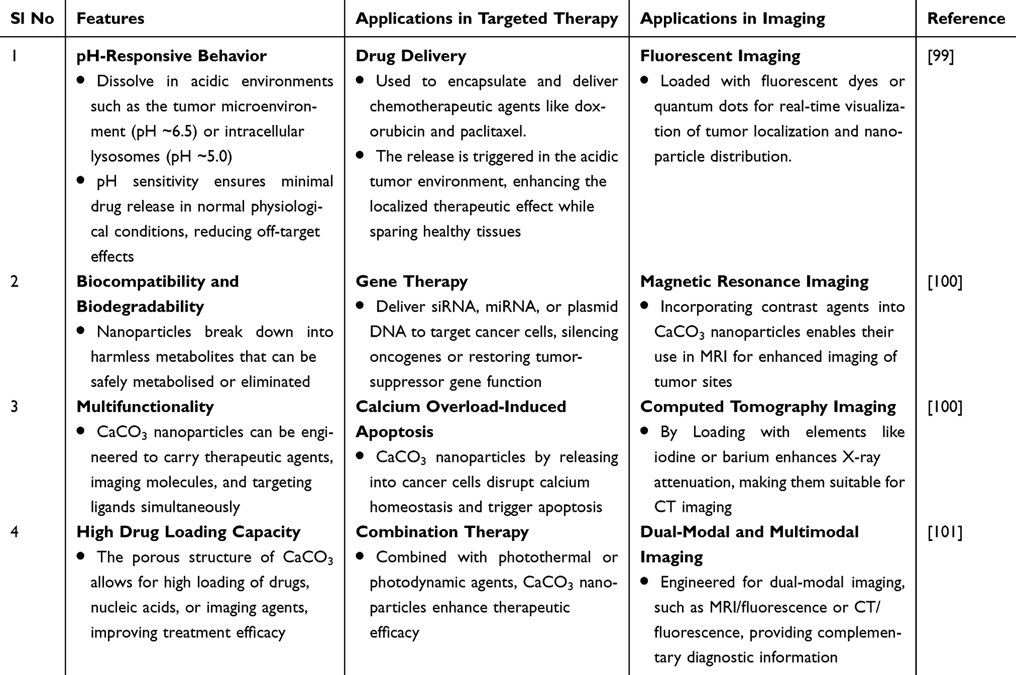 |
Table 3 Key Features of Multifunctional Calcium Carbonate Nanoparticles |
Recent Advances of Multifunctional Calcium Carbonate Nanoparticles
A new method for drug delivery that uses porous vaterite particles as carriers includes a two-part drug delivery solution that aligns with theragnostic requirements.101 In this study, researchers developed a new therapeutic tool with excellent prospects. These multiple-purpose delivery vehicles combine loading with magnetic control and diagnostic system elements. Microscopic scans and X-rays helped scientists to discover how the carriers and the materials they contained.98 The carriers moved through magnetic fields and produced a biomolecule detection method that showed enhanced signal strength. The combined nanocarrier system was proven safe according to cell toxicity testing. The tweezers-shaped carriers combine drug storage with magnetically controlled movement, making them a valuable tool for future theragnostic applications. This study demonstrated that CaCO3 particles can deliver drugs while helping detect molecules, separate particles and absorb chemicals.102 A unique nanoparticle platform that targets tumors responds to multiple triggers for cancer treatment.103 The nanoparticles combine polymers with loaded photosensitizer ICG and chemotherapeutic DOX. They show response capability under declining tumor acidity and decreasing environmental conditions. The nanoparticles exhibited reliable drug storage and delivery under designed conditions. Under controlled conditions in test tubes, nanostructures display powerful heat generation behavior.104 They show enhanced cellular uptake and cytotoxicity in cancer cells. In in vivo studies, the nanoparticles demonstrated superior antitumor effects in mice when combined with photothermal therapy and chemotherapy. The authors reported that these multifunctional, tumor-targeted, and dual stimuli-responsive nanoparticles have significant potential for effective cancer treatment. This study presents a promising approach to cancer therapy by using a nanocarrier system that effectively delivers both chemotherapeutic drugs and photosensitizers, enhancing their therapeutic efficacy through targeted delivery and dual stimuli-responsive release. Multifunctional CaCO3 nanoparticles represent a promising avenue for integrated cancer therapy and diagnostics, paving the way for more effective and personalized treatment strategies in the future. They ensure stability in the bloodstream allowing them to reach the tumor site. Stability issues in the bloodstream are overcome by using surface coatings, such as polyethylene glycol (PEG), which can improve stability and reduce immune clearance. Mechanisms for refining stimuli-responsiveness to ensure precise drug release. Rigorous preclinical and clinical studies are required to establish safety, efficacy, and scalability for human applications.105,106
Hybrid Nanocomposites
Hybrid nanocomposites combining calcium-based materials (such as CaCO3 or CaP) with other functional materials, such as gold or graphene, have shown remarkable potential in cancer therapy. Their applications in cancer therapy are listed in Table 4. These hybrid systems leverage the complementary properties of their components to achieve enhanced antitumor efficacy through synergistic effects. Examples of hybrid nanocomposites are including calcium-gold, calcium-graphene, calcium-magnetic hybrid nanocomposites. First, the enhanced efficacy of these composites makes synergistic interactions between calcium and other materials improving therapeutic outcomes. Second reduced drug resistance in combined therapies (chemotherapy and PTT) prevents tumor cells from adapting to a single treatment modality. Third, the improved biocompatibility of calcium-based components ensures safety and biodegradability in the body. Finally real-time monitoring of imaging capabilities allows for precise targeting and monitoring of treatment progress. Hybrid nanocomposites combining calcium with materials like gold or graphene represent a powerful tool in the fight against cancer, offering precise, effective, and minimally invasive treatment options.107
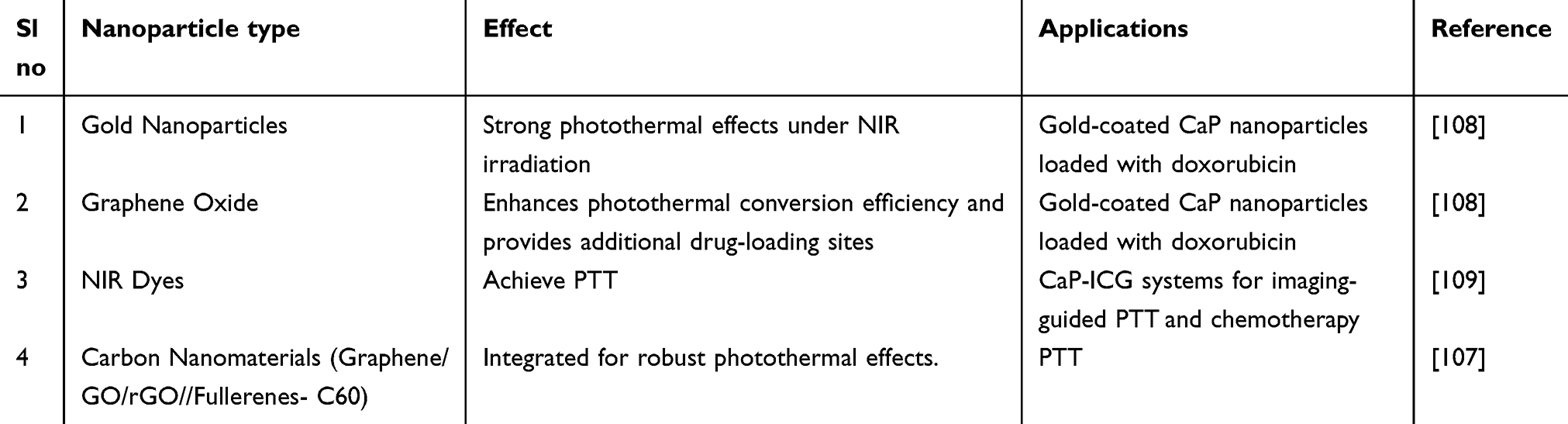 |
Table 4 Photothermal Agents Integrated with CaP Nanoparticles |
Hybrid Nanocomposites of Calcium Carbonate
Over the past few decades, researchers have studied gold nanoparticles because of their unique properties. The size, shape, and surface functionalization of these nanoparticles are used to enhance their biocompatibility, circulation time, tumor cell uptake, and targeting capability. Gold can easily endure biological environments because it is resistant to oxidation. To accomplish active targeting, gold nanoparticles are often coated with organic compounds. They are also coated with ligands that include sulphur, such as thiols and disulphides. The size of the gold nanoparticles and the targeting ligands caused them to exhibit greater cellular uptake and accumulation at the tumor site. In numerous studies, the use of EGFR-targeting gold nanoparticles has resulted in increased suppression of breast cancer cells. Compared to unconjugated nanoparticles, gold nanoparticles are significantly more stable because of surface conjugation. Consequently, several researchers have investigated different conjugations of gold nanoparticles and shared their findings. DOX-containing gold nanoparticles decrease multidrug resistance.110 The effectiveness of gold nanoparticles coated with anti-miR-155 on the MCF7 breast cancer cell line was altered the nanoparticles using the AS1411 aptamer, which attaches to nucleolin on cancer cells, to achieve tumor-targeted distribution.111 However, the main issue with using gold nanoparticles is that they biodegrade very little. Because of their unique features, such as customizable size and shape, gold nanoparticles can be altered to enhance cellular uptake, targeting, and circulation time. Their stability in biological contexts is improved by their resistance to oxidation. Owing to their special properties and adaptability in surface modification, gold nanoparticles present a promising platform for cancer treatment. However, a major obstacle that must be overcome for safe and successful clinical translation is their poor biodegradability.112
Interesting information about fluorescence and confocal imaging of Au-CsCaCO3sNPs-treated MCF-7 and NIH3T3 cells.113 The goal of the study was to determine the apoptosis and acceptance of the synthesized nanoparticles. The most notable result of fluorescence imaging was that the MCF-7 cells showed a higher rate of cell death than NIH3T3 cells. Furthermore, confocal imaging revealed that the cells may absorb nanoparticles within their cellular compartments. The bio-conjugated nanoparticles validated cellular internalization into MCF-7 and MDA-MB-231 over NIH3T3, which is consistent with their findings.114 This study also sought to evaluate the Au-CsCa CaCO3NPs’ bio cellular uptake and fluorescence imaging. Overall, their results suggest that the produced conjugated nanoparticles may have a role in advancing cancer imaging. This study advances understanding of Au-Cs CaCO3NP-based bioimaging and its possible use in diseased-cellular diagnosis.115
In Figure 5a, HOCN NPs have been designed to increase antigen cross-presentation by DCs when mitoxantrone (MTX) is used in chemotherapy.116 HOCN interferes with various intracellular barriers in the acidic tumor microenvironment. It facilitates the release of damage-associated molecular pattern (DAMP) by tumor cells killed by MTX, enables the uptake of tumor antigens by DCs, and induces autophagy-lysosome events to support antigen processing and cross-presentation on MHC class I. This enhances cytotoxic T lymphocytes (CTLs) and the development of memory T cells against primary and distant tumors as well as stimulating systemic immune responses. The synthetic pathway of a coordination-assembly nanoplatform (CTC) to be used as an ion-homeostasis perturbator is depicted in Figure 5b.117 The carrier co-loads ions or ion-releasing compounds in such a way that following cell internalization and TME-dependent degradation, it both increases intracellular Ca2+ and perturbs Mg2+ levels and leads to the amplification of oxidative stress and organelle dysfunction. This oxidative damage-enhanced Ca2+/Mg2+ interference therapy causes death of tumor cells and it produces immunogenic signals which can be synergized with other therapies. Figure 5c is dedicated to mechanistic validation: Calpain-1 and ATP2B4 which is a plasma membrane Ca2+ pump in 4T1 cancer cells under the influence of various treatments in the form of Western blot.118 The molecular evidence supporting calcium-overload-mediated apoptosis is the up-regulation of Calpain-1 and modulation of ATP2B4 levels, which proves the presence of calcium-overload-mediated apoptosis. These protein modifications are linked to increased cell death and therapeutic efficacy which is backed by quantitative analysis. Figure 5d demonstrates pH-responsive DNCaNPs, which respond to the acidic tumor extracellular environment and deliver chemotherapeutic and calcium-related moieties systematically.119 These NPs suppress acidic TME, induce immunogenic cell death, expose calreticulin and other eat-me signals, and eliminate immunosuppression of IDO and Treg pathways, thereby increasing DC maturation and CTL infiltration. This draws attention to the combination of ion dysregulation, pH-responsive release, and immune modulation as the means to transform the traditional chemotherapy into the potent chemo-immunotherapy. The Au/ CaCO3 nanocomposites were synthesized through eggshell templates at a low cost and without harming the environment.120 The open structure of the nanocomposites creates enhanced possibilities for luminescent probe placement. Strong sensing performance emerged from the immunosensor, which combined outstanding sensitivity with precise specificity and extended operation time. The ECL immunosensor demonstrated strong performance with the OTA-specific Nb28 antibody because of its robust binding affinity. This documentation details a methodology for Au and CaCO3 nanocomposite synthesis which employs eggshells as the template base. Au-CS CaCO3NPs with spherical gold-coated CaCO3 nanoparticle shapes were successfully synthesized by investigators using cockle shells. A basic, eco-friendly, and low-cost production method allowed researchers to synthesize conjoined nanoparticles. The results indicated that the manufactured nanoparticles maintained their stability and purity levels throughout the testing period. Scientists present evidence showing how gold nanoparticles link successfully to cockle shell materials. Laboratory results indicate that Au-CS CaCO3NPs demonstrate potential medical applications across several fields.120
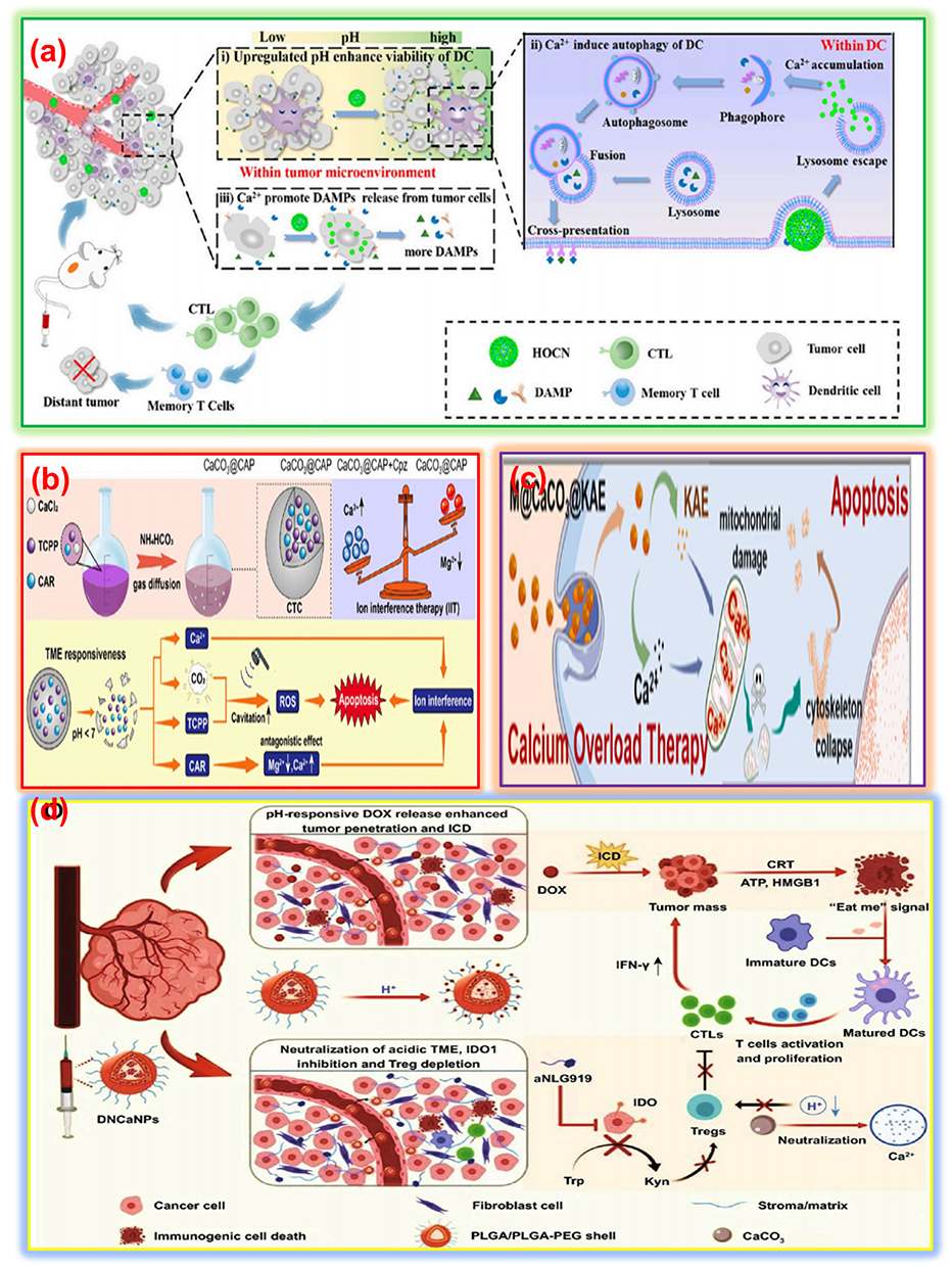 |
Figure 5 (a) Schematic diagram of HOCN disruption of multiple barriers in antigen cross-presentation of DCs for enhanced mitoxantrone (MTX)-mediated chemo-immunotherapy (Reproduced with permission from ref).116 (b) Schematic diagram to show the synthetic procedure of CTC as an ion homeostasis perturbator and its application for oxidative damage-augmented Ca2+/Mg2+ interference therapy (Reproduced with permission from ref.117) (c) Western blot analysis of Calpain-1 and ATP2B4 in 4T1 cancer cells after varied treatments, along with the corresponding quantitative analysis (Reproduced with permission from ref.118) (d) Schematic illustration of the preparation of tumor extracellular pH-responsive immune-modulatable DNCaNPs for effective cancer chemo- and immunotherapy (Reproduced with permission from ref).119 |
Mechanism of Combined Photothermal and Chemotherapy
Photothermal agents absorb NIR light and convert it into heat, thereby increasing the temperature of the tumor microenvironment. Localized hyperthermia (40–45°C) damages cancer cells by disrupting their protein structures and cellular membranes, thereby inducing apoptosis. Chemotherapeutic drugs encapsulated in CaP nanoparticles are released in a controlled manner, especially in acidic tumor environments or when triggered by heat generated during PTT.121 The drugs further enhance cancer cell death by targeting DNA replication or metabolic pathways. The heat generated during PTT enhances the uptake of chemotherapeutic drugs by increasing the permeability of the tumor cell membrane. Hyperthermia sensitizes cancer cells to chemotherapy, reducing the required drug dose and minimizing side effects.122 ICG is an effective photosensitizer that can promote the production of harmful ROS. ICG can also produce a potent photothermal effect because of its distinctive NIR light absorption. ICG’s two primary features can be used for noninvasive light-induced tumor ablation. ICG is prone to aggregation or early excretion from the body due to its instability in the blood circulation. Before ICG can be used as an anticancer agent, these challenges must be successfully resolved. Fe3O4@PDA/CaCO3ICG (FPCI) nanoparticles were nanocomposite that combines the photothermal and photodynamic characteristics of polydopamine (PDA) and ICG. It is composed of magnetic PDA nanoparticles modified with CaCO3 and loaded with ICG. Specifically, CaCO3 allowed for the controlled release of ICG through self-decomposition in response to an acidic tumor microenvironment and to prevent blood clearance by enclosing ICG in a stable aggregate. Based on in vitro and in vivo tumor models, this proof-of-concept study demonstrated that this multifunctional therapeutic agent allows the combination of photothermal (PTT) and photodynamic therapies (PDT) against tumors with the use of magnetic guiding.123 Researchers created hybrid nanoparticles by combining conducting polyaniline (PANI) with CaCO3 microparticles (CMPs). Carboxymethyl cellulose was used as a stabilizing agent. This is a simple and reproducible synthesis method with a high encapsulation efficiency of CMPs. The pH-responsive drug release in the acidic tumor microenvironment is an advantage of this method. CMP-PANI-Cys nanoparticles demonstrated higher efficiency in photothermal therapy than PANI-Cys alone.
In the realm of tumor therapy, research on treatment approaches based on nanomaterials that promote ROS generation has increased dramatically. These techniques include chemo dynamic therapy (CDT), electrodynamic therapy (EDT), radio dynamic therapy, sonodynamic treatment, and PDT. External fields, such as lasers, can activate nanomaterials to produce ROS. By damaging vital components of cancer cells such as proteins, lipids, or DNA, the generated ROS induce apoptosis or necrosis. A variety of chemicals, including acoustic sensitizers, and catalysts, can be delivered to tumor locations using calcium-based nanomaterials. They also contain large amounts of Ca2+, which can significantly increase and worsen calcium overload. During tumor treatment, this amplification mechanism typically works in concert with other mechanisms. Manganese-doped calcium phosphate (MnCaP) mineralized glucose oxidase was combined to create nanoparticles, spherical in shape (GOx-MnCaP NPs), through in situ biomimetic mineralization. These nanoparticles increase CDT and efficiently destroy tumors via a cascade effect that includes the release of Ca2+, the GOx-driven oxidation reaction, and the Mn2+ mediated Fenton-like reaction.124 Additionally, drug release can be tracked with specific calcium nanotherapeutics. ICG and chlorin Ce6 (Ce6), common PTT/PDT agents, were reversibly linked via ACC. ICG physically decreased Ce6’s photosensitivity and fluorescence during cycling at non- tumor locations, reducing PDT’s adverse effects. To track the quantitative release of the drug, they can be readily repaired in tumor tissues. When combined with photothermal therapy (PTT) and photodynamic therapy (PDT) to enhance their therapeutic efficacy, this synergistic approach involves the use of ICG-loaded calcium nanoparticles. ICG generates singlet oxygen for PDT and induces apoptosis upon exposure to NIR light. The calcium phosphate layer enhances the biocompatibility. Importantly, the controlled release of Ca2+ in response to the acidic tumor microenvironment disrupts the mitochondrial membrane potential, reducing oxygen consumption within the tumor. This combined strategy allows for effective cancer treatment at lower temperatures.125
Theragnostic Platforms
Multifunctional CaCO3 nanoparticles are being developed as theragnostic agents, integrating therapeutic and diagnostic functionalities into a single platform.126 Although the strategy of disrupting Ca2+ homeostasis for cancer treatment shows immense potential, achieving effective interference with Ca2+ homeostasis remains a challenge. It is necessary to combine the unique advantages of various Ca2+-based materials and design Ca2+-based nanomaterials tailored to the pathological characteristics of different types of tumors for specific tumor diagnosis and treatment. These tumor diagnosis and treatment strategies based on Ca2+-based materials may include Ca2+overload therapy through exogenous Ca2+ supply, amplifying Ca2+ overload therapy by supplying exogenous Ca2+ in conjunction with various Ca2+ channel/pump targets, regulating the TME in synergy with exogenous Ca2+ supply as a treatment modality, as well as enhancing tumor calcification for imaging and diagnostic capabilities.127
Development of a Dual-Mode Imaging and Therapeutic Nanoplatform
Contrast-enhanced ultrasonography (CEUS) is frequently used in clinical cancer diagnosis. However, the practical application of gas-filled contrast agents is limited by their short imaging time and blood instability. This study produced a diagnostic nanoparticle system for dual-mode imaging (fluorescent and ultrasound), which showed simultaneous therapeutic function for cancer treatment after being encapsulated with DOX. Therefore, CaCO3-DOX was created by encapsulating CaCO3 nanoparticles with DOX. It generates carbon dioxide (CO2) to improve ultrasound imaging and boost DOX release in acidic environments. When CaCO3 -DOX was injected intravenously into tumor-bearing animals, sufficient CO2 bubbles were formed at the tumor sites for echogenic reflectivity to occur in the presence of an ultrasonic field. ICG was used to capture tumor imaging while encapsulating it within CaCO3 nanoparticles. The theragnostic nanoparticle system was used to treat tumors elegantly in experimental animal models. This study presents a practical system to detect cancer in two ways.
Scientists created gold-attached CaCO3 nanoparticles from cockle shells (Au-CsCaCO3NPs) to observe and treat cancer using NIR light. The team analyzed and tested these nanoparticles in living cell environments to assess their compatibility. The tests revealed that normal cells easily accepted the nanoparticles without damage, while the nanoparticles killed cancer cells. The cells readily reacted with and brought these nanoparticles into their bodies. Researchers believe Au-CsCaCO3NPs can track cancer effectively and may become a better cancer medicine in the future. Their investigation aimed to assess how Au-CsCaCO3NPs enter living cells and how they display their fluorescence. The research determined that these easy-to-make conjugated nanoparticles show environmental safety and biocompatibility which support their use for bio-imaging applications. The particles enabled both normal and cancerous cell colonization and triggered cancer cells to die through mechanisms similar to those of normal cells. The analysis showed that the synthesized nanoparticles may help advance cancer imaging techniques. This study explained Au-CsCaCO3NP bioimaging details and demonstrated its potential for disease cell detection.
Calcium Phosphate-Based Nanomaterials
Calcium phosphate nanoparticles (CaPs) have demonstrated beneficial results in relation to the aforementioned requirements among various synthetic vectors. Since of their predictable bodily clearance channels and mechanisms, biodegradable nanoparticles are typically used for cancer therapy since they are safer options for clinical use. Size, charge, shape, composition, and surface chemistry are among the physical and chemical properties of CaP nanoparticles that have been demonstrated to be crucial in determining the path of internalization of nanomedicines. The cellular entry mechanism and the ultimate location inside the cells determine other crucial elements, including the dosage of bioagents and their functions. Given the large number of organelles in a cell, it is crucial that the delivery mechanism delivers the payload at the precise location to produce the intended result. Understanding how the cell transports its constituent parts to the various cellular compartments may help develop medications for more effective tumor treatment.128
Owing to the main inorganic component of bone, CaP-based nanomaterials have been widely applied in the biomedical field, particularly in bone repair and as drug carriers, due to their excellent biocompatibility, biodegradability, and chemical composition similar to that of bone tissue. The chemical composition of CaP nanomaterials matches that of bone tissue composition and provides better tissue attachment and stronger bone healing performance than that of CaCO3-based materials. Due to their high natural reactivity, CaP nanomaterials easily integrate with metal ions and other therapeutic agents, increasing their possible uses. The body uses CaP to build bones which explains why doctors use this substance to treat osteosarcoma. Doctors perform surgery to clear dead tumor areas and attach artificial bone parts directly to the bone to eliminate the disease. The implanted metals and ceramic devices break down and become worn over time creating fractures between the bone and implant that reduce their lifespan.129 The broken materials from wear and corrosion start diseases in both nearby and distant areas, which support cancer coming back. CaP nanomaterials demonstrate outstanding compatibility with living tissues, strong resistance to both wear and corrosion. A microwave-assisted method was used to synthesize hybrid CaP-containing calcium phosphate (CPPA) microspheres.130 The CPPA proved great at absorbing DOX while releasing it in response to changes in pH and worked successfully both inside real osteosarcoma tissue and during laboratory research. The CPPA material enhanced the process better than other options, where bone cells differentiate into bone tissue. CaP-based nanomaterials can treat osteosarcoma by releasing drugs locally while supporting new bone growth owing to their natural bone-building functions.
Calcium Peroxide-Based Nanomaterials
Researchers believe that calcium peroxide (CaO2) nanomaterials hold strong potential for tumor treatment because of their multiple beneficial functions. The multifunctionality of CaO2 nanomaterial results from their production of H2O2 and O2 alongside Ca2+ release during chemical breakdown.131 The body normally processes increases in released calcium ions, so cells remain resilient while they eventually die from excess calcium. The produced H2O2 can be transformed into harmful ROS through external or internal control, making it an effective tool for multiple biological applications. In addition, owing to the crucial role of its decomposition product, H2O2, in antibacterial and anticancer applications, CaO2 has been considered an excellent antitumor and antibacterial nanomaterial.131 In tumor therapy, CaO2 generates ROS and can be combined with other materials for tumor treatment. Its reaction with water to produce H2O2 enhances the catalytic effect of Fenton or Fenton-like reagents, intensifying oxidative stress to induce tumor cell death. Additionally, CaO2 generates O2, increasing O2 levels in anaerobic environments and facilitating starvation therapy. The CaO2-based nanospheres (CaO2@Cu-TCPP/CUR) could enhance PDT/ chemodynamic therapy and Ca2+overload synergistic tumor therapy through O2/H2O2/ Ca2+ self-supply and glutathione (GSH). Cu-TCPP prevented the degradation of CaO2, while Cu2+ could react with GSH, depleting it and generating Cu+ species.132 CaO2 could react with H2O to produce O2, H2O2, and Ca2+ achieving O2/H2O2/ Ca2+ self-supply, enabling TCPP-based PDT, Cu+ mediated CDT, and Cu-enhanced Ca2+ overload therapy.
The ability of CaO2 nanomaterials to generate H2O2 and O2 upon decomposition provides them with unique advantages in applications such as ROS-dependent cancer therapy and alleviating tumor hypoxia. In addition to the commonly used Ca2+-based materials in the biomedical field mentioned above, many Ca2+-based materials with excellent properties have received widespread attention from researchers, including HAP with excellent drug loading and osteogenic ability, CaF2 with enzymatic activity, and calcium hydride (CaH2) with H2 production capabilities.
Calcium-Based Inorganic Composite Materials
In addition to the individual advantages of Ca2+-based nanomaterials mentioned above, several studies have demonstrated the potential of combining Ca2+-based materials with other inorganic materials to construct different structures. Compared to organic nanomaterials, inorganic nanomaterials offer more unique physical and chemical properties, providing greater mechanical stability while maintaining a certain level of bioactivity.133 In addition, leveraging their optical, magnetic, acoustic, and X-ray absorption properties, as well as their chemical properties, such as pH-responsiveness, metal ion chelation, and catalytic activity, has led to the development of various multimodal imaging nanoprobes and physical therapy combined with treatment strategies targeting tumors. Various inorganic nanomaterials, including single metals, metal/transition metal oxides, silica, and rare earth metals, have been widely applied in antitumor therapy. Using a combination of Ca and Cu, intelligent Cu/CaCO3 nanoparticles loaded with Chlorin Ce6 (Ce6) (Cu/ CaCO3@Ce6, CCC NPs) were developed.134 These nanoparticles exhibited multi responsive release of Ca2+, Cu2+, and Ce6 in the TME, achieving multimodal combined therapy through induced Ca2+ overload, GSH depletion, Fenton-like reaction-promoted CDT, and Ce6-triggered SDT. In the presence of H2O2, some metal oxides (eg, MnO2) act as promoters of ROS generation, enhancing ROS production through catalytic reactions. pH-responsive nanoregulators were developed by combining curcumin (CUR) (Ca2+ enhancer) with CaCO3 and MnO2. The nano regulator enhanced tumor treatment efficacy through the release of Ca2+ and Mn2+, Ca2+-induced overload, and Mn2+-catalyzed ROS generation in H2O2. Furthermore, integrating multiple inorganic materials into a single nanoplatform can simultaneously exert multiple functions.135 In the TME-responsive CaCO3@Pt-TiO nanocomposite (CaPT), Ca2+ release can induce Ca 2+ overload and amplify oxidative stress, thereby enhancing the SDT effectiveness.136 The designed Ca2+-based inorganic composite materials further enrich their application width based on the excellent Ca2+ overload triggering ability of Ca2+- based materials.
Calcium-Based Organic Composite Materials
Current nanotechnology applications make widespread use of organic materials, including polymers, liposomes, protein membranes, and hydrogels, because these materials display unique properties and blend well with biological systems. Organic materials added to Ca2+ nanomaterials including CaCO3 and CaO2 improve their application range for cancer treatment while handling stability and aggregation issues in Ca2+ systems nanomaterials, such as CaCO3 and CaO2, which has enhanced their functional diversity and potential in cancer treatment, addressing issues of poor stability and aggregation in Ca2+-based materials.137 Spherical liposomes use lipid bilayers that function as protective capsules to shield both hydrophobic and hydrophilic medications from degradation and dilution processes. The drug transport properties of liposomes improve when they function as delivery carriers, boosting target tissue absorption while minimizing drug-related toxicity. Rationally designed liposome-mediated surface functionalization of Ca2+-NM’s contributes to enhanced applications in cancer therapy.138
The liposome system simultaneously encapsulates Ca2+ supplement CaO2, and DNA methyltransferase (DNMT) inhibitor decitabine (DAC) to induce ferroptosis under mitochondrial dysfunction conditions.139 The lipid layer effectively protected against premature decomposition of CaO2 and premature leakage of DAC after drug administration. Recently, the excellent photothermal properties of PDA were used to develop a novel cavity CaO2@PDA nanocomposite.140 The chelation-induced core Ca2+ diffused to the outer shell PDA, promoting multiple reflections of the near-infrared laser within the cavity, enhancing the photothermal conversion efficiency of PDA. PTT synergize with Ca2+ overload therapy to further exacerbate mitochondrial dysfunction, promoting antitumor effects. Materials that specifically target to tumor sites, including hyaluronic acid (HA) and biomimetic cell membranes, are widely used in tumor treatment because of their excellent immune evasion, prolonged in vivo circulation time, and homologous targeting ability. Multifunctional CaCO3 nanoparticles loaded with curcumin and the protein deacetylase (HDAC) inhibitor, QTX125, were coated with HA (CaCO3@Cur@QTX125@HA).141 These HA-coated nanoparticles specifically targeted the CD44 ligand on the cell surface, significantly enhancing cellular internalization. Hydrogel materials, characterized by high local drug concentration, sustained release, low invasiveness, and low systemic toxicity, play an important role in cancer therapy.142 Injectable composite hydrogels encapsulating CaCO3 nanoparticles and other active ingredients were designed to effectively inhibit the metastasis of head and neck squamous cell carcinomas. Accordingly, the design of Ca2+-based organic composite nanomaterials tailored to specific tumor characteristics and pathological needs may yield unexpected effects in cancer treatment.
Exploiting Calcium-Based Nanocomposites for Cancer Therapy
Calcium-based nanocomposites offer a promising platform for developing novel and effective cancer therapies. By leveraging their unique properties and exploring innovative design strategies, researchers can continue to advance the field of cancer nanotechnology and improve outcomes for patients with cancer.
Calcium-Based Nanomaterials as Drug Delivery Vehicles
Users select Ca2+-based nanoparticles for cargo delivery because these nanoparticles demonstrate several advantages, including good compatibility with living systems and easy manufacturing at low cost. The efficiency of Ca-NM’s for cancer treatment increases at higher levels when coupled with effective drug delivery systems (DDSs).143 These delivery methods send drugs exactly where they are needed and release them gradually, thereby enhancing drug efficacy and reducing unwanted effects. Tumor blood vessels that exhibit enhanced permeability and retention (EPR) help these Ca-NM’s selectively gather in the tumor tissue, where they deliver their maximum therapeutic effect.
Calcium‑based NM’ s for Antitumor Chemotherapy Drug Delivery
Effective cancer treatment is hindered by several challenges, including severe side effects, poor drug bioavailability, drug resistance, bone marrow suppression, GIT issues, difficulties in monitoring treatment progress, and limitations in drug delivery, such as the poor water solubility of many chemotherapy drugs.144 These factors significantly impact the delivery and absorption of therapeutic agents. These limitations underscore the need for innovative approaches to cancer treatment that address the shortcomings of conventional chemotherapy. Most of chemotherapeutic medications have poor absorption and may have harmful effects on the body. Hydrophobic drugs like Adriamycin and paclitaxel (PTX) can be effectively encapsulated by nanocarriers based on calcium that have a high specific surface area, increasing their bioavailability.145 This approach enhances drug delivery to cancer tissues while mitigating the adverse effects of emulsifiers commonly used in conventional formulations. Furthermore, incorporating PEG or utilizing a biocompatible Silk Fibroin coating can extend the circulation time of calcium nanocarrier materials within the body. Moreover, the inclusion of PEG or the use of a biocompatible Silk Fibroin coating can prolong the in vivo circulation lifetime of calcium nanocarrier materials. This method offers a potential solution to the issue of slow blood/renal clearance of chemotherapeutic drugs.146
Specialized calcium-based nanomaterials boost cancer therapy success by maximizing the EPR effect. Researchers have carefully adjusted nanomaterial designs to create suitable particles that naturally travel into cancerous regions. Nanocarriers that deliver cancer treatments can locate cancer cells better when targeting molecules attached to their surface, which helps the drugs make more effective contact with the target cells. Researchers have developed a biodegradable nanocarrier system to deliver cancer-fighting drugs that successfully treat a challenging type of liver cancer.147 Blood vessels in cancerous tumors allow calcium-based nanomaterials to improve EPR therapy delivery, which remains a major technique in cancer control. Engineered nanomaterials move toward tumors better when their dimensions match specific charges and remain water-absorbing. Targeted ligands attached to these surfaces improve the efficiency of cancer drugs, making them work better and helping them reach cancer cells specifically. The cancer-targeting system combines RGD peptides that focus on liver cancer treatment with aptamers that target PTX resistance in prostate cancer cells using PSMA-specific binding. Studies show that Ca2+- NM technology delivers cancer drugs better while targeting only the affected areas and decreasing side effects.148
When calcium nanoparticles are treated with chemotherapeutic drugs, adverse side effects can be reduced.149 Calcium nanoparticles are the primary approach for minimizing side effects because they limit the action of chemotherapeutic medicines to the tumor site and regulate drug release. This approach aims to minimize the exposure of healthy cells to therapeutic agents. Additionally, this aids in reducing peak plasma concentrations that could be harmful. A novel drug delivery system using PTX-loaded CaCO3-coated mesoporous silica nanoparticles minimizes the damage caused by paclitaxel (PTX) to healthy tissues during the treatment of tongue squamous cell carcinoma.150 The dual response of this nano system pH and redox, made it possible to control the release of drugs. Nanodrugs using Ca-NMs have demonstrated potential in lowering immunogenicity and hepatorenal toxicity, which are two unintended side effects. These nanodrugs also prevent the spread of tumors. A nanodrug with a combination of prolonged drug release, anticancer capabilities, and osteogenic effects can address the problem of tumor metastasis after surgery for in situ osteosarcoma.151 It includes PTX, HAp, and bovine serum albumin (BSA). Numerous tests, demonstrated that these nanoparticles not only support osteogenic differentiation but also have strong anticancer effects.152 Chemotherapeutic drug resistance severely restricts the use of gemcitabine and other chemotherapeutic drugs for the successful treatment of breast cancer. A promising method to overcome this restriction is to use calcium nanoparticles with a shell–core configuration. These nanoparticles allow resistance inhibitors (such as VER and verapamil) and chemotherapeutic drugs to be released subsequently, reducing chemotherapy efflux from multidrug-resistant cells in a synergistic manner. Chemotherapeutic sensitizers added to calcium nanoparticles help to tackle multidrug resistance in patients receiving chemotherapy. Successful tumor treatment requires immediate tracking procedures and effective medical interventions during chemotherapy. The combination of Adriamycin chemotherapy drugs with carbon dots and magnetite oxides, gadolinium, and manganese ions works effectively within calcium nanoparticle platforms. Image tracking agents reach their intended location in tumors which enables treatment evaluation in real-time with excellent diagnostic capabilities. The therapy shows prolonged cellular distribution coupled with improved tissue retention duration, which leads to enhanced cellular uptake when used against tumors.153
Utilizing Calcium‑based Nanomaterials for Peptide and Protein Delivery
Drugs based on peptides and proteins are important in cancer therapy because they are very effective in treating tumors, have low toxicity and immunogenicity, and provide comparatively off-target effects and interactions with other medications. Therapeutic protein delivery under control, however, is fraught with difficulties. Many proteins targeting intracellular targets face significant challenges, including poor cell membrane permeability, instability in the acidic environment within cells, rapid breakdown by the body, and degradation by enzymes. These factors significantly limit their therapeutic potential. This considerably reduces proteins’ and peptides’ bioavailability.154 Through electrostatic attraction, calcium-based nanoparticles can adsorb protein drugs such as peptide antigens, vaccinations, and antibacterial immunoglobulins. The adsorption capacity of these proteins can be managed through surface functionalization methods and alongside structural changes such as manipulating the growth direction and creating porous frameworks. Targeted calcium nanoparticles maintain pH responsiveness, enhancing the delivery efficiency of PTX and superoxide dismutase into tumor cells, leading to enhanced accumulation within the xenograft tumor areas of mice. This synergistic impact drives the development of combination medications. Researchers preloaded metal–organic frameworks (MOFs) with two T cell essential therapeutic proteins, granzyme B and perforin, for tumor treatment.155 The research team used CaCO3 in MOF mineralization to stabilize therapeutic proteins in multiple experiments. The tumor-fighting effectiveness of T cell immunotherapy improves as calcium ions are released upon calcium peroxide nanoparticle degradation. The combined use of CaO2 nanoparticles alongside different proteins and enzymes produces a synergistic outcome. Tumor penetration benefits from this combined method, which improves both photosensitizer delivery and T-lymphocyte cytotoxic activity while suppressing immunological reactions.
Calcium‑based Nanocarriers for Gene Therapy Applications
The benefit of gene therapy over traditional medications is that it can directly alter the genes linked to carcinogenesis at the genetic level, thereby guaranteeing long-term effectiveness. However, nucleic acids have a short half-life and are unstable in the bloodstream. Their huge negative charges and high molecular weights make it difficult for them to flow across biological membranes. Consequently, it is essential to develop suitable methods for nucleic acid delivery. Nanocarriers based on calcium show promise in enhancing the effectiveness of gene therapy.156 In vivo, these nanocarriers shield molecules from nuclease destruction. It has been reported that calcium lipid phosphate nanoparticles that target galactose, or Gal-LCP nano formulations, efficiently and precisely distribute miR-122 into liver cells, inhibiting the hepatic metastasis of colorectal cancer.157 Calcium nanoparticles have two functions: they prevent nucleic acids from degrading and provide receptor-specific ligands to the delivery system, including peptides, antibodies, aptamers, and affinities. A nanoplatform can be changed when exposed to specific pH levels via cell-penetrating peptides.158 Researchers designed a nanoplatform to transport two therapeutic agents, including siRNA and HCPT to tumors. After the nanostructure enters endo/lysosomes, the CaP shell dissolves under acidic conditions to release both HCPT and siRNA. This nanoplatform combines tumor immunotherapy and chemotherapeutic drugs to enhance cancer treatment. Cells can better uptake genes when nanoplatforms are wrapped in cancer cell membranes to deliver treatments. A new method was developed to transport interleukin-12 mRNA by encapsulating CaCO3 nanomaterials in a cancer cell membrane that received extra cRGD functionality.159 This distinct delivery method improves targeting and helps nanoparticles cross the blood-brain barrier. The cRGD-modified cell membrane enables nanoplatforms to penetrate brain tumors through the blood-brain barrier and deliver their cargo. Due to their sensitivity to acidity, CaCO3 nanoparticles dissolve inside cancer cells to release calcium ions and carbonate.160 When carbonates and hydrogen ions unite, they form carbon dioxide. Inside lysosomes the building of carbon dioxide pressurizes the compartment until it ruptures through its membrane. The scientific field is known as lysosomal membrane permeabilization (LMP). When LMP releases lysosomal enzymes into the cytosol, it alters cellular equilibrium and can initiate cell death processes such as necrosis and apoptosis. Unlike viral vectors, calcium-based nanocarriers work better for gene transfer because they provoke minimal immune responses. Because they activate weaker immune reactions calcium-based nanocarriers lead to fewer immune reactions in the host organism compared to viral delivery systems. The lower toxicity of calcium nanocarriers stands out when compared with other inorganic transport systems.
Therapy Using Ion Interference
Calcium is a prevalent metal ion in cells, and maintaining calcium ion homeostasis is essential for regular cellular functions. By altering Ca2+, ion-interfering treatment (IIT) upsets intracellular ion homeostasis.161
Ion Channels as a Therapeutic Target for the Treatment of Cancer
As regulators of calcium homeostasis, calcium channels and pumps manage complex variations in intracellular calcium levels and play a role in the growth of tumors. Significant remodeling of the relevant calcium channel proteins and changed expression of calcium transporter molecules are typically seen in conjunction with malignant transformation and tumor spread. Calcium channel inhibitors have been used in most tumor treatment investigations. It has been discovered that T-type inhibitors of calcium channels, like TTA-Q6 and azelnidipine, interfere with cancer cell’s ability to absorb calcium, leading to ER stress. Calreticulin is transferred to the cell surface as a result of this stress, which initiates massive phagocytosis and dendritic cell development.162 This approach leads to the activation of antitumor T lymphocytes due to efficient antigen presentation. Several studies have investigated the use of amplitude-modulated radiofrequency electromagnetic fields to stimulate Cav 3.2 T-type calcium channels in tumor cells, particularly in hepatocellular carcinoma (HCC).163,164 This activation induces a quiescent state in HCC cells, targeting cancer stem cells and potentially reducing tumor metastasis and primary tumor growth in advanced stages. Additionally, calcium-based nanozymes utilize CaF2 nanocrystals to release exogenous Ca2+ upon ultrasound exposure. These nanozymes generate ROS and exhibit peroxidase-like activity, demonstrating their potential for cancer treatment.165 When exogenous Ca2+ ions are introduced, and calcium ion channels are controlled in tumor cells, intracellular Ca2+ buildup is increased, leading to mitochondrial dysfunction caused by Ca2+ overload. Interestingly, the CaF2 nano enzymes’ POD-mimetic catalytic activity was greatly increased by ultrasound, which acted as an external energy input.166 Various NM’s with Ca2+ for cancer therapy are listed in Table 5.
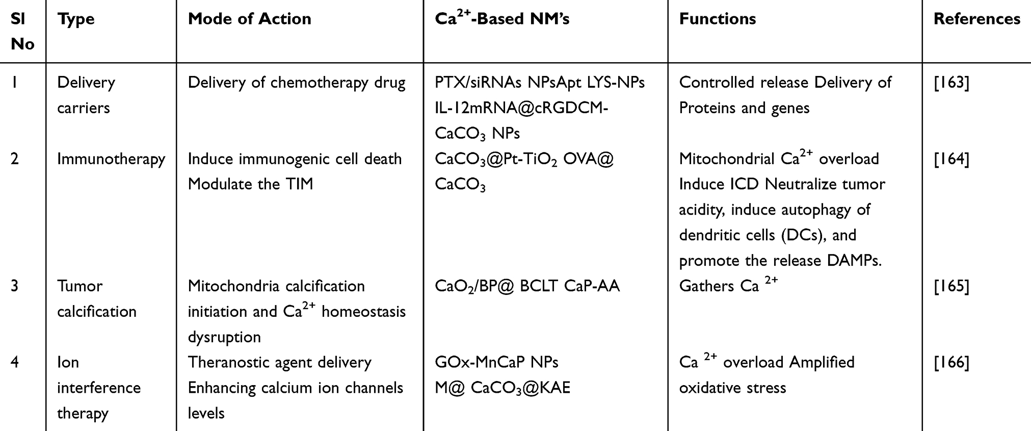 |
Table 5 Representative Ca2+ -Based NM’ s for Cancer Therapy |
Combined Ion Interference and Other Therapy
Owing to the complex nature of tumors, a single treatment approach often proves insufficient for effective inhibition or treatment. Immunotherapy is frequently combined with other cancer therapies to achieve synergetic effects. A TME-responsive nanoplatform was developed using dihydroartemisinin (DHA) encapsulated within Ca2+-doped mesoporous silica nanoparticles.167 This approach enhances Fenton reaction-mediated chemo dynamic therapy (CDT) by increasing intracellular calcium overload, triggered by DHA-induced calcium release, and reducing intracellular Fe3+ to Fe2+. This strategy may trigger pyroptosis in cancer cells, resulting in a strong antitumor immune response.
Targeted Functionalization
Folate-targeted cancer therapies leverage the ability of folic acid to bind to folate receptor-positive (FR-positive) tumor cells to deliver therapeutic agents. Medical professionals equip tumor cells with folate-hapten conjugates to elicit antibody responses.168 The immune system fights targeted tumor cells by identifying those cells after they have been coated with antibodies and their respective antibody detectors. The goal is to show hidden cancer cells to the immune system as these cells require an invisible status to stay alive. This immunotherapy involves a multi-step process.169 Vaccinating the patient with a foreign agent helps them develop a strong immune response to attack that agent. The vaccine trains the immune system to fight and destroy cancer cells more effectively.170 Folate-hapten conjugates are administered to attach to tumor cells that show FR presence specifically.171 Subsequently, regular anti-hapten antibodies bind to the haptens that decorate the tumor cells. During the last stage immune cells with Fc receptors identify and eliminate tumor cells by binding to antibody-coated surfaces.172–174 Immune cells that destroy tumors use proinflammatory cytokines as a boosting mechanism.175,176 Importantly, antibody-dependent cell death, unlike apoptosis, promotes the presentation of tumor antigens by antigen-presenting cells, further stimulating the immune response against cancer. This approach holds significant promise for overcoming immune evasion by cancer cells and effectively combating the disease. The folate mediated targeting mechanism is shown in Figure 6.
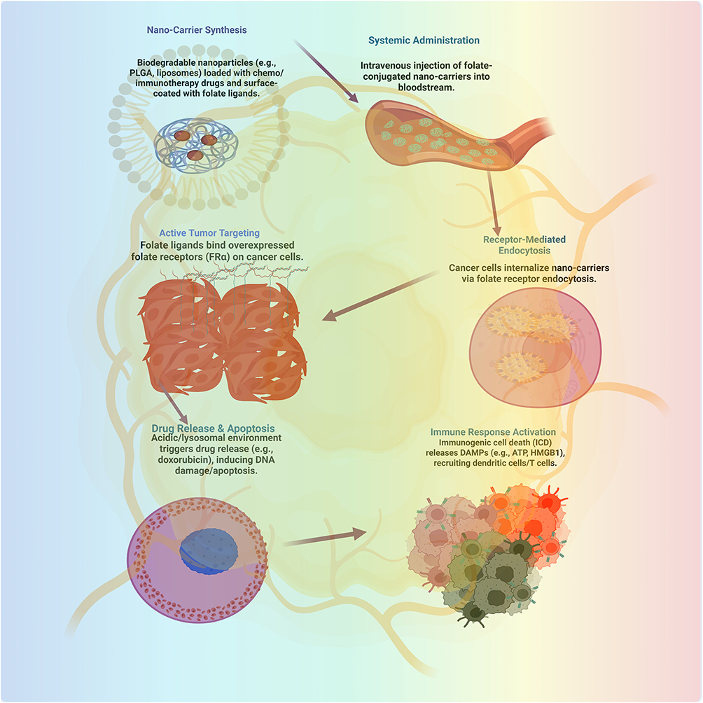 |
Figure 6 Diagrammatic representation of use of nano carrier in cancer cell death mediated via folate receptor [Image is created by Biorender.com]. |
Limitations of Calcium Nanomaterials for Homeostasis
Calcium nanomaterials can induce tumor cell apoptosis or sensitize cancer cells to other therapies through Ca2+ overload, causing mitochondrial dysfunction or disrupting signaling pathways. Nevertheless, their usefulness and safety in clinical translation are limited by several intrinsic and extrinsic factors. The major problem is that most calcium nanomaterials are not tumor-specific, as they produce low tumor-to-normal tissue discrimination. This may result in the non-specific release of Ca2+ and unintentional cytotoxicity in normal cells, negatively affecting the therapeutic window and results in off-target toxicity or inflammation of organs. The inherent buffering and compensatory processes in cancer and normal cells may cause an additional decrease in the effectiveness of calcium nanomaterials induced Ca2+ overload because these processes can quickly replenish ionic homeostasis, thereby blocking the desired apoptotic signals. Tumor heterogeneity, a dense extracellular matrix, and irregular vascularization may further impair the adequate penetration and retention of calcium nanomaterials within the tumor microenvironment.
The non-controllable release kinetics of Ca2+ nanoparticles may saturate not only tumor cells, but also normal cells, which may increase the risk of systemic toxicity and acute inflammatory reactions. Excessive gradual absorption or insufficiently absorbed release may not cause the required degree of calcium imbalance to be therapeutically effective. The hypoxia and acidity of the tumor microenvironment can be unpredictable for the dissolution of nanoparticles and release of Ca2+, which can result in variable results. Moreover, the long-term effects of Ca2+ stress can decrease the effectiveness of the treatment by activating survival pathways, initiating autophagy, and forming resistant tumor cell subpopulations. The toxicity and biocompatibility of the nanomaterial platform should also be considered. Synthesis issues including the possibility of reproducibility, scalability, and quality of calcium nanomaterials, as well as the absence of non-invasive methods to measure Ca2+ dynamics, further limit the implementation of this strategy in clinical practice. Thus, although manipulating tumor Ca2+ homeostasis using calcium nanomaterials holds potential, the problems associated with specificity, delivery, toxicity, resistance, and clinical viability are essential for implementing these nanomaterials as a part of a complex cancer treatment strategy.
Conclusion and Future Perspectives
This article reviews the relationship between Ca2+ homeostasis and tumor therapy, covering the mechanisms of Ca2+ regulation, its link to tumor progression, types of Ca 2+-based nanomaterials, and their diagnostic and therapeutic applications. Despite the advances made, challenges remain. These include inefficient accumulation of materials at tumor sites, insufficient Ca2+ release after internalization, and slow development of drugs targeting Ca2+ -regulated mechanisms. Enhancing intracellular Ca 2+ concentrations through drugs could address inadequate exogenous Ca2+ supply, but limited options exist. Biosafety concerns are crucial, as increasing doses or using novel materials may lead to adverse reactions. Ca2+ homeostasis needs to be studied through the control of upstream regulatory processes to comprehend effects beyond direct Ca2+ changes. The creation of advanced Ca2+ -based nanomaterials and innovative Ca2+ homeostasis mechanisms must come first for us to develop effective and safe medical tools.
The importance of calcium signaling in cancer treatment remains unclear because of its intricate relationship with different cancer cells. The ion interference method shows promise, but industry experts must continue to develop it because it causes problems when cells change and hit unintended targets. The need for more research on calcium and immune lasers depends on our success in translating preclinical findings into real medical treatments. The mechanism of tumor calcification in therapy requires further research, including how body systems respond best to these treatments. Better calcium probes and expanded analytical methods, including metabolomics and proteomics, need development to enhance calcium signaling research. To create effective cancer treatments using calcium, one must work as a team across several fields. A better understanding of how calcium interacts with cancer will help create better treatment methods for this generation. This review highlights essential findings while suggesting that further research is needed to fully achieve the therapeutic potential of calcium-based cancer therapies.
Data Sharing Statement
Review summarized references from the literature, and no new data were created.
Acknowledgments
Authors would like to thank the Deanship of Graduate Studies and Research, Ajman University, UAE, for their support in providing assistance in article processing charges for this review.
Author Contributions
Lekshmi Gangadhar (L.G): Writing—original draft preparation, Data curation. Siva Sankar Sana (S.S.S): conceptualization, software, writing—review and editing, Raja Venkatesan (R.V): writing—review and editing, Richie R. Bhandare (RRB): writing—review and editing, Seong-Cheol Kim (S.C.K).; Visualization, execution, writing—review and editing, supervision. Haya Khader Ahmad Yasin (K.A.Y).; acquisition of data, writing—review and editing, project administration. All authors made a significant contribution to the work reported, whether that is in the conception, study design, execution, acquisition of data, analysis and interpretation, or in all these areas; took part in drafting, revising or critically reviewing the article; gave final approval of the version to be published; have agreed on the journal to which the article has been submitted; and agree to be accountable for all aspects of the work.
Funding
This research received no external funding.
Disclosure
The authors declare no conflicts of interest in this work.
References
1. Panda S, Chatterjee O, Roy L, Chatterjee S. Targeting Ca2+ signaling: a new arsenal against cancer. Drug Discovery Today. 2022;27(3):923–34. doi:10.1016/j.drudis.2022.01.034
2. Chi Y, Sun P, Gao Y, Zhang J, Wang L. Ion interference therapy of tumors based on inorganic nanoparticles. Biosensors. 2022;12(2):100–113. doi:10.3390/bios12020100
3. Liu C, Guo L, Wang Y, Zhang J, Fu C. Delivering metal ions by nanomaterials: turning metal ions into drug-like cancer theranostic agents. Coord Chem Rev. 2023;494:215332. doi:10.1016/j.ccr.2023.215332
4. Durante M, Debus J, Loeffler JS. Physics and biomedical challenges of cancer therapy with accelerated heavy ions. Nat Rev Phys. 2021;3(12):777–790. doi:10.1038/s42254-021-00381-4
5. Rovituso M, La Tessa C. Nuclear interactions of new ions in cancer therapy: impact on dosimetry. Transl Cancer Res. 2017;6(5):914–933. doi:10.21037/tcr.2017.05.05
6. Sundararaman A, Amirtham U, Rangarajan A. Calcium-oxidant signaling network regulates AMP-activated protein kinase (AMPK) activation upon matrix deprivation. J Biol Chem. 2016;291(28):14410–14429. doi:10.1074/jbc.M116.731257
7. Song L. Calcium and bone metabolism indices. Adv Clin Chem. 2017;82:1–46. doi:10.1016/bs.acc.2017.06.005
8. Kudla J, Becker D, Grill E, Hedrich R, Hippler M, Schumacher K. Advances and current challenges in calcium signaling. New Phytol. 2018;218(2):414–431. doi:10.1111/nph.14966
9. Kang Y, Xu L, Dong J, et al. Calcium-based nanotechnology for cancer therapy. Coord Chem Rev. 2023;481:215050. doi:10.1016/j.ccr.2023.215050
10. Tiwari H, Gupta P, Verma A, et al. Advancing era and rising concerns in nanotechnology-based cancer treatment. ACS Chem Health Saf. 2024;31(2):153–161. doi:10.1021/acs.chas.3c00104
11. Cagel M, Grotz E, Bernabeu E, Moretton MA, Chiappetta DA. Doxorubicin: nanotechnological overviews from bench to bedside. Drug Discovery Today. 2017;22(2):270–281. doi:10.1016/j.drudis.2016.11.005
12. Chavda VP, Patel AB, Mistry KJ, et al. Nano-drug delivery systems entrapping natural bioactive compounds for cancer: recent progress and future challenges. Front Oncol. 2022;12:867655. doi:10.3389/fonc.2022.867655
13. Shahin M, Munir K, Wen C, Li Y. Magnesium matrix nanocomposites for orthopedic applications: a review from mechanical, corrosion, and biological perspectives. Acta Biomater. 2019;96:1–19. doi:10.1016/j.actbio.2019.06.007
14. Emadi H, Karevan M, Masoudi Rad M, et al. Bioactive and biodegradable polycaprolactone-based nanocomposite for bone repair applications. Polymers. 2023;15(17):3617. doi:10.3390/polym15173617
15. Zhao Y, Hu X, Liu Y, et al. ROS signaling under metabolic stress: cross-talk between AMPK and AKT pathway. Mol Cancer. 2017;16:79. doi:10.1186/s12943-017-0648-1
16. Thazhackavayal B, Murahar A, Kumar P, Gayam R, Harikumar KB, Mathew J. Beyond cyclopamine: targeting Hedgehog signaling for cancer intervention. Arch Biochem Biophys. 2024;754:109952. doi:10.1016/j.abb.2024.109952
17. Bruce JIE, James AD. Targeting the calcium signalling machinery in cancer. Cancers. 2020;12(9):2351. doi:10.3390/cancers12092351
18. Zheng S, Wang X, Zhao D, Liu H, Hu Y. Calcium homeostasis and cancer: insights from endoplasmic reticulum-centered organelle communications. Trends Cell Biol. 2023;33(4):312–323. doi:10.1016/j.tcb.2022.07.004
19. Moccia F. Endothelial Ca2+ signaling and the resistance to anticancer treatments: partners in crime. Int J Mol Sci. 2018;19(1):217. doi:10.3390/ijms19010217
20. Dimitrova N, Zamudio JR, Jong RM, et al. Public Access NIH Public Access. PLoS One. 2017;12(7):e0178059. doi:10.1371/journal.pone.0178059
21. Tokunaga R, Cao S, Naseem M, et al. AMPK variant, a candidate of novel predictor for chemotherapy in metastatic colorectal cancer: a meta-analysis using TRIBE, MAVERICC and FIRE3. Int J Cancer. 2019;145(8):2082–2090. doi:10.1002/ijc.32261
22. Prevarskaya N, Skryma R, Shuba Y. Ion channels in cancer: are cancer hallmarks oncochannelopathies? Physiol Rev. 2018;98(2):559–621. doi:10.1152/physrev.00044.2016
23. Yu T, Cai Z, Chang X, et al. Research progress of nanomaterials in chemotherapy of osteosarcoma. Orthopaed Surg. 2023;15(9):2244–2259. doi:10.1111/os.13837
24. Chen SMY, Krinsky AL, Woolaver RA, Wang X, Chen Z, Wang JH. Tumor immune microenvironment in head and neck cancers. Mol Carcinogenesis. 2020;59(7):766–774. doi:10.1002/mc.23181
25. Zhang M, Song R, Liu Y, et al. Calcium-overload-mediated tumor therapy by calcium peroxide nanoparticles. Chem. 2019;5(8):2171–2182. doi:10.1016/j.chempr.2019.06.003
26. Chen LF, Ye YT, Meng RY, et al. Precisely designed calcium-based nanomaterials: from regulation of cellular Ca2+ homeostasis to cancer therapy. ACS Mater Lett. 2024;6(10):4819–4840. doi:10.1021/acsmaterialslett.4c01432
27. Wong KK, Banham AH, Yaacob NS, Nur Husna SM. The oncogenic roles of TRPM ion channels in cancer. J Cell Physiol. 2019;234(9):14556–14573. doi:10.1002/jcp.28168
28. Liu J, Xie Y, Ma J, Chu H. New Ca2+ based anticancer nanomaterials trigger multiple cell death targeting Ca2+ homeostasis for cancer therapy. Chem Biol Interact. 2024;393:110948. doi:10.1016/j.cbi.2024.110948
29. Romito O, Guéguinou M, Raoul W, et al. Calcium signaling: a therapeutic target to overcome resistance to therapies in cancer. Cell Calcium. 2022;108:102673. doi:10.1016/j.ceca.2022.102673
30. Chien CC, Wu MS, Chou SW, Jargalsaikhan G, Chen YC. Roles of reactive oxygen species, mitochondrial membrane potential, and p53 in evodiamine-induced apoptosis and G2/M arrest of human anaplastic thyroid carcinoma cells. ChinMed. 2021;16:87–98. doi:10.1186/s13020-021-00505-3
31. Zheng P, Ding J. Calcium ion nanomodulators for mitochondria-targeted multimodal cancer therapy. Asian J Pharm Sci. 2021;17:10–12. doi:10.1016/j.ajps.2021.10.004
32. Li QH, Wang YZ, Tu J, et al. Anti-EGFR therapy in metastatic colorectal cancer: mechanisms and potential regimens of drug resistance. Gastroenterol Rep. 2020;8(3):179–191. doi:10.1093/gastro/goaa026
33. Bonsignore G, Martinotti S, Ranzato E. Endoplasmic reticulum stress and cancer: could unfolded protein response be a druggable target for cancer therapy? Int J Mol Sci. 2023;24(2):1566. doi:10.3390/ijms24021566
34. Phadwal K, Vrahnas C, Ganley IG, MacRae VE. Mitochondrial dysfunction: cause or consequence of vascular calcification? Front Cell Develop Biol. 2021;9:611922. doi:10.3389/fcell.2021.611922
35. Hu M, Guo J, Du J, et al. Development of Ca2+-based, ion-responsive superabsorbent hydrogel for cement applications: self-healing and compressive strength. J Colloid Interface Sci. 2019;538:397–405. doi:10.1016/j.jcis.2018.12.004
36. Hunt EG, Andrews AM, Larsen SR, Thaxton JE. The ER-mitochondria interface as a dynamic hub for T cell efficacy in solid tumors. Front Cell Develop Biol. 2022;10:867341. doi:10.3389/fcell.2022.867341
37. Avolio R, Matassa DS, Criscuolo D, Landriscina M, Esposito F. Modulation of mitochondrial metabolic reprogramming and oxidative stress to overcome chemoresistance in cancer. Biomolecules. 2020;10(1):135. doi:10.3390/biom10010135
38. Crul T, Maléth J. Endoplasmic reticulum‐plasma membrane contact sites as an organizing principle for compartmentalized calcium and cAMP signaling. Int J Mol Sci. 2021;22(9):4703. doi:10.3390/ijms22094703
39. Peruzzo R, Costa R, Bachmann M, Leanza L, Szabò I. Mitochondrial metabolism, contact sites and cellular calcium signaling: implications for tumorigenesis. Cancers. 2020;12(9):2574. doi:10.3390/cancers12092574
40. Godfraind T. Discovery and development of calcium channel blockers. Front Pharmacol. 2017;8:286. doi:10.3389/fphar.2017.00286
41. Shi H, Sadler PJ. How promising is phototherapy for cancer? Br J Cancer. 2020;123(6):871–873. doi:10.1038/s41416-020-0985-7
42. Striessnig J, Ortner NJ. Ca2+ channel blockers. Encyclop Mol Pharmacol. 2022;1:375–383. doi:10.1007/978-3-030-57401-7_444
43. Wang C, Peng J, Xiao Y, et al. Advances in nanotherapeutics for tumor treatment by targeting calcium overload. Colloids Surf B. 2025;245:114190. doi:10.1016/j.colsurfb.2024.114190
44. Carvalho TSS, Torres PMC, Belo JH, Mano J, Olhero SM. Bioactive magnetic materials in bone tissue engineering: a review of recent findings in CaP-based particles and 3D-printed scaffolds. Adv NanoBiomed Res. 2023;3(9):2300035. doi:10.1002/anbr.202300035
45. Qin S, Ma L, Li R, et al. Multifunctional calcium nanomodulators regulate type I interferon pathway to remodel tumor microenvironment for cancer immunotherapy. Chem Eng J. 2023;477:147118. doi:10.1016/j.cej.2023.147118
46. Ru X, Guo Y, Bai Z, et al. Synthesis of calcium carbonate nanoparticles in erythrocytes enables efficient removal of extracellular lead ions. Commun Chem. 2019;2(1):105. doi:10.1038/s42004-019-0199-z
47. Liu X, Wu Z, Cavalli R, Manzoli M, Cravotto G. Ultrasonic preparation of nano-CaCO3 templates and hollow mesoporous SiO2 nanoparticles for voriconazole loading. AAPS Pharm Sci Tech. 2024;25:165. doi:10.1208/s12249-024-02948-3
48. Jones CA, Hazlehurst LA. Role of calcium homeostasis in modulating EMT in cancer. Biomedicines. 2021;9(9):1200. doi:10.3390/biomedicines9091200
49. Dong Z, Liu Y, Wang C, et al. Tumor microenvironment modulating CaCO3‐based colloidosomal microreactors can generally reinforce cancer immunotherapy. Adv Mater. 2024;36(9):2308254. doi:10.1002/adma.202308254
50. He J, Wang Y, Ren Y, et al. Calcium-mediated cell adhesion enhancement-based antimetastasis and synergistic antitumor therapy by conjugated polymer–calcium composite nanoparticles. ACS Nano. 2024;18(36):24953–24967. doi:10.1021/acsnano.4c05771
51. Li Y, Wang J, Zhu T, et al. A ROS storm generating nanocomposite for enhanced chemodynamic therapy through H2O2 self-supply, GSH depletion and calcium overload. Nanoscale. 2024;16(17):8479–8494. doi:10.1039/D4NR00890B
52. Bożyk A, Wojas-Krawczyk K, Krawczyk P, Milanowski J. Tumor microenvironment—a short review of cellular and interaction diversity. Biology. 2022;11(6):929. doi:10.3390/biology11060929
53. Chen H, Zhang F, Zhang J, Zhang X, Guo Y, Yao Q. A holistic view of berberine inhibiting intestinal carcinogenesis in conventional mice based on microbiome-metabolomics analysis. Front Immunol. 2020;11:588079. doi:10.3389/fimmu.2020.588079
54. Lv B, Wang Y, Ma D, et al. Immunotherapy: reshape the tumor immune microenvironment. Front Immunol. 2022;13:844142. doi:10.3389/fimmu.2022.844142
55. Gautam J, Khedgikar V, Kushwaha P, et al. Formononetin, an isoflavone, activates AMP-activated protein kinase/β-catenin signalling to inhibit adipogenesis and rescues C57BL/6 mice from high-fat diet-induced obesity and bone loss. Br J Nutr. 2017;117(5):645–661. doi:10.1017/S0007114517000149
56. Gao J, Liang Y, Wang L. Shaping polarization of tumor-associated macrophages in cancer immunotherapy. Front Immunol. 2022;13:887713. doi:10.3389/fimmu.2022.887713
57. van Elsas MJ, Middelburg J, Labrie C, et al. Immunotherapy-activated T cells recruit and skew late-stage activated M1-like macrophages that are critical for therapeutic efficacy. Cancer Cell. 2024;42(6):1032–1050. doi:10.1016/j.ccell.2024.04.011
58. Luo G, Li X, Lin J, et al. Multifunctional calcium–manganese nanomodulator provides antitumor treatment and improved immunotherapy via reprogramming of the tumor microenvironment. ACS Nano. 2023;17(16):15449–15465. doi:10.1021/acsnano.3c01215
59. Lee J, Im K-I, Gil S, et al. TLR5 agonist in combination with anti-PD-1 treatment enhances anti-tumor effect through M1/M2 macrophage polarization shift and CD8+ T cell priming. Cancer Immunology. Immunotherapy. 2024;73:1–10. doi:10.1007/s00262-024-03679-5
60. Zhao P, Tian Y, Lu Y, et al. Biomimetic calcium carbonate nanoparticles delivered IL-12 mRNA for targeted glioblastoma sono-immunotherapy by ultrasound-induced necroptosis. J Nanobiotechnol. 2022;20(1):525. doi:10.1186/s12951-022-01731-z
61. Walkon LL, Strubbe-Rivera JO, Bazil JN. Calcium overload and mitochondrial metabolism. Biomolecules. 2022;12(12):1891. doi:10.3390/biom12121891
62. Pravina P, Sayaji D, Avinash M. Calcium and its role in human body. Int J Res Pharmaceut Biomed Sci. 2013;4(2):659–669.
63. Wu X, Han X, Guo Y, et al. Application prospect of calcium peroxide nanoparticles in biomedical field. Rev Adv Mater Sci. 2023;62(1):1–24. doi:10.1515/rams-2022-0308
64. Khalifehzadeh R, Arami H. Biodegradable calcium phosphate nanoparticles for cancer therapy. Adv Colloid Interface Sci. 2020;279:102157. doi:10.1016/j.cis.2020.102157
65. Casi G, Neri D. Noninternalizing targeted cytotoxics for cancer therapy. Mol Pharmaceut. 2015;12(6):1880–1884. doi:10.1021/mp500798y
66. Yuan K, Zhang C, Pan X, Hu B, Zhang J, Yang G. Immunomodulatory metal-based biomaterials for cancer immunotherapy. J Control Release. 2024;375:249–268. doi:10.1016/j.jconrel.2024.09.008
67. He C, Zhang S, Liu X, et al. CaO2 nanomedicines: a review of their emerging roles in cancer therapy. Nanotechnology. 2023;34(48):482002. doi:10.1088/1361-6528/acf381
68. Niu YQ, Liu JH, Aymonier C, et al. Calcium carbonate: controlled synthesis, surface functionalization, and nanostructured materials. Chem Soc Rev. 2022;51(18):7883–7943. doi:10.1039/d1cs00519g
69. Kiranda HK, Mahmud RB, Mahmood SK, Zakaria ZA. Fluorescent imaging and bio-cellular uptake assessment of gold-near infrared dye conjugated cockle shell calcium carbonate nanoparticles. Iraqi J Vet Sci. 2024;38(4):721–730. doi:10.33899/ijvs.2024.146804.3470
70. Zhou X, Wang Q, Lei Z, et al. Calcium carbonate-based nanoplatforms for cancer therapeutics: current state of art and future breakthroughs. ACS Omega. 2024;9(11):12539–12552. doi:10.1021/acsomega.3c09987
71. Yu J, Wang L, Xie X, et al. Multifunctional nanoparticles codelivering doxorubicin and amorphous calcium carbonate preloaded with indocyanine green for enhanced chemo-photothermal cancer therapy. Int J Nanomed. 2023;18:323–337. doi:10.2147/IJN.S394896
72. Perrone M, Patergnani S, Di Mambro T, et al. Calcium homeostasis in the control of mitophagy. Antioxid Redox Signaling. 2023;38(9):581–598. doi:10.1089/ars.2022.0122
73. Parakhonskiy BV, Abalymov A, Ivanova A, Khalenkow D, Skirtach AG. Magnetic and silver nanoparticle functionalized calcium carbonate particles-dual functionality of versatile, movable delivery carriers which can surface-enhance Raman signals. J Appl Phys. 2019;126(20):203102. doi:10.1063/1.5111973
74. Thomas J, Worch H, Kruppke B, Gemming T. Contribution to understanding the biomineralization of bones. J Bone Mineral Metab. 2020;38(4):456–468. doi:10.1007/s00774-020-01083-4
75. Shi Q, Yang Z, Yang H, et al. Targeting ion channels: innovative approaches to combat cancer drug resistance. Theranostics. 2025;15(2):521–545. doi:10.7150/thno.103384
76. Tew LS, Lee T-H, Lo L-W, Khung YL, Chen N-T. Aptamerized silica/gold nanocapsules for stimulated release of doxorubicin through remote two-photon excitation. Int J Smart Nano Mater. 2022;13(1):79–99. doi:10.1080/19475411.2022.2033874
77. Curcio M, Vittorio O, Bell JL, Iemma F, Nicoletta FP, Cirillo G. Hyaluronic acid within self-assembling nanoparticles: endless possibilities for targeted cancer therapy. Nanomaterials. 2022;12(16):2851. doi:10.3390/nano12162851
78. Feng J, Armillei MK, Yu AS, Liang BT, Runnels LW, Yue L. Ca2+ signaling in cardiac fibroblasts and fibrosis-associated heart diseases. J Cardiovasc Dev Dis. 2019;6(4):34. doi:10.3390/jcdd6040034
79. Li Y, Zhu Z, Hua S, et al. Metal-based nanoparticles promote the activation of cGAS-STING pathway for enhanced cancer immunotherapy. Nano Today. 2024;58:102445. doi:10.1016/j.nantod.2024.102445
80. Hak A, Shinde VR, Rengan AK. A review of advanced nanoformulations in phototherapy for cancer therapeutics. Photodiagn Photodyn Ther. 2021;33:102205. doi:10.1016/j.pdpdt.2021.102205
81. Neira-Carrillo A, Yslas E, Marini YA, et al. Hybrid biomaterials based on calcium carbonate and polyaniline nanoparticles for application in photothermal therapy. Colloids Surf B. 2016;145:634–642. doi:10.1016/j.colsurfb.2016.05.060
82. Xue P, Hou M, Sun L, et al. Calcium-carbonate packaging magnetic polydopamine nanoparticles loaded with indocyanine green for near-infrared induced photothermal/photodynamic therapy. Acta Biomater. 2018;77:242–255. doi:10.1016/j.actbio.2018.09.045
83. Xie W, Ye J, Guo Z, et al. TME-responded full-biodegradable nanocatalyst for mitochondrial calcium overload-induced hydroxyl radical bursting cancer treatment. Chem Eng J. 2022;438:135372. doi:10.1016/j.cej.2022.135372
84. Chen C, Sun J, Chen S, et al. A multifunctional-targeted nanoagent for dual-mode image-guided therapeutic effects on ovarian cancer cells. Int J Nanomed. 2019;14:753–769. doi:10.2147/IJN.S187929
85. Ambrosio L, Raucci MG, Vadalà G, Ambrosio L, Papalia R, Denaro V. Innovative biomaterials for the treatment of bone cancer. Int J Mol Sci. 2021;22(15):8214. doi:10.3390/ijms22158214
86. Tang B, Huang R, Ma W. Advances in nanotechnology-based approaches for the treatment of head and neck squamous cell carcinoma. RSC Adv. 2024;14(52):38668–38688. doi:10.1039/D4RA07193J
87. Wu J, Yi S, Cao Y, et al. Dual-driven nanomotors enable tumor penetration and hypoxia alleviation for calcium overload-photo-immunotherapy against colorectal cancer. Biomaterials. 2023;302:122332. doi:10.1016/j.biomaterials.2023.122332
88. Davoodvandi A, Sadeghi S, Alavi SMA, et al. The therapeutic effects of berberine for gastrointestinal cancers. Asia-Pac J Clini Oncol. 2024;20(2):152–167. doi:10.1111/ajco.13941
89. Beena TB, Mathew AJ, Harikumar KB, Safeeda E. Integrative analysis of the AMPK subunits in colorectal adeno carcinoma. Asian Pac J Cancer Prev. 2023;24(4):1159–1171. doi:10.31557/APJCP.2023.24.4.1159
90. Xie Z, Fan T, An J, et al. Emerging combination strategies with phototherapy in cancer nanomedicine. Chem Soc Rev. 2020;49(22):8065–8087. doi:10.1039/D0CS00349H
91. Zhai X, Sterea AM, El Hiani Y. Lessons from the endoplasmic reticulum Ca2+ transporters- A cancer connection. Cells. 2020;9(6):1536. doi:10.3390/cells9061536
92. Zhou Y, Zhang Z, Zhou C, Ma Y, Huang H, Liu J. Nanotechnology-fortified manipulation of cell Ca2+ signaling. Small Sci. 2024;4(3):2400169. doi:10.1002/smsc.202400169
93. Manyangadze M, Chikuruwo NM, Narsaiah TB, et al. Adsorption of lead ions from wastewater using nano silica spheres synthesized on calcium carbonate templates. Heliyon. 2020;6(11). doi:10.1016/j.heliyon.2020.e05309
94. An L, Cheng Y, Wang G, et al. Boric acid induced self-assembly of clinoatacamite for modulating tumor redox and immune homeostasis. Chem Eng J. 2025;Article166463. doi:10.1016/j.cej.2025.166463
95. Amit M, Eichwald T, Roger A, et al. Neuro-immune cross-talk in cancer. Nat Rev Cancer. 2025;1–17. doi:10.1038/s41568-025-00831-w
96. Wang Z, Deng T, Cheng R, Luo W, Bai Y. Apoptosis and mitochondrial DNA damaged induced by Zn2+/Ca2+ dual-ions overload for colorectal cancer improved immunotherapy. Colloids Surf B. 2025;Article114932. doi:10.1016/j.colsurfb.2025.114932
97. Yang WF, Guo WM, Luo QT, et al. Transcription factor TCF3 promotes bladder cancer development via TMBIM6-Ca2+-dependent ferroptosis. Cell Death Discovery. 2025;11(1):303. doi:10.1038/s41420-025-02585-8
98. Tan EW, Singh SK, Dua K, et al. Cancer stem cells: mitochondria signalling pathway and strategies for therapeutic interventions. Mol Biol Rep. 2025;52(1):671.
99. Szewczyk A, Rembiałkowska N, Saczko J, Daczewska M, Novickij V, Kulbacka J. Calcium electroporation induces stress response through upregulation of HSP27, HSP70, aspartate β-hydroxylase, and CD133 in human colon cancer cells. Biol Res. 2025;58(1):10. doi:10.1186/s40659-025-00591-9
100. Chagas PS, Garcia CB, Chagas HIS, et al. Suppression of SIGMAR1 hinders oral cancer cell growth via modulation of mitochondrial Ca2+ dynamics. Mol Biol Rep. 2025;52(1):220. doi:10.1007/s11033-024-08951-y
101. Zhang Z, Rana I, Nam J. Metal coordination polymer nanoparticles for cancer therapy. Essays Biochem. 2025;69(2):. doi:10.1042/EBC20253012
102. Zhang Q, Ma RF, Chen SW, Cao K, Wang Y, Xu ZR. Biomineralized and metallized small extracellular vesicles encapsulated in hydrogels for mitochondrial-targeted synergistic tumor therapy. Acta Biomater. 2025;194:428–441.
103. He Y, Hong Q, Chen S, Zhou J, Qiu S. Reprogramming tumor-associated macrophages in gastric cancer: a pathway to enhanced immunotherapy. Front Immunol. 2025;16:1558091.
104. Wu C, Gao M, Xiao W, et al. Light-activatable manganese carbonate nanocubes elicit robust immunotherapy by amplifying endoplasmic reticulum stress-meditated pyroptotic cell death. J Exp Clin Cancer Res. 2025;44(1):147. doi:10.1186/s13046-025-03044-5
105. Lin J. Intelligent drug delivery systems applying cell death mechanisms in cancer therapy. Adv Eng Technol Res. 2025;13(1):1144.
106. Çakmak E, Bilgici B. The relationship between cancer and endoplasmic reticulum stress-induced apoptosis. J Kirikkale Univ Faculty Med. 2025;27(1):111–119.
107. Liao W, Matsuda S, Takimoto T, et al. DCF-resistant ESCC redevelops drug sensitivity when combined with calcium channel blockers due to ER stress response. Esophagus. 2025;22:1–10.
108. Awad AMAM, Abdul Karim N. Dysregulation of mitochondrial function in cancer cells. Int J Mol Sci. 2025;26(14):6750. doi:10.3390/ijms26146750
109. Steiner P, Melek K, Andosch A, et al. Thapsigargin triggers a non-apoptotic, caspase-independent programmed cell death in basophilic leukaemia cells. Cell Death Discovery. 2025;11(1):313. doi:10.1038/s41420-025-02595-6
110. Liang JL, Cao Y, Lv K, Xiao B, Sun J. Amplifying Ca2+ overload by engineered biomaterials for synergistic cancer therapy. Biomaterials. 2025;316:123027. doi:10.1016/j.biomaterials.2024.123027
111. Alharbi AF, Parrington J. TPC2 in drug development: Emerging target for cancer, viral infections, cardiovascular diseases, and neurological disorders. Pharmacol Res. 2025;107655. doi:10.1016/j.phrs.2024.107655.
112. Verma R, Zeyaullah M, Singh V, Saxena PS, Koch B, Kumar M. Chitosan-functionalized fluorescent calcium carbonate nanoparticle loaded with methotrexate: future theranostics for triple negative breast cancer. ACS Biomater Sci Eng. 2025;11(2):981–999. doi:10.1021/acsbiomaterials.5c00034
113. Yin D, Wu X, Chen X, et al. Enhanced anticancer effect of carfilzomib by codelivery of calcium peroxide nanoparticles targeting endoplasmic reticulum stress. Mater Today Bio. 2025;32:101649. doi:10.1016/j.mtbio.2025.101649
114. Wang Z, Wu M, Jiang Y, et al. Biomimetic calcium-chelation nanoparticles reprogram tumor metabolism to enhance antitumor immunity. J Control Release. 2025;380:362–374. doi:10.1016/j.jconrel.2024.10.043
115. Ren T, Wang J, Ma Y, et al. Preparation of pH-responsive tanshinone IIA-loaded calcium alginate nanoparticles and their anticancer mechanisms. Pharmaceutics. 2025;17(1):66. doi:10.3390/pharmaceutics17010066
116. An J, Zhang K, Wang B, et al. Nanoenabled disruption of multiple barriers in antigen cross-presentation of dendritic cells via calcium interference for enhanced chemo-immunotherapy. Acs Nano. 2020;14(6):7639–7650. doi:10.1021/acsnano.0c03881
117. Huang J, He J, Wang J, et al. Calcium carbonate-actuated ion homeostasis perturbator for oxidative damage-augmented Ca2+/Mg2+. Biomaterials. 2023;302:122340. doi: 10.1016/j.biomaterials.2023.122340
118. Li Y, Zhou S, Song H, Yu T, Zheng X, Chu Q. CaCO3 nanoparticles incorporated with KAE to enable amplified calcium overload cancer therapy. Biomaterials. 2021;277:121080. doi:10.1016/j.biomaterials.2021.121080
119. Zhu Y, Yang Z, Dong Z, et al. CaCO3-assisted preparation of pH-responsive immune-modulating nanoparticles for augmented chemo-immunotherapy. Nano-Micro Lett. 2021;13(1):29. doi:10.1007/s40820-020-00549-4
120. Chirio D, Sapino S, Chindamo G, et al. Doxorubicin-loaded lipid nanoparticles coated with calcium phosphate as a potential tool in human and canine osteosarcoma therapy. Pharmaceutics. 2022;14(7):1362. doi:10.3390/pharmaceutics14071362
121. Prigol AN, Silva AH, Rode MP, et al. Calcium phosphate nanoparticles efficiently deliver miR-205-5p to modulate oncogenic targets in prostate cancer cells. Int J Pharm. 2025;125984. doi:10.1016/j.ijpharm.2025.125984
122. Xie B, Zhao H, Ding YF, et al. Drug-free tumor therapy via spermine-responsive intracellular biomineralization. J Control Release. 2023;357:572–579. doi:10.1016/j.jconrel.2023.05.041
123. Zhang J, Zhou Y, Guo J, Yan M, Liu C, Du B. Core–shell nanoparticles with sequential drug release depleting cholesterol for reverse tumor multidrug resistance. ACS Appl Mater Interfaces. 2025;17(4):6689–6702. doi:10.1021/acsami.4c22045
124. Ahmad I, Altameemi KKA, Hani MM, et al. Shifting cold to hot tumors by nanoparticle-loaded drugs and products. Clin Transl Oncol. 2025;27(1):42–69. doi:10.1007/s12094-024-03346-2
125. Wang D, Zhang L, Yang WH, et al. Arginine‐loaded nano‐calcium‐phosphate‐stabilized lipiodol pickering emulsions potentiates transarterial embolization‐immunotherapy. Adv Sci. 2025;12(6):2410484. doi:10.1002/advs.202410484
126. Maghraby N, El-Baz MA, Hassan AM, Abd-Elghaffar SK, Ahmed AS, Sabra MS. Metformin alleviates Doxorubicin-induced cardiotoxicity via preserving mitochondrial dynamics balance and calcium homeostasis. Appl Biochem Biotechnol. 2025;197(4):2713–2733. doi:10.1007/s12010-024-04945-4
127. Komarova EG, Akimova EB, Kondranova AM, et al. Laser interference microscopy study of the morphometric features of MCF-7 cancer cells contacted with 5-Fluorouracil-loaded calcium phosphate coatings on titanium implants. Mater Chem Phys. 2025;333:130299. doi:10.1016/j.matchemphys.2024.130299
128. Mohd Nor MA, Nik Mohd Kamal NNS, Khairuddean M, et al. Nanotechnology-based combination approach using essential oils and chemotherapeutic drugs for targeting cancer cells. Int J Polym Mater Polym Biomater. 2025;1–17. doi:10.1080/00914037.2024.2337433
129. Raineri F, Larue L, Cojocaru C, et al. The nucleolin antagonist N6L and paclitaxel combination treatment could be a new promising therapeutic strategy for pancreatic ductal adenocarcinoma therapy. Eur J Pharmacol. 2025;991:177310. doi:10.1016/j.ejphar.2024.177310
130. Han W, Xiong N, Huang L. Probiotics and nanoparticle-mediated nutrient delivery in the management of transfusion-supported diseases. Front Cell Infect Microbiol. 2025;15:1575798. doi:10.3389/fcimb.2025.1575798
131. Appidi T, China D, Ștefan GR, et al. Engineered multifunctional nanoparticles for enhanced radiation therapy: three-in-one approach for cancer treatment. Mol Cancer. 2025;24(1):68. doi:10.1186/s12943-025-01968-x
132. Du H, Zhang L, Feng J, Zhou M, Yang C. Enhancing photodynamic therapy efficacy via photo-triggered calcium overload and oxygen delivery in tumor hypoxia management. ACS Appl Mater Interfaces. 2025;17(5):8403–8416. doi:10.1021/acsami.4c26216
133. He M, Chen S, Yu H, et al. Advances in nanoparticle-based radiotherapy for cancer treatment. iScience. 2025;28(1). doi:10.1016/j.isci.2025.107351
134. Hamid AT, Oudah NN, Abdullah DA. Evaluation of the Caspase-3, Calpain-2, GSH, and MDA in AML and ALL patients undergoing chemotherapy. Yemeni J Med Sci. 2025;19(2):1–17.
135. Zhang Z, Hu Y, Ding Y, et al. Dual-enzyme-instructed peptide self-assembly to boost immunogenic cell death by coordinating intracellular calcium overload and chemotherapy. ACS Nano. 2025;19(1):488–503. doi:10.1021/acsnano.2c09393
136. Bai W, Chen H, Li J, Cai W, Kong Y, Zuo X. Calcium carbonate hollow microspheres encapsulated cellulose nanofiber/sodium alginate hydrogels as a sequential delivery system. Int J Biol Macromol. 2025;309:142839. doi:10.1016/j.ijbiomac.2024.142839
137. Sadat-Shojai M, Khorasani MT, Dinpanah-Khoshdargi E, Jamshidi A. Synthesis methods for nanosized hydroxyapatite with diverse structures. Acta Biomater. 2013;9(8):7591–7621. doi:10.1016/j.actbio.2013.05.004
138. Burdusel AC, Neacsu IA, Birca AC, et al. Microwave-assisted hydrothermal treatment of multifunctional substituted hydroxyapatite with prospective applications in bone regeneration. J Funct Biomat. 2023;14(7):378. doi:10.3390/jfb14070378
139. Hwang ET, Tatavarty R, Chung JY, Gu MB. New functional amorphous calcium phosphate nanocomposites by enzyme-assisted biomineralization. ACS Appl Mater Interfaces. 2013;5(3):532–537. doi:10.1021/am302706k
140. Hu XZ, Liu JL, Wang YX, et al. Composite nanoplatforms based on nanocalcium peroxide for cancer treatment: from monotherapy to combination therapy. Adv Ther. 2023;6(8):2300034. doi:10.1002/adtp.202300034
141. Mosmeri H, Alaie E, Shavandi M, Dastgheib SMM, Tasharrofi S. Benzene-contaminated groundwater remediation using calcium peroxide nanoparticles: synthesis and process optimization. Environ Monit Assess. 2017;189(9):452. doi:10.1007/s10661-017-6122-0
142. Zhang MG, Kiratiwongwan T, Shen W. Oxygen-releasing polycaprolactone/calcium peroxide composite microspheres. J Biomed Mater Res Part B. 2020;108(3):1097–1106. doi:10.1002/jbm.b.34474
143. Lin CT, Akhtar M, Li YJ, Ji M, Huang RQ. Recent developments in CaCO3 nano-drug delivery systems: advancing biomedicine in tumor diagnosis and treatment. Pharmaceutics. 2024;16(2):275. doi:10.3390/pharmaceutics16020275
144. Dong Z, Feng L, Hao Y, et al. Synthesis of CaCO3-based nanomedicine for enhanced sonodynamic therapy via amplification of tumor oxidative stress. Chem. 2020;6(6):1391–1407. doi:10.1016/j.chempr.2020.05.010
145. Levingstone TJ, Herbaj S, Dunne NJ. Calcium phosphate nanoparticles for therapeutic applications in bone regeneration. Nanomaterials. 2019;9(11):1570. doi:10.3390/nano9111570
146. Pylostomou A, Demir Ö, Loca D. Calcium phosphate bone cements as local drug delivery systems for bone cancer treatment. Biomat Adv. 2023;148:213367. doi:10.1016/j.bioadv.2023.213367
147. Zeng YZ, Hoque J, Varghese S. Biomaterial-assisted local and systemic delivery of bioactive agents for bone repair. Acta Biomater. 2019;93:152–168. doi:10.1016/j.actbio.2019.05.038
148. Zhou ZF, Sun TW, Chen F, et al. Calcium phosphate-phosphorylated adenosine hybrid microspheres for anti-osteosarcoma drug delivery and osteogenic differentiation. Biomaterials. 2017;121:1–14. doi:10.1016/j.biomaterials.2017.02.022
149. Shi YR, Dai Z, Wang Y, et al. Engineering crystallinity gradients for tailored CaO2 nanostructures: enabling alkalinity-reinforced anticancer activity with minimized Ca2+/H2O2 production. Nano Lett. 2023;23(23):10657–10666. doi:10.1021/acs.nanolett.3c03418
150. Yu H, Xu GY, Wen C, Yu BB, Jin YX, Yin XB. Multi-level reactive oxygen species amplifier to enhance photo-/chemo-dynamic/Ca2+ overload synergistic therapy. ACS Appl Mater Interfaces. 2024;16(15):18459–18473. doi:10.1021/acsami.3c21921
151. Dong CH, Dai XY, Wang X, et al. A calcium fluoride nanozyme for ultrasound-amplified and Ca2+-overload-enhanced catalytic tumor nanotherapy. Adv Mater. 2022;34(43):2205680. doi:10.1002/adma.202205680
152. Gong F, Xu JC, Liu B, et al. Nanoscale CaH2 materials for synergistic hydrogen-immune cancer therapy. Chem. 2022;8(1):268–286. doi:10.1016/j.chempr.2021.11.014
153. Zhao YJ, Bian YL, Xiao X, et al. Tumor microenvironment-responsive Cu/CaCO3-based nanoregulator for mitochondrial homeostasis disruption-enhanced chemodynamic/sonodynamic therapy. Small. 2022;18(38):2204047. doi:10.1002/smll.202204047
154. Tang C, Li H, Sha M, et al. A tumor microenvironment responsive titanium-based nanosonosensitizer for combination sonodynamic-immunotherapy with calcium ion overload. Chem Eng J. 2023;475:146054. doi:10.1016/j.cej.2023.146054
155. Toplak Ž, Hendrickx LA, Abdelaziz R, Shi X, Peigneur S, Tomašič T. Overcoming challenges of HERG potassium channel liability through rational design: eag1 inhibitors for cancer treatment. Med Res Rev. 2022;42(1):183–226. doi:10.1002/med.21874
156. Jahchan NS, Dudley JT, Mazur PK, et al. A drug repositioning approach identifies tricyclic antidepressants as inhibitors of small cell lung cancer and other neuroendocrine tumors. Cancer Discovery. 2013;3(12):1364–1377. doi:10.1158/2159-8290.CD-13-0235
157. Martínez R, Stühmer W, Martin S, Schell J, Reichmann A, Rohde V. Analysis of the expression of Kv10.1 potassium channel in patients with brain metastases and glioblastoma multiforme: impact on survival. BMC Cancer. 2015;15:839. doi:10.1186/s12885-015-1834-6
158. Agarwal JR, Griesinger F, Stühmer W, Pardo LA. The potassium channel Ether à go-go is a novel prognostic factor with functional relevance in acute myeloid leukemia. Mol Cancer. 2010;9:18. doi:10.1186/1476-4598-9-18
159. Huang X, He Y, Dubuc AM, Hashizume R, Zhang W, Reimand J. EAG2 potassium channel with evolutionarily conserved function as a brain tumor target. Nat Neurosci. 2015;18(9):1236–1246. doi:10.1038/nn.4088
160. Jacquemet G, Baghirov H, Georgiadou M, et al. L-type calcium channels regulate filopodia stability and cancer cell invasion downstream of integrin signalling. Nat Commun. 2016;7:13297. doi:10.1038/ncomms13297
161. Liu S, Khan AR, Yang X, Dong B, Ji J, Zhai G. The reversal of chemotherapy-induced multidrug resistance by nanomedicine for cancer therapy. J Control Release. 2021;335:1–20. doi:10.1016/j.jconrel.2021.04.032
162. Rodrigues T, Sieglitz F, Bernardes GJL. Natural product modulators of transient receptor potential (TRP) channels as potential anti-cancer agents. Chem Soc Rev. 2016;45(21):6130–6137. doi:10.1039/C6CS00182J
163. Nabissi M, Morelli MB, Santoni M, Santoni G. Triggering of the TRPV2 channel by cannabidiol sensitizes glioblastoma cells to cytotoxic chemotherapeutic agents. Carcinogenesis. 2013;34(1):48–57. doi:10.1093/carcin/bgs305
164. Morelli MB, Offidani M, Alesiani F, et al. The effects of cannabidiol and its synergism with bortezomib in multiple myeloma cell lines. A role for transient receptor potential vanilloid type-2. Int J Cancer. 2014;134(10):2534–2546. doi:10.1002/ijc.28518
165. Xu S, Cheng X, Wu L, et al. Capsaicin induces mitochondrial dysfunction and apoptosis in anaplastic thyroid carcinoma cells via TRPV1-mediated mitochondrial calcium overload. Cell Signalling. 2020;75:109733. doi:10.1016/j.cellsig.2020.109733
166. Wu L, Xu S, Cheng X, et al. Capsaicin inhibits the stemness of anaplastic thyroid carcinoma cells by triggering autophagy-lysosome mediated OCT4A degradation. Phytother Res. 2022;36(3):938–950. doi:10.1002/ptr.7363
167. Yeh JH, Chou CT, Chen IS, Lu T, Lin KL, Yu CC. Effect of thymol on Ca2⁺ homeostasis and viability in PC3 human prostate cancer cells. Chin J Physiol. 2017;60(1):32–40. doi:10.4077/CJP.2017.BAF362
168. Qin S, Su Q, Li X, Shao M, Zhang Y, Yu F. Curcumin suppresses cell proliferation and reduces cholesterol absorption in Caco-2 cells by activating the TRPA1 channel. Lipids Health Dis. 2023;22:6. doi:10.1186/s12944-023-01750-6
169. Rumpa MM, Maier C. TRPV1-dependent antiproliferative activity of dioecious Maclura pomifera extracts in estrogen receptor-positive breast cancer cell lines involves multiple apoptotic pathways. Int J Mol Sci. 2024;25(11):5258. doi:10.3390/ijms25115258
170. Lai H, Liu C, Hou L, Lin W, Chen T, Hong A. TRPM8-regulated calcium mobilization plays a critical role in synergistic chemosensitization of Borneol on doxorubicin. Theranostics. 2020;10(22):10154–10170. doi:10.7150/thno.44495
171. Law BYK, Michelangeli F, Qu YQ, et al. Neferine induces autophagy-dependent cell death in apoptosis-resistant cancers via ryanodine receptor and Ca(2+)-dependent mechanism. Sci Rep. 2019;9:20034. doi:10.1038/s41598-019-56514-7
172. Shi S, Bai X, Ji Q, et al. Molecular mechanism of ion channel protein TMEM16A regulated by natural product of narirutin for lung cancer adjuvant treatment. Int J Biol Macromol. 2022;223:1145–1157. doi:10.1016/j.ijbiomac.2022.01.194
173. Wang X, Hao A, Song G, et al. Inhibitory effect of daidzein on the calcium-activated chloride channel TMEM16A and its anti-lung adenocarcinoma activity. Int J Biol Macromol. 2023;253:127261. doi:10.1016/j.ijbiomac.2023.127261
174. Guo S, Bai X, Shi S, Deng Y, Kang X, An H. TMEM16A, a homoharringtonine receptor, as a potential endogenic target for lung cancer treatment. Int J Mol Sci. 2021;22(19):10930. doi:10.3390/ijms221910930
175. Ma B, Shi S, Guo W, Zhang H, Zhao Z, An H. Liensinine, a novel and food-derived compound, exerts potent antihepatoma efficacy via inhibiting the Kv10.1 channel. J Agri Food Chem. 2024;72(15):4689–4702. doi:10.1021/acs.jafc.3c08473
176. Ma B, Shi S, Ren S, Qu C, Zhao Z, An H. Corydaline binds to a druggable pocket of hEAG1 channel and inhibits hepatic carcinoma cell viability. Eur J Pharmacol. 2024;962:176240. doi:10.1016/j.ejphar.2024.176240
 © 2026 The Author(s). This work is published and licensed by Dove Medical Press Limited. The
full terms of this license are available at https://www.dovepress.com/terms
and incorporate the Creative Commons Attribution
- Non Commercial (unported, 4.0) License.
By accessing the work you hereby accept the Terms. Non-commercial uses of the work are permitted
without any further permission from Dove Medical Press Limited, provided the work is properly
attributed. For permission for commercial use of this work, please see paragraphs 4.2 and 5 of our Terms.
© 2026 The Author(s). This work is published and licensed by Dove Medical Press Limited. The
full terms of this license are available at https://www.dovepress.com/terms
and incorporate the Creative Commons Attribution
- Non Commercial (unported, 4.0) License.
By accessing the work you hereby accept the Terms. Non-commercial uses of the work are permitted
without any further permission from Dove Medical Press Limited, provided the work is properly
attributed. For permission for commercial use of this work, please see paragraphs 4.2 and 5 of our Terms.
Recommended articles

A Mini-Review of Diagnostic and Therapeutic Nano-Tools for Pancreatitis

Zhang Q, Li S, Yu Y, Zhu Y, Tong R
International Journal of Nanomedicine 2022, 17:4367-4381
Published Date: 19 September 2022
Research and Application Prospect of Nanomedicine in Kidney Disease: A Bibliometric Analysis From 2003 to 2024

Meng Y, Sui L, Xu T, Zhao H, Yuan Q, Sun L
International Journal of Nanomedicine 2025, 20:3007-3030
Published Date: 12 March 2025
Nano-Reagents in Acute Pancreatitis: Diagnostic, Therapeutic, and Theranostic Advances
Yang X, Li Q, Tang L, Huang L, Zhu C, Chen Q
International Journal of Nanomedicine 2026, 21:562220
Published Date: 19 February 2026
Extracellular Vesicles in Pancreatic Cancer Function and Potential Clinical Applications
Li D, Fang JJ, Jiang ZL
OncoTargets and Therapy 2026, 19:591622
Published Date: 8 April 2026