Back to Journals » Journal of Inflammation Research » Volume 18
Network Pharmacology, Experimental Validation Explored Kakkalide to Ameliorate Endothelial Cell Dysfunction and Inflammatory Response
Authors Li J, Yang S, Pan J, Qin Y ![]() , Ye H, Liao S
, Ye H, Liao S ![]() , Shao Z, Rong G, Xia Z, Sun J
, Shao Z, Rong G, Xia Z, Sun J ![]() , Shao Y
, Shao Y ![]()
Received 4 February 2025
Accepted for publication 9 August 2025
Published 28 August 2025 Volume 2025:18 Pages 11737—11753
DOI https://doi.org/10.2147/JIR.S519660
Checked for plagiarism Yes
Review by Single anonymous peer review
Peer reviewer comments 2
Editor who approved publication: Dr Tara Strutt
Jiafu Li,1 Shuai Yang,1 Jiahui Pan,1 Yuting Qin,1 Huiyun Ye,1 Shuanglin Liao,1 Zixuan Shao,1 Guangli Rong,1 Zhengyuan Xia,2 Jianbo Sun,3 Yiming Shao1,4
1Dongguan Key Laboratory of Sepsis Translational Medicine, The Intensive Care Unit, The First Dongguan Affiliated Hospital, Guangdong Medical University, Dongguan, Guangdong, People’s Republic of China; 2The First Dongguan Affiliated Hospital, Guangdong Medical University, Dongguan, Guangdong, People’s Republic of China; 3Dongguan Key Laboratory of Chronic Inflammatory Diseases, The First Dongguan Affiliated Hospital, Guangdong Medical University, Dongguan, Guangdong, People’s Republic of China; 4The Key Laboratory of Sepsis Translational Medicine, Guangdong Medical University, Zhanjiang, Guangdong, People’s Republic of China
Correspondence: Yiming Shao, The First Dongguan Affiliated Hospital, Guangdong Medical University, Jiaoping Road 42, Tangxia Town, Dongguan, Guangdong, 523710, People’s Republic of China, Tel +86 18826690188, Email [email protected]
Background: Sepsis continues to represent a significant challenge due to its detrimental effects and high mortality rate. The protection of endothelial function and the attenuation of the excessive inflammatory response are pivotal in the reduction of mortality risk. Flos Puerariae is the flower of Pueraria montana var. lobata (Willd). Flos Puerariae was recorded as ameliorating loathing of cold with high fever and clearing heat and purging the lung from “Minnan Materia Medica”. Recent findings showed its potential efficacy of inflammatory diseases. However, the precise underlying mechanisms are produced remain to be elucidated.
Purpose: This study aimed to identify the key active compound in Flos Puerariae for sepsis treatment and its pharmacological targets.
Methods: Network pharmacology analysis identified potential targets of Flos Puerariae against sepsis. Cecal ligation and puncture (CLP) mouse models assessed survival, inflammation, and tissue damage via ELISA and H&E staining. Transwell assays, immunofluorescence (IF), and flow cytometry evaluated human umbilical vein endothelial cell (HUVEC) protection. Molecular docking and Western blot (WB) confirmed key signaling pathway targets.
Results: At a dose of 20 mg/kg, the Flos Puerariae-derived compound Kakkalide notably reduced mortality and serum inflammatory factor levels while protecting lung and renal tissues. In vitro experiments further showed that treatment with 5 μM Kakkalide in HUVEC decreased inflammatory factors, reactive oxygen species, and apoptosis, while promoting cell migration and proliferation. The aforementioned effects were associated with alterations in the phosphorylation levels of Tumor Necrosis Factor (TNF), extracellular regulated protein kinases (ERK), and protein kinase B (AKT).
Conclusion: Our study demonstrated that Kakkalide as the primary ingredient of Flos Puerariae enhanced the survival of CLP mice and protected lipopolysaccharides (LPS)-stimulated HUVEC function. This study offers a novel perspective on the effects and molecular mechanisms of Kakkalide in sepsis.
Keywords: Flos Puerariae, Kakkalide, sepsis, inflammation, cyberpharmacology
Graphical Abstract:

Introduction
Sepsis is a fatal organ dysfunction caused by dysregulation of the host response to infection. In severe infections, patients experience septic shock,1 a condition in which the body’s response to infection is so overwhelming such that it causes organ failure. Sepsis is a significant contributor to morbidity in patients receiving intensive care. Annually, approximately 50 million individuals worldwide are afflicted with sepsis, with 11 million succumbing to the condition. This equates to a mortality rate of approximately one death per 2.8 seconds.2 Given its high prevalence, rapid progression, high mortality rate, and high cost of treatment, sepsis represents a significant public health concern that requires urgent attention. Presently, the treatment of sepsis is primarily centered on the understanding of its pathogenesis, early resuscitation, anti-infection measures, hormonal therapy, mechanical ventilation, nutritional support, immunomodulation, and symptomatic treatment.3–5 However, the mortality rate remains alarmingly high.6 The pathogenesis of sepsis is highly intricate, encompassing a multitude of factors including infection, inflammation, immunity, coagulation, and tissue damage. Among the most significant pathophysiologic mechanisms of sepsis is the excessive release of pro-inflammatory factors, which results in extensive microvascular endothelial damage, microcirculatory disorders, and ultimately, functional damage to target organs.7,8 Consequently, the prognosis of sepsis and the mortality rate of septic patients can be enhanced by identifying effective methods to regulate the inflammatory response and avert endothelial injury.
Network pharmacology has recently been used to elucidate the potential target or mechanism of action of herbal compounds or single-flavored Chinese medicines.9,10 From the perspective of “component-target-pathway-disease”, network pharmacology is currently regarded as an efficacious methodology for elucidating the mechanism of action of herbal medicines. This is achieved through an investigation of the components, drug targets, and their role and connection with diseases, with the aim of identifying the potential mechanism of action of Chinese herbal medicines in treating diseases through multi-target and multi-pathway mechanisms.11,12
Flower of Lobed Kudzuvine named Flos Puerariae is obtained after drying of the leguminosae of Pueraria lobata (Willd). Ohwi and Puearia thomsonii Benth.13 Kakkalide, active ingredient in Flos Puerariae, has been shown that Kakkalide inhibits macrophage nuclear factor kappa-B (NF-κB) activation and prostaglandin E2 expression, and ameliorates reactive oxygen species-associated inflammatory insulin resistance in endothelial cells. However, the impact of Kakkalide on sepsis is unclear.
The Medical Forest Compendium indicates that Flos Puerariae has the effect of moistening the lungs. Additionally, Southern Materia Medica has documented that Flos Puerariae can alleviate a dislike of cold, hiccups, hematemesis, and fever. As a traditional Chinese medicine, Flos Puerariae is sun-dried into a medicine and has a sweet taste and a warm nature. It has a channel tropism for the stomach and has various effects, including heat-clearing and detoxifying, relieving stranguria by diuresis, and activating the spleen by relieving alcoholism.14,15 Additionally, it influences endothelial cell oxidative stress levels by stimulating the synthesis of eNOS. Modern pharmacological studies have demonstrated that Flos Puerariae exerts a range of pharmacological effects, including anti-inflammatory and anti-oxidative stress properties.16–18 However, the therapeutic potential of Flos Puerariae in sepsis and the underlying pharmacological mechanisms remain uncertain.
The objective of this study was to conduct a comprehensive investigation into the potential of Kakkalide to mitigate the excessive inflammatory response and vascular endothelial cell dysfunction in sepsis, and to elucidate the underlying mechanisms using a mouse model of sepsis. Firstly, network pharmacology and molecular docking were employed to analyze the potential interaction mechanisms between the potential active ingredients of Flos Puerariae and the candidate targets of sepsis. Secondly, the effects of the active ingredients of Flos Puerariae on the survival rate of septic mice, pathological hematoxylin-eosin staining (H&E) of the lungs and kidneys, and the expression levels of inflammatory factors in the serum were evaluated through in vivo animal experiments. Subsequently, the mechanism of action of Kakkalide on inflammatory response and vascular endothelial cell dysfunction was validated through in vitro cellular experiments.
Materials and Methods
Collection of Potential Active Components and Prediction of Candidate Targets of Puerariae Flos and Construction of the Protein-Protein Interaction (PPI) Network
We used Traditional Chinese Medicine Systems Pharmacology Database and Analysis Platform (TCMSP, https://old.tcmsp-e.com/tcmsp.php), entered the keywords of Chinese medicine “Gehua” or “Flos Puerariae” to obtain all the chemical constituents of Puerariae Flos, and screened them according to pharmacokinetic parameters, oral taking bio-availability (OB) and drug-likeness (DL) as the screening conditions (S1), and added other compounds reported in the literature to generate a list of active ingredients of Puerariae Flos, and the drug targets were normalized to the corresponding gene names.
Secondly, the prediction targets related to sepsis were searched in the network databases, including Genecards (https://www.genecards.org/), DisGeNET (https://www.disgenet.org/) and OMIM (https://omim.org/), with the keyword “Sepsis” and normalized by the UniProtKB (https://www.uniprot.org/uniprotkb) database.
Finally, with the selection of the species as “Homo sapiens”, the above target genes were imported into the STRING database (https://string-db.org/) to screen. Then we used the Venn diagram to show those intersected genes. Protein-protein interaction network was obtained by the overlapping targets, and those were imported into the software Cytoscape V3.9.1 for the construction of the “active ingredient-target-disease” were visualized interaction network.
GO Function Enrichment Analysis and KEGG Pathway Enrichment Analysis
The DAVID (https://david.ncifcrf.gov) database was used to draw GO enrichment and KEGG analysis. Biological process (BP), cellular component (CC) and molecular function (MF) were performed in GO enrichment analysis. The top lists of enriched pathways and their cellular processes were shown in the KEGG network.
Molecular Docking
AutoDock Vina-1.5.7 was used for all molecular docking. The crystallin structures of potential active ingredients (Kakkalide, Kakkatin, 8-o-Methylreyusi, 3ʹ-Methoxydaidzein, Daidzein-4,7-diglucoside) and key targets from the PDB (https://www1.rcsb.org/) database were used as docking targets. The tightness of protein-ligand binding was expressed in terms of the binding energy for docking binding, with lower binding free energies indicating stronger ligand-receptor interactions. Finally, we used PyMOL-2.5.5 was used demonstrate results of docking.
Sepsis Animal Model
Male C57BL/6J mice (6–8 weeks old, 20–22 g) were purchased from Guangzhou Tengke Biomedical Technology Co. All animals were housed in the Laboratory Animal Center of Guangdong Medical University. Animals’ protocols were approved by the Animal Care and Use Committee of Guangdong Medical University (License and Approval No. GDMU-2024-000039) and were conducted in accordance with the institutional guidelines of Guangdong Medical University.
Forty mice were randomly assigned to four groups of ten: a sham group, a Kakkalide group, a CLP group and a CLP + Kakkalide group. The mice were fasted for 12 hours prior to inducing CLP. Following intraperitoneal anaesthesia with sodium pentobarbital, a midline abdominal incision was performed on all mice. In the sham and Kakkalide groups, the incision was sutured directly after the procedure. In the CLP and CLP+Kakkalide groups, the cecum was located and ligated approximately 1 cm from its distal end following the incision. A sterile 5-gauge needle was then used to puncture the cecum, gently expressing a small amount of feces before returning it to the abdominal cavity. After closure of the abdomen, all mice received immediate subcutaneous injection of 0.6mL 0.9% normal saline for fluid resuscitation, as well as access to adequate food and water. Following surgery, mice in the Kakkalide and CLP+Kakkalide groups underwent an intraperitoneal injection of 10 or 20 mg/kg Kakkalide one hour after the operation. The dosage of Kakkalide was determined based on the minimum effective dose recommended in the study by Min et al.19 Kakkalide was procured from Shanghai Tauto Bio-Technology Co., Ltd, (China).
HUVEC Cell Culture
HUVEC purchased from Shanghai Institute of Cell Biology (Shanghai, China) were cultured in 1640 supplemented with 5% FBS at 37°C, 5% CO2 and passaged every 2–3 days. Furthermore, we obtained 1640 basic medium from Thermo Fisher, (USA) and fetal bovine serum from Zeta life, (USA). HUVECs were plated in T25 flasks at a density of 2×10⁵ cells and cultured until reaching sub-confluence (70–80%), then processed for downstream assays. Lipopolysaccharide (LPS) was obtained from Sigma-Aldrich Trading Co., Ltd. (Shanghai, China). LPS treatment for 12 hours was applied on HUVEC for the in vitro model. Then, Kakkalide was applied to investigate the regulatory role of Kakkalide on sepsis.
ELISA for Inflammation Cytokines
We utilized the ELISA kit (Elabscience, Wuhan, China) to measure the levels of Creatinine, Blood urea nitrogen (BUN) and inflammatory cytokines like IL-1β, IL-6, and TNF-α in serum or medium. According to the experimental protocol, the standard product or sample was added to the wells of the ELISA plate, and the optical density (OD) was measured using spectrophotometry at a wavelength of 450 nm. The OD value is directly proportional to the concentration of the analyte being detected. The concentration of the analyte in the sample was determined by comparing its OD with a standard curve.
MDA Levels in Lung Tissues
The Malondialdehyde (MDA) Colorimetric Assay Kit (TBA Method, CAT# E-BC-K025-M) was used to detect MDA levels in lung tissues. The specific procedures were as follows: After homogenization of lung tissue samples, the homogenate was centrifuged at 10,000 ×g for 10 min at 4°C. The supernatant was collected and placed on ice for subsequent analysis. A portion of the supernatant was reserved for protein concentration determination. Corresponding reagents were added according to the kit instructions and mixed thoroughly. The EP tube openings were sealed with cling film (with small holes retained) and incubated in a 100°C water bath for 40 min. After cooling to room temperature under running water, the samples were centrifuged at 9569 ×g for 10 min. Then, 0.25 mL of the supernatant was transferred to a microplate, and the OD value was measured at 532 nm using a microplate reader.
Hematoxylin-Eosin Staining (H&E) to Detect Organoleptic Changes
Lung, kidney, and liver tissues were fixed with 4% paraformaldehyde, and the tissues were then dehydrated and embedded into wax blocks, and the blocks were cut into 2.5μm sections using a microtome. It was then deparaffinized and hydrated and stained with hematoxylin and eosin. Subsequently, ethanol gradient dehydration was performed, and the sections were sealed with neutral glue. Finally, the pathological changes of the tissues were observed and scanned by optical microscope.
The Wet-to-Dry Weight Ratio of Lung
Fresh left lung tissues from each group were obtained, washed with PBS, and blotted dry with filter paper. The initial measurement and recording of the lung tissue weight was conducted in a wet state (W). Subsequently, the lung tissue was desiccated in an oven maintained at 60 °C for a period of 24 hours. Subsequently, the lung tissue was re-weighed and documented as the dry weight (D). Subsequently, the ratio of W/D was calculated to quantify the degree of lung tissue oedema.
WB for VE-Cadherin Expression
Cells from each group were collected or tissues from mice were homogenized, lysed using RIPA buffer containing phosphatase inhibitors and protease inhibitors, protein concentration was determined by BCA and quantified, denatured at 100 °C for 10 min and then separated by electrophoresis under conditions of 80 V, 30 min; 120 V, 1 h; 400 mA, 38 min to polyvinylidene fluoride (PVDF) membranes (Millipore); The Quick Blocking Solution (Epizyme Biotech, shanghai, China) was blocked at room temperature for 40 min; then incubated overnight at 4 °C with the specific primary antibodies; antibodies were recovered, washed membranes with TBST; corresponding appropriate horseradish peroxidase (HRP)-conjugated secondary antibodies were incubated at room temperature for 1 h and washed membranes with TBST again. Finally, signals were detected using the multifunctional gel imaging analysis system (Azure 600, Azure Biosystems, United States). The following primary antibodies were used: VE-cadherin (1:50000; ab33168, abcam), NF-κB (1:500; sc-514451, Santa Cruz), p-NF-κB (1:200; sc-166748, Santa Cruz), AKT (1:2000; 10,176-2-AP, Proteintech), p-AKT (1:1000; 28,731-1-AP, Proteintech), ERK (1:2000; sc-166748, Proteintech), p-ERK (1:1000; 80,031-1-RR, Proteintech), β-action (1:1000; ab33168, Abcam).
Ribonucleic Acid Isolation and Quantitative Real-Time Polymerase Chain Reaction for ICAM-1 mRNA Expression
Total RNA from HUVEC was isolated using Trizol reagent (Invitrogen, MA, USA). For cDNA synthesis, 1 μg of total RNA was reverse-transcribed with 5 μL of 5× HiScript III RT SuperMix and 4 μL of 4× gDNA wiper (Vazyme, Nanjing, China). Quantitative RT-PCR was performed using 10 μL of 2× ChamQ Universal SYBR qPCR Master Mix Kit (Vazyme, Nanjing, China) under the following conditions: 40 cycles, each consisting of 10 seconds at 95°C and 15 seconds at 60°C. β-actin was served as a normalized gene, and mRNA levels were normalized by the method of 2−ΔΔCT. The list of primers is the following: ICAM-1: F-CTTGAGGGCACCTACCTCTG; R-GGCTGCTACCACAGTGATGA, β-actin: F-TCCCTGGAGAAGAGCTACGA; R-AGCACTGTGTTGGCGTACAG.
In vitro Cells Viability and Cytotoxicity Assay
In vitro cytotoxicity of Kakkalide on HUVEC were examined using MTT assay. Approximately 5000 cells with 100ul medium were added to each well of 96-well plate. After the cells adhere for 8 hours, added into the medicated culture medium with Kakkalide at concentrations of 0, 1, 3, 5, 10, 20, 30 and 50 μM to culture, and at the same time, set control group without drug and zero setting well with culture medium only. Each group was treated with more than three replicates. After stimulation, added 10 μL MTT working solution to each well, and continued incubation in the cell culture chamber for 4 h. After a small number of purple crystals appeared at the bottom of the well plate, the culture was stopped. The supernatant solution in the well plate was carefully and slowly aspirated, taking care not to disturb the purple crystals at the bottom. Then, 100 μL of Formazan dissolving solution was added, and the plate was incubated in an incubator at 37°C for approximately 15 minutes. The incubation was continued until the purple crystals were completely dissolved, after which the absorbance was measured at 570 nm.
Transwell Assay for HUVEC
700 μL complete medium was added to a 24-well plate, and the inserts were carefully placed in a 24-well plate to avoid bubbles at the bottom of the inserts. After the pretreated HUVEC were counted, 300 μL serum-free medium containing 40,000 HUVEC were added to each insert, and the 24-well plate was placed in an incubator to continue incubation for 12 h. The medium in the inserts was discarded, and the inserts were fixed in 4% paraformaldehyde fixative at room temperature for 30 min; the inserts were washed with PBS, and then the inserts were placed in crystal violet solution at room temperature for 15 min. After being PBS was washed for three times, the inner side of the inserts was carefully wiped with a cotton swab to remove residual cells, and the migration of the cells was observed under a microscope.
Cell Scratching Experiment
HUVEC were seeded in 6-well plates, and after the cells were treated with intervention, sterile 200 μL pipette tips were used at the bottom of the plates to draw cells along the direction perpendicular to the horizontal scratch line, washed three times with PBS, and the medium was replaced with serum-free medium; the initial location of cell scratches was observed and recorded under a microscope; the cells were placed in an incubator to continue the culture, and the location of cell migration was observed and recorded under a microscope at 12 h and 24 h. It was necessary to ensure that the field of view photographed each time was completely consistent with the field of view photographed at 0 h; finally, the average migration area of each field of view was statistically analyzed using Image-J 1.8.0 software.
Detection of Cell Apoptosis
Annexin V-FITC and propidium iodide (PI) apoptosis Kit (E-CK-A211; Elabscience) were used to label membranes and nuclei of apoptotic HUVEC. Pretreated samples were collected and resuspended in 500ul of Annexin V Binding Buffer. Samples were mixed with 4 μL of Annexin V-FITC and 4 μL of PI and incubated for 15 minutes away from light. Collect flow cytometry data on a NovoCyte (Agilent).
Immunofluorescence Staining
The treated cells were washed three times with PBS after removing the medium, then fixed with 4% paraformaldehyde fixative for 15 min at room temperature and washed three times with PBS. The cells were closed with 5% BSA at room temperature for 1 h. The primary antibody was incubated at 4 degrees overnight and then washed three times with PBS, and the secondary antibody was incubated at room temperature for 1 h and washed three times with PBS. The nuclei were stained with DAPI and then visualized under a confocal microscope. The list of antibodies used is the following: ZO-1 (1:100; sc-33725, Santa Cruz), claudin-5 (1:100; sc-374221, Santa Cruz), β-catenin (1:200; sc-53483, Santa Cruz), VE-cadherin (1:400; ab33168, abcam).
Monocyte-Endothelial Cell Adhesion Assay
HUVEC were seeded in 6-well plates and allowed to reach 70% confluency for group intervention. Subsequently, HUVEC were rinsed twice with PBS and calcein pre-stained THP-1 was added at a certain ratio and incubated for another 1 h. At the end of the incubation period, medium was aspirated and endothelial cells were washed three times with PBS to remove unattached THP-1. Photographs were taken with a fluorescence microscope at excitation and emission wavelengths of 490 nm and 515 nm, respectively. THP-1 adhesion to endothelial cells was expressed as fluorescence intensity, and three fields were captured under each experimental condition, and this experiment was repeated three times to calculate the ratio of cell adhesion rate in different treatment groups to that in the control group.
Intracellular Reactive Oxygen Species Assay
Briefly, DCFDA/H2DCFDA-cellular ROS Assay Kit (Abcam, Cambridge, USA) was utilized,2ʹ,7ʹ-dichlorodihydrofluoresce in diacetate (DCFH-DA) molecular fluorescent probe (LABLEAD, Beijing, China) was used assay intracellular reactive oxygen species content. HUVEC were inoculated in 6-well plates and incubated with 10 μM DCFH-DA for 30 min at 37 °C under 5% CO2, and then the fluorescence intensity was observed using a fluorescence microscope. The relative fluorescence intensity of DCFH-DA was used to reflect as the reactive oxygen species level.
Statistical Analysis
Data were analyzed using GraphPad PRISM version 10.0 software and presented as mean ± standard deviation (SD). Difference between results were evaluated using one-way analysis of variance (ANOVA). Survival curves were conducted with the Kaplan Meier method and comparisons were made by the log rank (Mantel-Cox) tests. p < 0.05 was represented as a statistically significant result.
Results
Network Pharmacological Analysis of Flos Puerariae Treatment for Sepsis
We obtained 4990 sepsis targets after searching Genecards, DisGeNET and OMIM database for predicted targets and removing duplicate values from the three databases. By combining the three target sets, a total of 85 common targets were obtained (to generate PPI maps) (Figure 1A). We screened the active ingredients according to TCMSP and obtained 138 active compounds. With OB 30% and DL 0.18 as the subsequent screening steps, 18 active substances were obtained (Supplementary Table 1). We continued to summarize possible associated targets based on 18 active substances and removed duplicate targets, obtaining a total of 501 potential action targets. Venn maps of potential targets of TCM and sepsis targets, and the middle overlapping part were the intersection targets of Flos Puerariae and sepsis, with a total of 288 (Figure 1B).
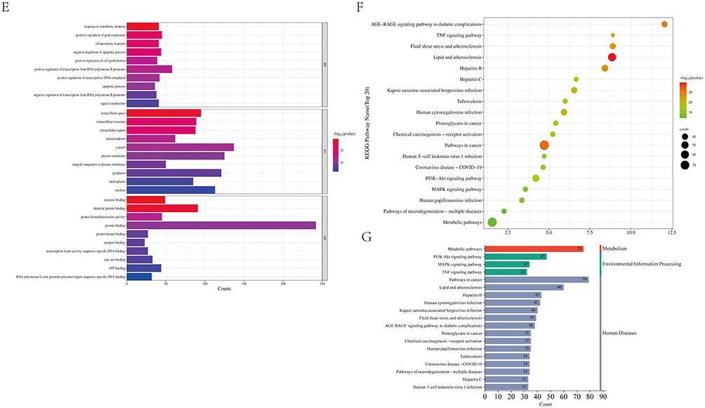
Figure 1 Continued. Figure 1 Network pharmacology analysis of potential mechanisms of Flos Puerariae against sepsis. (A) Venn diagram of overlapping Sepsis-related targets from OMIM, Genecards and DisGeNET database. (B) Venn diagram of overlapping genes between compound targets of Flos Puerariae and Sepsis-related genes. PPI network with visualized analysis (C) and compounds’ predicted hub targets network (D) were constructed through the analysis of overlapping genes. The size and darkness of the nodes were proportional to the degree, larger circles indicate higher levels of degree. (E) The analysis of Go annotation including biological processes (BP), cellular components (CC), and molecular function (MF). (F and G) The analysis of KEGG pathway enrichment and second-level categorization.
We drew PPI network with 288 intersection targets of sepsis by Cytoscape 10.0 software, which constructed 285 nodes and 7856 edges, then screened by Closeness> 0.001872551, Betweenness> 260.58 and Degree> 55.13 as screening conditions, and finally constructed a complex-sepsis target gene network with 66 nodes and 1707 edges and visualized analysis (Figure 1C). The size and color of the nodes were positively correlated with the degree values. Larger Degree values had larger nodes and darker colors (Figure 1D).
GO and KEGG enrichment analysis was performed using the DAVID database. The final analysis (Figure 1E) included biological processes (BP), cellular components (CC), and molecular function (MF). GO enrichment and KEGG analysis based on P <0.05 and FDR <0.05 were found Flos Puerariae to be strongly associated with inflammation (Figure 1F and G). Nevertheless, it remains unclear which of these active ingredients may be involved.
Effect of Kakkalide from Flos Puerariae on Inflammatory Response and Organ Damage in CLP Mice
As previously demonstrated, our findings suggested that a specific active ingredient in Flos Puerariae might have a role in the treatment of sepsis, potentially through anti-inflammatory mechanisms. However, the precise active ingredient remains uncertain. Kakkalide was a crucial active ingredient in the bioactivity screening of Flos Puerariae, with those exhibiting OD and DL values at the upper end of the ranking demonstrating notable activity. Additionally, previous literature had indicated that Kakkalide can induce colitis in mice and exhibit insulin-resistant activity.20 However, there is no evidence of Kakkalide involvement in septic diseases. Accordingly, the impact of Kakkalide on a murine model of CLP was examined. Figure 2A is a flowchart of the animal experiment. The results demonstrated that 20 mg/kg Kakkalide enhanced the survival rate of CLP mice. Conversely, 10 mg/kg Kakkalide had no impact on the survival of CLP mice (Figure 2B). Concurrently, we discovered that Kakkalide diminished the secretion of inflammatory mediators, including IL-1β, IL-6, and TNF-α, in the serum of CLP mice (Figure 2C–E). Subsequently, we conducted further investigations into the effects of Kakkalide on tissue damage. The serum levels of blood urea nitrogen (BUN) and Creatinine (Cr) were elevated in CLP mice, and Kakkalide reversed this trend (Figure 2F–G). Furthermore, VE-cadherin in mouse serum is decrease after Kakkalide treatment (Figure 2H). In addition, we found that Kakkalide reduced the edema of lung tissue and reduced the level of oxidative stress in the lung tissue of CLP mice (Figure 2I and J). Additionally, tissue HE staining revealed that Kakkalide mitigated the destruction of lung and kidney tissues in CLP mice (Figure 2K and L).
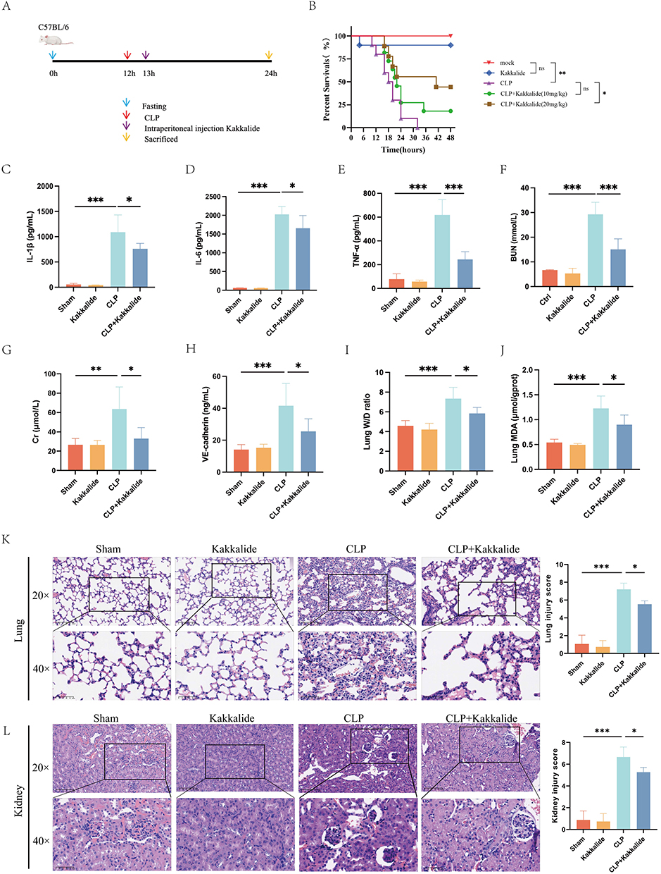 |
Figure 2 Kakkalide protected a mouse model of CLP-induced sepsis. (A) Schematic diagram of animal experiment. (B) The sepsis model in vivo was established via the CLP method. Subsequently, the experimental mice were divided into five groups (Sham, Kakkalide, CLP, CLP+10 mg/kg Kakkalide and CLP+20 mg/kg Kakkalide). Following a 12-hour modelling period, the survival percentage of mice was calculated. The expression of inflammatory factors, including IL-1β (C) IL-6 (D) TNF-α (E) was quantified in mouse serum via ELISA 12 hours following an intraperitoneal injection of 20 mg/kg Kakkalide in CLP mice. BUN (F) and Cr (G) levels are measured to assess kidney function. (H) VE-cadherin in mouse serum was used to assess endothelial direct adhesion levels. (I) W/D to assess oedema in the lung tissue. (J) ELISA for MDA to assess the level of oxidative stress in the lung tissue. The extent of damage to the lungs (K) and kidneys (L) in these groups was investigated through the use of haematoxylin and eosin (HE) staining. Data are expressed as the mean± SD (n ≥ 5 mice in every group). *p < 0.05, **p < 0.01, ***p < 0.001. |
Kakkalide Inhibited LPS-Induced Inflammatory Response and Injury in HUVEC
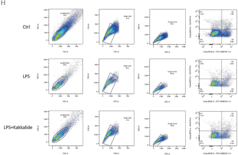
Our findings indicate that Kakkalide administration resulted in a reduction of CLP-induced release of inflammatory factors and tissue damage in mice. Additionally, it led to a decrease in tissue damage and a reduction in endothelial SDC-1 expression. It has been demonstrated that the primary pathophysiologic mechanisms of sepsis encompass an exaggerated inflammatory response, coagulation dysfunction, and microcirculatory disorders.21 All the aforementioned factors are related to vascular endothelial function. Vascular endothelial injury represents a pivotal factor in the pathogenesis of microcirculatory disorders in sepsis, underscoring the necessity to preserve the integrity of vascular endothelial cells. Accordingly, the impact of Kakkalide on HUVEC cells was examined in an in vitro setting. The proliferative effects of Kakkalide on HUVEC cells were initially evaluated, and it was observed that 5 μM of Kakkalide significantly promoted HUVEC proliferation, whereas 50 μM significantly inhibited HUVEC cell proliferation (Figure 3A). Similarly, the administration of 5 μM Kakkalide was observed to reduce the secretion of inflammatory factors triggered by LPS-stimulated HUVEC, including Interleukin (IL) −1β, IL-6, and TNF-α (Figure 3B–D). Regarding the impact on endothelial damage markers, our results indicated that Kakkalide safeguarded against Syndecan-1 (SDC-1) disruption and mitigated the LPS-induced elevation in intercellular cell adhesion molecule (ICAM) −1 mRNA expression in HUVEC (Figure 3E and F). Additionally, the impact of Kakkalide on endothelial cell apoptosis and reactive oxygen species was investigated. The findings revealed that Kakkalide mitigated the elevation in apoptosis resulted from LPS stimulation, diminished the production of reactive oxygen species, and safeguarded HUVEC from damage (Figure 3G and H).
Figure 3 Continued. Figure 3 Kakkalide mitigates the inflammatory response and endothelial cell dysfunction elicited by LPS stimulation of HUVEC. (A) The cellular activity of different concentrations of Kakkalide treated with HUVEC. 5 μM Kakkalide was to deal with HUVEC, and ELISA was used to evaluate the level of inflammatory factors like IL-1β (B) IL-6 (C) TNF-α (D) indicators of endothelial cell dysfunction SDC-1 (E) and ICAM-1 mRNA (F). (G) Fluorescence microscopy was used to examine the interaction between Kakkalide and reactive oxygen species in LPS-stimulated HUVEC. (H) Investigate the effect of Kakkalide on apoptosis in HUVEC stimulated by LPS through flow cytometry. Data are expressed as the mean± SD (n ≥ 3). * p < 0.05, **p < 0.01, ***p < 0.001.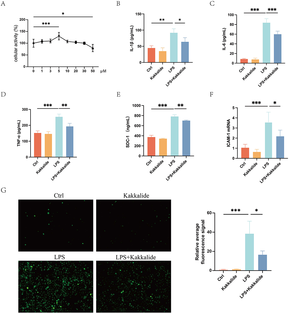
Kakkalide Promoted HUVEC Migration
Similarly, the adhesion and migration of endothelial cells represents a crucial function of HUVEC.22 In the event of endothelial injury, there is an increase in THP-1 adhesion levels, which serves to exacerbate endothelial dysfunction. Our findings revealed that Kakkalide could effectively reverse this phenomenon and safeguard endothelial cells from further damage (Figure 4A). Regarding the impact of Kakkalide on the migratory capacity of HUVEC, the findings showed that the number of migrated cells was found to be reduced in the LPS group in comparison to the control group. However, the number of migrated cells increased significantly following the administration of Kakkalide (Figure 4B). The cell scratch assay was employed to ascertain the migratory capacity of cells in distinct groups. Results showed that the stimulation of HUVEC with 1 mg/mL of LPS for 12 hours resulted in a statistically significant reduction in the rate of scratch wound confluence when compared to the control group. The intervention of LPS stimulation using 5 μM Kakkalide resulted in a notable increase in the rate of scratch trauma confluence, when compared to the LPS alone group (Figure 4C).
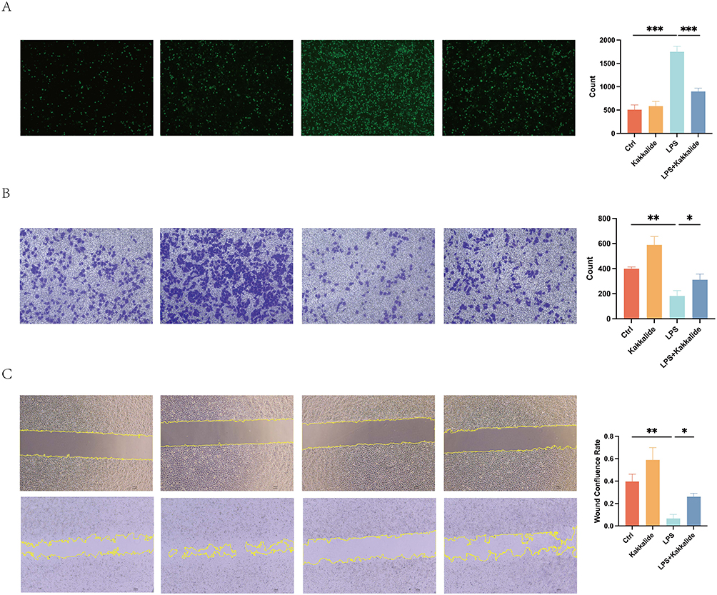 |
Figure 4 Kakkalide promotes migration in cultured HUVEC. (A) Calcein staining procedure assesses the adhesion of THP-1 cells to HUVEC after stimulation by 5 μM Kakkalide or/and 1 mg/mL LPS with 12 hours.The number of THP-1 cells adhered in different groups was counted. (B) Transwell was used to evaluate the migration ability on HUVEC. The number of migrated HUVEC cells was counted and compared. (C) The effect of Kakkalide on the confluence of scratch wounds in cultured HUVEC. The ratio of the migrated area to the initial area was statistically analyzed and compared. Data are expressed as the mean± SD (n ≥ 3). * p < 0.05, **p < 0.01, ***p < 0.001. |
Kakkalide Ameliorated LPS-Induced Disruption of Tight Junctions
Tight junction proteins and adhesion proteins serve as the safeguards of endothelial integrity.23,24 Our results demonstrated that LPS significantly disrupted the tight junction ZO-1 and adhesion protein VE-cadherin between HUVEC cells. However, this disruption was effectively mitigated by the intervention of Kakkalide (Figure 5A–D).
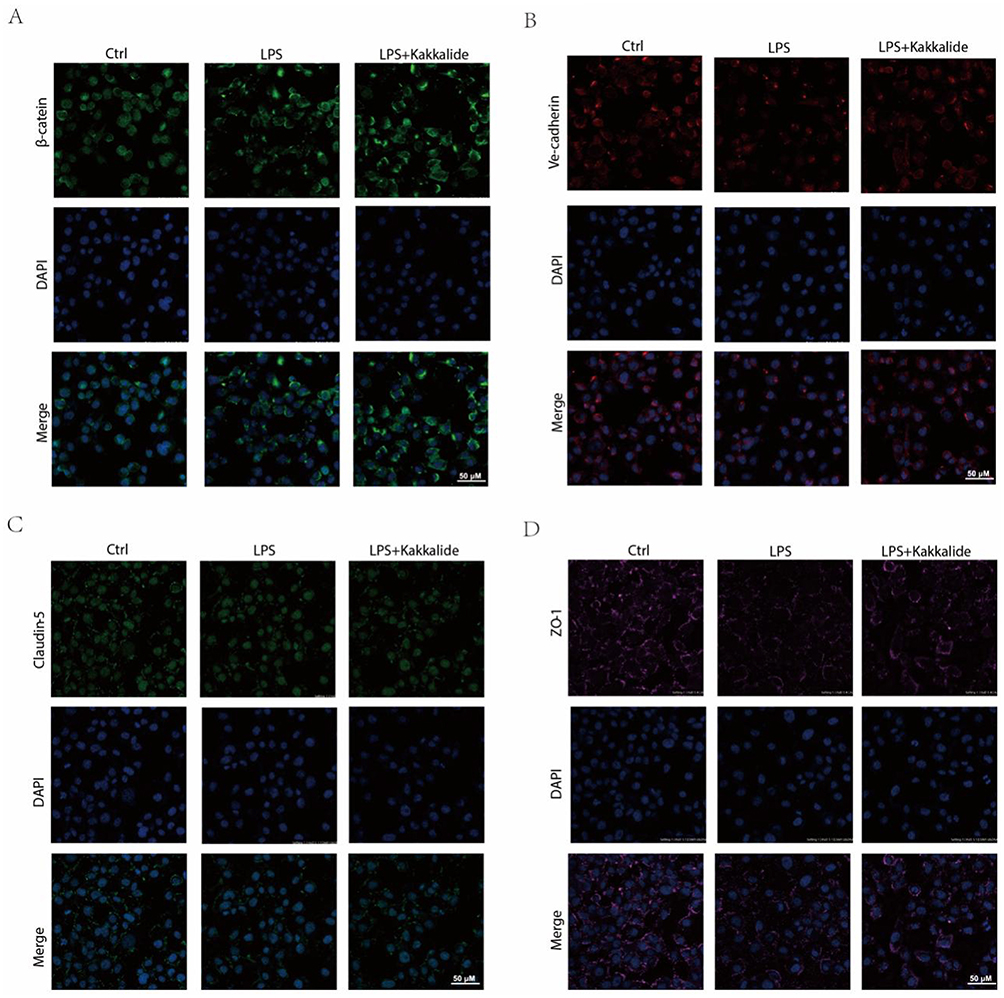 |
Figure 5 Kakkalide facilitates the restoration of endothelial junctions induced by LPS. Representative immunofluorescence images for green β-catenin (A) and red VE-cadherin (B) green claudin-5 (C) pink ZO-1 (D) all with blued DAPI, in the HUVECs. Data are expressed as the mean± SD (n ≥ 3). Scale bars: 50 μm. |
Molecular Docking and Target Validation
Through the above network pharmacological analysis, we found that Kakkalide may be the most promising effective component in the Flos Puerariae.
A lack of analysis of the key genes on which Kakkalide may act has prompted us to propose target validation through the upper layer of Kakkalide, that is, Flos Puerariae. We used the software of Pymol and Autodock to establish molecular docking of the five targets. The results (Table 1) showed that, the binding energy of Gand TNF (Figure 6A) is −8.086 kcal / mol, the binding energy of Kakkalide and ERK (Figure 6B) is −9.314 kcal / mol, the binding energy of Kakkalide and Akt (Figure 6C) is −8.455 kcal / mol, the binding energy of Kakkalide and JNK (Figure 6D) is −7.267 kcal / mol; the binding energy of Kakkalide and IL-1 (Figure 6E) is −3.915 kcal / mol. The more stable the ligand and protein conformation, the lower the corresponding binding energy. At the same time, the binding affinity between them is greater. Therefore, Kakkalide may be the most effective active ingredient for treating sepsis through the targets described above. We confirmed the aforementioned possibility through Western blot analysis, demonstrating that Kakkalide could indeed decrease the phosphorylation levels of Akt, p65 (NF-κB) and ERK (Figure 7).
 |
Table 1 Molecular Docking of Kakkalide with TNF, ERK, Akt, JNK and IL-1 |
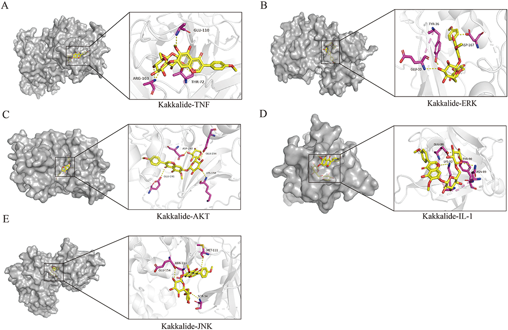 |
Figure 6 Molecular docking of Kakkalide to key targets. Visualization of Kakkalide with receptor protein complex molecules of TNF (A) ERK (B) AKT (C) IL-1 (D) and JNK (E). The affinity of TNF-Kakkalide is −8.086 kcal/mol; ERK-Kakkalide is −9.314 kcal/mol; ERK-Kakkalide is −8.455 kcal/mol; Akt-Kakkalide is −7.267 kcal/mol; IL-1-Kakkalide is −3.915 kcal/mol. |
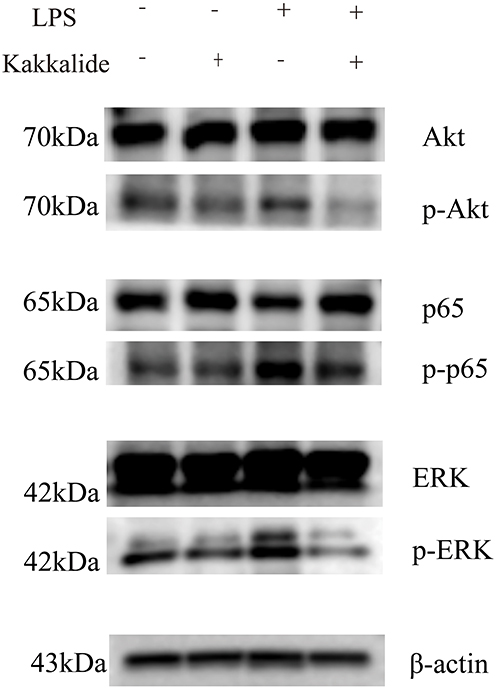 |
Figure 7 Western blot analysis confirmed the validity of the predicted key targets. Western blot analyses demonstrate the impact of Kakkalide on the protein expression levels of Akt, p-Akt, p65 (NF-κB), p-p65, ERK and p-ERK in HUVEC. |
Discussion
This study explored the therapeutic potential of the active ingredient Kakkalide in the Flos Puerariae for the sepsis-induced inflammatory response and endothelial damage. Sepsis is a lethal organ dysfunction syndrome caused by a dysregulated host response to infection, and effective clinical treatments remain lacking due to its extremely complex pathogenesis.25,26 The most important pathogenic link in the process of sepsis is considered to be the excessive release of proinflammatory factors that leads to extensive microvascular endothelial damage, microcirculation dysfunction, and then causes the functional impairment of target organs.27,28 Controlling the inflammatory response and preventing vascular endothelial damage may improve sepsis outcome and reduce case fatality. In our study, treatment with Kakkalide significantly improved the excessive inflammatory response and endothelial damage in HUVEC as well as lung and kidney function impairment in septic mice induced by CLP.
Pueraria lobata has hugely been proved to alleviate organ damage and inflammatory response in sepsis, but the other part is rarely studied.29,30 Flos Puerariae is the flower of Pueraria lobata. Flos Puerariae is a highly significant herbal remedy that has been demonstrated to possess therapeutic properties regarding antioxidant stress and colitis. Oxidative stress and inflammatory response are important pathological manifestations of sepsis.31,32 However, there is a paucity of evidence regarding its efficacy in the context of septic diseases. Our preliminary experiments demonstrated that Flos Puerariae could improve septic mice.
Network pharmacology is a novel research model that integrates pharmacology, bioinformatics, computer science, and other disciplines. It can help us to look for the active ingredients of herbs that can act on diseases, as well as the key pathways and targets that act on diseases, and then discover more effective herbs that can treat diseases. Therefore, we firstly explored the effective components according to the network pharmacology method and found that Kakkalide may be the most promising potential effective component of Flos Puerariae for the treatment of sepsis. Our findings suggest that it may intervene in the progression of sepsis through key targets, including TNF, ERK, Akt, JNK, and IL-1, through molecular docking and other means. TNF, ERK, Akt, JNK and IL-1 are all closely associated with the pathogenesis and progression of sepsis, playing a key role in the inflammatory responses and cellular signalling pathways associated with the condition. TNF is a key pro-inflammatory cytokine that is released during sepsis. When overproduced, it triggers a systemic inflammatory response syndrome (SIRS), leading to fever, increased vascular permeability, and organ dysfunction.33,34 Another core pro-inflammatory cytokine, IL-1, works with TNF to increase systemic inflammation, causing fever, acute-phase protein synthesis, and vascular endothelial damage.35 ERK, Akt and JNK are signalling kinases involved in cellular stress responses, inflammation and survival. They collectively drive sepsis pathogenesis by regulating inflammatory cascades, cell survival/apoptosis and endothelial function.36–38 Therefore, therapeutic strategies targeting these molecules may offer promising interventions for sepsis by interrupting its pathological progression.
Subsequently, we validated the anti-inflammatory efficacy of Kakkalide in vivo by demonstrating its capacity to reduce IL-1β, IL-6, and TNF-α levels in the serum of CLP-induced septic mice. It also could reduce the formation of edema and the level of oxidative stress in the lungs of CLP mice, thereby reducing mortality and mitigating renal and pulmonary tissue damage. Furthermore, in vitro experiments demonstrated that by reducing the expression levels of IL-1β, IL-6, and TNF-α in LPS-induced endothelial cells, it also promoted cell proliferation, reduced THP-1 adhesion, ameliorated oxidative stress, decreased apoptosis, and ameliorated disruption of adhesion molecules and tight junction proteins on the membrane surface of endothelial cells. These findings suggest that Kakkalide is a promising candidate for further investigation as a means of treating sepsis.
It should be noted that this study is not without limitations. A comprehensive understanding of the specific targets, pharmacodynamic effects, and signaling pathways of Kakkalide remains elusive. It is therefore recommended that future research efforts prioritize elucidating the effects of Kakkalide on molecular signal transduction mechanisms and the pharmacodynamic implications thereof.
Conclusion
In conclusion, the present study suggests that Kakkalide, the active ingredient in Flos Puerariae, exerts a protective effect against sepsis-induced endothelial cell dysfunction and inflammatory responses. This may be achieved through several key targets, including TNF, ERK, and Akt. The findings of our study offer novel insights into the mechanisms underlying endothelial cell dysfunction and excessive inflammatory response in sepsis, while also providing a new avenue for the identification of efficacious targets within the domain of traditional Chinese medicine.
Abbreviations
BP, Biological processes; BUN, Blood urea nitrogen; CC, Cellular components; CLP, Cecal ligation and puncture; Cr, Creatinine; DL, Drug-likeness; ELISA, Enzyme linked immunosorbent assay; eNOS, Endothelial nitric oxide synthase; HE, Hematoxylin-eosin staining; HUVEC, Human Umbilical Vein Endothelial Cells; ICAM-1, Intercellular cell adhesion molecule-1; IL-1β, Interleukin-1β; IL-6, Interleukin-6; GO, Gene Ontology; KEEG, Kyoto Encyclopedia of Genes and Genomes; LPS, Lipopolysaccharide; MDA, Malondialdehyde; MF, Molecular function; OB, Bio-availability; PI, Propidium iodide; SDC-1, Syndecan-1; TNF-α, Tumor necrosis factor-α; W/D, Wet weigh/dry weight ratioma.
Acknowledgments
We would like to express our profound gratitude to all members of the Dongguan Key Laboratory of Sepsis Translational Medicine and the Dongguan Key Laboratory of Chronic Inflammatory Diseases for their invaluable assistance and insightful counsel.
Funding
The authors’ research was supported by the National Nature Science Foundation of China (82072151), the Guangdong Basic and Applied Basic Research Fund (Guangdong-Dongguan Joint Fund) (2023A1515140177), The Talent Development Foundation of The First Dongguan Affiliated Hospital of Guangdong Medical University and the Foundation of State Key Laboratory of Pathogenesis, Prevention and Treatment of High Incidence Diseases in Central Asia (GCC2023005, SKL-HIDCA-2024-GD4) also provided financial support, as did the Affiliated Hospital of Guangdong Medical University Clinical Research Program (LCYJ2019A002).
Disclosure
The authors report no conflicts of interest in this work.
References
1. Singer M, Deutschman CS, Seymour CW, et al. The third international consensus definitions for sepsis and septic shock (Sepsis-3). JAMA. 2016;315(8):801–810. doi:10.1001/jama.2016.0287
2. Rudd KE, Johnson SC, Agesa KM, et al. Global, regional, and national sepsis incidence and mortality, 1990-2017: analysis for the global burden of disease study. Lancet. 2020;395(10219):200–211. doi:10.1016/S0140-6736(19)32989-7
3. Tindal EW, Armstead BE, Monaghan SF, et al. Emerging therapeutic targets for sepsis. Expert Opin Ther Targets. Mar. 2021;25(3):175–189.
4. Zampieri FG, Bagshaw SM, Semler MW. Fluid therapy for critically Ill adults with sepsis: a review. JAMA. 2023;329(22):1967–1980. doi:10.1001/jama.2023.7560
5. Liu D, Huang SY, Sun JH, et al. Sepsis-induced immunosuppression: mechanisms, diagnosis and current treatment options. Mil Med Res. 2022;9(1):56. doi:10.1186/s40779-022-00422-y
6. Vincent JL. Current sepsis therapeutics. EBioMedicine. 2022;86:104318. doi:10.1016/j.ebiom.2022.104318
7. Ince C, Mayeux PR, Nguyen T, et al. The endothelium in sepsis. Shock. 2016;45(3):259–270. doi:10.1097/SHK.0000000000000473
8. Johansson PI, Stensballe J, Ostrowski SR. Shock induced endotheliopathy (SHINE) in acute critical illness - a unifying pathophysiologic mechanism. Crit Care. 2017;21(1):25. doi:10.1186/s13054-017-1605-5
9. Zhao L, Zhang H, Li N, et al. Network pharmacology, a promising approach to reveal the pharmacology mechanism of Chinese medicine formula. J Ethnopharmacol. 2023;309:116306. doi:10.1016/j.jep.2023.116306
10. Wang YX, Yang Z, Wang WX, et al. Methodology of network pharmacology for research on Chinese herbal medicine against COVID-19: a review. J Integr Med. 2022;20(6):477–487. doi:10.1016/j.joim.2022.09.004
11. Zhang P, Zhang D, Zhou W, et al. Network pharmacology: towards the artificial intelligence-based precision traditional Chinese medicine. Brief Bioinform. 2023;25(1). doi:10.1093/bib/bbad518
12. Li X, Liu Z, Liao J, et al. Network pharmacology approaches for research of traditional Chinese medicines. Chin J Nat Med. 2023;21(5):323–332. doi:10.1016/S1875-5364(23)60429-7
13. Hirayama K, Matsuzuka Y, Kamiya T, et al. Metabolism of isoflavones found in the Pueraria thomsonii flower by human intestinal microbiota. Biosci Microflora. 2011;30(4):135–140. doi:10.12938/bifidus.30.135
14. Han NR, Nam SY, Hong S, et al. Improvement effects of a mixed extract of flowers of Pueraria thomsonii Benth. and peels of Citrus unshiu Markovich on postmenopausal symptoms of ovariectomized mice. Biomed Pharmacother. 2018;103:524–530. doi:10.1016/j.biopha.2018.04.070
15. Kamiya T, Nagamine R, Sameshima-Kamiya M, et al. The isoflavone-rich fraction of the crude extract of the Puerariae flower increases oxygen consumption and BAT UCP1 expression in high-fat diet-fed mice. Glob J Health Sci. 2012;4(5):147–155. doi:10.5539/gjhs.v4n5p147
16. Kim Y, Kim J, Son SR, et al. Chemical constituents of the flowers of Pueraria lobata and their cytotoxic properties. Plants. 2022;11(13).
17. Yuan D, Xie YY, Bai X, et al. Inhibitory activity of isoflavones of Pueraria flowers on nitric oxide production from lipopolysaccharide-activated primary rat microglia. J Asian Nat Prod Res. 2009;11(6):471–481. doi:10.1080/10286020902819822
18. Chen X, Zhang J, Li R, et al. Flos puerariae-semen hoveniae medicinal pair extract ameliorates DSS-induced inflammatory bowel disease through regulating MAPK signaling and modulating gut microbiota composition. Front Pharmacol. 2022;13:1034031. doi:10.3389/fphar.2022.1034031
19. Min SW, Park YJ, Kim DH. Kakkalide and its metabolite irisolidone ameliorate carrageenan-induced inflammation in mice by inhibiting NF-κB pathway. Inflammation. 2011;34(5):344–351. doi:10.1007/s10753-010-9240-1
20. Zhang D, Gao X, Wang Q, et al. Kakkalide ameliorates endothelial insulin resistance by suppressing reactive oxygen species-associated inflammation. J. Diabetes. 2013;5(1):13–24. doi:10.1111/1753-0407.12017
21. Yao YM, Luan YY, Zhang QH, et al. Pathophysiological aspects of sepsis: an overview. Methods Mol Biol. 2015;1237:5–15.
22. Cuschleri J, Gourlay D, Garcia I, et al. Endotoxin-induced endothelial cell proinflammatory phenotypic differentiation requires stress fiber polymerization. Shock. 2003;19(5):433–439. doi:10.1097/01.shk.0000051762.08171.ea
23. Lee W, Ku SK, Bae J-S. Bae JS. Vascular barrier protective effects of orientin and isoorientin in LPS-induced inflammation in vitro and in vivo. Vasc Pharmacol. 2014;62(1):3–14. doi:10.1016/j.vph.2014.04.006
24. Zodio S, Serreli G, Melis MP, et al. Protective effect of hydroxytyrosol and tyrosol metabolites in LPS-induced vascular barrier derangement in vitro. Front Nutr. 2024;11:1350378. doi:10.3389/fnut.2024.1350378
25. Chousterman BG, Swirski FK, Weber GF. Cytokine storm and sepsis disease pathogenesis. Semin Immunopathol. 2017;39(5):517–528. doi:10.1007/s00281-017-0639-8
26. Leligdowicz A, Richard-Greenblatt M, Wright J, et al. Endothelial activation: the ang/tie axis in sepsis. Front Immunol. 2018;9:838. doi:10.3389/fimmu.2018.00838
27. Colbert JF, Schmidt EP. Endothelial and microcirculatory function and dysfunction in sepsis. Clin Chest Med. 2016;37(2):263–275. doi:10.1016/j.ccm.2016.01.009
28. Joffre J, Hellman J, Ince C, et al. Endothelial responses in sepsis. Am J Respir Crit Care Med. 2020;202(3):361–370. doi:10.1164/rccm.201910-1911TR
29. Cheung DW, Koon CM, Wat E, et al. A herbal formula containing roots of Salvia miltiorrhiza (Danshen) and Pueraria lobata (Gegen) inhibits inflammatory mediators in LPS-stimulated RAW 264.7 macrophages through inhibition of nuclear factor κB (NFκB) pathway. J Ethnopharmacol. 2013;145(3):776–783. doi:10.1016/j.jep.2012.12.011
30. Jin SE, Son YK, Min BS, et al. Anti-inflammatory and antioxidant activities of constituents isolated from Pueraria lobata roots. Arch Pharm Res. 2012;35(5):823–837. doi:10.1007/s12272-012-0508-x
31. Joffre J, Hellman J. Oxidative stress and endothelial dysfunction in sepsis and acute inflammation. Antioxid Redox Signal. 2021;35(15):1291–1307. doi:10.1089/ars.2021.0027
32. Mun SJ, Cho E, Kim HK, et al. Enhancing acute inflammatory and sepsis treatment: superiority of membrane receptor blockade. Front Immunol. 2024;15:1424768. doi:10.3389/fimmu.2024.1424768
33. Zhou Y, Yu Z, Lu Y. To explore the influencing factors of clinical failure of anti-tumor necrosis factor-α (TNF-α) therapy in sepsis. Life Sci. 2025;369:123556. doi:10.1016/j.lfs.2025.123556
34. Gao XM, Zhou XH, Jia MW, et al. Identification of key genes in sepsis by WGCNA. Prev Med. 2023;172:107540.
35. Bhatia M, He M, Zhang H, et al. Sepsis as a model of SIRS. Front Biosci. 2009;14(12):4703–4711. doi:10.2741/3561
36. Liang J, Zhang J, Fan J, et al. ANXA3 interference inactivates ERK/ELK1 pathway to mitigate inflammation and apoptosis in sepsis-associated acute lung injury. Mol Immunol. 2024;167:25–33. doi:10.1016/j.molimm.2024.01.006
37. Jiang L, Yang D, Zhang Z, et al. Elucidating the role of Rhodiola rosea L. in sepsis-induced acute lung injury via network pharmacology: emphasis on inflammatory response, oxidative stress, and the PI3K-AKT pathway. Pharm Biol. 2024;62(1):272–284. doi:10.1080/13880209.2024.2319117
38. Nie Z, Xia X, Zhao Y, et al. JNK selective inhibitor, IQ-1S, protects the mice against lipopolysaccharides-induced sepsis. bioorg. Med Chem. 2021;30:115945.
 © 2025 The Author(s). This work is published and licensed by Dove Medical Press Limited. The
full terms of this license are available at https://www.dovepress.com/terms
and incorporate the Creative Commons Attribution
- Non Commercial (unported, 4.0) License.
By accessing the work you hereby accept the Terms. Non-commercial uses of the work are permitted
without any further permission from Dove Medical Press Limited, provided the work is properly
attributed. For permission for commercial use of this work, please see paragraphs 4.2 and 5 of our Terms.
© 2025 The Author(s). This work is published and licensed by Dove Medical Press Limited. The
full terms of this license are available at https://www.dovepress.com/terms
and incorporate the Creative Commons Attribution
- Non Commercial (unported, 4.0) License.
By accessing the work you hereby accept the Terms. Non-commercial uses of the work are permitted
without any further permission from Dove Medical Press Limited, provided the work is properly
attributed. For permission for commercial use of this work, please see paragraphs 4.2 and 5 of our Terms.