")
Back to Journals » International Journal of Nanomedicine » Volume 18
Nanomaterials Modulating the Fate of Dental-Derived Mesenchymal Stem Cells Involved in Oral Tissue Reconstruction: A Systematic Review
Authors Li X , Wang Y, Huang D, Jiang Z , He Z, Luo M, Lei J, Xiao Y
Received 27 April 2023
Accepted for publication 3 September 2023
Published 21 September 2023 Volume 2023:18 Pages 5377—5406
DOI https://doi.org/10.2147/IJN.S418675
Checked for plagiarism Yes
Review by Single anonymous peer review
Peer reviewer comments 2
Editor who approved publication: Professor Lei Yang
Xingrui Li,1,* Yue Wang,1,* Denghao Huang,1 Zhonghao Jiang,1 Zhiyu He,1 Maoxuan Luo,2 Jie Lei,1,2 Yao Xiao1– 3
1Oral & Maxillofacial Reconstruction and Regeneration of Luzhou Key Laboratory, the Affiliated Stomatological Hospital of Southwest Medical University, Institute of Stomatology, Southwest Medical University, Luzhou, People’s Republic of China; 2Department of Orthodontics, the Affiliated Stomatological Hospital of Southwest Medical University, Luzhou, People’s Republic of China; 3Department of Chengbei Outpatient, the Affiliated Stomatological Hospital of Southwest Medical University, Luzhou, People’s Republic of China
*These authors contributed equally to this work
Correspondence: Yao Xiao; Jie Lei, Email [email protected]; [email protected]
Abstract: The critical challenges in repairing oral soft and hard tissue defects are infection control and the recovery of functions. Compared to conventional tissue regeneration methods, nano-bioactive materials have become the optimal materials with excellent physicochemical properties and biocompatibility. Dental-derived mesenchymal stem cells (DMSCs) are a particular type of mesenchymal stromal cells (MSCs) with great potential in tissue regeneration and differentiation. This paper presents a review of the application of various nano-bioactive materials for the induction of differentiation of DMSCs in oral and maxillofacial restorations in recent years, outlining the characteristics of DMSCs, detailing the biological regulatory effects of various nano-materials on stem cells and summarizing the material-induced differentiation of DMSCs into multiple types of tissue-induced regeneration strategies. Nanomaterials are different and complementary to each other. These studies are helpful for the development of new nanoscientific research technology and the clinical transformation of tissue reconstruction technology and provide a theoretical basis for the application of nanomaterial-modified dental implants. We extensively searched for papers related to tissue engineering bioactive constructs based on MSCs and nanomaterials in the databases of PubMed, Medline, and Google Scholar, using keywords such as “mesenchymal stem cells”, “nanotechnology”, “biomaterials”, “dentistry” and “tissue regeneration”. From 2013 to 2023, we selected approximately 150 articles that align with our philosophy.
Keywords: nano-material, stem cell, tissue reconstruction, cell differentiation
Introduction
Trauma, tumor, infection, and congenital malformation can cause soft and hard tissue defects in the oral and maxillofacial region, thus impairing facial aesthetics, physical function, and social communication ability and seriously affecting patients’ physiological and psychological health and social status.1 However, due to the complex three-dimensional anatomical structure, various tissue layers, and the tendency to form scar healing, it is not easy to repair completely and regain function with existing repair methods such as flap transfer repair.2,3 Exogenous bone replacement materials are prone to disease transmission and immune reactions, and artificial material grafts are challenging to obtain ideal therapeutic results due to the lack of osteogenic induction characteristics.4 Autologous bone grafting was once considered the gold standard for bone filling, but donor shortages and complications have limited its development.3 With the advancement of tissue engineering technology and stem cell research, various bioactive materials and odontogenic stem cells have received widespread attention and have been gradually applied in clinical practice.
Bioactive nanomaterials are specialized biomaterials used at the nanoscale to obtain better therapeutic efficacy than conventional inert materials by inducing bioactive reactions or modulating biological functions.5 Various natural and artificial materials have ideal avenues of application in nanomaterials. Their unique nano-size effect and bioactive efficacy resulting from their interaction with the organism allow for biomedical applications such as bone tissue engineering, drug delivery, and wound dressings.5–7 In oral and maxillofacial soft and complex tissue defect repair, nano-bioactive materials can be obtained from a wide range of sources with significant bioefficacy. They can mimic the natural process of embryonic development by guiding the formation of functional tissues and organs from undifferentiated mesenchymal stem cells, thus reducing the occurrence of immune reactions.8
DMSCs have biological properties such as self-renewal, multidirectional differentiation, and high growth potential. They can proliferate, homogenize, and differentiate under the guidance of cytokines and scaffolding materials to achieve tissue transformation, which includes dental pulp stem cells (DPSCs),9 stem cells from human exfoliated deciduous teeth (SHEDs),10 and periodontal membrane stem cells (PDLSCs)11, root apical papillary stem cells (SCAPs),12 and dental follicular stem cells (DFSCs),13 which have received widespread attention for their wide variety of differentiable cells,14–17 long in vitro storage time,18,19 and good interaction with scaffolds and growth factors.20–22
Notably, the repair effect mediated by MSCs depends on the modulation of the bionic microenvironment established by tissues and biomimetic materials;23,24 in addition to the fibrous structure of nanomaterials, which has been shown to mimic the design of the extracellular matrix (ECM), thereby inducing the adhesion, proliferation, and differentiation of MSCs,25 some of the nanomaterials, such as calcium phosphates, can induce the remineralization capacity of MSCs by mediating the release of Ca and P ions by the release of Ca and P ions from functional groups to activate the remineralization ability of MSCs;26 nanofibrous sponge microspheres made of l-lactic acid can also induce up-regulation of vascular endothelial growth factor (VEGF) genes through hypoxia to enhance the vasculogenic ability of MSCs.27 Graphene-based materials can also stimulate the osteogenic differentiation of MSCs by increasing the osteoinductive conductivity.28 More and more studies have demonstrated the great relevance of nanomaterials to cell growth and development, metabolism, differentiation, and immunomodulation.29–31
In addition, nanomaterials are gradually gaining prominence for applications in dental caries, endodontic diseases, periodontal diseases, and intraoral implants. Silver nanoparticles (AgNPs) and zinc nanoparticles (ZnNPs), which do not form oxides when they come into contact with oxygen in the oral environment, have enhanced biocompatibility and antimicrobial properties and are more suitable for the prevention and treatment of dental caries than conventional materials.32–34 Several studies have shown that the antimicrobial effect of AgNPs is also suitable for root canal disinfection.35,36 A multifunctional material based on the nano-Si/Ca(Sr) system has been developed, which achieves acid resistance, bacterial inhibition, low cytotoxicity, and promotes dentin biomimetic remineralization, which can reduce dentin hypersensitivity.37 Nanomaterials are also increasingly used in periodontitis diagnosis, treatment, and tissue engineering, not only developing magnetic nanoparticle-based sensors for a comprehensive assessment of periodontitis, but also drug delivery in the form of nanoparticles, nanocapsules, and nanofibers, and modulation of stem cells using nanocomposite scaffolds as carriers.38 Nanoengineering, which is a new platform to enhance the bioactivity of dental implants, has been used to enhance the antimicrobial and bioactive properties of conventional implants and improve the long-term therapeutic outcome by fabricating titanium dioxide nanotubes (TNT), nanoparticles (NPs), and various types of biopolymers through physical modification, chemical modification, mechanical modification, and biomolecular modification.39
In summary, nanomaterials modulating the fate of MSCs have become a hot research topic in tissue repair and regeneration, and the modulation of different MSCs by different materials is also a topic worthy of investigation, so this paper outlines the latest developments in the fabrication of implantable biomaterials with nanostructures and evaluates them.
Dental-Derived Mesenchymal Stem Cells
Please refer to Figure 140 and Table 1.
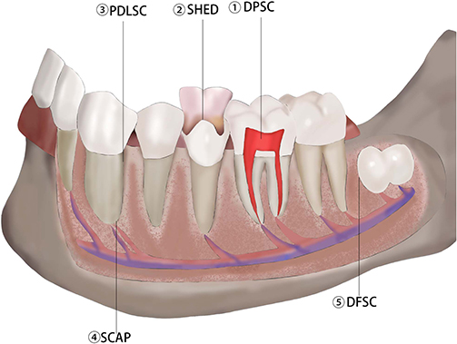 |
Figure 1 The origin of DMSCs. Notes: Dental pulp stem cells (DPSC). Mostly derived from healthy milk teeth and wisdom teeth, they have the potential for multidirectional differentiation. In addition to the ability to form mineralized nodule-capable cells, they can also differentiate into cell lineage types such as adipose, bone, cartilage, muscle, vascular endothelium, hepatic, and neural cell types after induction by different cytokines. The main advantages include abundant sources, side effects, and no ethical controversies. Human mammary dental stem cells (SHED). Most of the tissues are extracted from the healthy milk teeth of the affected children, with multi-directional differentiation potentials such as osteogenic differentiation, lipogenic differentiation, and differentiation in the direction of neuronal cells. Periodontal stem cells (PDLSC). PDLSCs are derived from normal human dental tissues and express Stro-1, a marker of MSCs, and have multidirectional differentiation potential. Stem cells of the apical papilla (SCAP). SCAPs are primarily developed from the apical part of young permanent teeth with immature roots and secrete exosomes that play an essential role in the repair of tissue damage, organ development, and immunomodulation of the body. Dental Follicular Stem Cells (DFSC). Derived from the cranial neural crest, DFSCs are mesenchymal precursor cells around the dental embryo that form osteoid, periodontium, and alveolar bone during tooth development. Adapted from Chalisserry EP, Nam SY, Park SH, Anil S. Therapeutic potential of dental stem cells. J Tissue Eng. 2017;8. Creative Commons and with Permission of SAGE Publications.40 |
 |
Table 1 Classification and Research Progress of DMSCs |
DPSC
As an essential component of odontogenic MSCs, DPSCs have a unique position in repairing maxillofacial injuries in the body under their high differentiation characteristics and ability to regenerate the dentin/pulp complex. In 2000, Gronthos et al isolated a type of fibroblast with a multidirectional differentiation potential and named it dental pulp stem cells while studying dental pulp tissues.9 As neural crest-derived cells, DPSCs can be used for cell therapy along several cell lineages.51 Currently, many studies have demonstrated the ability of DPSCs in neural differentiation, angiogenesis, dentinogenesis, osteogenesis, and chondrogenesis,41,42,52–56 not only in the maxillofacial region but also in tissues throughout the body (eg, the brain, heart, cornea). Recently, their secreted factors have shown therapeutic potential in various diseases.41,42 The differentiation process is often influenced by the scaffolds and growth factors used, eg, embedded collagen/chitosan scaffolds promote dentin differentiation of DPSCs,52 a bioengineered scaffold provided by porcine decellularized dental pulp has also been found to have a pro-dentin/enamel differentiation effect,53 transforming growth factor (TGF-β) enhances the proliferation and chondrogenic differentiation of DPSCs,55 and nerve growth factor (NGF) affects the neurogenic ability of DPSCs.56 The excellent proliferation and differentiation ability of DPSCs protects patients in maxillofacial restoration and provides more possibilities for stem cell research in the biomedical field.
SHED
Miura M et al found that human deciduous teeth contain highly proliferative clonal cells known as SHEDs.57 Originating from perivascular neuronal cells and neuroglia, SHEDs have strong angiogenic, neurogenic, and odontogenic differentiation abilities and have great potential in the repair of oral pulp tissues.58 Xiong H et al incorporated SHEDs into gelatin/bioactive glass (GEL/BGM) scaffolds and found that they could significantly repair bone defects and promote the formation of new bone.43 Similarly, Hagar MN et al induced osteogenesis of SHEDs on granular hydroxyapatite scaffolds (gHA) and found that alkaline phosphatase (ALP) concentration was significantly increased, and osteogenic potential was enhanced considerably.44
Compared with other mesenchymal cells, such as DPSCs and bone marrow mesenchymal stem cells (BMSCs), SHEDs have superior proliferative capacity and faster colony formation efficiency. In addition to dental pulp regeneration, in recent years, SHEDs have made significant progress in neurological research. Some studies have reported that SHEDs can not only alleviate trigeminal neuralgia by inhibiting endoplasmic reticulum stress but also prevent apoptosis of neural progenitor cells to a certain extent and promote facial nerve regeneration.59,60 Although the proliferation and differentiation of SHEDs share similarities with many odontogenic MSCs, their natural shedding and non-invasive acquisition add new possibilities for stem cell applications, and storing SHEDs when children shed their milk teeth may be beneficial in treating future injuries or diseases.59,61–65
PDLSC
Since the first extraction of PDLSCs from periodontal membranes by Seo BM,11 research on the differentiation of this cell has been a hot topic. More and more researchers have discovered its excellent differentiation potential to be able to differentiate into many types of mesenchymal lineage cells, such as osteoblasts, chondrocytes, cementoblasts, and adipocytes in vitro, and to regenerate PDL-like tissues in vivo.46,47,66 Zhang Y et al found that gold nanoparticles (AuNPs) activated the autophagy pathway by upregulating β-microtubulin III and down-regulating chelator 1 / p62, enhancing osteogenesis in PDLSCs slices.45 Li X et al, on the other hand, noted that the reactive oxygen species (ROS) encapsulated in menaquinone (MitoQ; an autophagy enhancer) released the nanoparticle, released menaquinone effectively induced mitochondrial autophagy via the PINK1-Parkin pathway and successfully reduced oxidative stress by decreasing the number of reactive oxygen species, which was beneficial for periodontal regeneration.67 The study of PDLSCs exosomes (PDLSCs-Exos) has also been gradually increasing. FOR THE FIRST TIME, Liu T et al revealed that miRNAs originating from PDLSCs-Exos might promote the osteogenic differentiation of BMSCs.68 Liu M et al found that PDLSCs-Exos could also alleviate high glucose-induced senescence in PDLSCs by transferring miR-3-1p to activate the KEAP2-NRF signaling pathway.69 More and more studies have shown that the cell fate of PDLSCs is capable of targeted regulation and that epigenetic modifications, endoplasmic reticulum-mitochondrial coupling, and diabetic metabolites have inhibitory effects on osteogenic differentiation of periodontal stem cells,70–72 which undoubtedly provides significant value for stem cell transplantation therapy.
SCAP
Stem cells derived from the apical papillae of immature permanent teeth (SCAPs), since Huang GT discovered that SACPs could differentiate into pulp tissue,73 Bakopoulou A and Han C et al explored their differentiation and proliferation potentials respectively. Surprisingly, SCAPs were superior to DPSCs in differentiation ability and to PDLSCs in proliferation ability which makes SCAPs a promising candidate for periodontal tissue regeneration.48,49,74–76 Wu X et al, on the other hand, co-cultured DFSCs with SCAPs and found that the osteogenic and fibrotic abilities of DFSCs were inhibited, suggesting that SCAPs regulate the fate of DFSCs.77 Hilkens P et al, on the other hand, found that transplantation of SCAPs-containing of 3D-printed hydroxyapatite scaffolds resulted in the successful formation of vascularized dentin/pulp-like tissues, opening up a new frontier in the field of dental tissue engineering.78
DFSC
DFSCs are derived from a loose connective tissue capsule with specific markers of MSCs, such as Nestin, stromal cell antigen 1 (Stro-1), CD105, and CD90, and have been shown in recent years to have similar cell stemness to DPSCs and PDLSCs. Rezai-Rad M et al demonstrated the osteogenic potential of DFSCs;79 Bok JS et al co-cultured human umbilical vein endothelial cells (HUVECs) with DFSCs and found that their secretion of angiogenic factors stimulated the osteogenic differentiation of DFSCs.50 Rezai-Rad M et al demonstrated that DFSCs could repair craniofacial defects and that PCL could be used as a scaffolding material implanted into DFSCs for bone regeneration.79 However, more studies on the co-culture of bioactive nanomaterials with such cells are, and their interaction has yet to be proved.
Modulation of Tooth-Derived Stem Cells by Various Materials
Please refer to Table 2.
 |
Table 2 Effect of Nanomaterials on DMSCs |
Natural Materials (Alginate, Chitosan)
In today‘s rapidly evolving biomedical field, natural materials have been gradually developed for biomimetic scaffolds, drug delivery, and tissue repair. They have been extensively utilized for their wide range of sources and biocompatibility.118 In recent years, more and more cases of natural materials have been reported for dental biomaterials. The study of natural materials at the nanoscale has promoted interdisciplinary academic exchanges, with natural polysaccharides, such as chitosan and alginate, being the most popular materials for current research.119,120
Alginate-Based Materials
Alginate is an unbranched polysaccharide composed of β-D-mannuronic acid and its C-5 differential isomer α-L-guluronic acid,121 a salt derivative of alginate. As a natural polymer, alginate has excellent biocompatibility and plasticity, implying that the solubility, hydrophilicity, viscosity tunability, and mechanical properties of alginate can be adjusted to different degrees under different modifications, making it a promising biomaterial.122 Nano-spongy alginate scaffolds obtained by freeze-drying technique have high porosity and interconnectivity,123 which can provide sufficient sites for cells to use for their survival and growth, and have different effects on the growth and differentiation of tooth-derived MSCs when modified with other materials. Silvia Sancilio et al showed that alginate mixed with hydroxyapatite in water to form a composite scaffold using the calcium release method (ALg/HA) modulated cyclooxygenase 2 (COX2) and prostaglandin E2 (PGE2) expression of dental pulp stem cells and, in line with the trend of biocompatibility of this scaffold, reduced the release of interleukin 6 (IL-6), down-regulated the level of inflammation, modulated cellular oxidative stress, and promotes the expression of proteins associated with cell survival and proliferation.124 Vahideh Raeisdasteh Hokmabad et al showed that the highly porous structure formed by embedding E-caprolactone with different nanohydroxyapatite contents into alginate structure and then freeze-drying it could effectively improve the survival and proliferation of DPSCs and upregulate ALP levels, elevate the expression levels of the osteogenic markers bone gamma carboxy glutamate protein (BGLAP), bone morphogenetic protein (BMP-2), and RUNX family transcription factor 2 (RUNX2) expression levels, promote cellular osteogenesis, and show some hydroxyapatite content dependence within a specific range;80 furthermore, the Mahdieh Alipour et al further revealed that the addition of gelatin to the complex of alginate and hydroxyapatite to form an alginate/gelatin/nano hydroxyapatite (ALg/Gel/nHA) microencapsulated structure also possessed a rougher and denser surface morphology, which implied that the pulp stem cells could better adhere to its surface and proliferate more favorably, whereas ALP, BMP-2, osteocalcin (OCN), osteonectin (ON), RUNX-2 and dentin salivary phosphoprotein (DSPP) content changes also proved that this material can better promote osteogenic differentiation of odontogenic MSCs.81 Sahar Ansari et al showed that a novel hyaluronic acid/alginate nanomaterial could promote the growth and proliferation of periodontal stem cells by encapsulating them.
In contrast, varying the content of its constituents could influence their biological properties and further affect cell proliferation. It was demonstrated by the cellular expression of β-microtubulin III and glial fibrillary acidic protein (GFAP) that hyaluronic acid/alginate composites were able to influence the neurogenic differentiation of odontogenic MSCs. The expression of differentiation markers was higher in PDLSCs than in GMSCs.82 These materials offer further possibilities for the direction of alginate research.
Alginate can conform to irregularly shaped bone defects, has good biocompatibility, and is favorable for ligand attachment; however, its poor mechanical strength has driven the development of alginate-based composites. Alginate and HA composites have significant advantages in bone formation. Alginate composite hydrogels have a better drug slow-release effect, and the introduction of antimicrobial agents such as nano-silver can significantly improve the antibacterial performance; if the combination of chitin and chitosan is combined, it can affect its mechanical properties to make its application more widely. The vital inclusiveness of alginate provides more selective directions for developing composite materials.
Chitosan-Based Materials
As a derivative of chitin, chitosan has sound antibacterial and anti-inflammatory effects; it has a structure similar to that of the dentin component glycosaminoglycans and has been widely studied in nanomaterials in recent years.125 It is worth noting that chitosan is highly hydrophobic, which adds limitations to its research in the biological field. At the same time, more and more scholars have begun to modify and process it. Kaviya Baskar et al showed that they obtained a porous bio-mineralized composite scaffold by magnetically stirring and freeze-drying nano-apatite and carboxymethyl chitosan together with a Ca/P molar ratio of 1.67, which explained the high proliferation and high cellular activity of DPSCs. In contrast, the increased expression of two osteogenic and vasculogenic genes, DSPP and VEGF, indicated that the scaffold could promote the osteogenic and vasculogenic differentiation of DPSCs under different conditions.83 While Ishwarya Gurucharan et al used the same material to add nano-hydroxyapatite (n-HAp) and carboxymethyl cellulose (CMC) in aqueous medium, followed by freeze-drying of the product, which also enhanced the expression of ALP, osteoblasts (OPN), which also suggests that this chitosan material, improves the osteogenic and differentiation potential of DPSCs, and regulates the cellular life process.84 Golnaz Navidi et al invented a chitosan scaffold containing Ca-SAPO (aluminum silicate phosphate)-34 and Fe-Ca-SAPO-34, which was fabricated by freeze-drying method, and co-culturing the scaffolds with the cells resulted in increased ALP activity in PDLSCs, and the osteogenic markers BGLAP, BMP-2, RUNX-2, and secreted protein acidic and rich in cysteine (SPARC) were all increased and correlated with the concentrations of Ca and Fe.85 Wen-Ta Su et al added strontium (Sr) element to chitosan fiber scaffolds, which led to a change in the morphology of SCAPs adhering to the scaffolds and a significant increase in ALP activity, and the expression of osteogenic genes, such as ON and OCN, confirmed that the material could promote the osteogenic differentiation of SHEDs.86
Chitosan and its derivatives can lower cholesterol and blood lipids and have sound immunomodulatory effects and anti-tumor activity. They are better wound healing materials as they promote healing and hemostasis and prevent tissue adhesion in wounds. The new chitosan nanomaterials have developed their advantages. They can be further regulated by extensive chemical modifications (deacetylation, complexation, acylation) to control the differentiation of stem cells, promote cell proliferation, and change their physical properties, such as water absorption and mechanical properties through inter-group interactions, which is conducive to their development in wound healing.
Bioceramics Class (Glass-Ceramic Class, Hydroxyapatite)
As an inorganic biocompatible material, bioceramics have the characteristics of being chemically stable and non-corrosive. They thus can be used for endodontic treatment in dentistry in direct contact with living tissues.126 Many reports have investigated the biotoxicity and biocompatibility of bioceramic materials on tooth-derived mesenchymal stem cells,127 with differentiation towards osteoblasts and fibroblasts, ie, pulp cells, being the most extensively studied.
Nano-Hydroxyapatite
Hydroxyapatite (Hap), similar to human bone tissue‘s inorganic components, has been commonly recognized for its excellent osteoconductive and integrative properties. However, Hap also suffers from low fracture resistance, low resorption rate, fatigue failure, and brittleness.128 The now commonly used nano-hydroxyapatite (n-HAp) improves on this. N-HAp has a higher surface area and exhibits enhanced resorbability, higher bioactivity, and cell adhesion, in addition to its superior properties of improving stem cell proliferation and differentiation into osteoblasts leading to bone formation.87,129 In pursuing more demanding biomaterials, many researchers have combined them with various natural/synthetic materials to form composites to solve some potential problems of pure n-Hap biomaterials.130
Nanohydroxyapatite particle size is close to apatite crystals naturally occurring in human bone tissue, which has better biocompatibility and is commonly used to guide tissue regeneration and bone regeneration. Ahmed Khaled Hanafy et al simultaneously evaluated the promotional effects of mineral trioxide aggregates (MTA) and n-HAp on the odontogenic differentiation of DPSCs by examining the gene expression of ALP, OPN, RUNX family transcription factor 1 (RUNX1), OCN, and collagen genes. They found that both groups of genes were expressed. However, nanohydroxyapatite odontogenic genes were more highly expressed than MTA. In contrast, n-Hap’s increased potential for odontogenic differentiation may be due to its chemical composition and surface morphology.87
Biodegradable natural polymers synthesize composite structures with good mechanical properties and biocompatibility, while HAp can support alginate and enhance the osteoconductivity of composite scaffolds. Turco G et al successfully developed alginate/hydroxyapatite (Alg/HAp) composite scaffolds.131 Sancilio S et al evaluated the cytotoxic response of DPSCs to Alg/HAp scaffolds with lactate dehydrogenase (LDH) assay, moreover verified the potential of mineralization and differentiation based on DPSCs on Alg/HAp composite scaffolds, and not only evaluated the biocompatibility of the new constructs but also found that through the experiments related to the osteogenic differentiation of DPSCs and the deposition of mineralized matrix, their physiological functions are closely related to redox homeostasis. This homeostasis is regulated by activating the enzymatic antioxidant catalase.88 On this basis, they further found that in the Alg/Hap scaffold/DPSCs model, the degree of inflammation was increased within seven days, but after seven days, inflammation and cytokine production were significantly reduced, but to varying degrees, which may be attributed to the fact that the cells avoided inflammation by increasing the level of phosphorylated extracellular regulated protein kinases (Erk) 1/2, in addition, Alg/Hap biocomposites increased the DPSCs survival and proliferation-associated protein expression and was shown to favor the intracellular balance that exists between oxidative stress response and DPSCs differentiation by changes in the molecular axis NF-E2-related factor 2 (Nrf2)/ PGE2 / IL-6.
In recent years, the research on nanoscale hydroxyapatite materials has made significant progress, and researchers have mainly improved the synthesis method, perfected the molecular design, and utilized the principle of bionics to regulate the composite materials to match the bone growth rate. Previous investigations have shown the role of nano Hap materials in stimulating stem cell osteogenesis and triggering angiogenesis and osteogenesis through immunomodulatory mechanisms, respectively. Hap‘s excellent osseointegration ability and bioactivity have led to a wide range of directions for its development, which can be applied in different biomedical fields.
Bioactive Glass-Like Materials
Bioactive glass is a bioactive material capable of bonding to bone; it has been shown to stimulate cell proliferation and influence new bone formation through the release of ions and surface apatite crystals; it is essentially a product of a network-like polymerization of silicates and phosphates; with different modifying compositions, to obtain other mechanics of machinery and biological properties.132 Reza MooMoonesi Rad Rnesi Rad et al received a new bioactive glass by performing boron modification; they found that this new material, with larger particle size, contains a calcium Ca/P ratio closer to that of hydroxyapatite, and increasing the boron content brings this ratio closer to hydroxycarbonate apatite. This finding explains the excellent degree of adhesion and growth of DPSCs on it. In addition, boron-containing bioactive glasses have been shown to promote osteogenic differentiation of DPSCs by upregulating ATPase activity and the degree of calcium deposition.89 Ahmed El-Fiqi et al, on the other hand, fabricated amine-functionalized Cu-doped bioactive glass nanospheres by doping Cu. Under the co-culture of DPSCs at 37°C, the osteogenic/dentogenic genes collagen type I (COL-1), dentin matrix protein 1 (DMP-1), DSPP, and OCN of the cells were significantly upregulated, which proved that such nanospheres could promote DPSCs’ osteogenic-dentinogenic differentiation, which they interpreted as a role of silicate and calcium ions.90 Jae Hwa Ahn’s study found that a mesoporous bioactive glass nanoparticle (MBN)/GO composite was fabricated by the sol-gel method combined with graphene oxide (GO) after modification of bioactive glass. After autoclaving, this powder was made into a coating and co-cultured with cells at 37°C. This material absorbed the advantages of both and was able to promote the proliferation of DPSCs better and increase the expression of ALP activity, as well as osteoblastic genes, such as DSPP, DMP-1, RUNX-2, BMP-2, and matrix extracellular phosphoglycoprotEin (MEPE), which suggests that this novel material can regulate the life course of DPSCs and improve the differentiation potential.91 Yeonju Choi A novel mesoporous nanoparticle was fabricated by adding Zn, a protein hydrolase inhibitor capable of degrading ECM, to bioactive glass, which was sterilized and placed in tubes with DMEM for co-culturing with cells. It had a significant effect on enhancing both cellular activity and ALP activity.133 Maryam Khoroushi et al fabricated polyhydroxy butyrate (PHB)/chitosan/nano-bio-glass (nBG) nanofibrous scaffolds that were able to improve the adhesion and cell viability of SHEDs and were effective in increasing the expression of DSPP and ALP to promote tooth-forming differentiation.92
Synthetic Class Materials (Polycaprolactone)
Synthetic-based materials are a class of materials that polymerize small organic molecules into macromolecular substances through chemical synthesis, which makes these materials promising in terms of regenerative medicine fields, such as tissue engineering, drug delivery, and in vivo implants, since they seldom induce immune responses in vivo, as well as their excellent biocompatibility and biological properties.134
Polycaprolactone
Poly(ε-caprolactone) (PCL), a synthetic aliphatic polyester and a biocompatible absorbable material, is commonly used in biomedical fields. However, its application has some limitations due to the weak adhesion of cells on its surface.135,136 To overcome its shortcomings, PCL nano synthesis materials, which are biodegradable, non-toxic, and stable, are often used in practical applications and have been shown to have different degrees of positive effects on the proliferation, viability, adhesion, and differentiation of various cells.137–139 In the context of these studies, the application of PCL nanomaterials in repairing oral and maxillofacial tissue defects is also expected.
Hosseini FS et al prepared PCL-poly(ethylene oxide) (PEO) blends filled with β-glycerophosphate (β-GP) by electrostatic spinning method and showed that the scaffolds did not have any cytotoxicity. Β-gp could be released for an extended period after the cultivation of DPSCs. The scaffolds promoted osteogenic differentiation of DPSCs by causing RUNX-2, COL-1, ON, and osteocalcin (OSC) Upregulation of RUNX-2, COL-1, ON, and OSC gene expression promotes osteogenic differentiation of DPSCs, in which β-GP plays a significant role.93 Furthermore, Ma L et al prepared berberine/polycaprolactone/collagen (BBR/PCL/COL) scaffolds by applying emerging PCL synthetic materials by electrostatic spinning. They used the scaffolds with 50 and 75 μg/mL BBR, which had better biocompatibility and pro-differentiation ability, to conduct an in-depth study of DPSCs. The results showed that the scaffolds promoted DPSCs’ osteogenic differentiation through the up-regulation of osteogenesis-related genes ALP BMP-2, OCN, and COL-1 expression contributing to bone differentiation, facilitating the acceleration of maxillofacial bone defect repair.94 To enhance cellular responses using biodegradable scaffolds and stimulating factors, Alipour M et al also evaluated the effect of zeolite on DPSCs cultured on poly(e-caprolactone)-poly(ethylene glycol)-poly(e-caprolactone) (PCL-PEG-PCL) nanofibers, and the results showed that its highly porous structure favored the growth and proliferation of DPSCs, and the expression of OCN, BMP-2, RUNX −2 and DSPP, and the up-regulation of the expression of critical genes suggesting the promotion of the osteogenic differentiation potential of DPSCs. However, there was no significant change in the presentation of the odontogenic differentiation gene DSPP, indicating that the scaffolds had almost no effect on the odontogenic differentiation of DPSCs.95
Although composites can affect cell survival by releasing relevant biological factors, it has been found that adding nHA to PCL improves the physicochemical properties and biological effects of the scaffolds.140 Azaryan E et al further compared the effects of PCL/nHA and PCL/Elaeagnus angustifolia extract (nHAEA) on DPSCs. The latter was found to not only promote the activity and proliferation of DPSCs by improving their adhesion to scaffolds but also increase the expression of marker genes for odontogenic differentiation (BMP2, RUNX2, and DSPP) to enhance osteogenic/dental differentiation of DPSCs.96 In addition to this, a novel polyvinyl alcohol (PVA)-polycaprolactone (PCL) bioceramic (HAB) electrostatically spun composite scaffold has also attracted the attention of researchers due to its ability to overcome the defects of PCL’s hydrophobicity. Prabha RD et al observed the uniform distribution, survival, and proliferation of DPSCs on scaffolds, which were tested to enhance DPSCs’ ability to differentiate from other scaffolds by increasing ALP activity, osteoblast gene expression, and the ability to form a mineralized matrix to promote osteogenic differentiation of DPSCs, providing an ideal material for maxillofacial bone tissue engineering.97 Wang W et al fabricated an injectable nanofibrous microsphere (NF-MS), which, with the addition of the osteogenic factor BMP-2, was effective in increasing the expression of the osteogenic and odontogenic markers of SCAPs, Col-1, bone sialoprotein (BSP), OCN, and DSPP, suggesting that NF-MS can promote the osteogenic differentiation of SCAPs.141 Wen-Ta Su et al added strontium phosphate to polycaprolactone to form a novel nanofiber material that increased ALP activity and promoted the expression of osteogenic genes in SHEDs.98
The advantages of PCL are its adequate cell adhesion and proliferation, good mechanical strength, and slow degradation rate, and the biodegradation of PCL-based scaffolds is driven by a hydrolysis reaction without the need for enzymes or catalysts.142 Several studies have shown that PCL nanocomposites may improve the adsorption of osteogenic factors on their surfaces, thereby improving the mineralization process of bone tissue regeneration. Compared with PCL, composite nanomaterials can enhance cell spreading and adhesion, have more powerful healing efficacy, and have great potential for oral soft and hard tissue repair.
Bio-Organic Materials (Nucleic Acid-Based Materials, Protein-Peptide-Based Materials)
Bioorganic materials, represented by nucleic acids and protein peptides, have unique biological properties, interact with living organisms, precisely control their bodily functions by their complex three-dimensional structures,143 and have unique advantages in biodegradation and recyclability.
Nucleic Acid-Based Materials
Tetrahedral DNA nanostructures (TDN) are composed of four single-stranded DNA (ssDNA) molecules, and unlike pristine DNA, TDN is permeable, biocompatible, and biodegradable to cellular membranes, and it can enter the cell and direct gene expression through caveolin-mediated endocytosis.144,145 The research on TDN for cells has become a hot topic nowadays. It has been demonstrated that TDN is inextricably linked to several signaling pathways, including Wnt/β-catenin, Akt/Nrf2/HO-1, PI3K/AKT/mTOR, which implies that TDN can regulate cell proliferation, migration, apoptosis, oxidative stress, anti-inflammatory, anti-inflammatory, anti-infective and immunomodulatory activities.146–149 Mi Zhou et al indicated that after adding TDN to conventional DMEM to reach a concentration of 250 nmol/L, dental pulp stem cells exposed to TDN had altered cell cycle progression and accelerated cell proliferation process; in addition, TDN upregulated the content of osteogenic markers, such as RUNX-2, ALP, OPN, OCN, and promoted the process of cellular osteogenesis. Meanwhile, the notch signaling pathway was also somewhat activated, promoting osteogenic/odontogenic differentiation of DPSCs.99 Meanwhile, Mi Zhou et al also explored the effect of TDN on PDLSCs. They found that after exposing PDLSCs to 250 nmol/L TDNs solution, the content of osteogenic marker proteins, such as OPN and RUNX-2, was significantly increased. The expression of Wnt/β-catenin signaling pathway proteins was elevated considerably, explaining the TDN mechanism promoting osteogenesis. Interestingly, under the inflammatory environment, TDN could also protect PDLSCs by down-regulating pro-inflammatory factors, such as tumor necrosis factor-α (TNF-α), interleukin-1β (IL-1β), and IL-6, and inhibiting the mitogen-activated protein kinase (MAPK)/extracellular regulated protein (ERK) signaling pathway, which indicated that TDN could affect cellular processes and handle the fate of tooth-derived MSCs from multiple tracks.100,101 In addition, Yang Wang et al formed a stable nanoscale complex, BSA-CAa2+-siRNA nanocomposite (5 μM siRNA, 125 μg/mL BSA, and 250 mM CaCl2), by chelating bovine serum albumin (BSA) and calcium ions (Ca2+) and then combining them with small interfering RNAs (siRNAs), which were added to α -MEM at a 1:50 fresh dilution and the cells were incubated for an additional 3.5 hours. This improved the biocompatibility of Ca2+ and promoted the proliferation of PDLSCs. At the same time, BSA- Ca2+ -siRNA could be transfected with the WW domain-containing E3 ubiquitin-protein ligase (WWP1) gene under the action of the inflammatory factor TNF-α to enhance the osteogenesis of PDLSCs and to prevent the destruction of tissues and cells by inflammation.102
Currently, the research of nucleic acid-based nanomaterials is more popular with TDN, whose unique three-dimensional structure responds to stimuli from the surrounding environment and thus regulates biological behaviors such as cell differentiation, anti-inflammatory response, and value-added migration and can be used for molecular and targeted drug delivery.TDN, a new type of nanomaterial that can significantly affect cell behavior, has great potential in bone tissue engineering based on PDLSCs.
Protein and Peptide Materials
Collagen is an essential ECM component and the most abundant protein in bone. Recently, a newly synthesized collagen apatite that can mimic natural bone components has become a hot research topic due to its excellent mechanical properties, strength, and biocompatibility.150–152 Ci Zhang et al fabricated a bionic bone-like protofibrillar internal mineralized collagen (IMC) with type I collagen as a scaffold and nano-apatite fibers deposited therein. Their study showed that by sterilizing the scaffold and invading it into PBS, and then inoculating it with porcine PDLSCs, the cells on the surface of this scaffold could better secrete ECM and express more RUNX-2, transforming growth factor-β1 (TGF-β1), and osterix (OSX), which were not occurring on the scaffolds of hydroxylated apatite alone. Apatite scaffolds did not happen, implying that IMC scaffolds can promote adhesion and growth proliferation of PDLSCs and regulate osteogenic differentiation of PDLSCs.103,104
Keratin is an intermediate filament that can be formed in various epithelial cells, and it has recently been shown that keratin can regulate multiple processes of cell fate.153,154 Wu-Ya Chen The synthesized mineralized keratins (M-keratins) further increased the particle size of keratins into nanoparticles separated from each other compared to normal keratins and blank control groups, which, together with the effect on calcium and phosphorus ion concentration, explained their promotion of adhesion and proliferation of DPSCs. In contrast, the increased expression levels of ALP, RUNX-2, DSPP, OSX, and DMP-1 proved that M-keratin could promote the differentiation of DPSCs into dentinogenic cells, and these could be related to the PI3K/Akt signaling pathway and Ca2+ signaling pathway.105 Lan Ma et al formed a novel nano scaffold by combining berberine (BBR), polycaprolactone, and collagen, which had good biocompatibility and the best induction of the osteogenic genes ALP, BMP-2, OCN, and COL-1 when BBR was at a moderate concentration, which resulted in an effective enhancement of the osteogenic differentiation potential of DPSCs.94 Milena Radunovic showed that enriching a layer of GO on collagen membrane could effectively increase the proliferative activity of DPSCs, and could increase the expression of BMP-2, RUNX-2, and transcription factor SP7, which indicated that this material could promote the osteogenesis of DPSCs. It is worth noting that after increasing the concentration of GO, the expression of TNF-α and COX-2 genes was down-regulated accordingly, which indicated that this material has a role in controlling inflammation.106
Also, Ajay Kumar’s multiple studies showed that secreted histones upregulated the levels of nerve growth factor (NGF), brain-derived neurotrophic factor (BDNF), and neurotrophin 3 (NT-3) in DPSCs, suggesting a regulated and accelerated process of neural differentiation, they also demonstrated that in the presence of osteogenic inducing factors, the DPSCs’ osteogenic markers DSPP, ALP, and RUNX-2, OCN were highly expressed. Thus, it indicates that secreted histones can regulate the type of differentiation of DPSCs under different conditions, providing clues for further studies.107,108 Self-assembled peptide (SAP) is a nanomolecular material synthesized by automated solid-phase synthesis. Different amino acid sequences can obtain various biological activities.155
Kun Xia et al fabricated a novel nano scaffold by combining the amino acid sequence RAD/PAG/KLT with SAP, which effectively promoted the proliferation of DPSCs and could significantly increase the expression of VEGF, CD1, and von Willebrand factor (vWF) in DPSCs, and the high level of these vasculogenic genes provided strong evidence for the material’s ability to promote vasculogenic differentiation of DPSCs provides strong evidence.109 Krisztina Nagy et al investigated the effect of aqueous matrix (HydM) synthetic peptide nanofiber scaffolds on PDLSCs. They found that PDLSCs on the surface of HydM at higher concentrations had more active proliferation characteristics and high levels of expression of osteogenic genes, such as ALP, RUNX-2, and OSX, which suggests that HydM can promote the proliferation and osteogenic differentiation of PDLSCs and that it is positively correlated with the concentration within a specific range, a positive correlation with concentration was observed.110 Beom Soo Jo et al developed a bioactive calcium-accumulating peptide nanomaterial (CAP) by chemically coupling a functional partially synthesized peptide to a hydrogel. Their study showed that PDLSCs increased ALP activity and calcium deposition in the CAP environment. The osteogenic markers RUNX-2, pSmad1/5/8, OCN, and OPN were expressed to varying degrees, suggesting that the CAP can promote the osteogenic differentiation of PDLSCs.111
The essence of bio-nanotechnology lies in applying nanotechnology/nanomaterials to solve biological problems, eg, protein nanotubes can fulfill various functions in drug delivery, biosensors, and energy storage, and their different structures have different applications in multiple fields. Protein and peptide nanocomposites have better biocompatibility and bioavailability than other materials and can promote osteogenic and odontogenic differentiation of cells and control inflammatory responses to facilitate repair. Unique peptide materials such as antimicrobial peptide nanocomposites have shown remarkable efficacy in tissue engineering biomedical applications as they are antibacterial, anti-biofilm, and promote wound healing.156
Bioinorganic Materials (Graphene-Like Materials, Metal Nanomaterials)
Bioinorganic materials include bioinorganic nonmetallic materials and metallic materials, the former represented by nanocarbon, which has been widely used in biosensing, drug carriers, and disease diagnosis;157 the latter is commonly used in tissue replacement, such as titanium for orthopedic implants. Such materials have great potential in the clinic.158
Graphene-Based Family
Graphene is a single atomic layer form of carbon, and its family has various derivatives, GO, reduced graphene oxide (RGO), and graphene nanosheets, which are widely used in biomedical engineering due to their excellent biological properties.159,160 In recent years, more and more researchers have been integrating graphene and its derivatives with drugs, antibodies, and other biomolecules and designing nanocomposite carriers utilizing their good ductility, elasticity, and biocompatibility.161 The role of graphene in the specific differentiation of human mesenchymal stem cells has been reported, and graphene and its derivative nanocomposites can also promote the osteogenic and proliferative potential of stem cells due to their physicochemical and biological properties.162,163
Based on the study of graphene-based nanomaterials to promote osteogenic differentiation of bone marrow mesenchymal stem cells,162,164 more reports based on tooth-derived mesenchymal stem cells appeared in the public eye. Graphene has been reported to be able to aggregate osteogenic differentiation precursors through non-covalent π-bonds, thus promoting osteogenic differentiation of MSCs.Zhang L et al chose to investigate how graphene affects the proliferation and differentiation of DPSCs using the poly(4-vinyl pyridine) (P4VP) system. They found that graphene additions were extensively mineralized on the planar surfaces. In contrast, on the microfibrous surfaces, mineralization was significantly reduced due to the suppression of the expression of OCN genes, which could be because graphene on flat scaffolds can be in direct contact with DPSCs. Its π-bonding allows growth factors released by DPSCs to be better absorbed by the surface, thus accelerating biomineralization on the scaffolds.165
Graphene oxide is a highly oxidized form of graphene. Still, in contrast to graphene’s low solubility, GO is highly soluble in water and easily binds to other biomolecules due to the presence of functional groups such as carbonyl, carboxyl, and epoxy groups because of its surface functionalization. GO has lower cytotoxicity than graphene and other derivatives, which are properties that confer great potential in biomedical fields.106,166 Radunovic M et al showed that graphene oxide-coated membranes were not cytotoxic and promoted the adhesion and proliferation of DPSCs, increased PGE2 secretion at higher concentrations, and induced a faster differentiation of DPSCs into dentinogenic/osteoblastic cells, as compared to conventional guided tissue regeneration (GBR) membranes.106,112 A study by Di Carlo et al found that positive effects on the adhesion and viability of DPSCs during their differentiation may also be induced by a highly folded surface associated with a slight increase in stiffness obtained during GO enrichment.113 The research and application of composites are also increasing year by year. Ahn JH et al synthesized mesoporous bioactive glass nanoparticles (MBN)/GO composites using the sol-gel method and colloidal treatment and found that the expression of DSPP and RUNX-2 were both significantly upregulated when DPSCs were cultured and concluded that MBN/GO composites promote, through the Wnt/β-linker protein signaling pathway, the odontogenic differentiation and induced dentin formation.91 Liu J et al fabricated a novel multi-walled carbon nanotube material that significantly upregulated hypoxia-inducible factor-1 (HIF-1α), and VEGF expression, which promoted vasculogenic differentiation of SCAPs.49,76 Liquid-phase exfoliation of graphene films prepared by Simonovic J et al for SCAPs with neurogenic induction, the high expression of neural differentiation markers such as neurogenin-2 (Ngn2), neurofilament triplet M (NF-M), Nestin, microtubule-associated protein-2 (MAP2), and mammalian achaete-scute homolog-1 (MASH1) demonstrated that this type of material could induce neural differentiation of SCAPs;114 they also investigated the effect of graphene dispersion and water-soluble single-walled carbon nanotubes on the SCAPs neural differentiation, and like the above neuro markers, such materials can also promote SCAPs neural differentiation.115
Research on carbon nanomaterials has been emphasized in various fields. Standard methods to improve functionality include improved hydrophilicity, biocompatibility, and functionalization for cellular uptake and selectivity.167 Graphene nanomaterials, as one of the popular materials, can influence the adhesion, proliferation, and differentiation of dental-derived stem cells to promote bone regeneration and wound healing. This material has some antimicrobial activity and can interact with immune system cells to increase biocompatibility and decrease the amount of apoptosis. Existing dental materials show improved properties with the addition of graphene.168 In conclusion, graphene-based nanomaterials have emerged as promising scaffolds for various biomedical applications.
Metallic Nanomaterials
Metal nanomaterials at the nanoscale have properties such as melting point and ductility that are far different from the macroscopic scale, and they can also have different effects on cells when they come into contact with each other.169 Metal nanomaterials such as Ti, Mg, Zn, and alloys have a broad scope of development in biomedicine under their high strength, good biodegradability, antimicrobial activity, and stem cell-inducing properties. Their stem cell-induced properties have a better application prospect in the repair of oral and maxillofacial tissue defects. Degradable metal nanomaterials also mean that the implants can be metabolized and absorbed by human tissues or cells, reducing the risk of sequelae in the human body.170
Titanium (Ti) and its alloys have good biocompatibility, mechanical properties, and corrosion resistance.39 However, their excellent corrosion resistance makes them often exhibit the defects of slow biological response, low osseointegration, and lack of antimicrobial properties, and nanoscale surface modification is an effective means to facilitate the solution of the above problems and to confer functionality to implants (eg, enhancement of osteoconductivity).170 Nanomorphs are challenging to form on dentin sections, but various nanomorphs are easily prepared on Ti. Based on the observation of the effects of titanium and its derivative nano morphisms on the function of periodontal membrane stem cells (PDLSCs) and periodontal regeneration, Gao H et al found that titanium dioxide nanotubes (NTs) enhanced the initial adhesion, diffusion, and collagen secretion of PDLSCs, and that there was a size effect, ie, NT5 and NT10 tended to be more potent than NT20. In contrast, ectopic implantation of a Ti/cell slice/HA complex model was able to regenerate periodontal tissue and produce dense collagen fiber bundles. Regarding modifying Ti for better performance, Li Z et al observed that rutile TiO2 nanorod arrays (TNRs) have good physicochemical and differentiation-promoting properties, TNRs modified Ti. The obtained TNRs-modified Ti matrix effectively promoted the adhesion, proliferation, and osteogenic differentiation of PDLSCs, and the differentiation-promoting properties could be attributed to two reasons: ① Mechanical factors: the TNRs good roughness and wettability of the surface ② Biological factors promote osteogenic differentiation of PDLSCs by inducing cytoskeletal F-actin reorganization and cell shape change.116
AgNPs are powerful disinfectant antimicrobial agents widely used in the environmental and medical fields. Dentistry commonly uses them to fight patient infections and osseointegration.171 AuNPs have the characteristics of simple synthesis, easy surface modification, and unique physicochemical properties. Therefore, they have aroused great interest in biomedicine. Although there have been some reports on the toxicity of AuNPs, this mainly depends on the shape, size, and surface chemistry at the time of preparation. It has been reported that AuNPs can promote the osteogenic differentiation of MSCs.172–174 Zhang Y et al treated PDLSCs with 45 nm AuNPs based on a previous study and found that they exhibited better osteogenic differentiation and ECM mineralization, which may be due to the fact that: i) mechanical nanoparticle stimulation of nanoparticles induces biochemical changes and regulates p38 / MAPK, ERK / MAPK or Wnt/β-catenin signaling pathways to promote osteogenic differentiation ② AuNPs may affect cytoskeletal structure through endocytosis, which then promotes osteogenic differentiation through autophagy activation.45 In addition to pure metal nanomaterials, metal composite carriers have also received extensive attention, Thanasrisuebwong P et al have developed dense monodisperse bioactive glass nanoparticles (Zn-BAGNPs) containing zinc, and they found that Zn-BAGNPs bring toxic effects to tooth-derived MSCs to a certain extent, which is perhaps since Ca, Sr, and Zn incorporation altered the silica network structure and accelerated its degradation. Zn-BAGNPs also promoted the osteogenic differentiation of PDLSCs by upregulating the expression levels of ALP, COL1A1, OSX, ON, and RUNX2 genes.117
There is a wide variety of nano-metallic materials, each showing different properties and efficacy. Silver nanoparticles are known for their antimicrobial properties, gold nanoparticles for their anti-inflammatory effects, and copper nanoparticles for their antimicrobial effects. Studies have shown that various nano-metallic materials have shown better stem cell induction and can inhibit biofilm formation, which has a more significant potential for application in the dental field.
Tooth-Derived Stem Cell and Material Regeneration Strategies for Various Types of Tissues
Based on the complex three-dimensional anatomical structure and the bacterial environment connected with the outside world, oral and maxillofacial tissue repair always needs to rely on the joint action of multiple tissues and cells and the regeneration of bone, blood vessels, nerves, and odontogenic stem cells in pulp tissue induced by nanomaterials provides a more comprehensive restoration option in the clinic.
Please refer to Table 3.
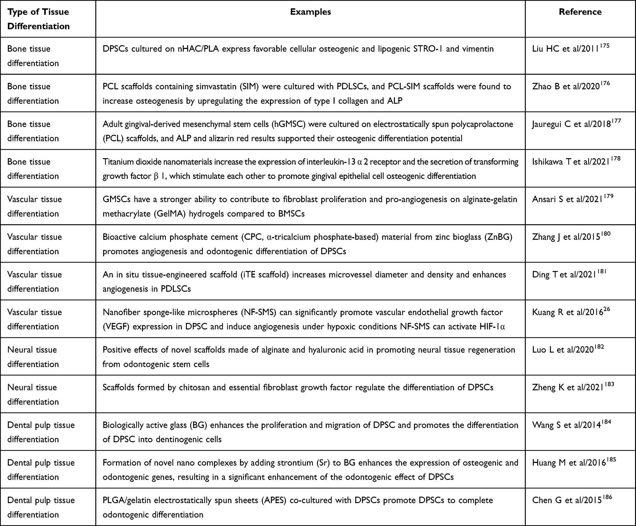 |
Table 3 Study on DMSCs in Tissue Differentiation |
Bone Tissue Differentiation
Bone regeneration is one of the most important means of restoring facial aesthetics and regaining oral function, which often requires regenerative materials with excellent biocompatibility and osteoblast inducibility. Based on the primitive osteogenic ability of cells such as PDLSCs and DPSCs, nanomaterials such as collagen-based nanohybrids (nHAC), PLA, and graphene have a solid potential to enhance osteogenic differentiation. Liu HC et al found that DPSCs cultured on nHAC/PLA expressed STRO-1 and vimentin, which are favorable for cellular osteogenesis and lipogenesis, and that the additional addition of recombinant human bone morphogenetic protein 2 (rhBMP-2), it was more able to increase autologous bone formation by rising gene expression.175 As a widely used scaffold material, PCL also has a more significant effect on PDLSCs; Zhao B et al cultured PDLSCs on PCL scaffolds containing simvastatin (SIM) and found that PCL-SIM scaffolds increased osteogenesis by upregulating the expression of COL-1 and ALP.176 Jauregui C et al decided to spin PCL scaffolds in the cultured adult GMSCs electrostatically, and the ALP and alizarin red results supported their osteogenic differentiation potential.177
In addition, Ishikawa T et al probed the osteogenic differentiation potential of gingival epithelial cells with titanium dioxide nanomaterials. They showed that this resulted in a rise in the expression of interleukin-13 α 2 receptor and an increase in the secretion of TGF-β1. The two stimulated each other, promoting gingival epithelial cell osteogenic differentiation.178
Vascular Tissue Differentiation
Vascular regeneration has a more critical position in soft tissue reconstruction; a rich blood supply provides nutrition to promote repair and helps prevent all kinds of complications due to nutritional disorders. Many biomaterials have been widely studied in the field of vasculogenesis. Ansari S et al developed an alginate-gelatin methacrylate (GelMA) hydrogel, which, compared to BMSCs, gingival mesenchymal stem cells (GMSCs) promoting TGF-β1, primary fibroblast growth factor (bFGF), and VEGF, with a more vital ability to contribute to fibroblast proliferation and pro-angiogenesis.179 Zhang J et al, on the other hand, developed a bioactive calcium phosphate cement (CPC, α-tricalcium phosphate-based) material incorporating zinc bioglass (ZnBG), and the results showed that the material, through the up-regulation of integrins and activating signaling pathways such as Wnt, MAPK, and NF-κB, thereby promoting angiogenesis and odontogenic differentiation of DPSCs.180 Ding T et al prepared core-shelled fiber scaffolds releasing bFGF and BMP-2 enhanced angiogenesis of PDLSCs, macrophage polarization, and enhanced osteogenic differentiation of PDLSCs through immune mechanisms.181 Furthermore, Kuang R et al assembled nanofiber spongy microspheres (NF-SMS). They found that under hypoxic conditions, NF-SMS activated HIF-1α, significantly promoted VEGF expression in DPSCs, and induced angiogenesis, possibly due to cell-cell interactions.27
Neural Tissue Differentiation
As a popular nanomaterial for influencing the neural differentiation of stem cells, hydrogel-like materials have been shown to affect the processes of growth, proliferation, and differentiation of a wide range of cells, including spinal cord neural stem cells,187 macrophages,188 and human neuroblastoma cells.189 While Luo L et al showed that a novel material consisting of hydrogel, recombinant human essential fibroblast growth factor, and DPSCs filled with cellulose was able to effectively promote the proliferation of DPSCs, and the anti-human neural-specific markers of GFAP, β-microtubulin III, S-100, and MBP were all highly expressed;182 Ansari S et al simultaneously applied alginate and hyaluronic acid at the same time to make a novel scaffold, which also had an effect on the proliferative activity of PDLSCs, and the expression of β-microtubulin III and GFAP was increased under the impact of continuously released β-nerve growth factor (β-NGF), confirming the positive effect of hydrogel in promoting the regeneration of neural tissues of odontogenic stem cells;179 in addition to the above materials, Zheng K et al have also explored the effect of scaffolds formed by chitosan and primary FGF on DPSCs; in their study, cell viability was unaffected, and the expression levels of the neural differentiation markers, GFAP, central neural specific Protein (S1-00β), and β-microtubulin III were significantly increased, demonstrating that this material can also regulate the differentiation process of DPSCs.183
Pulp Tissue Differentiation
Regeneration and repair of dental pulp have always been a challenge for treating endodontic diseases, and the excellent prospects of stem cells and nano-bioactive materials bring new hope for this challenge. Wang S et al explored the effect of bioactive glass (BG) on DPSCs, and they found that under the influence of BG, the proliferation and migration ability of DPSCs was enhanced, and the newborn dentin was visible, which indicated that BG promoted the differentiation of DPSCs into dentin-forming cells;184 in addition, Huang M et al added Sr to BG to form a novel nano-complex, which was able to enhance the expression of osteogenic genes, such as RUNX-2, OCN, MEPE, BMP-2, and ON, as well as the face of DSPP and DMP-1 dentin-forming genes, which undoubtedly indicated that, under the condition, the DPSCs The tooth-forming effect of DPSCs was significantly enhanced;185 Xia K et al developed The Arginine - Glycine - Aspartic acid (Arg-Gly-Asp, RGD) and VEGF-assembled peptide that was able to provide 3D scaffolds for DPSCs, upregulate the levels of their osteogenic and tooth-forming genes, such as ALP, DMP14, and DSPP, and was able to significantly increase the proportion of coronal dentin in a rat molar model, which suggests that this material has a positive effect on dentin differentiation;109 Chen G et al on the other hand, fabricated a PLGA/gelatin electrostatically spun sheet (APES), and by co-culturing it with DPSCs, demonstrated that DPSCs secreted positive ECM proteins COL-1, collagen type III (Col-3), fibronectin (FN), and Laminin (LN), upregulated the odontogenic markers DMP-1, DSPP, and VEGF, and markedly down-regulated from the early osteogenic markers ALP, BSP, and RUNX-2, which suggests that DPSCs are able to accomplish odontogenic differentiation in the presence of this material.186 In the field of endodontic differentiation, nano complexes composed of BGs have become an emerging material as a direction of the investigation.
Summary and Prospects
In 1959, American physicist Richard Feynman, “the father of modern nanotechnology”, put forward the concept of nanotechnology, nanotechnology, and nanomaterials began to get attention, but also in 1990, after the rapid development, in recent years, due to the complexity of tissue repair in the oral and maxillofacial tissues and susceptibility to infection, nano-bioactive materials have also been used more and more widely.190,191 As seen previously, at the nanoscale, various biomaterials exhibit cell-inducing properties that are not visible from a macroscopic perspective. For example, n-HAp can have osteogenic/dental differentiation effects on DPSCs through pro-gene expression and modulation of oxidative stress. In contrast, graphene oxide can have different inducing effects on DPSCs due to changes in its surface morphology. And n-HAp can be used as a cell-inducing agent for DPSCs: adhesion, proliferation, and osteogenic differentiation of PDLSCs. In comparison with BMSCs, although DPSCs were stronger than BMSCs in terms of cell proliferation, growth activity, and availability, ALP activity and osteogenic gene expression showed that their osteogenic differentiation potential was weaker than that of BMSCs.192 Therefore, utilizing nano-bioactive materials is indispensable if the restorative capacity of tooth-derived MSCs is to be fully demonstrated.
The adverse effects of these materials cannot be ignored to ensure the safety and efficacy of nanomaterials used in clinical applications for patients. In the comparison between dental composite nanomaterials and traditional materials, although the former has a higher modulus of elasticity, the defects of its mechanical properties cannot be ignored, which may be due to the unbalanced structure and properties of the material.193 In the process of material-cell interaction, the toxic response caused by material-induced production of biotoxic substances or disruption of the cytoskeletal network is also an essential factor affecting the repair of the organism.194 Among them, high levels of endogenously/exogenously generated ROS often evoke the cellular autophagy pathway, and it has been shown that TGF-β induces senescence in PDLSCs by increasing ROS production.195 Based on this, Li Y et al developed a conductive alginate/gelatin (AG) scaffold doped with graphene oxide (PGO) and hydroxyapatite nanoparticles (PHA) with antioxidant properties and flow cytometry demonstrated that ROS scavenging of PGO-PHA-AG scaffolds could protect the cells from damage.196 Several studies have found that the graphene family mostly has low cytotoxicity and is dose-dependent and time-dependent; some reported that GO has no toxic effect on cell behavior, but others found that the surface charge and lateral size of GO are responsible for cytotoxicity.197,198 In addition to possible cellular toxicity, nanomaterials have some potential risk in inducing and influencing disease development, with some studies suggesting that engineered nanoparticles (NPs) may accelerate the progression of asthma through mechanisms such as altered oxidative stress, activation or inhibition of inflammatory vesicles, and interactions with antigen-presenting cells, and exacerbate such effects when co-exposed with other risk factors.199 Despite these drawbacks, nano bioactive materials have a place in oral and maxillofacial prosthetics due to their unique advantages. However, as a practical matter, the high cost of nanomaterials and the long production cycle are challenges that need to be overcome in social medicine.200 In addition, the precision and stability of the produced materials are also a significant concern for most patients.
In recent years, the application of extracellular vesicles (EVs) in stem cell-based therapeutic approaches in dentistry has also become increasingly widespread.EVs are phospholipid bilayer-encapsulated particles that can be secreted and released by almost all types of cells and are mainly classified into exosomes and microvesicles (MVs).EVs are essential information carriers and are often used as biomarkers for diagnosing, prognosis, and treating disease.201 Sundaram et al developed ginger-derived nanovesicles (GiNVs) that have remarkable stability and can be used as therapeutic agents to ameliorate or prevent chronic periodontal disease, target Porphyromonas gingivalis, and minimize the occurrence of bone loss and inflammation.202 Yin B et al investigated mesenchymal stem cells or platelet-rich plasma-derived EVs (MSC-Evs or PRP-Evs). They found that both could inhibit the inflammatory microenvironment and reduce chondrocyte apoptosis.203 Nanovesicles have unique advantages over nanocomposite scaffolds.AuNPs are a commonly used nanomaterial. However, most lack in vivo tumor specificity, whereas ncRNA-enriched EVs can carry drugs or nanoparticles to improve specificity and significantly enhance their efficacy.204 For example, for protein molecules, autologous protein cargoes of EVs can be transferred to recipient cells to induce various cellular functions.205 While aging impacts endodontic physiological changes, Iezzi I et al found that miRNAs and exosomes derived from endodontic stem cells constitute a significant nanovesicle source that can treat age-related dental lesions.206 In addition, exosomes have an enormous potential for application in wound healing and scar attenuation. Zhao W et al used exosomes derived from mesenchymal stem cells (MSC-Exo) to down-regulate the expression of SIRT1 and inhibit the biological behaviors of fibroblasts, thereby directly or indirectly modulating the immune response during pathologic scarring, which can be applied to skin scarring, organ fibrosis, and other related diseases.207 However, exosome production, isolation, and utilization efficiency remain challenging. Adopting strategies to enhance exosome production and activity is urgent in the current biomedical field.
The use of living organisms to create inorganic nanoscale particles is a potential new development in biotechnology, and green nanomaterials have emerged for the rational utilization of biological resources. Studies have shown that green nanomaterials are particularly suitable for drug and DNA delivery, eg, cellulose, a readily available biomolecule, is often used as a green alternative to chemical nonviral gene delivery systems.208
The goal of reconstructive surgery is to restore form and function, and commonly used methods such as microvascular free flap transfers often result in severe pain, sensory nerve disorders, or concomitant scar formation. On the other hand, tissue engineering techniques based on stem cell research can promote the regeneration of hard and soft tissues and induce the reconstruction of defective areas, promising an alternative to current surgical repair techniques. Cellular scaffolds have their unique biodegradability, which can provide space for the growth of cells in the surrounding tissues and guide tissue regeneration and repair by inducing osteogenic, odontogenic, and angiogenic differentiation of cells and promoting cell proliferation.209 Multifunctionality is a prominent advantage of nanocomposites compared to existing conventional materials, and multiple functions such as targeted ligands, drug therapy, infection prevention, and imaging markers can be concentrated in a single entity, which together participates in the diagnosis and treatment of diseases. Several studies have shown that nanomaterials can be used as intrinsic antimicrobial agents (eg, AgNPs and ZnO NPs) in the promotion of wound healing and as nanocarriers for therapeutic agents to help wound healing (eg, combining antibiotics, NO), and nanomaterial scaffolds are even more widely used by virtue of their unique physical and structural properties. Nanomaterials-based wound-healing growth factors can regulate cell growth, differentiation, and migration and play an essential role in tissue repair.210 Nowadays, nanomaterials have a wide range of applications as potential solutions for regenerative medicine in inducing mesenchymal stem cells to promote tissue differentiation. They have also gained more attention in the field of drug delivery. Therefore, this paper categorizes and summarizes the specific regulatory functions of different nano-bioactive materials and the research mechanisms on the basis of the existing studies to provide a theoretical basis for the research and development of new nano-scaffold materials.211,212
As more and more nano bioactive materials are being investigated for their physicochemical properties, various types of drugs and techniques are combined for disease treatment. Currently, hard tissues of the oral and maxillofacial region are mainly repaired by resetting and bone grafting. In contrast, soft tissues depend more on the body’s healing and tissue grafting, which in most cases can only partially restore the morphology and function of the defective area. The demand for aesthetics is forcing researchers to look for a new direction. The use of bioactive materials and stem cell transplantation in combination to induce tissue repair in traumatized and pathologically damaged tissues has become an essential tool for future surgical repair,213,214 and the influence of immune response and stem cell activity has made it a hot topic to consider the coexistence of bioactive materials and stem cells. In the future, with the development of modern nanoengineering technology, innovative tissue reconstruction protocols and transplantation techniques will continue to emerge into a new era of nanomaterials research.
Author Contributions
Xingrui Li and Yue Wang are co first authors, Yao Xiao and Jie Lei are the corresponding authors of this article. All authors made a significant contribution to the work reported, whether that is in the conception, study design, execution, acquisition of data, analysis and interpretation, or in all these areas; took part in drafting, revising or critically reviewing the article; gave final approval of the version to be published; have agreed on the journal to which the article has been submitted; and agree to be accountable for all aspects of the work.
Funding
This publication of this review paper was supported by Sichuan Province Technology and Innovation Seedling Program (grant number 2021032), the Applied Basic Research of Luzhou Science and Technology Talent Bureau (grant number 2020-JYJ-40), and the National Undergraduate Innovation and Entrepreneurship Training Program of the Ministry of Education (grant number 202210632265), and Undergraduate Research Training at Southwestern Medical University School of Dentistry (grant numbe2022URTP02).
Disclosure
The authors declare that the research was conducted without any commercial or financial relationships that could be construed as a potential conflict of interest.
References
1. Zhang Q, Wu W, Qian C, et al. Advanced biomaterials for repairing and reconstruction of mandibular defects. Mater Sci Eng C Mater Biol Appl. 2019;103:109858. doi:10.1016/j.msec.2019.109858
2. Zhao Z, Tao Y, Xiang X, Liang Z, Zhao Y. Identification and Validation of a Novel Model: predicting Short-Term Complications After Local Flap Surgery for Skin Tumor Removal. Med Sci Monit. 2022;28:e938002. doi:10.12659/MSM.938002
3. Liu P, Zhang Y, Ma Y, et al. Application of dental pulp stem cells in oral maxillofacial tissue engineering. Int J Med Sci. 2022;19(2):310–320. doi:10.7150/ijms.68494
4. Liu Y, Sun X, Yu J, et al. Platelet-Rich Fibrin as a Bone Graft Material in Oral and Maxillofacial Bone Regeneration: classification and Summary for Better Application. Biomed Res Int. 2019;2019:3295756. doi:10.1155/2019/3295756
5. Pei Z, Lei H, Cheng L. Bioactive inorganic nanomaterials for cancer theranostics. Chem Soc Rev. 2023;352. doi:10.1039/d2cs00352j
6. Bramhill J, Ross S, Ross G. Bioactive Nanocomposites for Tissue Repair and Regeneration: a Review. Int J Environ Res Public Health. 2017;14(1):66. doi:10.3390/ijerph14010066
7. Masne N, Ambade R, Bhugaonkar K. Use of Nanocomposites in Bone Regeneration. Cureus. 2022;14(11):e31346. doi:10.7759/cureus.31346
8. Rahman SU, Nagrath M, Ponnusamy S, Arany PR. Nanoscale and Macroscale Scaffolds with Controlled-Release Polymeric Systems for Dental Craniomaxillofacial Tissue Engineering. Materials. 2018;11(8):1478. doi:10.3390/ma11081478
9. Gronthos S, Mankani M, Brahim J, Robey PG, Shi S. Postnatal human dental pulp stem cells (DPSCs) in vitro and in vivo. Proc Natl Acad Sci U S A. 2000;97(25):13625–13630. doi:10.1073/pnas.240309797
10. Gronthos S, Zhao M, Lu B, Fisher LW, Robey PG, Shi S. SHED: stem cells from human exfoliated deciduous teeth. Proc Natl Acad Sci U S A. 2003;100(10):5807–5812. doi:10.1073/pnas.0937635100
11. Seo BM, Miura M, Gronthos S, et al. Investigation of multipotent postnatal stem cells from human periodontal ligament. Lancet. 2004;364(9429):149–155. doi:10.1016/S0140-6736(04)16627-0
12. Liu Y, Yamaza T, Tuan RS, Wang S, Shi S, Huang GT. Characterization of the apical papilla and its residing stem cells from human immature permanent teeth: a pilot study. J Endod. 2008;34(2):166–171. doi:10.1016/j.joen.2007.11.021
13. Lin X, Li Q, Hu L, Jiang C, Wang S, Wu X. Apical Papilla Regulates Dental Follicle Fate via the OGN-Hh Pathway. J Dent Res. 2022;14:220345221138517. doi:10.1177/00220345221138517
14. Kim BC, Bae H, Kwon IK, et al. Osteoblastic/cementoblastic and neural differentiation of dental stem cells and their applications to tissue engineering and regenerative medicine. Tissue Eng Part B Rev. 2012;18(3):235–244. doi:10.1089/ten.TEB.2011.0642
15. Zhang C, Zhang Y, Feng Z, et al. Therapeutic effect of dental pulp stem cell transplantation on a rat model of radioactivity-induced esophageal injury. Cell Death Dis. 2018;9(7):738. doi:10.1038/s41419-018-0753-0
16. Raza SS, Wagner AP, Hussain YS, Khan MA. Mechanisms underlying dental-derived stem cell-mediated neurorestoration in neurodegenerative disorders. Stem Cell Res Ther. 2018;9(1):245. doi:10.1186/s13287-018-1005-z
17. Yamada Y, Nakamura-Yamada S, Kusano K, Baba S. Clinical Potential and Current Progress of Dental Pulp Stem Cells for Various Systemic Diseases in Regenerative Medicine: a Concise Review. Int J Mol Sci. 2019;20(5):1132. doi:10.3390/ijms20051132
18. Hata M, Omi M, Kobayashi Y, et al. Transplantation of cultured dental pulp stem cells into the skeletal muscles ameliorated diabetic polyneuropathy: therapeutic plausibility of freshly isolated and cryopreserved dental pulp stem cells. Stem Cell Res Ther. 2015;6(1):162. doi:10.1186/s13287-015-0156-4
19. Papaccio G, Graziano A, d’Aquino R, et al. Long-term cryopreservation of dental pulp stem cells (SBP-DPSCs) and their differentiated osteoblasts: a cell source for tissue repair. J Cell Physiol. 2006;208(2):319–325. doi:10.1002/jcp.20667
20. Liang C, Liang Q, Xu X, et al. Bone morphogenetic protein 7 mediates stem cells migration and angiogenesis: therapeutic potential for endogenous pulp regeneration. Int J Oral Sci. 2022;14(1):38. doi:10.1038/s41368-022-00188-y
21. Zhang Z, Oh M, Sasaki JI, Nör JE. Inverse and reciprocal regulation of p53/p21 and Bmi-1 modulates vasculogenic differentiation of dental pulp stem cells. Cell Death Dis. 2021;12(7):644. doi:10.1038/s41419-021-03925-z
22. Wang J, Liu X, Jin X, et al. The odontogenic differentiation of human dental pulp stem cells on nanofibrous poly(L-lactic acid) scaffolds in vitro and in vivo. Acta Biomater. 2010;6(10):3856–3863. doi:10.1016/j.actbio.2010.04.009
23. Zheng C, Chen J, Liu S, Jin Y. Stem cell-based bone and dental regeneration: a view of microenvironmental modulation. Int J Oral Sci. 2019;11(3):23. doi:10.1038/s41368-019-0060-3
24. Zhang X, Li H, Sun J, et al. Cell-derived micro-environment helps dental pulp stem cells promote dental pulp regeneration. Cell Prolif. 2017;50(5):e12361. doi:10.1111/cpr.12361
25. Ma W, Zhan Y, Zhang Y, Mao C, Xie X, Lin Y. The biological applications of DNA nanomaterials: current challenges and future directions. Signal Transduct Target Ther. 2021;6(1):351. doi:10.1038/s41392-021-00727-9
26. Liang K, Wang S, Tao S, et al. Dental remineralization via poly(amido amine) and restorative materials containing calcium phosphate nanoparticles. Int J Oral Sci. 2019;11(2):15. doi:10.1038/s41368-019-0048-z
27. Kuang R, Zhang Z, Jin X, et al. Nanofibrous spongy microspheres for the delivery of hypoxia-primed human dental pulp stem cells to regenerate vascularized dental pulp. Acta Biomater. 2016;33:225–234. doi:10.1016/j.actbio.2016.01.032
28. Tahriri M, Del Monico M, Moghanian A, et al. Graphene and its derivatives: opportunities and challenges in dentistry. Mater Sci Eng C Mater Biol Appl. 2019;102:171–185. doi:10.1016/j.msec.2019.04.051
29. Bachhuka A, Delalat B, Ghaemi SR, Gronthos S, Voelcker NH, Vasilev K. Nanotopography mediated osteogenic differentiation of human dental pulp derived stem cells. Nanoscale. 2017;9(37):14248–14258. doi:10.1039/c7nr03131a
30. Yang Y, Zhang H, Komasa S, et al. Immunomodulatory Properties and Osteogenic Activity of Polyetheretherketone Coated with Titanate Nanonetwork Structures. Int J Mol Sci. 2022;23(2):612. doi:10.3390/ijms23020612
31. Ayadilord M, Nasseri S, Emadian Razavi F, Saharkhiz M, Rostami Z, Naseri M. Immunomodulatory effects of phytosomal curcumin on related-micro RNAs, CD200 expression and inflammatory pathways in dental pulp stem cells. Cell Biochem Funct. 2021;39(7):886–895. doi:10.1002/cbf.3659
32. Yin IX, Zhao IS, Mei ML, Li Q, Yu OY, Chu CH. Use of Silver Nanomaterials for Caries Prevention: a Concise Review. Int J Nanomedicine. 2020;15:3181–3191. doi:10.2147/IJN.S253833
33. Santos VE, Vasconcelos Filho A, Targino AG, et al. A new “silver-bullet” to treat caries in children--nano silver fluoride: a randomised clinical trial. J Dent. 2014;42(8):945–951. doi:10.1016/j.jdent.2014.05.017
34. Angel Villegas N, Silvero Compagnucci MJ, Sainz Ajá M, et al. Novel Antibacterial Resin-Based Filling Material Containing Nanoparticles for the Potential One-Step Treatment of Caries. J Healthc Eng. 2019;2019:6367919. doi:10.1155/2019/6367919
35. Ioannidis K, Niazi S, Mylonas P, Mannocci F, Deb S. The synthesis of nano silver-graphene oxide system and its efficacy against endodontic biofilms using a novel tooth model. Dent Mater. 2019;35(11):1614–1629. doi:10.1016/j.dental.2019.08.105
36. Razumova S, Brago A, Serebrov D, et al. The Application of Nano Silver Argitos as a Final Root Canal Irrigation for the Treatment of Pulpitis and Apical Periodontitis. In Vitro Study. Nanomaterials. 2022;12(2):248. doi:10.3390/nano12020248
37. Liu C, Hao Z, Yang T, Wang F, Sun F, Teng W. Anti-Acid Biomimetic Dentine Remineralization Using Inorganic Silica Stabilized Nanoparticles Distributed Electronspun Nanofibrous Mats. Int J Nanomedicine. 2021;16:8251–8264. doi:10.2147/IJN.S331321
38. Totu EE, Isildak I, Nechifor AC, Cristache CM, Enachescu M. New sensor based on membranes with magnetic nano-inclusions for early diagnosis in periodontal disease. Biosens Bioelectron. 2018;102:336–344. doi:10.1016/j.bios.2017.11.003
39. Zhang Y, Gulati K, Li Z, Di P, Liu Y. Dental Implant Nano-Engineering: advances, Limitations and Future Directions. Nanomaterials. 2021;11(10):2489. doi:10.3390/nano11102489
40. Chalisserry EP, Nam SY, Park SH, Anil S. Therapeutic potential of dental stem cells. J Tissue Eng. 2017;8. doi:10.1177/2041731417702531
41. Zhang W, Vazquez B, Oreadi D, Yelick PC. Decellularized Tooth Bud Scaffolds for Tooth Regeneration. J Dent Res. 2017;96(5):516–523. doi:10.1177/0022034516689082
42. Hu L, Gao Z, Xu J, et al. Decellularized Swine Dental Pulp as a Bioscaffold for Pulp Regeneration. Biomed Res Int. 2017;2017:9342714. doi:10.1155/2017/9342714
43. Xiong H, Zhao F, Peng Y, Li M, Qiu H, Chen K. Easily attainable and low immunogenic stem cells from exfoliated deciduous teeth enhanced the in vivo bone regeneration ability of gelatin/bioactive glass microsphere composite scaffolds. Front Bioeng Biotechnol. 2022;10:1049626. doi:10.3389/fbioe.2022.1049626
44. Hagar MN, Yazid F, Luchman NA, Ariffin SHZ, Wahab RMA. Comparative evaluation of osteogenic differentiation potential of stem cells derived from dental pulp and exfoliated deciduous teeth cultured over granular hydroxyapatite based scaffold. BMC Oral Health. 2021;21(1):263. doi:10.1186/s12903-021-01621-0
45. Zhang Y, Wang P, Wang Y, et al. Gold Nanoparticles Promote the Bone Regeneration of Periodontal Ligament Stem Cell Sheets Through Activation of Autophagy. Int J Nanomedicine. 2021;16:61–73. doi:10.2147/IJN.S282246
46. Mohebichamkhorami F, Fattahi R, Niknam Z, et al. Periodontal ligament stem cells as a promising therapeutic target for neural damage. Stem Cell Res Ther. 2022;13(1):273. doi:10.1186/s13287-022-02942-9
47. Kwack KH, Ji JY, Park B, Heo JS. Fucoidan (Undaria pinnatifida)/Polydopamine Composite-Modified Surface Promotes Osteogenic Potential of Periodontal Ligament Stem Cells. Mar Drugs. 2022;20(3):181. doi:10.3390/md20030181
48. Cui Y, Xie J, Fu Y, et al. Berberine mediates root remodeling in an immature tooth with apical periodontitis by regulating stem cells from apical papilla differentiation. Int J Oral Sci. 2020;12(1):18. doi:10.1038/s41368-020-0085-7
49. Liu J, Zou T, Zhang Y, et al. Three-dimensional electroconductive carbon nanotube-based hydrogel scaffolds enhance neural differentiation of stem cells from apical papilla. Biomater Adv. 2022;138:212868. doi:10.1016/j.bioadv.2022.212868
50. Bok JS, Byun SH, Park BW, et al. The Role of Human Umbilical Vein Endothelial Cells in Osteogenic Differentiation of Dental Follicle-Derived Stem Cells in In Vitro Co-cultures. Int J Med Sci. 2018;15(11):1160–1170. doi:10.7150/ijms.27318
51. Aurrekoetxea M, Garcia-Gallastegui P, Irastorza I, et al. Dental pulp stem cells as a multifaceted tool for bioengineering and the regeneration of craniomaxillofacial tissues. Front Physiol. 2015;6:289. doi:10.3389/fphys.2015.00289
52. Ranganathan K, Lakshminarayanan V. Stem cells of the dental pulp. Indian J Dent Res. 2012;23:558.
53. Tsutsui TW. Dental Pulp Stem Cells: advances to Applications. Stem Cells Cloning. 2020;13:33–42. doi:10.2147/SCCAA.S166759
54. Ogata K, Moriyama M, Matsumura-Kawashima M, Kawado T, Yano A, Nakamura S. The Therapeutic Potential of Secreted Factors from Dental Pulp Stem Cells for Various Diseases. Biomedicines. 2022;10(5):1049. doi:10.3390/biomedicines10051049
55. Karbanová J, Soukup T, Suchánek J, Pytlík R, Corbeil D, Mokrý J. Characterization of dental pulp stem cells from impacted third molars cultured in low serum-containing medium. Cells Tissues Organs. 2011;193(6):344–365. doi:10.1159/000321160
56. Mead B, Logan A, Berry M, Leadbeater W, Scheven BA. Intravitreally transplanted dental pulp stem cells promote neuroprotection and axon regeneration of retinal ganglion cells after optic nerve injury. Invest Ophthalmol Vis Sci. 2013;54(12):7544–7556. doi:10.1167/iovs.13-13045
57. Miura M, Gronthos S, Zhao M, et al. SHED: stem cells from human exfoliated deciduous teeth. Proc Natl Acad Sci U S A. 2003;100(10):5807–5812. doi:10.1073/pnas.0937635100
58. Shi X, Mao J, Liu Y. Pulp stem cells derived from human permanent and deciduous teeth: biological characteristics and therapeutic applications. Stem Cells Transl Med. 2020;9(4):445–464. doi:10.1002/sctm.19-0398
59. Zainuri M, Purba J, Wa Jusman S, Bachtiar EW. Conditioned-medium of stem cells from human exfoliated deciduous teeth prevent apoptosis of neural progenitors. Saudi Dent J. 2022;34(7):565–571. doi:10.1016/j.sdentj.2022.08.005
60. Pereira LV, Bento RF, Cruz DB, et al. Stem Cells from Human Exfoliated Deciduous Teeth (SHED) Differentiate in vivo and Promote Facial Nerve Regeneration. Cell Transplant. 2019;28(1):55–64. doi:10.1177/0963689718809090
61. Naz S, Khan FR, Khan I, et al. Comparative analysis of dental pulp stem cells and stem cells from human exfoliated teeth in terms of growth kinetics, immunophenotype, self-renewal and multi lineage differentiation potential for future perspective of calcified tissue regeneration. Pak J Med Sci. 2022;38(5):1228–1237. doi:10.12669/pjms.38.5.5187
62. Kunimatsu R, Nakajima K, Awada T, et al. Comparative characterization of stem cells from human exfoliated deciduous teeth, dental pulp, and bone marrow-derived mesenchymal stem cells. Biochem Biophys Res Commun. 2018;501(1):193–198. doi:10.1016/j.bbrc.2018.04.213
63. Yang Z, Wang C, Zhang X, et al. Stem cells from human exfoliated deciduous teeth attenuate trigeminal neuralgia in rats by inhibiting endoplasmic reticulum stress. Korean J Pain. 2022;35(4):383–390. doi:10.3344/kjp.2022.35.4.383
64. Bai X, Zhang X, Wang C, et al. Stem Cells from Human Exfoliated Deciduous Teeth Attenuate Trigeminal Neuralgia in Rats. Stem Cells Int. 2021;2021:8819884. doi:10.1155/2021/8819884
65. Arora V, Arora P, Munshi AK. Banking stem cells from human exfoliated deciduous teeth (SHED): saving for the future. J Clin Pediatr Dent. 2009;33(4):289–294. doi:10.17796/jcpd.33.4.y887672r0j703654
66. Zhao Y, Shi Y, Yang H, et al. Stem cell microencapsulation maintains stemness in inflammatory microenvironment. Int J Oral Sci. 2022;14(1):48. doi:10.1038/s41368-022-00198-w
67. Li X, Zhao Y, Peng H, et al. Robust intervention for oxidative stress-induced injury in periodontitis via controllably released nanoparticles that regulate the ROS-PINK1-Parkin pathway. Front Bioeng Biotechnol. 2022;10:1081977. doi:10.3389/fbioe.2022.1081977
68. Liu T, Hu W, Zou X, et al. Human Periodontal Ligament Stem Cell-Derived Exosomes Promote Bone Regeneration by Altering MicroRNA Profiles. Stem Cells Int. 2020;2020:8852307. doi:10.1155/2020/8852307
69. Liu M, Chen R, Xu Y, Zheng J, Wang M, Wang P. Exosomal miR-141-3p from PDLSCs Alleviates High Glucose-Induced Senescence of PDLSCs by Activating the KEAP1-NRF2 Signaling Pathway. Stem Cells Int. 2023;2023:7136819. doi:10.1155/2023/7136819
70. Gao H, Li B, Zhao L, Jin Y. Influence of nanotopography on periodontal ligament stem cell functions and cell sheet based periodontal regeneration. Int J Nanomedicine. 2015;10:4009–4027. doi:10.2147/IJN.S83357
71. Liang Y, Shakya A, Liu X. Biomimetic Tubular Matrix Induces Periodontal Ligament Principal Fiber Formation and Inhibits Osteogenic Differentiation of Periodontal Ligament Stem Cells. ACS Appl Mater Interfaces. 2022;14(32):36451–36461. doi:10.1021/acsami.2c09420
72. Jia L, Li D, Wang YN, Zhang D, Xu X. PSAT1 positively regulates the osteogenic lineage differentiation of periodontal ligament stem cells through the ATF4/PSAT1/Akt/GSK3β/β-catenin axis. J Transl Med. 2023;21(1):70. doi:10.1186/s12967-022-03775-z
73. Huang GT, Yamaza T, Shea LD, et al. Stem/progenitor cell-mediated de novo regeneration of dental pulp with newly deposited continuous layer of dentin in an in vivo model. Tissue Eng Part A. 2010;16(2):605–615. doi:10.1089/ten.TEA.2009.0518
74. Bakopoulou A, Leyhausen G, Volk J, et al. Comparative analysis of in vitro osteo/odontogenic differentiation potential of human dental pulp stem cells (DPSCs) and stem cells from the apical papilla (SCAP). Arch Oral Biol. 2011;56(7):709–721. doi:10.1016/j.archoralbio.2010.12.008
75. Han C, Yang Z, Zhou W, et al. Periapical follicle stem cell: a promising candidate for cementum/periodontal ligament regeneration and bio-root engineering. Stem Cells Dev. 2010;19(9):1405–1415. doi:10.1089/scd.2009.0277
76. Liu J, Zou T, Yao Q, Zhang Y, Zhao Y, Zhang C. Hypoxia-mimicking cobalt-doped multi-walled carbon nanotube nanocomposites enhance the angiogenic capacity of stem cells from apical papilla. Mater Sci Eng C Mater Biol Appl. 2021;120:111797. doi:10.1016/j.msec.2020.111797
77. Wu X, Hu L, Li Y, et al. SCAPs Regulate Differentiation of DFSCs During Tooth Root Development in Swine. Int J Med Sci. 2018;15(4):291–299. doi:10.7150/ijms.22495
78. Hilkens P, Bronckaers A, Ratajczak J, Gervois P, Wolfs E, Lambrichts I. The Angiogenic Potential of DPSCs and SCAPs in an In Vivo Model of Dental Pulp Regeneration. Stem Cells Int. 2017;2017:2582080. doi:10.1155/2017/2582080
79. Rezai-Rad M, Bova JF, Orooji M, et al. Evaluation of bone regeneration potential of dental follicle stem cells for treatment of craniofacial defects. Cytotherapy. 2015;17(11):1572–1581. doi:10.1016/j.jcyt.2015.07.013
80. Hokmabad VR, Davaran S, Aghazadeh M, Rahbarghazi R, Salehi R, Ramazani A. Fabrication and characterization of novel ethyl cellulose-grafted-poly (ɛ-caprolactone)/alginate nanofibrous/macroporous scaffolds incorporated with nano-hydroxyapatite for bone tissue engineering. J Biomater Appl. 2019;33(8):1128–1144. doi:10.1177/0885328218822641
81. Alipour M, Firouzi N, Aghazadeh Z, et al. The osteogenic differentiation of human dental pulp stem cells in alginate-gelatin/Nano-hydroxyapatite microcapsules. BMC Biotechnol. 2021;21(1):6. doi:10.1186/s12896-020-00666-3
82. Ansari S, Diniz IM, Chen C, et al. Human Periodontal Ligament- and Gingiva-derived Mesenchymal Stem Cells Promote Nerve Regeneration When Encapsulated in Alginate/Hyaluronic Acid 3D Scaffold. Adv Healthc Mater. 2017;6(24):
83. Baskar K, Saravana Karthikeyan B, Gurucharan I, et al. Eggshell derived nano-hydroxyapatite incorporated carboxymethyl chitosan scaffold for dentine regeneration: a laboratory investigation. Int Endod J. 2022;55(1):89–102. doi:10.1111/iej.13644
84. Gurucharan I, Saravana Karthikeyan B, Mahalaxmi S, et al. Characterization of nano-hydroxyapatite incorporated carboxymethyl chitosan composite on human dental pulp stem cells. Int Endod J. 2022. doi:10.1111/iej.13885
85. Navidi G, Allahvirdinesbat M, Al-Molki SMM, et al. Design and fabrication of M-SAPO-34/chitosan scaffolds and evaluation of their effects on dental tissue engineering. Int J Biol Macromol. 2021;187:281–295. doi:10.1016/j.ijbiomac.2021.07.104
86. Su WT, Wu PS, Ko CS, Huang TY. Osteogenic differentiation and mineralization of human exfoliated deciduous teeth stem cells on modified chitosan scaffold. Mater Sci Eng C Mater Biol Appl. 2014;41:152–160. doi:10.1016/j.msec.2014.04.048
87. Hanafy AK, Shinaishin SF, Eldeen GN, Aly RM. Nano Hydroxyapatite & Mineral Trioxide Aggregate Efficiently Promote Odontogenic Differentiation of Dental Pulp Stem Cells. Open Access Maced J Med Sci. 2018;6(9):1727–1731. doi:10.3889/oamjms.2018.368
88. Sancilio S, Gallorini M, Di Nisio C, et al. Alginate/Hydroxyapatite-Based Nanocomposite Scaffolds for Bone Tissue Engineering Improve Dental Pulp Biomineralization and Differentiation. Stem Cells Int. 2018;2018:9643721. doi:10.1155/2018/9643721
89. Moonesi Rad R, Atila D, Evis Z, Keskin D, Tezcaner A. Development of a novel functionally graded membrane containing boron-modified bioactive glass nanoparticles for guided bone regeneration. J Tissue Eng Regen Med. 2019;13(8):1331–1345. doi:10.1002/term.2877
90. El-Fiqi A, Mandakhbayar N, Jo SB, Knowles JC, Lee JH, Kim HW. Nanotherapeutics for regeneration of degenerated tissue infected by bacteria through the multiple delivery of bioactive ions and growth factor with antibacterial/angiogenic and osteogenic/odontogenic capacity. Bioact Mater. 2020;6(1):123–136. doi:10.1016/j.bioactmat.2020.07.010
91. Ahn JH, Kim IR, Kim Y, et al. The Effect of Mesoporous Bioactive Glass Nanoparticles/Graphene Oxide Composites on the Differentiation and Mineralization of Human Dental Pulp Stem Cells. Nanomaterials. 2020;10(4):620. doi:10.3390/nano10040620
92. Khoroushi M, Foroughi MR, Karbasi S, Hashemibeni B, Khademi AA. Effect of Polyhydroxybutyrate/Chitosan/Bioglass nanofiber scaffold on proliferation and differentiation of stem cells from human exfoliated deciduous teeth into odontoblast-like cells. Mater Sci Eng C Mater Biol Appl. 2018;89:128–139. doi:10.1016/j.msec.2018.03.028
93. Hosseini FS, Enderami SE, Hadian A, et al. Efficient osteogenic differentiation of the dental pulp stem cells on β-glycerophosphate loaded polycaprolactone/polyethylene oxide blend nanofibers. J Cell Physiol. 2019;234(8):13951–13958. doi:10.1002/jcp.28078
94. Ma L, Yu Y, Liu H, et al. Berberine-releasing electrospun scaffold induces osteogenic differentiation of DPSCs and accelerates bone repair. Sci Rep. 2021;11(1):1027. doi:10.1038/s41598-020-79734-9
95. Alipour M, Aghazadeh M, Akbarzadeh A, Vafajoo Z, Aghazadeh Z, Raeisdasteh Hokmabad V. Towards osteogenic differentiation of human dental pulp stem cells on PCL-PEG-PCL/zeolite nanofibrous scaffolds. Artif Cells Nanomed Biotechnol. 2019;47(1):3431–3437. doi:10.1080/21691401.2019.1652627
96. Azaryan E, Hanafi-Bojd MY, Alemzadeh E, Emadian Razavi F, Naseri M. Effect of PCL/nHAEA nanocomposite to osteo/odontogenic differentiation of dental pulp stem cells. BMC Oral Health. 2022;22(1):505. doi:10.1186/s12903-022-02527-1
97. Prabha RD, Kraft DCE, Harkness L, et al. Bioactive nano-fibrous scaffold for vascularized craniofacial bone regeneration. J Tissue Eng Regen Med. 2018;12(3):e1537–e1548. doi:10.1002/term.2579
98. Su WT, Wu PS, Huang TY. Osteogenic differentiation of stem cells from human exfoliated deciduous teeth on poly(ε-caprolactone) nanofibers containing strontium phosphate. Mater Sci Eng C Mater Biol Appl. 2015;46:427–434. doi:10.1016/j.msec.2014.10.076
99. Zhou M, Liu NX, Shi SR, et al. Effect of tetrahedral DNA nanostructures on proliferation and osteo/odontogenic differentiation of dental pulp stem cells via activation of the notch signaling pathway. Nanomedicine. 2018;14(4):1227–1236. doi:10.1016/j.nano.2018.02.004
100. Zhou M, Liu N, Zhang Q, et al. Effect of tetrahedral DNA nanostructures on proliferation and osteogenic differentiation of human periodontal ligament stem cells. Cell Prolif. 2019;52(3):e12566. doi:10.1111/cpr.12566
101. Zhou M, Gao S, Zhang X, et al. The protective effect of tetrahedral framework nucleic acids on periodontium under inflammatory conditions. Bioact Mater. 2020;6(6):1676–1688. doi:10.1016/j.bioactmat.2020.11.018
102. Wang Y, Song W, Cui Y, Zhang Y, Mei S, Wang Q. Calcium-siRNA Nanocomplexes Optimized by Bovine Serum Albumin Coating Can Achieve Convenient and Efficient siRNA Delivery for Periodontitis Therapy. Int J Nanomedicine. 2020;15:9241–9253. doi:10.2147/IJN.S278103
103. Zhang C, Yan B, Cui Z, et al. Bone regeneration in minipigs by intrafibrillarly-mineralized collagen loaded with autologous periodontal ligament stem cells. Sci Rep. 2017;7(1):10519. doi:10.1038/s41598-017-11155-7
104. Liu Y, Li N, Qi YP, et al. Intrafibrillar collagen mineralization produced by biomimetic hierarchical nanoapatite assembly. Adv Mater. 2011;23(8):975–980. doi:10.1002/adma.201003882
105. Chen WY, Li X, Feng Y, Lin S, Peng L, Huang D. M-keratin nano-materials create a mineralized micro-circumstance to promote proliferation and differentiation of DPSCs. J Mater Sci Mater Med. 2020;31(12):124. doi:10.1007/s10856-020-06465-8
106. Radunovic M, De Colli M, De Marco P, et al. Graphene oxide enrichment of collagen membranes improves DPSCs differentiation and controls inflammation occurrence. J Biomed Mater Res A. 2017;105(8):2312–2320. doi:10.1002/jbm.a.36085
107. Kumar A, Kumar V, Rattan V, Jha V, Bhattacharyya S. Secretome Cues Modulate the Neurogenic Potential of Bone Marrow and Dental Stem Cells. Mol Neurobiol. 2017;54(6):4672–4682. doi:10.1007/s12035-016-0011-3
108. Kumar A, Kumar V, Rattan V, Jha V, Bhattacharyya S. Secretome proteins regulate comparative osteogenic and adipogenic potential in bone marrow and dental stem cells. Biochimie. 2018;155:129–139. doi:10.1016/j.biochi.2018.10.014
109. Xia K, Chen Z, Chen J, et al. RGD- and VEGF-Mimetic Peptide Epitope-Functionalized Self-Assembling Peptide Hydrogels Promote Dentin-Pulp Complex Regeneration. Int J Nanomedicine. 2020;15:6631–6647. doi:10.2147/IJN.S253576
110. Nagy K, Láng O, Láng J, et al. A novel hydrogel scaffold for periodontal ligament stem cells. Interv Med Appl Sci. 2018;10(3):162–170. doi:10.1556/1646.10.2018.21
111. Jo BS, Lee Y, Suh JS, et al. A novel calcium-accumulating peptide/gelatin in situ forming hydrogel for enhanced bone regeneration. J Biomed Mater Res A. 2018;106(2):531–542. doi:10.1002/jbm.a.36257
112. Ruiz ON, Fernando KA, Wang B, et al. Graphene oxide: a nonspecific enhancer of cellular growth. ACS Nano. 2011;5(10):8100–8107. doi:10.1021/nn202699t
113. Di Carlo R, Zara S, Ventrella A, et al. Covalent Decoration of Cortical Membranes with Graphene Oxide as a Substrate for Dental Pulp Stem Cells. Nanomaterials. 2019;9(4):604. doi:10.3390/nano9040604
114. Simonovic J, Toljic B, Lazarevic M, et al. The Effect of Liquid-Phase Exfoliated Graphene Film on Neurodifferentiation of Stem Cells from Apical Papilla. Nanomaterials. 2022;12(18):3116. doi:10.3390/nano12183116
115. Simonovic J, Toljic B, Nikolic N, et al. Differentiation of stem cells from apical papilla into neural lineage using graphene dispersion and single walled carbon nanotubes. J Biomed Mater Res A. 2018;106(10):2653–2661. doi:10.1002/jbm.a.36461
116. Li Z, Qiu J, Du LQ, Jia L, Liu H, Ge S. TiO2 nanorod arrays modified Ti substrates promote the adhesion, proliferation and osteogenic differentiation of human periodontal ligament stem cells. Mater Sci Eng C Mater Biol Appl. 2017;76:684–691. doi:10.1016/j.msec.2017.03.148
117. Thanasrisuebwong P, Jones JR, Eiamboonsert S, Ruangsawasdi N, Jirajariyavej B, Naruphontjirakul P. Zinc-Containing Sol-Gel Glass Nanoparticles to Deliver Therapeutic Ions. Nanomaterials. 2022;12(10):1691. doi:10.3390/nano12101691
118. Lapidot S, Meirovitch S, Sharon S, Heyman A, Kaplan DL, Shoseyov O. Clues for biomimetics from natural composite materials. Nanomedicine. 2012;7(9):1409–1423. doi:10.2217/nnm.12.107
119. Torres FG, Troncoso OP, Pisani A, Gatto F, Bardi G. Natural Polysaccharide Nanomaterials: an Overview of Their Immunological Properties. Int J Mol Sci. 2019;20(20):5092. doi:10.3390/ijms20205092
120. Choukaife H, Seyam S, Alallam B, Doolaanea AA, Alfatama M. Current Advances in Chitosan Nanoparticles Based Oral Drug Delivery for Colorectal Cancer Treatment. Int J Nanomedicine. 2022;17:3933–3966. doi:10.2147/IJN.S375229
121. Pawar SN, Edgar KJ. Alginate derivatization: a review of chemistry, properties and applications. Biomaterials. 2012;33(11):3279–3305. doi:10.1016/j.biomaterials.2012.01.007
122. Rastogi P, Kandasubramanian B. Review of alginate-based hydrogel bioprinting for application in tissue engineering. Biofabrication. 2019;11(4):042001. doi:10.1088/1758-5090/ab331e
123. Shapiro L, Cohen S. Novel alginate sponges for cell culture and transplantation. Biomaterials. 1997;18(8):583–590. doi:10.1016/s0142-9612(96)00181-0
124. Sancilio S, Marsich E, Schweikl H, Cataldi A, Gallorini M. Redox Control of IL-6-Mediated Dental Pulp Stem-Cell Differentiation on Alginate/Hydroxyapatite Biocomposites for Bone Ingrowth. Nanomaterials. 2019;9(12):1656. doi:10.3390/nano9121656
125. Hussein H, Kishen A. Engineered Chitosan-based Nanoparticles Modulate Macrophage-Periodontal Ligament Fibroblast Interactions in Biofilm-mediated Inflammation. J Endod. 2021;47(9):1435–1444. doi:10.1016/j.joen.2021.06.017
126. Wang Z, Shen Y, Haapasalo M. Antimicrobial and Antibiofilm Properties of Bioceramic Materials in Endodontics. Materials. 2021;14(24):7594. doi:10.3390/ma14247594
127. Sanz JL, Rodríguez-Lozano FJ, Llena C, Sauro S, Forner L. Bioactivity of Bioceramic Materials Used in the Dentin-Pulp Complex Therapy: a Systematic Review. Materials. 2019;12(7):1015. doi:10.3390/ma12071015
128. Rajula MPB, Narayanan V, Venkatasubbu GD, Mani RC, Sujana A. Nano-hydroxyapatite: a Driving Force for Bone Tissue Engineering. J Pharm Bioallied Sci. 2021;13(Suppl 1):S11–S14. doi:10.4103/jpbs.JPBS_683_20
129. Molino G, Palmieri MC, Montalbano G, Fiorilli S, Vitale-Brovarone C. Biomimetic and mesoporous nano-hydroxyapatite for bone tissue application: a short review. Biomed Mater. 2020;15(2):022001. doi:10.1088/1748-605X/ab5f1a
130. Mo X, Zhang D, Liu K, Zhao X, Li X, Wang W. Nano-Hydroxyapatite Composite Scaffolds Loaded with Bioactive Factors and Drugs for Bone Tissue Engineering. Int J Mol Sci. 2023;24(2):1291. doi:10.3390/ijms24021291
131. Turco G, Marsich E, Bellomo F, et al. Alginate/Hydroxyapatite biocomposite for bone ingrowth: a trabecular structure with high and isotropic connectivity. Biomacromolecules. 2009;10:1575–1583. doi:10.1021/bm900154b
132. Brauer DS. Bioactive glasses—structure and properties. Angew Chem Int Ed Engl. 2015;54(14):4160–4181. doi:10.1002/anie.201405310
133. Choi Y, Sun W, Kim Y, et al. Effects of Zn-Doped Mesoporous Bioactive Glass Nanoparticles in Etch-and-Rinse Adhesive on the Microtensile Bond Strength. Nanomaterials. 2020;10(10):1943. doi:10.3390/nano10101943
134. Mamidi N, García RG, Martínez JDH, et al. Recent Advances in Designing Fibrous Biomaterials for the Domain of Biomedical, Clinical, and Environmental Applications. ACS Biomater Sci Eng. 2022;8(9):3690–3716. doi:10.1021/acsbiomaterials.2c00786
135. Coudane J, Nottelet B, Mouton J, Garric X, Van Den Berghe H. Poly(ε-caprolactone)-Based Graft Copolymers: synthesis Methods and Applications in the Biomedical Field: a Review. Molecules. 2022;27(21):7339. doi:10.3390/molecules27217339
136. Wesełucha-Birczyńska A, Kołodziej A, Świętek M, et al. Early Recognition of the PCL/Fibrous Carbon Nanocomposites Interaction with Osteoblast-like Cells by Raman Spectroscopy. Nanomaterials. 2021;11(11):2890. doi:10.3390/nano11112890
137. Qian Y, Song J, Zhao X, et al. 3D Fabrication with Integration Molding of a Graphene Oxide/Polycaprolactone Nanoscaffold for Neurite Regeneration and Angiogenesis. Adv Sci. 2018;5(4):1700499. doi:10.1002/advs.201700499
138. Lin Y, Zhang L, Liu NQ, et al. In vitro behavior of tendon stem/progenitor cells on bioactive electrospun nanofiber membranes for tendon-bone tissue engineering applications. Int J Nanomedicine. 2019;14:5831–5848. doi:10.2147/IJN.S210509
139. Mantecón-Oria M, Diban N, Berciano MT, et al. Hollow Fiber Membranes of PCL and PCL/Graphene as Scaffolds with Potential to Develop In Vitro Blood-Brain Barrier Models. Membranes. 2020;10(8):161. doi:10.3390/membranes10080161
140. Thomas V, Jagani S, Johnson K, et al. Electrospun bioactive nanocomposite scaffolds of polycaprolactone and nanohydroxyapatite for bone tissue engineering. J Nanosci Nanotechnol. 2006;6:487–493. doi:10.1166/jnn.2006.097
141. Wang W, Dang M, Zhang Z, et al. Dentin regeneration by stem cells of apical papilla on injectable nanofibrous microspheres and stimulated by controlled BMP-2 release. Acta Biomater. 2016;36:63–72. doi:10.1016/j.actbio.2016.03.015
142. Bharadwaz A, Jayasuriya AC. Recent trends in the application of widely used natural and synthetic polymer nanocomposites in bone tissue regeneration. Mater Sci Eng C Mater Biol Appl. 2020;110:110698. doi:10.1016/j.msec.2020.110698
143. Gagner JE, Kim W, Chaikof EL. Designing protein-based biomaterials for medical applications. Acta Biomater. 2014;10(4):1542–1557. doi:10.1016/j.actbio.2013.10.001
144. Shao X, Lin S, Peng Q, et al. Tetrahedral DNA Nanostructure: a Potential Promoter for Cartilage Tissue Regeneration via Regulating Chondrocyte Phenotype and Proliferation. Small. 2017;13(12). doi:10.1002/smll.201602770
145. Li Q, Zhao D, Shao X, et al. Aptamer-Modified Tetrahedral DNA Nanostructure for Tumor-Targeted Drug Delivery. ACS Appl Mater Interfaces. 2017;9(42):36695–36701. doi:10.1021/acsami.7b13328
146. Zhang Q, Lin S, Shi S, et al. Anti-inflammatory and Antioxidative Effects of Tetrahedral DNA Nanostructures via the Modulation of Macrophage Responses. ACS Appl Mater Interfaces. 2018;10(4):3421–3430. doi:10.1021/acsami.7b17928
147. Ma W, Xie X, Shao X, et al. Tetrahedral DNA nanostructures facilitate neural stem cell migration via activating RHOA/ROCK2 signalling pathway. Cell Prolif. 2018;51(6):e12503. doi:10.1111/cpr.12503
148. Ma W, Shao X, Zhao D, et al. Self-Assembled Tetrahedral DNA Nanostructures Promote Neural Stem Cell Proliferation and Neuronal Differentiation. ACS Appl Mater Interfaces. 2018;10(9):7892–7900. doi:10.1021/acsami.8b00833
149. Dou Y, Cui W, Yang X, Lin Y, Ma X, Cai X. Applications of tetrahedral DNA nanostructures in wound repair and tissue regeneration. Burns Trauma. 2022;10:tkac006. doi:10.1093/burnst/tkac006
150. Xia Z, Villa MM, Wei M. A Biomimetic Collagen-Apatite Scaffold with a Multi-Level Lamellar Structure for Bone Tissue Engineering. J Mater Chem B. 2014;2(14):1998–2007. doi:10.1039/C3TB21595D
151. Hu C, Zilm M, Wei M. Fabrication of intrafibrillar and extrafibrillar mineralized collagen/apatite scaffolds with a hierarchical structure. J Biomed Mater Res A. 2016;104(5):1153–1161. doi:10.1002/jbm.a.35649
152. Saska S, Teixeira LN, de Castro Raucci LMS, et al. Nanocellulose-collagen-apatite composite associated with osteogenic growth peptide for bone regeneration. Int J Biol Macromol. 2017;103:467–476. doi:10.1016/j.ijbiomac.2017.05.086
153. Nelson WG, Sun TT. The 50- and 58-kdalton keratin classes as molecular markers for stratified squamous epithelia: cell culture studies. J Cell Biol. 1983;97(1):244–251. doi:10.1083/jcb.97.1.244
154. Kirfel J, Magin TM, Reichelt J. Keratins: a structural scaffold with emerging functions. Cell Mol Life Sci. 2003;60(1):56–71. doi:10.1007/s000180300004
155. Rubert Pérez CM, Stephanopoulos N, Sur S, Lee SS, Newcomb C, Stupp SI. The powerful functions of peptide-based bioactive matrices for regenerative medicine. Ann Biomed Eng. 2015;43(3):501–514. doi:10.1007/s10439-014-1166-6
156. Wang Y, Sun H. Polymeric Nanomaterials for Efficient Delivery of Antimicrobial Agents. Pharmaceutics. 2021;13(12):2108. doi:10.3390/pharmaceutics13122108
157. Panwar N, Soehartono AM, Chan KK, et al. Nanocarbons for Biology and Medicine: sensing, Imaging, and Drug Delivery. Chem Rev. 2019;119(16):9559–9656. doi:10.1021/acs.chemrev.9b00099
158. Brammer KS, Frandsen CJ, Jin S. TiO2 nanotubes for bone regeneration. Trends Biotechnol. 2012;30(6):315–322. doi:10.1016/j.tibtech.2012.02.005
159. Thangamuthu M, Hsieh KY, Kumar PV, Chen GY. Graphene- and Graphene Oxide-Based Nanocomposite Platforms for Electrochemical Biosensing Applications. Int J Mol Sci. 2019;20(12):2975. doi:10.3390/ijms20122975
160. Bellet P, Gasparotto M, Pressi S, et al. Graphene-Based Scaffolds for Regenerative Medicine. Nanomaterials. 2021;11(2):404. doi:10.3390/nano11020404
161. Han S, Sun J, He S, Tang M, Chai R. The application of graphene-based biomaterials in biomedicine. Am J Transl Res. 2019;11(6):3246–3260.
162. Nayak TR, Andersen H, Makam VS, et al. Graphene for controlled and accelerated osteogenic differentiation of human mesenchymal stem cells. ACS Nano. 2011;5(6):4670–4678. doi:10.1021/nn200500h
163. Zhao W, Zhang S, Yang Q, Jiang D. Research Progress of Graphene and Derivatives Nanocomposite in Orthopedics Application. Sheng Wu Yi Xue Gong Cheng Xue Za Zhi. 2016;33(3):604–608.
164. Lee WC, Lim CHYX, Shi H, et al. Origin of Enhanced Stem Cell Growth andDifferentiation on Graphene and Graphene Oxide. ACS Nano. 2011;5(9):7334–7341.
165. Zhang L, Feng KC, Yu Y, et al. Effect of Graphene on Differentiation and Mineralization of Dental Pulp Stem Cells in Poly(4-vinylpyridine) Matrix in Vitro. ACS Appl Bio Mater. 2019;2(6):2435–2443. doi:10.1021/acsabm.9b00127
166. Halim A, Luo Q, Ju Y, Song G. A Mini Review Focused on the Recent Applications of Graphene Oxide in Stem Cell Growth and Differentiation. Nanomaterials. 2018;8(9):736. doi:10.3390/nano8090736
167. Burdanova MG, Kharlamova MV, Kramberger C, Nikitin MP. Applications of Pristine and Functionalized Carbon Nanotubes, Graphene, and Graphene Nanoribbons in Biomedicine. Nanomaterials. 2021;11(11):3020. doi:10.3390/nano11113020
168. Guazzo R, Gardin C, Bellin G, et al. Graphene-Based Nanomaterials for Tissue Engineering in the Dental Field. Nanomaterials. 2018;8(5):349. doi:10.3390/nano8050349
169. Shende P, Patel D. Potential of Tribological Properties of Metal Nanomaterials in Biomedical Applications. Adv Exp Med Biol. 2020;1237:121–134. doi:10.1007/5584_2019_440
170. Fu Y, Ui S C, Luo D, Liu Y. Novel Inorganic Nanomaterial-Based Therapy for Bone Tissue Regeneration. Nanomaterials. 2021;11(3):789. doi:10.3390/nano11030789
171. Sakthi Devi R, Girigoswami A, Siddharth M, Girigoswami K. Applications of Gold and Silver Nanoparticles in Theranostics. Appl Biochem Biotechnol. 2022;194(9):4187–4219. doi:10.1007/s12010-022-03963-z
172. Xia Q, Huang J, Feng Q, et al. Size- and cell type-dependent cellular uptake, cytotoxicity and in vivo distribution of gold nanoparticles. Int J Nanomedicine. 2019;14:6957–6970. doi:10.2147/IJN.S214008
173. Sani A, Cao C, Cui D. Toxicity of gold nanoparticles (AuNPs): a review. Biochem Biophys Rep. 2021;26:100991. doi:10.1016/j.bbrep.2021.100991
174. Yi C, Liu D. Gold nanoparticles promote osteogenic differentiation of mesenchymal stem cells through p38 MAPK pathway. ACS Nano. 2010;4(11):6439–6448. doi:10.1021/nn101373r
175. Liu HC, Wang DS, Su F, et al. Reconstruction of alveolar bone defects using bone morphogenetic protein 2 mediated rabbit dental pulp stem cells seeded on nano-hydroxyapatite/collagen/poly(L-lactide). Tissue Eng Part A. 2011;17(19–20):2417–2433. doi:10.1089/ten.TEA.2010.0620
176. Zhao B, Chen J, Zhao L, Deng J, Li Q. A simvastatin-releasing scaffold with periodontal ligament stem cell sheets for periodontal regeneration. J Appl Biomater Funct Mater. 2020;18:2280800019900094. doi:10.1177/2280800019900094
177. Jauregui C, Yoganarasimha S, Madurantakam P. Mesenchymal Stem Cells Derived from Healthy and Diseased Human Gingiva Support Osteogenesis on Electrospun Polycaprolactone Scaffolds. Bioengineering. 2018;5(1):8. doi:10.3390/bioengineering5010008
178. Ishikawa T, Sugawara S, Kihara H, et al. Titanium nanoparticles potentially affect gingival tissue through IL-13α2 receptor expression. J Oral Sci. 2021;63(3):263–266. doi:10.2334/josnusd.21-0130
179. Ansari S, Pouraghaei Sevari S, Chen C, Sarrion P, Moshaverinia A. RGD-Modified Alginate-GelMA Hydrogel Sheet Containing Gingival Mesenchymal Stem Cells: a Unique Platform for Wound Healing and Soft Tissue Regeneration. ACS Biomater Sci Eng. 2021;7(8):3774–3782. doi:10.1021/acsbiomaterials.0c01571
180. Zhang J, Park YD, Bae WJ, et al. Effects of bioactive cements incorporating zinc-bioglass nanoparticles on odontogenic and angiogenic potential of human dental pulp cells. J Biomater Appl. 2015;29(7):954–964. doi:10.1177/0885328214550896
181. Ding T, Kang W, Li J, Yu L, Ge S. An in situ tissue engineering scaffold with growth factors combining angiogenesis and osteoimmunomodulatory functions for advanced periodontal bone regeneration. J Nanobiotechnology. 2021;19(1):247. doi:10.1186/s12951-021-00992-4
182. Luo L, He Y, Jin L, et al. Application of bioactive hydrogels combined with dental pulp stem cells for the repair of large gap peripheral nerve injuries. Bioact Mater. 2020;6(3):638–654. doi:10.1016/j.bioactmat.2020.08.028
183. Zheng K, Feng G, Zhang J, et al. Basic fibroblast growth factor promotes human dental pulp stem cells cultured in 3D porous chitosan scaffolds to neural differentiation. Int J Neurosci. 2021;131(7):625–633. doi:10.1080/00207454.2020.1744592
184. Wang S, Gao X, Gong W, Zhang Z, Chen X, Dong Y. Odontogenic differentiation and dentin formation of dental pulp cells under nanobioactive glass induction. Acta Biomater. 2014;10(6):2792–2803. doi:10.1016/j.actbio.2014.02.013
185. Huang M, Hill RG, Rawlinson SC. Strontium (Sr) elicits odontogenic differentiation of human dental pulp stem cells (hDPSCs): a therapeutic role for Sr in dentine repair? Acta Biomater. 2016;38:201–211. doi:10.1016/j.actbio.2016.04.037
186. Chen G, Chen J, Yang B, et al. Combination of aligned PLGA/Gelatin electrospun sheets, native dental pulp extracellular matrix and treated dentin matrix as substrates for tooth root regeneration. Biomaterials. 2015;52:56–70. doi:10.1016/j.biomaterials.2015.02.011
187. Li X, Zhang C, Haggerty AE, et al. The effect of a nanofiber-hydrogel composite on neural tissue repair and regeneration in the contused spinal cord. Biomaterials. 2020;245:119978. doi:10.1016/j.biomaterials.2020.119978
188. Chedly J, Soares S, Montembault A, et al. Physical chitosan microhydrogels as scaffolds for spinal cord injury restoration and axon regeneration. Biomaterials. 2017;138:91–107. doi:10.1016/j.biomaterials.2017.05.024
189. Entekhabi E, Haghbin Nazarpak M, Moztarzadeh F, Sadeghi A. Design and manufacture of neural tissue engineering scaffolds using hyaluronic acid and polycaprolactone nanofibers with controlled porosity. Mater Sci Eng C Mater Biol Appl. 2016;69:380–387. doi:10.1016/j.msec.2016.06.078
190. Bayda S, Adeel M, Tuccinardi T, Cordani M, Rizzolio F. The History of Nanoscience and Nanotechnology: from Chemical-Physical Applications to Nanomedicine. Molecules. 2019;25(1):112. doi:10.3390/molecules25010112
191. Virlan MJ, Miricescu D, Radulescu R, et al. Organic Nanomaterials and Their Applications in the Treatment of Oral Diseases. Molecules. 2016;21(2):207. doi:10.3390/molecules21020207
192. Lee YC, Chan YH, Hsieh SC, Lew WZ, Feng SW. Comparing the Osteogenic Potentials and Bone Regeneration Capacities of Bone Marrow and Dental Pulp Mesenchymal Stem Cells in a Rabbit Calvarial Bone Defect Model. Int J Mol Sci. 2019;20(20):5015. doi:10.3390/ijms20205015
193. Jandt KD, Watts DC. Nanotechnology in dentistry: present and future perspectives on dental nanomaterials. Dent Mater. 2020;36(11):1365–1378. doi:10.1016/j.dental.2020.08.006
194. Vimbela GV, Ngo SM, Fraze C, Yang L, Stout DA. Antibacterial properties and toxicity from metallic nanomaterials. Int J Nanomedicine. 2017;12:3941–3965. doi:10.2147/IJN.S134526
195. Fan C, Ji Q, Zhang C, Xu S, Sun H, Li Z. TGF-β induces periodontal ligament stem cell senescence through increase of ROS production. Mol Med Rep. 2019;20(4):3123–3130. doi:10.3892/mmr.2019.10580
196. Li Y, Yang L, Hou Y, et al. Polydopamine-mediated graphene oxide and nanohydroxyapatite-incorporated conductive scaffold with an immunomodulatory ability accelerates periodontal bone regeneration in diabetes. Bioact Mater. 2022;18:213–227. doi:10.1016/j.bioactmat.2022.03.021
197. Singh Z. Applications and toxicity of graphene family nanomaterials and their composites. Nanotechnol Sci Appl. 2016;9:15–28. doi:10.2147/NSA.S101818
198. Rhazouani A, Gamrani H, El Achaby M, et al. Synthesis and Toxicity of Graphene Oxide Nanoparticles: a Literature Review of In Vitro and In Vivo Studies. Biomed Res Int. 2021;2021:5518999. doi:10.1155/2021/5518999
199. Deng R, Zhu Y, Wu X, Wang M. Toxicity and Mechanisms of Engineered Nanoparticles in Animals with Established Allergic Asthma. Int J Nanomedicine. 2023;18:3489–3508.
200. Zakrzewski W, Dobrzyński M, Zawadzka-Knefel A, et al. Nanomaterials Application in Endodontics. Materials. 2021;14(18):5296. doi:10.3390/ma14185296
201. Zhang H, Zhang Q, Deng Y, Chen M, Yang C. Improving Isolation of Extracellular Vesicles by Utilizing Nanomaterials. Membranes. 2021;12(1):55. doi:10.3390/membranes12010055
202. Sundaram K, Miller DP, Kumar A, et al. Plant-Derived Exosomal Nanoparticles Inhibit Pathogenicity of Porphyromonas gingivalis. iScience. 2019;21:308–327. doi:10.1016/j.isci.2019.10.032
203. Yin B, Ni J, Witherel CE, et al. Harnessing Tissue-derived Extracellular Vesicles for Osteoarthritis Theranostics. Theranostics. 2022;12(1):207–231. doi:10.7150/thno.62708
204. Yang Z, Liu D, Zhou H, et al. A New Nanomaterial Based on Extracellular Vesicles Containing Chrysin-Induced Cell Apoptosis Through Let-7a in Tongue Squamous Cell Carcinoma. Front Bioeng Biotechnol. 2021;9:766380. doi:10.3389/fbioe.2021.766380
205. Karamanidou T, Tsouknidas A. Plant-Derived Extracellular Vesicles as Therapeutic Nanocarriers. Int J Mol Sci. 2021;23(1):191. doi:10.3390/ijms23010191
206. Iezzi I, Pagella P, Mattioli-Belmonte M, Mitsiadis TA. The effects of ageing on dental pulp stem cells, the tooth longevity elixir. Eur Cell Mater. 2019;37:175–185. doi:10.22203/eCM.v037a11
207. Zhao W, Zhang H, Liu R, Cui R. Advances in Immunomodulatory Mechanisms of Mesenchymal Stem Cells-Derived Exosome on Immune Cells in Scar Formation. Int J Nanomedicine. 2023;18:3643–3662.
208. Singh H, Kumar V. Cellulosic Nanowhiskers: preparation and Drug Delivery Application. Curr Drug Deliv. 2021;18(10):1426–1434. doi:10.2174/1567201818666210525154345
209. Zeng A, Li H, Liu J, Wu M. The Progress of Decellularized Scaffold in Stomatology. Tissue Eng Regen Med. 2022;19(3):451–461. doi:10.1007/s13770-022-00432-w
210. Naskar A, Kim KS. Recent Advances in Nanomaterial-Based Wound-Healing Therapeutics. Pharmaceutics. 2020;12(6):499. doi:10.3390/pharmaceutics12060499
211. Khalilov R. A comprehensive review of advanced nano-biomaterials in regenerative medicine and drug delivery. Adv Biol Earth Sci. 2023;8(1):18.
212. Shahi S. Effect of gelatinous spongy scaffold containing nano-hydroxyapatite on the induction of odontogenic activity of dental pulp stem cells. J King Saud Univ Sci. 2022;34(8):102340.
213. Jazayeri HE, Tahriri M, Razavi M, et al. A current overview of materials and strategies for potential use in maxillofacial tissue regeneration. Mater Sci Eng C Mater Biol Appl. 2017;70(Pt 1):913–929. doi:10.1016/j.msec.2016.08.055
214. Ding Q, Cui J, Shen H, et al. Advances of nanomaterial applications in oral and maxillofacial tissue regeneration and disease treatment. Wiley Interdiscip Rev Nanomed Nanobiotechnol. 2020;8:e1669. doi:10.1002/wnan.1669
 © 2023 The Author(s). This work is published and licensed by Dove Medical Press Limited. The full terms of this license are available at https://www.dovepress.com/terms.php and incorporate the Creative Commons Attribution - Non Commercial (unported, v3.0) License.
By accessing the work you hereby accept the Terms. Non-commercial uses of the work are permitted without any further permission from Dove Medical Press Limited, provided the work is properly attributed. For permission for commercial use of this work, please see paragraphs 4.2 and 5 of our Terms.
© 2023 The Author(s). This work is published and licensed by Dove Medical Press Limited. The full terms of this license are available at https://www.dovepress.com/terms.php and incorporate the Creative Commons Attribution - Non Commercial (unported, v3.0) License.
By accessing the work you hereby accept the Terms. Non-commercial uses of the work are permitted without any further permission from Dove Medical Press Limited, provided the work is properly attributed. For permission for commercial use of this work, please see paragraphs 4.2 and 5 of our Terms.