Back to Journals » International Journal of Nanomedicine » Volume 21
Nanomaterial-Based Precision Drug Delivery for Advanced Nephrology Therapy: A Systematic Review
Authors Feng J, Zhang R, Chen X, Jia M, Li L, Zhang L, Fu Y ![]()
Received 29 November 2025
Accepted for publication 10 March 2026
Published 25 March 2026 Volume 2026:21 583744
DOI https://doi.org/10.2147/IJN.S583744
Checked for plagiarism Yes
Review by Single anonymous peer review
Peer reviewer comments 2
Editor who approved publication: Dr Kamakhya Misra
Jiayi Feng,1– 4,* Rongrong Zhang,1– 4,* Xu Chen,1– 4 Meng Jia,5 Liang Li,6 Lu Zhang,7 Yi Fu1– 4
1Department of Nephrology, The First Affiliated Hospital of Shandong First Medical University, Jinan, Shandong, People’s Republic of China; 2Biomedical Sciences College & Shandong Medicinal Biotechnology Centre, Shandong First Medical University & Shandong Academy of Medical Sciences, Jinan, Shandong, People’s Republic of China; 3Key Laboratory for Biotech-Drugs of National Health Commission, Jinan, Shandong, People’s Republic of China; 4Key Laboratory for Genetic Engineering and Synthetic Biology of Shandong Province, Jinan, Shandong, People’s Republic of China; 5Department of Microbiology, Perelman School of Medicine, University of Pennsylvania, Philadelphia, PA, USA; 6Department of Cancer Biology, Perelman School of Medicine, University of Pennsylvania, Philadelphia, PA, USA; 7Department of Cardiology, Xiamen Cardiovascular Hospital of Xiamen University, School of Medicine, Fujian Branch of National Clinical Research Center for Cardiovascular Diseases, Xiamen, Fujian, People’s Republic of China
*These authors contributed equally to this work
Correspondence: Yi Fu, Department of Nephrology, The First Affiliated Hospital of Shandong First Medical University, Jinan, Shandong, People’s Republic of China, Email [email protected] Lu Zhang, Department of Cardiology, Xiamen Cardiovascular Hospital of Xiamen University, School of Medicine, Fujian Branch of National Clinical Research Center for Cardiovascular Diseases, Xiamen, Fujian, People’s Republic of China, Email [email protected]
Abstract: Kidney disease represents a serious global health challenge. Current therapeutic strategies often lack the precision to deliver drugs specifically to the kidneys, potentially leading to systemic side effects. Nanomaterials offer a promising alternative, as their size and surface properties can be engineered to facilitate renal accumulation and retention. This systematic review summarizes recent advances in four primary nanomaterial platforms for nephrology therapy, namely lipid nanoparticles, polymeric nanoparticles, inorganic nanoparticles, and exosome delivery system. It focuses on how these rationally designed nanomaterials overcome inherent physiological barriers of the kidney (eg, the glomerular filtration barrier) and how their surface modification with targeting ligands enables response to pathological microenvironments to achieve controlled drug release. Furthermore, the theranostic potential of nanomaterials in kidney disease diagnosis, such as in advanced imaging and disease monitoring, is also discussed. Finally, this review identifies key challenges to the clinical translation of renal nanomedicines, including biosafety, limitations of murine models, insufficient long-term safety, suboptimal targeting accuracy, and scalable manufacturing hurdles, and suggests potential strategies to facilitate their bench-to-bedside translation and improve kidney disease management. In conclusion, this review provides a comprehensive overview of nanomaterial applications in nephrology, clarifying their potential and challenges to lay a foundation for promoting clinical translation and improving kidney disease care.
Keywords: nanomaterials, kidney disease, drug delivery systems
Introduction
Kidney disease is a major noncommunicable disease (NCD) that affects more people globally than heart disease or cancer.1 Based on the onset and duration of renal dysfunction, kidney disease is broadly categorized into acute kidney injury (AKI) and chronic kidney disease (CKD).2 AKI is marked by acute tubular necrosis and glomerulonephritis, accompanied by robust inflammation involving macrophage infiltration and polarization.3 Currently, AKI treatment remains supportive with limited options, contributing to high short-term mortality; for those with pre-existing CKD, AKI accelerates progression to end-stage kidney disease by 1.86-fold.4 In contrast, CKD is characterized by progressive damage to the glomerular filtration barrier (GFB), including endothelial and podocyte injury, along with tubular atrophy and interstitial fibrosis.5 Both AKI and CKD are driven by persistent inflammation and oxidative stress, which promote disease progression and contribute to unfavorable clinical outcomes.6 Despite recent progress in pharmacotherapy, conventional treatments such as ACE inhibitors and SGLT2 inhibitors fail to achieve site‑specific delivery, adequately halt disease progression, or reverse established renal injury.5
Compared with traditional supportive therapy, nanomaterials offer unique advantages in treating AKI and CKD by precisely regulating immune responses and oxidative stress, thereby improving renal function more efficiently and reducing side effects. However, this therapeutic precision relies on overcoming several inherent physiological barriers to renal drug delivery. First, the GFB acts as a size-selective filter that only permits the passage of very small molecules (typically < 6–8 nm), excluding most drug carriers. Second, the kidney’s efficient clearance mechanism rapidly removes any small particles that do manage to filtrate, resulting in insufficient residence time for therapeutic action.7,8 Third, the kidney’s complex architecture comprises numerous specialized cell types (eg, damaged tubular cells or inflamed glomerular cells). Therefore, effective treatment requires carriers capable of specific recognition and targeted entry into these particular cells.9–11 Nanomaterials are uniquely positioned to address these challenges. They possess tunable size, surface charge, and chemical properties that can be engineered to overcome renal delivery barriers. By controlling particle dimensions, they can be designed for glomerular passage or prolonged circulation. Surface modifications with targeting ligands (eg, peptides, antibodies) enable selective binding to receptors expressed on specific renal cell types, such as injured tubular epithelial cells or activated glomerular endothelium. Furthermore, nanocarriers can be fabricated to respond to pathological microenvironments (eg, pH, enzyme activity) for triggered drug release at the disease site.12 These capabilities collectively enhance therapeutic precision and reduce off-target effects.
This review provides a comprehensive and updated overview of nanomaterial-based strategies for kidney-targeted therapy, with a comparative focus on four major classes of nanotherapeutics: lipid-based, polymeric, inorganic, and exosome-derived systems. Lipid-based nanoparticles allow modular design and surface modification for targeting oxidative stress, inflammation, and fibrosis. Polymeric nanoparticles enable site-specific delivery and controlled release through ligand conjugation or microenvironment-responsive features.7,13,14 Inorganic nanoparticles, such as gold, silver, selenium, and silica, possess intrinsic antioxidant, anti-inflammatory, or enzyme-like activities. Exosome-based systems leverage natural biological tropism and low immunogenicity to deliver drugs and genetic material with high specificity.15
Despite these promising design advantages, advancing nanomaterials from the laboratory to the clinic faces significant translational hurdles. Key challenges include ensuring long-term biosafety, achieving scalable and reproducible manufacturing, and attaining the precise in vivo targeting required for effective and safe treatment; accordingly, this review will discuss these limitations of nanomaterials in detail.
Unlike previous reviews that simply categorize nanomaterials by their composition, this review emphasizes that the design of these nanomaterials is closely oriented to the pathophysiological mechanisms of kidney diseases. This review examines how rationally designed nanomaterials overcome renal delivery barriers to enable precision therapy for AKI and CKD by targeting oxidative stress, inflammation, and fibrosis via controlled release. We also examine the diagnostic potential of nanomaterials, their protective roles in kidney injury, and ongoing challenges in biosafety and targeting accuracy. Furthermore, we discuss the challenges faced by nanomaterials and propose future strategies to accelerate the clinical translation of these nanomaterial-based interventions for kidney disease.
In summary, this review systematically summarizes the recent advances of nanomaterials in nephrology therapy, clarifies their unique advantages and existing challenges, and provides a practical roadmap for clinical translation. It is expected to lay a foundation for promoting the development and clinical application of nanomedicine in kidney diseases, and ultimately improve the diagnosis and treatment level of kidney diseases.
Nanomaterial-Based Strategies for Kidney Disease Treatment: Classification, Mechanisms, and Applications
Effective renal targeting requires nanocarriers to overcome the physiological barriers outlined in the Introduction. The design of these nanomaterials must account for the glomerular filtration barrier’s size and charge selectivity, rapid renal clearance, and the need for specific cellular uptake. The following sections detail how lipid-based, polymeric, inorganic, and exosome-derived nanoparticles are engineered to address these challenges, through precise control of particle size, surface charge, ligand modification, and stimuli-responsive behavior.
Lipid Nanoparticles (LNPs)
Lipid nanoparticles (LNPs) are highly adaptable nanoplatforms whose lipid composition and surface characteristics can be precisely engineered. These tunable features support applications in drug delivery, imaging, and combination therapy. Their in vivo behavior and therapeutic performance in kidney disease depend largely on three modifiable attributes: lipid composition, surface properties, and interactions at the biological interface. Several advantages make LNPs particularly suitable for renal applications. First, rational lipid design allows control over particle size, zeta potential, and drug-release kinetics, which directly influences their ability to cross the GFB. For instance, Dong et al demonstrated that kidney-targeted liponanoparticles with optimized sizes (30–80 nm) and lipid surface modifications (DSPE-PEG-KTP) efficiently traverse the GBM and accumulate in renal cells, offering a promising strategy for renal disease therapy.16 Second, surface ligand modification enhances cellular and subcellular specificity, helping to overcome off-target distribution associated with renal cellular heterogeneity.17 Third, the lipid shell improves biocompatibility and reduces immune clearance, thus extending systemic circulation.18 As illustrated in Figure 1, LNPs enable kidney targeting and controlled release for the efficient delivery of both small-molecule drugs and nucleic acid therapeutics. Through drug therapy and gene intervention (including mRNA-mediated protein expression and siRNA/miRNA-mediated gene silencing), LNPs reduce renal inflammation and fibrosis, ultimately improving kidney function.
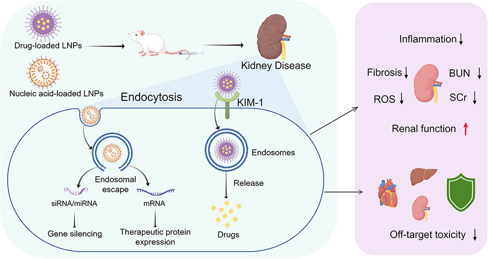 |
Figure 1 LNP-based nanoplatforms for renal-targeted therapy. LNPs deliver small-molecule drugs or nucleic acids (eg, mRNA, siRNA, miRNA) to treat kidney disorders. In preclinical models, targeted LNPs recognize kidney cell markers (eg, KIM-1 on injured renal tubular epithelial cells) for specific uptake via endocytosis. After endocytosis, nucleic acids escape endosomes for gene regulation, while drugs are released to exert effects. By reducing inflammation, fibrosis, and ROS, lowering off-target toxicity, and enhancing renal targeting, LNPs improve kidney function (evidenced by decreased BUN and SCr levels). The upward arrow (↑) indicates improvement, while the downward arrows (↓) indicate a decrease or inhibition. |
LNPs have shown significant advantages in the treatment of kidney diseases, with their core reflected in effectively reducing drug toxicity and realizing precise renal-targeted delivery, thus providing a new approach for the treatment of AKI and CKD. For instance, the novel stearic acid-gambogic acid (SA-GA)-modified solid lipid nanoparticles (SLNs) utilize gambogic acid (GA, a xanthonoid) with high affinity for the transferrin receptor (TfR) without competing with endogenous transferrin; after loading urolithin-A (UA) (particle size <250 nm, the highest encapsulation efficiency), they attain an uptake rate of approximately 50% in HK2 renal cells in vitro, and reduce inflammatory markers such as TLR4, NF-κB, and IL-1β to alleviate cisplatin-induced renal damage, highlighting their innovation as a TfR-targeted oral delivery system for AKI.19 In addition, to address the poor stability and solubility of astaxanthin (AST), AST-loaded SLNs have been designed as a novel formulation, which possess favorable physicochemical properties (eg, particle size <400 nm, high entrapment efficiency). They can effectively accomplish renal-targeted delivery via intraperitoneal administration (5 and 10 mg/kg), exert antioxidant effects in rats, improve renal function, reduce renal tissue damage, and thereby alleviate ischemia-reperfusion (I/R)-induced AKI.20 Importantly, to secure more precise kidney targeting and enhance therapeutic efficacy, LNPs can mediate drug endocytosis by binding to renal receptors. Biomimetic high-density lipoprotein (bHDL) lipid nanoparticles enable targeted release of triptolide (TP) and nintedanib (BIBF) in injured renal tubular epithelial cells via kidney injury molecule-1 (KIM-1)-mediated internalization, thereby synergistically alleviating renal fibrosis and improving CKD therapy with enhanced efficacy and reduced toxicity.21
LNPs with controlled drug release capabilities restore cellular homeostasis through diverse mechanisms. Kidney-targeted rhein (RH)-loaded liponanoparticles (KLPPR), a yolk-shell liponanoparticle (~60 nm) with a polycaprolactone-polyethyleneimine (PCL-PEI) core and kidney targeting peptide (KTP)-modified lipid shell, achieves kidney-specific controlled release of rhein by crossing the glomerular filtration membrane via size optimization and promoting non-lysosomal uptake in tubular cells, thereby suppressing fibronectin and TGF-β1 accumulation to alleviate diabetic kidney disease (DKD).16 Beyond enabling precise renal targeting, LNPs also effectively reduce the toxicity of drugs. Peptide-coupled celastrol (CLT, an active ingredient of Tripterygium wilfordii)-phospholipid lipid nanoparticles (PC-PLNs), designed based on the unique structure and pathological characteristics of the glomerulus, can specifically deliver toxic CLT to damaged glomerular endothelial cells and podocytes, effectively alleviating CKD through anti-inflammatory and endothelial protective effects while significantly reducing CLT toxicity.22
Regarding gene and nucleic acid delivery, LNPs enable precise genetic interventions to modulate disease progression. siRNA Selective ORgan Targeting lipid nanoparticles (siRNA SORT LNPs), a lipid nanoparticle formulation, can mediate durable extrahepatic gene silencing through systemic intravenous administration. This formulation overcomes the challenge of siRNA delivery to non-liver tissues, allowing siRNA to be effectively delivered to the kidneys (with a 15% reduction in TdTomato fluorescence in mouse kidneys), lungs, and spleen. It displays dose-dependent silencing of tissue-enriched endogenous targets, which may broaden the preclinical landscape of RNA interference (RNAi)-based therapeutic targets beyond the liver.23 Moreover, LNPs can efficiently achieve renal-targeted delivery of chemically modified SOD2 mRNA, and effectively ameliorate renal ischemia-reperfusion injury (IRI) by scavenging mitochondrial reactive oxygen species (ROS), restoring mitochondrial function and tissue integrity.24 Beyond targeting renal parenchymal cells, LNPs can also be engineered to target specific cell populations involved in renal disease pathogenesis, such as macrophages in CKD. Innovatively designed as macrophage-targeted lipid nanoparticles, these delivery systems enable precise renal disease-related delivery of Organic anion transporting polypeptide 2B1 (OATP2B1)/Solute carrier organic anion transporter family member 2B1 (Slco2b1) or Delta-like 4 (Dll4) siRNA, specifically inhibiting indoxyl sulfate-induced proinflammatory macrophage activation and atherosclerotic lesion development in CKD models.25
Taken together, LNPs with diversified targeting strategies (organ-, glomerulus-, and macrophage-specific) enable site-specific renal delivery of therapeutic agents and nucleic acids, highlighting their potential utility in the management of AKI and CKD.
Polymeric Nanoparticles (PNPs)
PNPs are nanoscale carriers constructed from synthetic or natural polymers that offer excellent biocompatibility, predictable biodegradation, and adjustable particle size. Their surfaces can be readily functionalized, and they accommodate high drug-loading capacities. Together, these attributes contribute to improved pharmacokinetics and sustained colloidal stability.26
The development of PNPs has progressed from simple cell-targeted carriers to sophisticated systems capable of responding to pathological cues within the kidney. Early formulations were designed to deliver therapeutics selectively to specific renal cell populations. For example, light chain-conjugated nanoparticles (LC-NPs) preferentially accumulate in renal tubular epithelial cells and renal carcinoma cells, thereby increasing drug deposition in the kidney while minimizing off-target exposure.27 Liu et al further advanced this concept by engineering nanoparticles that target CD44 through ChS–Chol conjugates. These self-assembled core–shell structures release their therapeutic payload via hydrolysis of pH-sensitive carbonate ester bonds under the acidic conditions characteristic of injured renal tissue, restoring mitochondrial function and suppressing apoptosis.28
Beyond small-molecule delivery, PNPs hold considerable promise for cell-specific targeting and gene therapy. A notable example involves a layered nanoparticle architecture composed of PLGA/CS/HA-KTP siRNA. Modification of the outer layer with a kidney-targeting peptide (KTP) enhanced tubular localization and enabled efficient delivery of Arg2 siRNA, resulting in robust gene silencing and therapeutic benefit in a contrast-induced AKI model.29 Another approach uses multifunctional polymeric nanocomplexes engineered with pH-responsive behavior to promote selective accumulation within diseased renal microenvironments. To achieve targeted NAD⁺ delivery, one study developed pH-responsive nanoparticles formed by complexing gallic acid with NAD⁺. Their ultrasmall size (< 6 nm) allows them to traverse the GFB and reach injured tubules efficiently. This targeted NAD⁺ supplementation restores NAD⁺-dependent mitochondrial activity and ATP generation, offering a strategy to halt the progression of acute kidney injury.30
In summary, PNPs enable precise therapeutic intervention by delivering drugs to defined renal cell types, responding to disease-associated microenvironments, and preferentially accumulating in pathological regions. Continued progress will depend on the rational design of PNP platforms that enhance potency while improving safety, ultimately transforming the clinical management of kidney disease.
Inorganic NPs
Inorganic nanoparticles exhibit distinctive physicochemical properties—including high specific surface area, quantum size effects, and, in the case of metal systems, surface plasmon resonance—that support diverse applications in photo-electromagnetics and biomedicine. These materials can be broadly categorized into two groups: (1) metal nanoparticles, such as gold, selenium, and silver, which display electrical conductivity, optical responsiveness, and favorable biocompatibility, and (2) metal oxide nanoparticles, including silica and iron oxide, which are widely used in catalytic adsorption and biomedical systems. As illustrated in Figure 2, inorganic nanoparticles exert therapeutic effects in kidney injury through diverse mechanisms, including the regulation of fibrosis and oxidative stress, protection of mitochondria, inhibition of ferroptosis, remodeling of the extracellular matrix, and suppression of inflammatory responses. However, many early formulations accumulated in tissues because of their limited biodegradability, raising concerns regarding long-term safety. To address these issues, contemporary generations of inorganic NPs have been engineered with enhanced degradability, improved biocompatibility, and reduced potential for chronic bioaccumulation.31–34
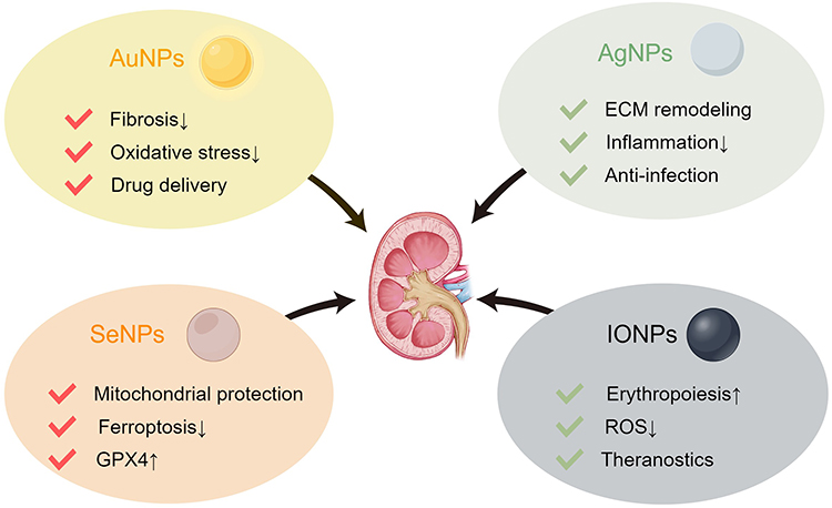 |
Figure 2 Renal protective actions of inorganic nanoparticles in kidney disease. Schematic of key inorganic nanoparticles (AuNPs, AgNPs, SeNPs, IONPs) and their distinct therapeutic roles in renal protection: AuNPs regulate fibrosis/oxidative stress and enable delivery; SeNPs protect mitochondria, inhibit ferroptosis, and activate GPX4; AgNPs remodel ECM, suppress inflammation, and prevent infection; IONPs support erythropoiesis, detoxify ROS, and enable theranostics. These activities underpin the potential of inorganic NPs for treating AKI and CKD. The upward arrows (↑) indicate promotion or activation, while the downward arrows (↓) indicate inhibition. |
Metal NPs
Gold nanoparticles (AuNPs) hold considerable promise for treating kidney disease because they combine direct antioxidant actions with programmable targeting.35 Studies have shown that glutathione modified AuNPs can be used as a kidney targeted drug delivery platform to precisely deliver anti fibrotic drugs to the site of kidney injury for responsive release and effective treatment of renal fibrosis.36 When decorated as targeted carriers, glycine–sarcosine–modified AuNPs present multivalent ligands that bind PEPT1/2 receptors and markedly improve renal targeting efficiency.37 These targeted systems reduce inflammation and promote tissue repair, in part through intrinsic immunomodulatory activity. Moreover, artichoke phenolic extracts have shown protective effects against AKI by inhibiting oxidative stress and regulating the COX-2/PGE2 pathway.38 This suggests that if developed into new formulations such as gold nanocapsules, they could potentially be applied in the treatment of renal fibrosis, although the specific mechanisms require further validation. However, clinical translation requires rigorous toxicological assessment, since AuNP safety depends strongly on particle size, shape and surface chemistry.39
Selenium nanoparticles (SeNPs) also show renal tropism and therapeutic activity. Prior work demonstrated preferential SeNP accumulation in renal tubules with subsequent uptake by tubular cells.40 SeNPs exert potent antioxidant and anti-inflammatory effects by activating the GPX-1 antioxidant pathway and suppressing NLRP3 inflammasome–Caspase-1 signaling.40,41 Consistent with these mechanisms, in vivo studies found that SeNP treatment ameliorates ischemia–reperfusion injury (IRI) and reduces the progression to renal fibrosis.40,42
Silver nanoparticles (AgNPs) exert reno-protective effects through multiple complementary pathways. AgNP treatment improves key renal functional biomarkers, including BUN and SCr, and enhances the activity of endogenous antioxidant enzymes such as SOD and GSH.43,44 In addition to their well-established antibacterial activity, specifically prepared AgNPs reduce inflammatory responses by lowering TNF-α and IL-6 levels, thereby further enhancing their therapeutic value.45 Despite these benefits, AgNPs can induce dose-dependent hepatorenal toxicity, which remains a major obstacle to clinical application. AgNPs can promote extracellular matrix deposition by activating the TGF β1/Smad signaling pathway and increasing the expression of collagen I, collagen III, and fibronectin.46 Co-administration of thymoquinone (TQ) substantially mitigates this toxicity. TQ provides synergistic antioxidant, anti-inflammatory, and anti-apoptotic protection, thereby reducing AgNP-induced damage.47 This strategy illustrates how pairing AgNPs with protective agents can balance therapeutic efficacy and safety, an essential step toward designing safer nanomedicines.
Metal Oxide NPs
Iron oxide nanoparticles (IONPs) have attracted substantial attention in biomedical research due to their superparamagnetic properties and favorable biocompatibility profiles.48 In kidney disease, IONPs provide both direct therapeutic benefits and versatile functionality as imaging and drug-delivery platforms.49 But IONPs can cause harm by increasing oxidative stress in cells. To solve this problem, a new type of nanoparticle called FeB was created. These FeB particles have an enzyme-like activity that breaks down harmful H2O2, and they also have a coating of glutathione (GSH) on their surface.50 This design removes the danger of oxidative stress and makes the particles safe for use in medicine. Clinically used IONPs, including the FDA approved iron supplement ferumoxytol for the treatment of anemia in chronic kidney disease, not only work as an iron supplement.51 Recent studies have also shown their potential for multiple uses in kidney disease. Through surface changes, IONPs can be used as drug delivery carriers to target damaged areas in the kidney. However, the clinical translation of IONPs remains constrained by dose-dependent hepatorenal toxicity, which is driven largely by oxidative stress and ferroptosis.52,53 Several strategies have been proposed to mitigate these toxicities. Surface polymer modification with PEG or antioxidant coatings can reduce oxidative stress and improve biocompatibility.54 Additional polymer engineering approaches aim to regulate ion release and minimize iron-mediated damage.55
Mesoporous silica nanoparticles (SiNPs) have emerged as promising carriers for renal therapeutics because they offer exceptionally high drug‐loading capacity and tunable release profiles, enabling them to overcome key biological delivery barriers.56,57 Despite these advantages, their potential nephrotoxicity warrants thorough investigation. In rat models, oral SiNP exposure elevates BUN and SCr levels, induces tubular necrosis, and leads to glomerular atrophy.58 Inhalation studies similarly demonstrate persistent tubulointerstitial inflammation and fibrosis.59 Subchronic SiNP exposure also triggers renal oxidative stress; notably, magnesium supplementation reverses this injury, suggesting that oxidative damage is a primary pathogenic mechanism and a viable point of intervention.60 Beyond their toxicological concerns, SiNPs have been innovatively adapted as fluorescent probes to monitor kidney injury biomarkers such as NAG and β-GAL, allowing dynamic visualization of renal microenvironments.61 Going forward, elucidating the molecular pathways involved including the roles of UPR activation will be important for optimizing biocompatibility.62 Developing targeted antioxidant strategies may further mitigate toxicity. Ultimately, the value of SiNPs in kidney therapy will depend on balancing their therapeutic utility with rigorous assurance of long-term safety.
Exosome Delivery System
Exosomes are nanoscale extracellular vesicles enclosed by a lipid bilayer, typically measuring 30–150 nm in diameter. They are secreted by many cell types and mediate intercellular communication by transferring diverse bioactive cargos, including proteins, lipids, and nucleic acids. Their lipid bilayer enables efficient passage across biological barriers, and their inherent structure supports substantial loading of therapeutic molecules.63 Combined with their low immunogenicity, high stability, and natural biological origin, these properties position exosomes as promising delivery vehicles for renal therapeutics.64–68 Figure 3 schematically summarizes these mechanisms and applications.
 |
Figure 3 Native, engineered, and drug-loaded exosomes in kidney disease therapy. Schematic of three exosome types (native, engineered, drug-loaded) and their distinct functional characteristics (renal tropism, enhanced targeting, controlled release). Exosomes mediate renal repair by delivering therapeutic cargos (including miRNAs, proteins, or small-molecule drugs), which exert key biological effects to suppress pathological stress responses (such as oxidative stress and inflammation), reduce fibrosis, promote tissue repair, and improve renal function in the treatment of AKI and CKD. The upward arrows (↑) indicate an increase or recovery, while the downward arrows (↓) indicate a decrease or inhibition. |
Mesenchymal stem cell (MSC)–derived exosomes from multiple tissue sources have shown potent reno-protective effects. These vesicles convey protective biomolecules directly to injured renal cells.69 For instance, human umbilical cord mesenchymal stem cell-derived exosomes (hucMSC-exos) deliver miR-29a-3p, which mitigates renal fibrosis and vascular rarefaction after renal ischemia-reperfusion injury by targeting collagen type I in fibroblasts and tumor necrosis factor receptor 1 (TNFR1) in endothelial cells.70 Exosomes derived from other MSC populations exhibit similar benefits. Adipose-derived MSC exosomes (ADMSC-Exos) activate the SIRT1 pathway to limit inflammation and apoptosis.71 Bone marrow MSC exosomes (BMMSC-Exos) shuttle miR-199a-3p and miR-223 to reduce fibrosis and inflammatory signaling.72 Exosomes from urine-derived and placenta-derived MSCs have likewise demonstrated therapeutic potential.73 Currently, there are many clinical trials worldwide investigating exosome-based therapeutics. These trials encompass a diverse range of formulations, including natural exosomes, drug-loaded variants, and genetically engineered products, highlighting their significant potential as a form of “cell-free therapy” in clinical applications.69,74
Nevertheless, the path toward clinical translation faces several critical challenges. The foremost issue lies in heterogeneity control and standardization: variations in cellular sources, culture conditions, and isolation methods result in significant batch-to-batch heterogeneity, directly impacting the reproducibility of therapeutic potency. In addition, the establishment of standardized and robust potency assays remains a significant hurdle. The diversity in mechanisms of action makes it difficult to define uniform potency indicators, and the lack of validated, quantitative methods for batch-to-batch activity comparison further complicates quality control and regulatory consistency.74,75 Furthermore, the precise mechanisms underlying targeted delivery remain incompletely elucidated, limiting accurate localization and efficient enrichment within renal tissues. The cost-effectiveness and scalability of large-scale production require further optimization to meet future clinical-grade demands. Finally, the regulatory classification of exosome-based products—whether as biologics, drugs, or combination products—lacks global consensus, introducing uncertainty into development pathways. Learning from regulatory frameworks such as those established by Japan’s PMDA and promoting GMP-standardized production alongside clear regulatory guidelines will accelerate IND submissions and clinical adoption.76–78
Researchers are increasingly engineering MSC-derived exosomes to address current limitations and improve therapeutic efficacy.79 These efforts rely on two principal strategies: (1) directly loading isolated exosomes with therapeutic cargo using techniques such as electroporation, incubation, or transfection, and (2) genetically modifying parent MSCs so they naturally secrete exosomes enriched with specific bioactive molecules.80 For example, transfection has been used to produce MSC-derived exosomes enriched with miR-let-7c, which suppresses TGF-β1 signaling and reduces renal fibrosis.81 For anti-inflammatory applications, IL-10 can be incorporated into exosomes either by direct loading or by engineering MSCs for endogenous IL-10 production;82 both approaches limit renal inflammation and promote M2 macrophage polarization. Electroporation or transfection has also been applied to generate exosomes that deliver miR-13474, which inhibits ADAM17 and attenuates renal fibrosis in vivo.83
A widely used engineering approach involves loading exosomes with therapeutic agents relevant to kidney injury. These agents include anti-inflammatory drugs (such as dexamethasone) and gene-regulating miRNAs. For example, encapsulating dexamethasone in extracellular vesicles has been shown to reduce kidney damage in an IgA nephropathy model.84 In addition, researchers are exploring the use of engineered exosomes for targeted delivery of specific microRNAs; one study showed that exosomes modified to deliver miR-218-5p could alleviate podocyte injury in diabetic kidney disease by activating mitophagy.85 In preclinical studies, these engineered exosomes improve renal function - evidenced by reduced BUN and SCr levels - by simultaneously modulating inflammation, apoptosis, and oxidative stress pathways.
Looking ahead, the development of the exosome field will focus on technological innovation and interdisciplinary integration. The incorporation of artificial intelligence, such as deep learning models to predict stem cell differentiation and exosome functionality, along with the design of intelligent responsive materials, holds promise for precise modulation of engineered exosome properties. Moreover, organoid-exosome combination therapies are emerging as a novel research direction, providing new platforms to model complex renal microenvironments and validate therapeutic efficacy. Concurrently, the application of exosomes in anti-aging and cosmetic medicine is accelerating their marketization, injecting additional momentum into the development of the therapeutic exosome industry.86–88
Nanomaterials in Kidney Disease Diagnosis
Beyond therapeutic nanoplatforms, nanomaterials also function as diagnostic regulators that shape therapeutic strategy selection and monitoring. Nanomaterials serve as a critical bridge between diagnosis and therapy in nephrology by enabling precise imaging that directly informs therapeutic decision-making across the entire clinical continuum, from early detection to treatment monitoring. In this context, advanced nanomaterial-based imaging platforms play a pivotal role in enhancing diagnostic accuracy and guiding personalized interventions. Currently, nanomaterials are being actively investigated as next-generation contrast agents for nephrologic imaging modalities, including magnetic resonance imaging (MRI), near-infrared fluorescence (NIRF), and integrated multimodal systems. Among them, superparamagnetic iron oxide nanoparticles (SPIONs), AuNPs, and carbon-based nanostructures represent the most extensively studied classes.89 Through ligand-guided surface engineering, these nanomaterials can be selectively targeted to specific renal compartments or pathological lesions, significantly improving signal-to-noise ratios and enhancing diagnostic sensitivity compared with conventional contrast agents. Such targeted imaging capability not only facilitates earlier and more accurate disease identification but also enables real-time evaluation of therapeutic response, thereby strengthening the diagnostic-therapeutic integration in nephrology.26
SPIONs in Kidney Disease Diagnosis
SPIONs provide a safer alternative to gadolinium-based contrast agents for renal MRI, particularly in patients with impaired kidney function where gadolinium exposure is associated with nephrogenic systemic fibrosis and tissue retention concerns. Ultrasmall SPION formulations, including ES-SPIONs (~3 nm core) and SNIOs (~1 nm core) coated with zwitterionic dopamine sulfonate, have demonstrated rapid renal clearance in preclinical studies while generating positive T1-weighted contrast suitable for high-resolution anatomical imaging.90 This improved safety profile expands access to contrast-enhanced MRI in vulnerable populations who would otherwise be excluded from such imaging. By enabling contrast evaluation in these patients, SPION-enhanced MRI supports clinical decision-making related to disease staging, assessment of disease activity, and timing of therapeutic intervention.
Beyond anatomical imaging, targeted SPION platforms enable functional and molecular assessments with potential therapeutic relevance. SPION-based blood volume mapping has been shown to detect early microvascular alterations in diabetic kidney disease models that precede elevations in serum creatinine.91 These imaging-derived indicators may facilitate earlier risk stratification and provide a rationale for timely intensification of renoprotective therapy at stages when structural damage may still be reversible. Similarly, antibody-directed SPIONs enable MRI-based detection and phenotyping of macrophage infiltration in acute kidney injury, offering insight into inflammatory endotypes that may inform immunomodulatory strategy selection.91 In this context, imaging findings extend beyond lesion detection to characterization of underlying pathophysiology relevant to treatment planning.
The favorable clearance and safety characteristics of ZDS-coated SPIONs also support longitudinal imaging for treatment response evaluation.90 Repeated administration enables monitoring of dynamic changes in microvascular perfusion or inflammatory burden over time, allowing therapeutic regimens to be maintained, intensified, or modified according to imaging-derived functional trends rather than delayed biochemical markers alone.91 Through this integration of safety, targeting capability, and functional readouts, SPION-enhanced MRI contributes to a decision-oriented imaging framework in nephrology, linking diagnostic information to adaptive therapeutic management.
AuNP in Kidney Disease Diagnosis
AuNPs, particularly renal-clearable formulations, provide functional imaging beyond conventional biochemical markers. Ning et al used glutathione-coated AuNPs as X-ray contrast agents to track nanoparticle transport in cisplatin-induced kidney injury.92 In mice with similarly elevated BUN and creatinine levels, AuNP retention in the outer stripe of the outer medulla (OSOM) showed marked heterogeneity and correlated with proximal tubular injury rather than serum biomarkers. These findings demonstrate that conventional renal function indices do not reliably reflect early tubular damage, whereas AuNP transport imaging resolves spatial and functional alterations. Such imaging has direct therapeutic relevance. In the setting of nephrotoxic chemotherapy, prolonged AuNP retention in the OSOM-despite modest biochemical changes-may indicate clinically significant tubular injury, supporting earlier treatment adjustment or implementation of renoprotective strategies before progression to more severe damage.
Complementing transport imaging, Tan et al developed renal-clearable luminescent AuNPs with pH-responsive surfaces that undergo charge conversion and enhanced tubular reabsorption in the acidic microenvironment of early renal injury.93 These nanoparticles produced enhanced ultrasound contrast in injured kidneys before substantial changes in BUN or creatinine were detectable. By exploiting injury-associated microenvironmental alterations rather than relying solely on filtration metrics, this approach enables earlier identification of kidney damage at a potentially reversible stage and may inform the timing of protective interventions prior to irreversible structural injury.
Collectively, nanomaterial-based imaging platforms redefine renal diagnostics by embedding therapeutic relevance directly into imaging readouts. Renal-clearable SPIONs and AuNPs move beyond static anatomical visualization to provide quantitative information on microvascular function, tubular transport dynamics, inflammatory activity, and injury‑associated microenvironmental changes. By enabling early detection, mechanistic phenotyping, risk stratification, and longitudinal monitoring of treatment response, these nanoparticle probes support imaging-guided therapy adjustment and early intervention. As surface engineering, multimodal integration, and quantitative imaging analytics continue to advance, nanomaterial-enabled imaging is poised to function as a continuous decision-support system, underpinning closed-loop diagnostic-therapeutic strategies and advancing precision nephrology.
Clinical Translation Challenges
Although drug delivery systems based on nanomaterials have shown enhanced targeting efficiency in preclinical models of AKI and CKD, several technical limitations have hindered their clinical translation. The in vivo behavior of nanoparticles is determined by key physicochemical characteristics including size, surface charge, and hydrophobicity which collectively influence biodistribution, organ retention, degradation kinetics, and immune activation.94,95 A primary challenge to translation is that the renal clearance threshold (fluid dynamic diameter of approximately 5.5 nanometers) is small enough for nanoparticles to pass through the kidneys and often exhibit rapid systemic clearance, thereby reducing the therapeutic exposure; while larger particles accumulate in the liver and spleen, increasing off-target toxicity.96 For example, inorganic nanoparticles such as gold- and silica-based structures can persist for months in the liver and spleen following systemic administration.97 Furthermore, in a comprehensive rat model study, Hamed et al investigated the renal safety of silver nanoparticles following repeated oral exposure, with results indicating potential renal concerns. They attributed this discrepancy to significant tubular structural damage, glomerular degeneration, inflammatory cell infiltration, and increased cell apoptosis, although the blood biochemical indicators did not show significant changes. However, the histopathological evidence clearly indicated the nephrotoxic effect.98
Another important consideration is that the long-term risks of targeted renal nanoparticles remain poorly understood. Of particular concern is chronic tubular accumulation, which has been detected in renal tubular epithelial cells in rodents up to 6 months after administration of AuNPs and carbon nanotubes. Further uncertainties include effects on cellular senescence and malignant transformation, as well as potential immunotoxicity. For patients requiring repeated dosing for chronic diseases, evidence suggests that anti-PEG antibodies form in 25–40% of the general population, which may trigger accelerated blood clearance and hypersensitivity reactions upon re-exposure.21
Moreover, although kidney-targeting nanomedicines have demonstrated remarkable therapeutic efficacy in preclinical rodent models, their progression to human clinical trials has encountered substantial obstacles. For instance, no kidney-specific nanomedicine has successfully advanced to clinical approval, with most systems remaining confined to preclinical rodent studies. This discrepancy might stem from the inherent limitations of murine models in replicating human renal pathophysiology, including species-specific differences in glomerular filtration rates, tubular reabsorption mechanisms, and the inability of rodent models to fully recapitulate the complex multi-organ comorbidities, metabolic disorders, and chronic disease progression patterns characteristic of human kidney diseases.99
To address these barriers, several strategies have been proposed. Surface modification with PEG or zwitterionic polymers can reduce immune recognition, minimize nonspecific protein adsorption, and extend circulation time. Targeting specificity can be enhanced by decorating nanoparticles with ligands such as anti-VCAM-1 or anti-PDGF-B antibodies, which direct accumulation to inflamed or fibrotic renal tissue.100 In parallel, the development of renal organoids and kidney-on-a-chip platforms offers more physiologically relevant human models for evaluating nanoparticle performance and mitigating discrepancies between conventional in vitro assays and animal studies.101 Nano-therapies guided by these design principles have demonstrated improved precision, reduced off-target toxicity, and enhanced tissue repair in preclinical models of nephropathy.102
In conclusion, although nanotechnology has provided innovative solutions for the management of kidney diseases, this field must shift from proof-of-concept research to rigorous, clinically translatable studies to clearly address these limitations. Future research priorities should include the development of biomimetic nanoparticles with improved renal permeability and reduced immunogenicity, the establishment of large animal models (pigs, non-human primates) that better simulate human renal physiology, and the implementation of adaptive clinical trial designs that incorporate pharmacokinetic-pharmacodynamic models. Through this balanced and prudent advancement, nanomaterials may offer further treatment prospects for patients with kidney diseases.
Conclusion
This review summarizes recent advances in four major nanomaterial platforms - lipid-based, polymeric, inorganic, and exosome-based delivery systems - in the context of kidney-targeted therapy. Each platform offers distinct physicochemical features, tunable size properties, and adaptable targeting strategies that support rational design for efficient drug delivery, reduced systemic toxicity, and integrated theranostic applications, as outlined in Table 1. Collectively, these nanotechnologies hold substantial promise for the treatment of both AKI and CKD, as they can simultaneously address key pathological processes, including inflammation, oxidative stress, and fibrosis. Their continued development may pave the way for more effective and personalized interventions in renal disease. Despite promising preclinical efficacy, the clinical translation of nanomedicines for kidney diseases is hindered by unresolved technical limitations, including suboptimal targeting, rapid clearance, and off-target accumulation-coupled with inherent pathophysiological discrepancies between rodent models and human disease, as well as insufficient long-term safety data. Accordingly, rational design optimization and rigorous safety evaluation will be essential for advancing kidney-targeted nanotherapies toward clinical reality.
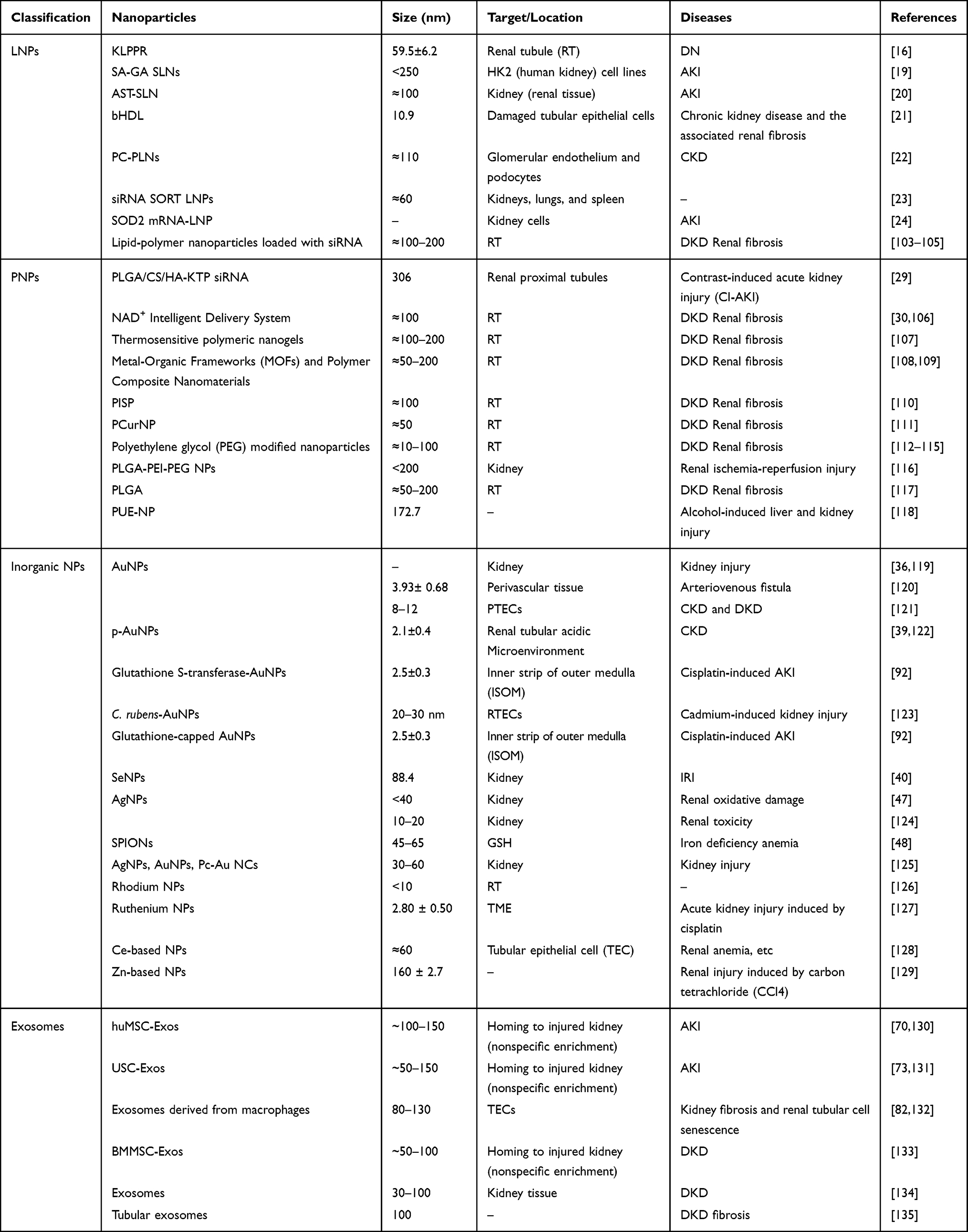 |
Table 1 Summary of Nanomaterials for Kidney Diseases: Size, Targets, and Therapeutic Applications |
Abbreviations
NCD, noncommunicable disease; AKI, acute kidney injury; CKD, chronic kidney disease; KIM-1, kidney injury molecule-1; LNPs, lipid nanoparticles; GFB, glomerular filtration barrier; ROS, reactive oxygen species; bHDL, biomimetic high-density lipoprotein; TP, triptolide; BIBF, nintedanib; ECM, extracellular matrix; PNPs, polymeric nanoparticles; LC-NPs, light chain-conjugated nanoparticles; ChS-Chol, chondroitin sulfate-cholesterol; KTP, kidney-targeting peptide; GSH, glutathione; AuNPs, gold nanoparticles; SeNPs, selenium nanoparticles; IRI, ischemia–reperfusion injury; TQ, thymoquinone; IONPs, iron oxide nanoparticles; SiNPs, silica nanoparticles; MSC, mesenchymal stem cell; huMSC-Exos, human umbilical cord-derived MSC exosomes; ADMSC-Exos, adipose-derived MSC exosomes; BMMSC-Exos, bone marrow MSC exosomes; NAC, N-acetylcysteine; MRI, magnetic resonance imaging; NIRF, near-infrared fluorescence; SPIONs, superparamagnetic iron oxide nanoparticles; NIR-II, the second near-infrared window; DKD, diabetic kidney disease; NPs, nanoparticles; Al2O3 NPs, aluminum oxide nanoparticles; AgNPs, silver nanoparticles; BUN, blood urea nitrogen; CD44, cluster of differentiation 44; CeO2 NPs, cerium oxide nanoparticles; COL1A1, collagen type I alpha 1 chain; GPX4/GPx-1, glutathione peroxidase 4/1; HO-1, heme oxygenase-1; ICAM-1, intercellular adhesion molecule-1; IL-1β, interleukin-1 beta; IL-6, interleukin-6; MAPK, mitogen-activated protein kinase; NAD⁺, nicotinamide adenine dinucleotide; NADPH, nicotinamide adenine dinucleotide phosphate; Nrf2, nuclear factor erythroid 2–related factor 2; p-AuNPs, pH-responsive gold nanoparticles; PCNA, proliferating cell nuclear antigen; PDGF-B, platelet-derived growth factor-B; PEG, polyethylene glycol; PGC-1α, peroxisome proliferator-activated receptor-γ coactivator-1 alpha; PI3K, phosphoinositide 3-kinase; PLGA, poly(lactic-co-glycolic acid); SCr, serum creatinine; SIRT1/SIRT3, sirtuin 1/3; SOD, superoxide dismutase; SR-B1, scavenger receptor class B type 1; TGF-β1/TGF-β, transforming growth factor-beta 1/beta; TNF-α, tumor necrosis factor-alpha; TNFR1, tumor necrosis factor receptor 1; USC-Exos, urine-derived stem cell exosomes; VCAM-1, vascular cell adhesion molecule-1; XBP1, X-box binding protein 1; ZnO NPs, zinc oxide nanoparticles; SGLT2, sodium-glucose cotransporter 2; MMP2, matrix metalloproteinase 2; BNIP3, BCL2 interacting protein 3; RTECs, Renal Tubular Epithelial Cells; KLPPR, rhubarb acid-loaded lipid nanoparticles; SOD2, superoxide dismutase 2; T-A-Ls, artemisinin Succinate-Nanoliposomes-TPP; T4LN, TKP 4-modified nystatin-loaded liposomal nanoparticles; DR5, death receptor 5; SORT, Selective ORgan Targeting; LyP-1-LNPs, LyP-1-modified cationic lipid nanoparticles; PEPT1/2, peptide transporter 1/2; COX-2, cyclooxygenase-2; PGE2, prostaglandin E2; GPX-1, glutathione peroxidase 1; NLRP3, NOD-like receptor family pyrin domain containing 3; H2O2, hydrogen peroxide; FDA, Food and Drug Administration; NAG, N-acetyl-β-D-glucosaminidase; β-GAL, β-galactosidase; UPR, unfolded protein response; miR, microRNA; PMDA, Pharmaceuticals and Medical Devices Agency; IL-10, interleukin 10; IgA, Immunoglobulin A; Smad, small mothers against decapentaplegic; Caspase-1, cysteine-aspartic protease 1.
Data Sharing Statement
All images included in this review were generated using Figdraw and Biorender.
Acknowledgments
During the preparation of this manuscript, the authors used ChatGPT solely for language polishing. All authors carefully reviewed and revised the generated text and accept full responsibility for the final content.
Author Contributions
All authors made a significant contribution to the work reported, whether that is in the conception, study design, execution, acquisition of data, analysis and interpretation, or in all these areas; took part in drafting, revising or critically reviewing the article; gave final approval of the version to be published; have agreed on the journal to which the article has been submitted; and agree to be accountable for all aspects of the work.
Funding
This work was supported by the National Natural Science Foundation of China (project no. 82200809), the Natural Science Foundation of Shandong Province (project nos. ZR2025LMB015, ZR2022QH176 and ZR2022QC245), the Taishan Scholars Program (tsqn202507251), the Youth Innovation Team Plan in Colleges and Universities of Shandong Province (2023KJ185), and project no. 3502Z20214ZD1183 from the Xiamen Medical and Health Guiding Project.
Disclosure
The authors declare no competing interests in this work.
References
1. Francis A, Harhay MN, Ong ACM, et al. Chronic kidney disease and the global public health agenda: an international consensus. Nat Rev Nephrol. 2024;20(7):473–17. doi:10.1038/s41581-024-00820-6
2. Chawla LS, Bellomo R, Bihorac A, et al. Acute kidney disease and renal recovery: consensus report of the Acute Disease Quality Initiative (ADQI) 16 Workgroup. Nat Rev Nephrol. 2017;13(4):241–257. doi:10.1038/nrneph.2017.2
3. Islamuddin M, Qin X. Renal macrophages and NLRP3 inflammasomes in kidney diseases and therapeutics. Cell Death Discov. 2024;10(1):229. doi:10.1038/s41420-024-01996-3
4. Wang X, He J, Wang Y. Long-term outcomes following acute kidney injury in individuals with pre-existing chronic kidney disease: a systematic review and meta-analysis. J Nephrol. 2025;38(9):2517–2537. doi:10.1007/s40620-025-02373-8
5. Boima V, Agyekum AB, Ganatra K, et al. Advances in kidney disease: pathogenesis and therapeutic targets. Front Med. 2025;12:1526090. doi:10.3389/fmed.2025.1526090
6. Jha PK, Nakano T, Itto LYU, et al. Vascular inflammation in chronic kidney disease: the role of uremic toxins in macrophage activation. Front Cardiovasc Med. 2025;12:1574489. doi:10.3389/fcvm.2025.1574489
7. Huang Y, Wang J, Jiang K, Chung EJ. Improving kidney targeting: the influence of nanoparticle physicochemical properties on kidney interactions. J Control Release. 2021;334:127–137. doi:10.1016/j.jconrel.2021.04.016
8. Peng C, Huang Y, Zheng J. Renal clearable nanocarriers: overcoming the physiological barriers for precise drug delivery and clearance. J Control Release. 2020;322:64–80. doi:10.1016/j.jconrel.2020.03.020
9. Mitchell MJ, Billingsley MM, Haley RM, Wechsler ME, Peppas NA, Langer R. Engineering precision nanoparticles for drug delivery. Nat Rev Drug Discov. 2021;20(2):101–124. doi:10.1038/s41573-020-0090-8
10. Williams RM, Shah J, Tian HS, et al. Selective nanoparticle targeting of the renal tubules. Hypertension. 2018;71(1):87–94. doi:10.1161/hypertensionaha.117.09843
11. Song Y, Peng Y, Wang X, et al. Impact of renal-targeting ligand density on kidney targeting, clearance, and off-target effects of renal-clearable nanoparticles. Nano Lett. 2025;25(24):9825–9833. doi:10.1021/acs.nanolett.5c02262
12. Qiu M, Singh A, Wang D, et al. Biocompatible and biodegradable inorganic nanostructures for nanomedicine: silicon and black phosphorus. Nano Today. 2019;25:135–155. doi:10.1016/j.nantod.2019.02.012
13. Rosales A, Blondel LO, Hull J, et al. Evolving adeno-associated viruses for gene transfer to the kidney via cross-species cycling of capsid libraries. Nat Biomed Eng. 2025;9(7):1086–1100. doi:10.1038/s41551-024-01341-0
14. Wang L, Zhou W, Chen H, et al. Barcoded screening identifies nanocarriers for protein delivery to kidney. Nat Commun. 2025;16(1):899. doi:10.1038/s41467-025-56257-3
15. Qin W, Huang J, Zhang M, Xu M, He J, Liu Q. Nanotechnology-based drug delivery systems for treating acute kidney injury. ACS Biomater Sci Eng. 2024;10(10):6078–6096. doi:10.1021/acsbiomaterials.4c01385
16. Wang G, Li Q, Chen D, et al. Kidney-targeted rhein-loaded liponanoparticles for diabetic nephropathy therapy via size control and enhancement of renal cellular uptake. Theranostics. 2019;9(21):6191–6208. doi:10.7150/thno.37538
17. Huang J, Guo J, Dong Y, et al. Self-assembled hyaluronic acid-coated nanocomplexes for targeted delivery of curcumin alleviate acute kidney injury. Int J Biol Macromol. 2023;226:1192–1202. doi:10.1016/j.ijbiomac.2022.11.233
18. Cheng X, Gao J, Ding Y, et al. Multi-functional liposome: a powerful theranostic nano-platform enhancing photodynamic therapy. Adv Sci. 2021;8(16):e2100876. doi:10.1002/advs.202100876
19. Ganugula R, Dinakar YH, Kurse A, Kumar M, Arora M. Design and in vitro evaluation of gambogic acid-conjugated stearic acid solid lipid nanoparticles for transferrin receptor-mediated drug delivery. Pharm Res. 2025;42(12):2235–2245. doi:10.1007/s11095-025-03946-9
20. Yarmohammadi A, Arkan E, Najafi H, et al. Protective effects of astaxanthin solid lipid nanoparticle as a promising candidate against acute kidney injury in rats. Naunyn Schmiedebergs Arch Pharmacol. 2025;398(4):4491–4502. doi:10.1007/s00210-024-03543-4
21. He S, Li X, He Y, et al. High-density lipoprotein nanoparticles spontaneously target to damaged renal tubules and alleviate renal fibrosis by remodeling the fibrotic niches. Nat Commun. 2025;16(1):1061. doi:10.1038/s41467-025-56223-z
22. Wu Q, Wang J, Wang Y, et al. Targeted delivery of celastrol to glomerular endothelium and podocytes for chronic kidney disease treatment. Nano Res. 2022;15(4):3556–3568. doi:10.1007/s12274-021-3894-x
23. Vaidya A, Moore S, Chatterjee S, et al. Expanding RNAi to kidneys, lungs, and spleen via Selective ORgan Targeting (SORT) siRNA lipid nanoparticles. Adv Mater. 2024;36(35):e2313791. doi:10.1002/adma.202313791
24. Hou Y, Lin S, Xia J, et al. Alleviation of ischemia-reperfusion induced renal injury by chemically modified SOD2 mRNA delivered via lipid nanoparticles. Mol Ther Nucleic Acids. 2023;34:102067. doi:10.1016/j.omtn.2023.102067
25. Nakano T, Katsuki S, Chen M, et al. Uremic toxin indoxyl sulfate promotes proinflammatory macrophage activation via the interplay of OATP2B1 and Dll4-Notch signaling. Circulation. 2019;139(1):78–96. doi:10.1161/circulationaha.118.034588
26. Deng Y, Liu Z, Zhu X, Wang Y, Feng X, Yang J. Kidney-targeted nanoplatforms: strategies and applications. Theranostics. 2026;16(6):3011–3031. doi:10.7150/thno.126217
27. Ordikhani F, Kasinath V, Uehara M, et al. Selective trafficking of light chain-conjugated nanoparticles to the kidney and renal cell carcinoma. Nano Today. 2020;35:100990. doi:10.1016/j.nantod.2020.100990
28. Zhao W, Zhou J, Xia Y, et al. Chondroitin sulfate-based ROS-responsive nanoparticles targeting activated myofibroblasts via CD44 receptors for the renal fibrosis therapy. Mol Pharmaceut. 2026;23:1785–1800. doi:10.1021/acs.molpharmaceut.5c01524
29. Gu XR, Tai YF, Liu Z, et al. Layer-by-layer assembly of renal-targeted polymeric nanoparticles for robust Arginase-2 knockdown and contrast-induced acute kidney injury prevention. Adv Healthcare Mater. 2024;13(20):e2304675. doi:10.1002/adhm.202304675
30. Kong Y, Chen X, Liu F, et al. Ultrasmall Polyphenol-NAD(+) nanoparticle-mediated renal delivery for mitochondrial repair and anti-inflammatory treatment of AKI-to-CKD progression. Adv Mater. 2024;36(30):e2310731. doi:10.1002/adma.202310731
31. Campos MT, Pires LS, Magalhães FD, Oliveira MJ, Pinto AM. Self-assembled inorganic nanomaterials for biomedical applications. Nanoscale. 2025;17(10):5526–5570. doi:10.1039/d4nr04537h
32. Oh JY, Villaseñor KE, Kian AC, Cormode DP. Advances in ultrasmall inorganic nanoparticles for nanomedicine: from diagnosis to therapeutics. ACS Appl Mater Interfaces. 2025;17(20):28982–29001. doi:10.1021/acsami.5c02810
33. Cave J, Christiono A, Schiavone C, et al. Rational design of safer inorganic nanoparticles via mechanistic modeling-informed machine learning. Res Square. 2025. doi:10.21203/rs.3.rs-5960303/v1
34. Percoco RM, Schirinzi S, Mandriota G, et al. Smart inorganic nanoparticles in nanomedicine: strategies for synthesis and functionalization. Int J Pharm. 2025;684:126192. doi:10.1016/j.ijpharm.2025.126192
35. Chan CKW, Szeto CC, Lee LKC, et al. A sub-10-nm, folic acid-conjugated gold nanoparticle as self-therapeutic treatment of tubulointerstitial fibrosis. Proc Natl Acad Sci. 2023;120(42):e2305662120. doi:10.1073/pnas.2305662120
36. Lai X, Geng X, Tan L, Hu J, Wang S. A pH-responsive system based on fluorescence enhanced gold nanoparticles for renal targeting drug delivery and fibrosis therapy. Int J Nanomed. 2020;15:5613–5627. doi:10.2147/ijn.S260069
37. Huang D, Tan Y, Tang J, He K, Zhou Y, Liu J. Transcytosis-based renal tubular reabsorption of luminescent gold nanoparticles for enhanced tumor imaging. Angew Chem. 2024;63(11):e202316900. doi:10.1002/anie.202316900
38. Elshamy AI, Abdallah HMI, Farrag ARH, et al. Artichoke phenolics confer protection against acute kidney injury. Rev Bras Farmacogn. 2020;30(1):34–42. doi:10.1007/s43450-020-00032-6
39. Milan J, Niemczyk K, Kus-Liskiewicz M. Treasure on the earth-gold nanoparticles and their biomedical applications. Materials. 2022;15(9):3355. doi:10.3390/ma15093355
40. Wang S, Chen Y, Han S, et al. Selenium nanoparticles alleviate ischemia reperfusion injury-induced acute kidney injury by modulating GPx-1/NLRP3/Caspase-1 pathway. Theranostics. 2022;12(8):3882–3895. doi:10.7150/thno.70830
41. Hassan I, Ebaid H, Al-Tamimi J, Habila MA, Alhazza IM, Rady AM. Selenium nanoparticles mitigate diabetic nephropathy and pancreatopathy in rat offspring via inhibition of oxidative stress. J King Saud Univ Sci. 2021;33(1):101265. doi:10.1016/j.jksus.2020.101265
42. Zuo Z, Luo M, Liu Z, et al. Selenium nanoparticles alleviate renal ischemia/reperfusion injury by inhibiting ferritinophagy via the XBP1/NCOA4 pathway. Cell Commun Signal. 2024;22(1):376. doi:10.1186/s12964-024-01751-2
43. Saud MA, Saud NA, Hamad MA, Farhan Gar L. Role of Salvia officinalis silver nanoparticles in attenuation renal damage in rabbits exposed to methotrexate. Arch Razi Inst. 2022;77(1):151–162. doi:10.22092/ari.2021.356313.1821
44. Barabadi H, Noqani H, Soltani M, Sabbagh Kashani A. Animal-based evidence supports protective activity of bioengineered silver and gold nanomaterials on hepatic and renal function profile parameters. Front Nanotechnol. 2025;6:1424562. doi:10.3389/fnano.2024.1424562
45. Pote S, Salve P, Kambi M. One-pot green synthesis, optimization, and characterization of silver nanoparticles from Mitragyna parvifolia: a novel therapeutic strategy for rheumatoid arthritis targeting COX-2 and TNF- α. Bioorg Chem. 2025;163:108739. doi:10.1016/j.bioorg.2025.108739
46. Assar DH, Mokhbatly AA, Ghazy EW, et al. Silver nanoparticles induced hepatoxicity via the apoptotic/antiapoptotic pathway with activation of TGFβ-1 and α-SMA triggered liver fibrosis in Sprague Dawley rats. Environ Sci Pollut Res Int. 2022;29(53):80448–80465. doi:10.1007/s11356-022-21388-3
47. Salama B, Alzahrani KJ, Alghamdi KS, et al. Silver nanoparticles enhance oxidative stress, inflammation, and apoptosis in liver and kidney tissues: potential protective role of thymoquinone. Biol Trace Elem Res. 2023;201(6):2942–2954. doi:10.1007/s12011-022-03399-w
48. Hsiao JK, Chen CL, Hsieh WY, Kuo KL. Theranostic role of iron oxide nanoparticle for treating renal anemia: evidence of efficacy and significance by MRI, histology and biomarkers. Pharmaceutics. 2023;15(6):1714. doi:10.3390/pharmaceutics15061714
49. Yang K, Shang Y, Yang N, Pan S, Jin J, He Q. Application of nanoparticles in the diagnosis and treatment of chronic kidney disease. Front Med. 2023;10:1132355. doi:10.3389/fmed.2023.1132355
50. Aydemir D, Arıbuğa D, Hashemkhani M, Acar HY, Balcı-çağıran Ö, Ulusu NN. Bioavailability assessment of the novel GSH-functionalized FeB nanoparticles via oxidative stress and trace element metabolism in vitro: promising tools for biomedical applications. J Nanopart Res. 2024;26(12):274. doi:10.1007/s11051-024-06191-0
51. Huang Y, Hsu JC, Koo H, Cormode DP. Repurposing ferumoxytol: diagnostic and therapeutic applications of an FDA-approved nanoparticle. Theranostics. 2022;12(2):796–816. doi:10.7150/thno.67375
52. Aboulhoda BE, Othman DA, Rashed LA, Alghamdi MA, Esawy AEWE. Evaluating the hepatotoxic versus the nephrotoxic role of iron oxide nanoparticles: one step forward into the dose-dependent oxidative effects. Heliyon. 2023;9(11):e21202. doi:10.1016/j.heliyon.2023.e21202
53. Luo C, Li X, Yan H, Guo Q, Liu J, Li Y. Iron oxide nanoparticles induce ferroptosis under mild oxidative stress in vitro. Sci Rep. 2024;14(1):31383. doi:10.1038/s41598-024-82917-3
54. Sanità G, Carrese B, Lamberti A. Nanoparticle surface functionalization: how to improve biocompatibility and cellular internalization. Front Mol Biosci. 2020;7:587012. doi:10.3389/fmolb.2020.587012
55. Abbasi R, Shineh G, Mobaraki M, Doughty S, Tayebi L. Structural parameters of nanoparticles affecting their toxicity for biomedical applications: a review. J Nanopart Res. 2023;25(3):43. doi:10.1007/s11051-023-05690-w
56. Xu B, Li S, Shi R, Liu H. Multifunctional mesoporous silica nanoparticles for biomedical applications. Signal Transduct Target Ther. 2023;8(1):435. doi:10.1038/s41392-023-01654-7
57. Janjua TI, Cao Y, Kleitz F, Linden M, Yu C, Popat A. Silica nanoparticles: a review of their safety and current strategies to overcome biological barriers. Adv Drug Deliv Rev. 2023;203:115115. doi:10.1016/j.addr.2023.115115
58. Azouz RA, Korany RMS. Toxic impacts of amorphous silica nanoparticles on liver and kidney of male adult rats: an in vivo study. Biol Trace Elem Res. 2021;199(7):2653–2662. doi:10.1007/s12011-020-02386-3
59. Sasai F, Rogers KL, Orlicky DJ, et al. Inhaled silica nanoparticles cause chronic kidney disease in rats. Am J Physiol Renal Physiol. 2022;323(1):F48–F58. doi:10.1152/ajprenal.00021.2022
60. Badawy MM, Sayed-Ahmed MZ, Almoshari Y, et al. Magnesium supplementation alleviates the toxic effects of silica nanoparticles on the kidneys, liver, and adrenal glands in rats. Toxics. 2023;11(4):381. doi:10.3390/toxics11040381
61. Ye X, Gao D, Mu X, Wu Q, Ma P, Song D. Dual-signal triple-mode optical sensing platform for assisting in the diagnosis of kidney disorders. Anal Chem. 2023;95(10):4653–4661. doi:10.1021/acs.analchem.2c04958
62. Liu N, Li M, Pang H, et al. Bioinformatics-driven discovery of silica nanoparticles induces apoptosis and renal damage via the unfolded protein response in NRK-52E cells and rat kidney. Comput Biol Med. 2024;168:107816. doi:10.1016/j.compbiomed.2023.107816
63. Tan F, Li X, Wang Z, Li J, Shahzad K, Zheng J. Clinical applications of stem cell-derived exosomes. Signal Transduct Target Ther. 2024;9(1):17. doi:10.1038/s41392-023-01704-0
64. Eweje F, Walsh ML, Ahmad K, et al. Protein-based nanoparticles for therapeutic nucleic acid delivery. Biomaterials. 2024;305:122464. doi:10.1016/j.biomaterials.2023.122464
65. Koh HB, Kim HJ, Kang SW, Yoo TH. Exosome-based drug delivery: translation from bench to clinic. Pharmaceutics. 2023;15(8):2042. doi:10.3390/pharmaceutics15082042
66. Wang L, Wang J, Xu A, et al. Future embracing: exosomes driving a revolutionary approach to the diagnosis and treatment of idiopathic membranous nephropathy. J Nanobiotechnology. 2024;22(1):472. doi:10.1186/s12951-024-02633-y
67. van Balkom BW, Pisitkun T, Verhaar MC, Knepper MA. Exosomes and the kidney: prospects for diagnosis and therapy of renal diseases. Kidney Int. 2011;80(11):1138–1145. doi:10.1038/ki.2011.292
68. Boussios S, Ovsepian SV. Exosomes in renal cell cancer: diagnostic and therapeutic nanovehicles. Technol Cancer Res Treat. 2024;23:15330338241275403. doi:10.1177/15330338241275403
69. Kalluri R, LeBleu VS. The biology, function, and biomedical applications of exosomes. Science. 2020;367(6478):eaau6977. doi:10.1126/science.aau6977
70. Huang J, Shi L, Yang Y, et al. Mesenchymal cell-derived exosomes and miR-29a-3p mitigate renal fibrosis and vascular rarefaction after renal ischemia reperfusion injury. Stem Cell Res Ther. 2025;16(1):135. doi:10.1186/s13287-025-04226-4
71. Gao F, Zuo B, Wang Y, Li S, Yang J, Sun D. Protective function of exosomes from adipose tissue-derived mesenchymal stem cells in acute kidney injury through SIRT1 pathway. Life Sci. 2020;255:117719. doi:10.1016/j.lfs.2020.117719
72. Hao Y, Wang R, Zhou Q, Ren J. Bone marrow mesenchymal stem cell-originated exosomes suppress activation of hepatic stellate cells through the miR-144-3p/SLC7A11 axis. Clin Exp Hepatol. 2024;10(3):197–210. doi:10.5114/ceh.2024.142898
73. Boysen AT, Whitehead B, Revenfeld ALS, Gupta D, Petersen T, Nejsum P. Urine-derived stem cells serve as a robust platform for generating native or engineered extracellular vesicles. Stem Cell Res Ther. 2024;15(1):288. doi:10.1186/s13287-024-03903-0
74. Herrmann IK, Wood MJA, Fuhrmann G. Extracellular vesicles as a next-generation drug delivery platform. Nat Nanotechnol. 2021;16(7):748–759. doi:10.1038/s41565-021-00931-2
75. Paolini L, Monguió-Tortajada M, Costa M, et al. Large-scale production of extracellular vesicles: report on the “massivEVs” ISEV workshop. J Extracell Biol. 2022;1(10):e63. doi:10.1002/jex2.63
76. Witwer KW, Goberdhan DC, O’Driscoll L, et al. Updating MISEV: evolving the minimal requirements for studies of extracellular vesicles. J Extracell Vesicles. 2021;10(14):e12182. doi:10.1002/jev2.12182
77. Xu G, Jin J, Fu Z, et al. Extracellular vesicle-based drug overview: research landscape, quality control and nonclinical evaluation strategies. Signal Transduct Target Ther. 2025;10(1):255. doi:10.1038/s41392-025-02312-w
78. Verma N, Arora S. Navigating the global regulatory landscape for exosome-based therapeutics: challenges, strategies, and future directions. Pharmaceutics. 2025;17(8):990. doi:10.3390/pharmaceutics17080990
79. Li L, Wang F, Zhu D, Hu S, Cheng K, Li Z. Engineering exosomes and exosome-like nanovesicles for improving tissue targeting and retention. Fundam Res. 2025;5(2):851–867. doi:10.1016/j.fmre.2024.03.025
80. Liang Y, Duan L, Lu J, Xia J. Engineering exosomes for targeted drug delivery. Theranostics. 2021;11(7):3183–3195. doi:10.7150/thno.52570
81. Wang B, Yao K, Huuskes BM, et al. Mesenchymal stem cells deliver exogenous MicroRNA-let7c via exosomes to attenuate renal fibrosis. Mol Ther. 2016;24(7):1290–1301. doi:10.1038/mt.2016.90
82. Tang TT, Wang B, Wu M, et al. Extracellular vesicle-encapsulated IL-10 as novel nanotherapeutics against ischemic AKI. Sci Adv. 2020;6(33):eaaz0748. doi:10.1126/sciadv.aaz0748
83. Shi L, Hu Y, Zeng H, et al. Mesenchymal stem cell-derived extracellular vesicles ameliorate renal interstitial fibrosis via the miR-13474/ADAM17 axis. Sci Rep. 2024;14(1):17703. doi:10.1038/s41598-024-67339-5
84. Zhang W, Yuan Y, Li X, et al. Orange-derived and dexamethasone-encapsulated extracellular vesicles reduced proteinuria and alleviated pathological lesions in IgA nephropathy by targeting intestinal lymphocytes. Front Immunol. 2022;13:900963. doi:10.3389/fimmu.2022.900963
85. Guo Z, Gao S, Wang Z, et al. Engineered RGD-Treg-Exos targeted delivery of miR-218-5p to activate mitophagy and attenuate podocyte injury in diabetic kidney disease. Adv Sci. 2025;12(37):e12034. doi:10.1002/advs.202412034
86. Tekguc M, Gaal RCV, Uzel SGM, et al. Kidney organoids: a pioneering model for kidney diseases. Transl Res. 2022;250:1–17. doi:10.1016/j.trsl.2022.06.012
87. Sreeraj H, AnuKiruthika R, Tamilselvi KS, Subha D. Exosomes for skin treatment: therapeutic and cosmetic applications. Nano TransMed. 2024;3:100048. doi:10.1016/j.ntm.2024.100048
88. Dong YJ, Hu JJ, Song YT, et al. Extracellular vesicles from urine-derived stem cell for tissue engineering and regenerative medicine. Tissue Eng Part B Rev. 2024;30(2):176–197. doi:10.1089/ten.TEB.2023.0100
89. Vithanarachchi SM, Allen MJ. Strategies for target-specific contrast agents for magnetic resonance imaging. Current Mol Imaging. 2012;1(1):12–25. doi:10.2174/2211555211201010012
90. Wei H, Bawendi MG. Superparamagnetic iron oxide nanoparticles - From synthesis to nanomedicine. Biochem Biophys Res Commun. 2025;783:152542. doi:10.1016/j.bbrc.2025.152542
91. Yao F, Gao W, Li L, Huang Y, Sang W, Zhang R. Nanomedical strategies for kidney disease: diagnostic innovations and therapeutic advancements. Adv Healthcare Mater. 2025;14(18):e2500657. doi:10.1002/adhm.202500657
92. Ning X, Zhong Y, Cai Q, et al. Gold nanoparticle transport in the injured kidneys with elevated renal function biomarkers. Adv Mater. 2024;36(36):e2402479. doi:10.1002/adma.202402479
93. Tan Y, Chen M, Chen H, Wu J, Liu J. Enhanced ultrasound contrast of renal-clearable luminescent gold nanoparticles. Angew Chem. 2021;60(21):11713–11717. doi:10.1002/anie.202017273
94. Chen G, Roy I, Yang C, Prasad PN. Nanochemistry and nanomedicine for nanoparticle-based diagnostics and therapy. Chem Rev. 2016;116(5):2826–2885. doi:10.1021/acs.chemrev.5b00148
95. Khlebtsov N, Dykman L. Biodistribution and toxicity of engineered gold nanoparticles: a review of in vitro and in vivo studies. Chem Soc Rev. 2011;40(3):1647–1671. doi:10.1039/c0cs00018c
96. Jakic K, Selc M, Razga F, et al. Long-term accumulation, biological effects and toxicity of BSA-coated gold nanoparticles in the mouse liver, spleen, and kidneys. Int J Nanomed. 2024;19:4103–4120. doi:10.2147/ijn.S443168
97. Xie G, Sun J, Zhong G, Shi L, Zhang D. Biodistribution and toxicity of intravenously administered silica nanoparticles in mice. Arch Toxicol. 2010;84(3):183–190. doi:10.1007/s00204-009-0488-x
98. Nosrati H, Hamzepoor M, Sohrabi M, et al. The potential renal toxicity of silver nanoparticles after repeated oral exposure and its underlying mechanisms. BMC Nephrol. 2021;22(1):228. doi:10.1186/s12882-021-02428-5
99. Schoales Z, Ghosh P, Vasylaki A, Jaimes EA, Williams R. Pathways to translation for nanomedicine in nephrology. Clin Kidney J. 2025;18(9):sfaf192. doi:10.1093/ckj/sfaf192
100. Yao P, Zheng Y, Li C. Precision nanotherapeutics for kidney disease: targeting inflammation and maladaptive repair. Int Urol Nephrol. 2025. doi:10.1007/s11255-025-04714-9
101. Tabibzadeh N, Morizane R. Advancements in therapeutic development: kidney organoids and organs on a chip. Kidney Int. 2024;105(4):702–708. doi:10.1016/j.kint.2023.11.035
102. Midgley AC, Wei Y, Zhu D, et al. Multifunctional natural polymer nanoparticles as antifibrotic gene carriers for CKD therapy. J Am Soc Nephrol. 2020;31(10):2292–2311. doi:10.1681/asn.2019111160
103. Su K, Shi L, Sheng T, et al. Reformulating lipid nanoparticles for organ-targeted mRNA accumulation and translation. Nat Commun. 2024;15(1):5659. doi:10.1038/s41467-024-50093-7
104. Jagaran K, Habib S, Singh M. Bio-inspired polymeric solid lipid nanoparticles for siRNA delivery: cytotoxicity and cellular uptake in vitro. Polymers. 2024;16(23):3265. doi:10.3390/polym16233265
105. Kulkarni JA, Witzigmann D, Chen S, Cullis PR, van der Meel R. Lipid nanoparticle technology for clinical translation of siRNA therapeutics. Acc Chem Res. 2019;52(9):2435–2444. doi:10.1021/acs.accounts.9b00368
106. Migaud ME, Ziegler M, Baur JA. Regulation of and challenges in targeting NAD(+) metabolism. Nat Rev Mol Cell Biol. 2024;25(10):822–840. doi:10.1038/s41580-024-00752-w
107. Ahadian S, Finbloom JA, Mofidfar M, et al. Micro and nanoscale technologies in oral drug delivery. Adv Drug Delivery Rev. 2020;157:37–62. doi:10.1016/j.addr.2020.07.012
108. Peng X, Xu L, Zeng M, Dang H. Application and development prospect of nanoscale iron based metal-organic frameworks in biomedicine. Int J Nanomed. 2023;18:4907–4931. doi:10.2147/ijn.S417543
109. Wang Z, Zhang C. Nanomaterials for targeted therapy of kidney diseases: strategies and advances. Mater Today Bio. 2025;31:101534. doi:10.1016/j.mtbio.2025.101534
110. Yao S, Wu D, Hu X, et al. Platelet membrane-coated bio-nanoparticles of indocyanine green/elamipretide for NIR diagnosis and antioxidant therapy in acute kidney injury. Acta Biomater. 2024;173:482–494. doi:10.1016/j.actbio.2023.11.010
111. Qin S, Liu C, Chen Y, et al. Cobaltosic oxide-polyethylene glycol-triphenylphosphine nanoparticles ameliorate the acute-to-chronic kidney disease transition by inducing BNIP3-mediated mitophagy. Kidney Int. 2023;103(5):903–916. doi:10.1016/j.kint.2023.01.025
112. Fazliyeva R, Makhov P, Uzzo RG, Kolenko VM. Targeting NPC1 in Renal Cell Carcinoma. Cancers. 2024;16(3):517. doi:10.3390/cancers16030517
113. Vallorz EL, Janda J, Mansour HM, Schnellmann RG. Kidney targeting of formoterol containing polymeric nanoparticles improves recovery from ischemia reperfusion-induced acute kidney injury in mice. Kidney Int. 2022;102(5):1073–1089. doi:10.1016/j.kint.2022.05.032
114. Wyss PP, Lamichhane SP, Abed A, et al. Renal clearance of polymeric nanoparticles by mimicry of glycan surface of viruses. Biomaterials. 2020;230:119643. doi:10.1016/j.biomaterials.2019.119643
115. Naumenko V, Nikitin A, Kapitanova K, et al. Intravital microscopy reveals a novel mechanism of nanoparticles excretion in kidney. J Control Release. 2019;307:368–378. doi:10.1016/j.jconrel.2019.06.026
116. Yang D, Tang M, Zhang M, et al. Downregulation of G protein-coupled receptor kinase 4 protects against kidney ischemia-reperfusion injury. Kidney Int. 2023;103(4):719–734. doi:10.1016/j.kint.2022.12.023
117. Yamaguchi S, Sedaka R, Kapadia C, et al. Rapamycin-encapsulated nanoparticle delivery in polycystic kidney disease mice. Sci Rep. 2024;14(1):15140. doi:10.1038/s41598-024-65830-7
118. Qiang S, Gu L, Kuang Y, Zhao M, You Y, Han Q. Changes in the content of Puerarin-PLGA nanoparticles in mice under the influence of alcohol and analysis of their antialcoholism. J Appl Biomater Funct Mater. 2023;21:22808000221148100. doi:10.1177/22808000221148100
119. Anadozie SO, Aduma AU, Adewale OB. Biologically synthesized gold nanoparticles mitigate aluminum chloride-induced nephrotoxicity via downregulation of iNOX, LCN2 and IL-1beta genes. Cell Biochem Biophys. 2024;82(3):2493–2502. doi:10.1007/s12013-024-01360-3
120. Barcena AJR, Perez JVD, Damasco JA, et al. Gold nanoparticles for monitoring of mesenchymal stem-cell-loaded bioresorbable polymeric wraps for arteriovenous fistula maturation. Int J Mol Sci. 2023;24(14):11754. doi:10.3390/ijms241411754
121. Peres RAS, Silva-Aguiar RP, Teixeira DE, et al. Gold nanoparticles reduce tubule-interstitial injury and proteinuria in a murine model of subclinical acute kidney injury. Biochim Biophys Acta Gen Subj. 2023;1867(4):130314. doi:10.1016/j.bbagen.2023.130314
122. Tai Y, Liu Z, Wang Y, et al. Enhanced glomerular transfection by BMP7 gene nanocarriers inhibits CKD and promotes SOX9-dependent tubule regeneration. Nano Today. 2024;59:102545. doi:10.1016/j.nantod.2024.102545
123. Adewale OB, Anadozie SO, Okpiri RT, et al. Synthesized gold nanoparticles mediated by Crassocephalum rubens extract down-regulate KIM-1/NGAL genes and inhibit oxidative stress in cadmium-induced kidney damage in rats. Drug Chem Toxicol. 2023;46(6):1154–1161. doi:10.1080/01480545.2022.2138427
124. Beus M, Pongrac IM, Capjak I, et al. Particle surface functionalization affects mechanism of endocytosis and adverse effects of silver nanoparticles in mammalian kidney cells. J Appl Toxicol. 2023;43(3):416–430. doi:10.1002/jat.4392
125. Abd Elhameed HAH, Attia MS, Mohamed AAA, et al. The role of Phthalocyanine-Gold Nanoconjugates (Pc-Au NCs) in ameliorating the hepatic and renal toxicity-induced by Silver Nanoparticles (Ag NPs) in male rats. Biol Trace Elem Res. 2024;202(12):5637–5652. doi:10.1007/s12011-024-04209-1
126. Zheng Y, Yi H, Zhan Z, et al. Reactive oxygen/nitrogen species scavenging and inflammatory regulation by renal-targeted bio-inspired rhodium nanozymes for acute kidney injury theranostics. J Colloid Interface Sci. 2024;662:413–425. doi:10.1016/j.jcis.2024.02.054
127. Zhu S, Huo L, Zeng J, et al. Differentiated management of ROS level in tumor and kidney to alleviate Cis-platinum induced acute kidney injury with improved efficacy. J Nanobiotechnol. 2024;22(1):436. doi:10.1186/s12951-024-02710-2
128. Gu J, Zhang P, Li H, et al. Cerium-luteolin nanocomplexes in managing inflammation-related diseases by antioxidant and immunoregulation. ACS Nano. 2024;18(8):6229–6242. doi:10.1021/acsnano.3c09528
129. El-Nekeety AA, Hassan MA, Abdel-Aziem SH, Hassan NS, Abdel-Wahhab MA. Zinc citrate-coated whey protein nanoparticles alleviate kidney damage and the disturbances in inflammatory gene expression in rats. J Biochem Mol Toxicol. 2023;37(12):e23495. doi:10.1002/jbt.23495
130. Mebarki M, Abadie C, Larghero J, Cras A. Human umbilical cord-derived mesenchymal stem/stromal cells: a promising candidate for the development of advanced therapy medicinal products. Stem Cell Res Ther. 2021;12(1):152. doi:10.1186/s13287-021-02222-y
131. Shi S, Zhu C, Shi S, et al. Human spindle-shaped urine-derived stem cell exosomes alleviate severe fatty liver ischemia-reperfusion injury by inhibiting ferroptosis via GPX4. Stem Cell Res Ther. 2025;16(1):81. doi:10.1186/s13287-025-04202-y
132. Yin Q, Tang TT, Lu XY, et al. Macrophage-derived exosomes promote telomere fragility and senescence in tubular epithelial cells by delivering miR-155. Cell Commun Signal. 2024;22(1):357. doi:10.1186/s12964-024-01708-5
133. Yu M, Liu W, Li J, et al. Exosomes derived from atorvastatin-pretreated MSC accelerate diabetic wound repair by enhancing angiogenesis via AKT/eNOS pathway. Stem Cell Res Ther. 2020;11(1):350. doi:10.1186/s13287-020-01824-2
134. Man X, Lin T, Xie Z, Jin J, He Q. Beneficial effects of cell-derived exosomes on diabetic nephropathy: a systematic review and meta-analysis of preclinical evidence. Acta Diabetol. 2025;62(5):607–620. doi:10.1007/s00592-025-02473-8
135. Wen J, Ma Z, Livingston MJ, et al. Decreased secretion and profibrotic activity of tubular exosomes in diabetic kidney disease. Am J Physiol Renal Physiol. 2020;319(4):F664–F673. doi:10.1152/ajprenal.00292.2020
 © 2026 The Author(s). This work is published and licensed by Dove Medical Press Limited. The
full terms of this license are available at https://www.dovepress.com/terms
and incorporate the Creative Commons Attribution
- Non Commercial (unported, 4.0) License.
By accessing the work you hereby accept the Terms. Non-commercial uses of the work are permitted
without any further permission from Dove Medical Press Limited, provided the work is properly
attributed. For permission for commercial use of this work, please see paragraphs 4.2 and 5 of our Terms.
© 2026 The Author(s). This work is published and licensed by Dove Medical Press Limited. The
full terms of this license are available at https://www.dovepress.com/terms
and incorporate the Creative Commons Attribution
- Non Commercial (unported, 4.0) License.
By accessing the work you hereby accept the Terms. Non-commercial uses of the work are permitted
without any further permission from Dove Medical Press Limited, provided the work is properly
attributed. For permission for commercial use of this work, please see paragraphs 4.2 and 5 of our Terms.
Recommended articles
How Efficient are Alendronate-Nano/Biomaterial Combinations for Anti-Osteoporosis Therapy? An Evidence-Based Review of the Literature
Klara J, Lewandowska-Łańcucka J
International Journal of Nanomedicine 2022, 17:6065-6094
Published Date: 6 December 2022
Nanomaterials in Targeted Immunotherapy for Chronic Spontaneous Urticaria: Focusing on Immune Cell Modulation and Precision Treatment
Yao W, Peng C, He Y, Yang G, Luo D, Zhang Z, Chen L, Huang X
International Journal of Nanomedicine 2026, 21:572074
Published Date: 13 January 2026