Back to Journals » International Journal of Nanomedicine » Volume 21
Nanoformulated Phytochemicals Against Pancreatic Cancer: Emerging Advances in Therapeutic Strategies
Authors Bravo-Vázquez LA, Rochefort García AP ![]() , Maciel-Alemán KA, Rodríguez-González GE
, Maciel-Alemán KA, Rodríguez-González GE ![]() , Sahare P, Luna-Bárcenas G, Duttaroy AK
, Sahare P, Luna-Bárcenas G, Duttaroy AK ![]() , Paul S
, Paul S
Received 2 October 2025
Accepted for publication 31 December 2025
Published 24 January 2026 Volume 2026:21 571767
DOI https://doi.org/10.2147/IJN.S571767
Checked for plagiarism Yes
Review by Single anonymous peer review
Peer reviewer comments 2
Editor who approved publication: Prof. Dr. RDK Misra
Luis Alberto Bravo-Vázquez,1 Ana Paola Rochefort García,1 Karla Andrea Maciel-Alemán,1 Giovanni Emmanuel Rodríguez-González,1 Padmavati Sahare,2 Gabriel Luna-Bárcenas,2 Asim K Duttaroy,3 Sujay Paul1
1School of Engineering and Sciences, Tecnologico de Monterrey, Campus Querétaro, Santiago de Querétaro, Querétaro, Mexico; 2Institute of Advanced Materials for Sustainable Manufacturing, Tecnologico de Monterrey, Campus Querétaro, Santiago de Querétaro, Querétaro, Mexico; 3Department of Nutrition, Institute of Basic Medical Sciences, Faculty of Medicine, University of Oslo, Oslo, Norway
Correspondence: Asim K Duttaroy, Email [email protected] Sujay Paul, Email [email protected]
Background: Pancreatic cancer (PC) is an aggressive malignancy that has become one of the leading causes of cancer-related death worldwide. Remarkably, phytochemical-based nanoformulations have demonstrated great potential in combating cancer progression. Therefore, the objective of this scoping review is to analyze the most recent advances in the application of nanoformulated phytochemicals against PC.
Methods: This scoping review included English-language articles published between 2018 and 2025 that reported advances in the development of phytochemical-based nanoformulations and their therapeutic evaluation in PC biological models. On the contrary, nanoformulation studies focused on cancers other than PC were excluded, as were those based solely on computational analyses or addressing a phytochemical or a nanoplatform without combining both into a nanoformulation. Different types of scientific communication, such as reviews, book chapters, commentaries, and news, were not considered. The literature searches were conducted across 6 databases, including Scopus, Web of Science, and PubMed.
Results: In this work, 26 eligible studies with preclinical data encompassing more than 20 distinct nanotechnological platforms were reviewed. Most of the conclusions from these investigations were drawn from cell proliferation assays, primarily involving the PC cell lines PANC-1, MIA PaCa-2, and HPAF-II. A smaller subset of investigations supplemented these findings with data from xenograft PC models treated with phytochemical-loaded nanoformulations. Among the phytochemicals most frequently incorporated into the nanoformulations were paclitaxel, curcumin, lawsone, and sulforaphane.
Conclusion: Phytochemical-containing nanoformulations hold considerable promise as innovative therapeutic alternatives for PC. However, many available studies present notable limitations, such as the use of preclinical models with limited translatability to humans and a lack of a standardized method for preparing nanoformulations. Therefore, further investigations are required to clarify the therapeutic efficacy, safety profile, pharmacodynamics, pharmacokinetics, and overall clinical potential of these nanotechnology-driven approaches.
Keywords: pancreatic cancer, nanoformulation, phytochemicals, nanomedicine, anticancer therapy
Introduction
Pancreatic cancer (PC) is an aggressive and heterogeneous disease whose global incidence has been rising over the last years. The early stages of PC are usually asymptomatic, and the pancreas’s deep anatomical position is one of the leading causes of delayed diagnosis.1–3 During PC pathogenesis, tumors tend to invade nearby tissues rapidly and show limited responsiveness to both chemotherapy and radiotherapy.4,5 In 2022, PC accounted for 510,566 new cases and 467,005 deaths worldwide, making it the sixth most common cause of death from cancer.6 Further, in 2024, 66,440 new cases of PC and 51,750 PC-related deaths were estimated in the United States.7 Remarkably, by 2050, the incidence of PC is projected to reach 998,663 new cases, representing an estimated 95.4% increase from 2022.8 Although the clinical presentation of PC can be variable and often lacks specificity for a definitive diagnosis, the most commonly reported symptoms include weight loss, pain, depression, and ascites.9 The poor prognosis and limited treatment success associated with PC also have a profound impact on the patients’ quality of life, often leading to marked deterioration, particularly in psychological well-being, cognitive functions, and the ability to cope with the disease.10
Accordingly, the development of effective and safe treatments for PC is of utmost relevance for researchers and global authorities. Currently, the most common strategies for PC management include surgery, chemotherapy, and radiotherapy. Surgery (usually pancreaticoduodenectomy/Whipple procedure) is the only potentially curative option and is typically performed in early-stage cases.11,12 Chemotherapy is frequently used either before (neoadjuvant) or after surgery (adjuvant), or as the primary treatment in advanced stages. Radiation therapy may also be combined with chemotherapy to help shrink tumors or alleviate symptoms.11,12 However, over the last decade, numerous novel therapeutic candidates against PC have emerged, including non-coding RNA (ncRNA)-based drugs,13,14 immunotherapeutic approaches,15 oncolytic viruses,16 phytochemicals,17 and nanotechnological platforms.18 Particularly, nanoparticles (NPs) have arisen as prospective therapeutic agents for cancer treatment due to their unique properties that allow them to function both as anticancer drugs and as delivery vehicles (nanocarriers).19–23
NPs are tiny particles (usually ranging from 1–100 nm) with unique properties that make them useful in medicine, especially for targeted drug delivery and combined therapy.24–28 Some examples of NP systems studied for PC treatment include solid lipid NPs, polymeric NPs, liposomes, mesoporous silica NPs, peptide-based NPs, and engineered exosomes, among others.29 On the other hand, phytochemicals comprise a diverse group of bioactive molecules produced by plants and are abundant in fruits, vegetables, grains, and other plant species. Most of these substances are not essential for primary physiological functions such as growth or reproduction and are therefore classified as secondary metabolites. Notably, extensive research over recent decades has demonstrated that many of these metabolites possess significant health-promoting properties, including the ability to reduce the risk of chronic conditions such as cancer, metabolic disorders, and inflammatory diseases, as well as to counteract oxidative stress.30–33 Phytochemicals are gaining attention for their promising role in PC therapy as these compounds can suppress cancer development through a wide range of mechanisms, such as promoting programmed cell death, suppressing antiapoptotic signals, or impeding cell proliferation by interrupting the cell cycle.17,34 Some of the key signaling pathways that mediate the anticancer activity of phytochemicals are nuclear factor kappa B (NF-κB) signaling, MAPK pathway, PI3K/AKT/mTOR pathway, and JAK-STAT pathway.35 In addition, the most representative phytochemicals studied for PC therapy include apigenin, curcumin, fisetin, kaempferol, luteolin, paclitaxel, quercetin, and resveratrol.17,34
Interestingly, integrating NPs with phytochemicals offers a promising strategy for developing nanoformulations, combining the bioactive potential of plant-derived compounds with the advanced delivery capabilities of nanotechnology. In this regard, nanoformulations are nanotechnology-based systems that enhance the delivery and effectiveness of therapeutic compounds.36 When used with phytochemicals, nanoformulations improve their stability, absorption, and bioavailability by protecting them and enabling controlled release.37,38 Indeed, emerging evidence indicates that the application of nanotechnology in combination with phytochemicals can optimize delivery within the tumor microenvironment (TME) and enhance PC treatment efficacy.39 Other advantages of nanoformulated phytochemicals comprise improved solubility, enhanced half-life of the bioactive compound, targeted delivery to cancer cells, minimized off-target effects, extended circulation time, and the possibility of combining the phytochemical with other anticancer therapies through the design of co-loaded nanoformulations.40,41 Some of the advantages and potential mechanisms of action of nanoformulated phytochemicals are depicted in Figure 1.
 |
Figure 1 Overview of the components of nanoformulated phytochemicals for PC, the advantages of using nanotechnology-based formulations of phytochemicals, and their potential anticancer effects. Nanocarriers enhance drug half-life, stability, solubility, and targeted delivery while reducing off-target effects. These nanoformulations cause various anticancer responses, including cell-cycle arrest, pro-apoptotic activity, anti-metastatic effects, and the modulation of key signaling pathways like NF-κB, MAPK, PI3K/AKT/mTOR, and JAK-STAT (created with a licensed version of BioRender.com). |
It is worth noting that there are currently some FDA-approved nanoformulated phytochemical drugs that are either licensed or under clinical trials for PC treatment, including Abraxane (a paclitaxel albumin-stabilized NP formulation) in combination with gemcitabine, Genexol-PM (also known as IG-001, cynviloq, or nant-paclitaxel; a paclitaxel-loaded micellar diblock copolymer), as well as Onivyde (an irinotecan hydrochloride liposome).42–45 Accordingly, these facts indicate that phytochemical-based nanoformulations have great potential to reach the clinical landscape of PC therapeutics in the near future, underscoring the importance of critically examining recent advances in this research arena. Some of the most representative registered clinical trials (https://clinicaltrials.gov/) evaluating nanoformulated phytochemicals against PC are shown in Supplementary Table 1.
Despite the fact that several noteworthy reviews have discussed the therapeutic applications of phytochemical-based nanoformulations against PC,39,46–48 the availability of reviews in this field that adhere to the strict Preferred Reporting Items for Systematic Reviews and Meta-Analyses (PRISMA) guidelines49,50 is still scarce. In addition, reports based on recent studies in PC biological models can provide a broad overview of data that can be of great benefit to oncologists and policymakers in developing innovative therapies for PC. Therefore, this scoping review aims to comprehensively discuss the novel advances in the exploration of the therapeutic potential of phytochemical nanoformulations for PC. Additionally, this work does not seek to replicate or update earlier reviews. By moving beyond a purely narrative synthesis and undertaking a more rigorous, in-depth examination of the available literature, our work focuses on analyzing recent progress and identifying areas requiring further refinement in the design of nanoformulated phytochemicals for PC therapeutics. Ultimately, drawing on our analysis, we outline a series of recommendations to strengthen the scientific rigor of forthcoming studies and promote greater uniformity in methodological descriptions and the reporting of experimental findings.
Materials and Methods
This scoping review was conducted in accordance with the PRISMA 2020 guidelines49 and the PRISMA-ScR guidelines.50 The entire methodological process was carried out without the use of automation tools and independently by four reviewers (LAB-V, APR-G, KAM-A, and GER-G). Any disagreements or ambiguities encountered during the process were discussed and resolved with the other four reviewers (PS, GL-B, AKD, and SP). No other protocol was developed or registered beyond the methods detailed in this article.
Eligibility Criteria
In this scoping review, we included articles published in English between 2018 and 2025. The study selection procedure was based on the PICO framework, determined as follows: biological models of PC (Population), treated with a phytochemical-based nanoformulation (Intervention), and the nanomedicine’s anticancer effect evaluated (Outcome). In this case, the Comparison element of the PICO framework was not explicitly considered, as the included studies inherently assessed the impact of the corresponding nanoformulations by contrasting their outcomes between treated biological models and their respective control groups (untreated). Thus, studies exploring the anticancer effects of nanoformulated phytochemicals in biological models of PC were included in this scoping review. Conversely, we did not include reports in which phytochemical nanoformulations were tested against other cancer types. Articles relying solely on bioinformatic approaches, or those focusing exclusively on either a phytochemical or a nanotechnological platform, without addressing the design and application of a nanoformulation, were also excluded. Other scientific materials were also omitted, including review papers, retracted articles, book chapters, commentaries, conference abstracts, and retracted studies, news, among others.
Search Strategy, Information Sources, and Study Selection
The literature searches for this scoping review were performed on February 3, 2025. Seeking to maximize the scope of our searches, we considered six bibliographic databases during the search methodology. Such databases were Scopus, Web of Science, PubMed, Taylor & Francis Journals, Wiley Online Library, and Gale Academic OneFile. General searches on Google Scholar complemented the strategy. Besides, to maintain the relevance and concordance of the collected studies with the focus of our investigation, our searches were limited to abstracts, or title and abstract in the case of PubMed, and restricted to publications from the past six years. The keyword combinations were carefully crafted and tested through preliminary searches to confirm their alignment with the established inclusion and exclusion criteria (no records were retrieved during these primary searches). Detailed information on the database searches, including keywords used, coverage periods, and specific search strings, is provided in Supplementary Table 2.
All the database searches were performed in triplicate using the previously designed search strings and date ranges to ensure reproducibility. Duplicate entries were removed using the Rayyan free web app for systematic reviews.51 The titles and abstracts of the remaining studies were manually screened in the same web app using the predetermined inclusion and exclusion criteria. Subsequently, the full-text versions of the potentially eligible articles were retrieved. The non-open-access articles were obtained either through institutional access credentials or via interlibrary loan services, both provided by our institution, ie, Tecnológico de Monterrey. Finally, the complete texts were manually screened in the Rayyan app and selected or excluded according to the established eligibility criteria.
Data Extraction and Data Items
The data extraction procedure was conducted through a thorough examination of the full texts of the selected articles. From this analysis, the data items collected from the articles consisted of the first author and year of publication, the type of nanotechnological platform and phytochemical used in the nanoformulation, the encapsulation/loading percentage of the phytochemical in the nanoformulation or any data related to the amount of phytochemical present within the nanoformulation, the PC biological model used for testing the nanoformulation, as well as the effective dose against PC or measurable outcome reported by the authors in the evaluated PC cell line (eg, IC50 or cell colony survival fraction).
Data Synthesis and Analysis
To perform data synthesis and analysis, the studies were classified by the type of nanotechnological platform used as the carrier in the nanoformulation. This was due to the wide variability in plant-derived molecules used in the studies, which made it difficult to categorize the research by phytochemical type. In contrast, classifying based on nanotechnological platforms yielded three sections. The content of each selected article was carefully analyzed and summarized in the main body of our review, and the data items were organized in a summary table.
Results
Selection of Sources of Evidence
At the first stage of the search and selection process, 214 records were extracted from the databases, and 94 duplicate entries were removed. Later, the titles and abstracts of the remaining 120 records were screened, and 88 were excluded because they were outside the scope of the review. The complete texts of the remaining 32 studies were sought for retrieval, and 1 of them could not be obtained. As this is a scoping review, aimed at mapping the existing evidence and providing recommendations for future publications in this field rather than conducting a statistical synthesis, the absence of this single study is unlikely to introduce meaningful bias into our overall suggestions and conclusions. Notwithstanding this, any potential impact could only be determined through a formal risk-of-bias assessment in a systematic review. Following full-text screening, 5 articles were excluded based on the predefined inclusion and exclusion criteria outlined in our methodology. Consequently, this scoping review ultimately comprised 26 eligible records. The selection methodology is illustrated in Figure 2.
 |
Figure 2 PRISMA flow diagram summarizing the identification and selection process of the studies included in this scoping review. A total of 214 records were identified across the databases (Scopus: 77, Web of Science: 55, PubMed: 49, Taylor & Francis Journals: 3, Wiley Online Library: 7, Gale Academic OnFile: 4, and Google Scholar: 16). After removing 94 duplicates, 120 records were screened, of which 88 were excluded based on their titles and abstracts (review articles, out-of-scope studies, book chapters, lack of experimental work, absence of nanoformulations, or absence of phytochemicals). Full-text retrieval was attempted for 32 articles; however, one could not be obtained despite searching in multiple sources. Of the 31 full texts retrieved, five were excluded (retracted or out of scope), resulting in 26 studies included in the review. |
General Overview of the Included Studies
According to the data analyzed in this review, the nanocarriers used to design different nanoformulations targeting PC include a wide range of platforms. Still, they are not limited to, metallic NPs, solid lipid NPs (SLNs), poly(lactic-co-glycolic acid) (PLGA) NPs, niosomes, liposomes, among others. The most frequently reported phytochemicals in these nanoformulations were paclitaxel (3 studies out of 26), curcumin (3 studies out of 26), lawsone (2 studies out of 26), and sulforaphane (2 studies out of 26). On the other hand, some of the less common phytochemicals used in the nanodrugs include camptothecin, nimbolide, herniarin, berbamine, and 4-farnesyloxycoumarin, amongst others. The most commonly used cell lines to test the cytotoxicity of the phytochemical-loaded nanoformulations were PANC-1 (15 out of 26 studies), MIA PaCa-2 (7 studies out of 26), and HPAF-II (3 studies out of 26), while less commonly used cell lines included AsPC-1, BxPC-3, and RIN-5F. However, not all studies extended the evaluation of phytochemical-based nanoformulations to assess their therapeutic efficacy and safety in animal models. In this regard, only 5 of 26 investigations tested the anticancer effect of their corresponding phytochemical nanoformulation in animal models, including Kras* orthotopic murine models of PC, genetically engineered mouse models of PC, xenograft PC mouse models, and xenograft PC zebrafish models. Table 1 enlists the included studies in chronological order and presents the data items extracted from each report, and a graphical summary of some key characteristics detected in the studies is presented in Supplementary Figure 1.
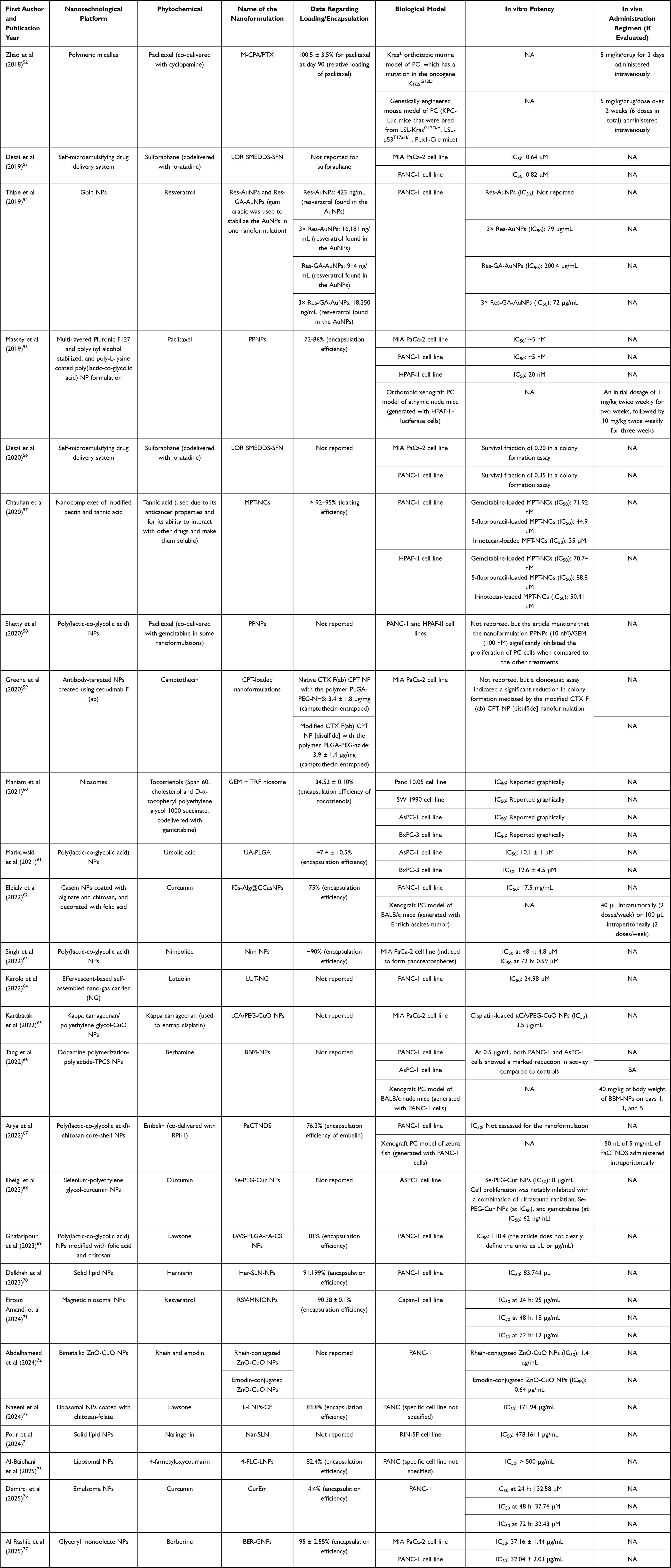 |
Table 1 Information Derived from Studies Addressing the Therapeutic Applications of Nanoformulated Phytochemicals Against PC |
Metallic Nanocarriers
Metallic NPs are gaining attention as powerful tools for cancer drug delivery, thanks to their ability to improve stability, solubility, and targeted release.78 On the other hand, phytochemicals, with their natural anticancer potential, often face challenges like poor absorption and rapid metabolism, which limit their clinical use.79 Incorporating phytochemicals into metallic NPs can help overcome these barriers, enhancing efficacy and potentially reducing side effects.80 In the context of PC, a malignancy characterized by late diagnosis, high resistance to conventional therapies, and poor prognosis, metallic NP-based delivery of phytochemicals offers a promising strategy to potentiate their anticancer potential and expand the therapeutic landscape for this challenging disease, as it has been evidenced in different remarkable reports.
For instance, in a study conducted by Thipe et al,54 resveratrol was employed both as a reducing and capping agent to synthesize resveratrol‑conjugated gold NPs (Res‑AuNPs), with gum arabic (GA) incorporated to enhance the colloidal stability of Au NPs and to serve as a supportive matrix for improved trans-resveratrol delivery. These authors systematically varied the thickness of the resveratrol layer coating the Au NP surface to examine its impact on anti‑tumor activity against breast (MDA‑MB‑231), pancreatic (PANC‑1), and prostate (PC‑3) cancer cell lines. As a result, the nanoformulation with a higher resveratrol coating, ie, 3× Res-AuNPs and 3× Res-GA-AuNPs, demonstrated significantly superior antiproliferative effects, attributed to enhanced cellular uptake and increased resveratrol bioavailability. Lower doses did not achieve effective IC50 values; these formulations were the only ones capable of reaching effective IC50 values of 79 μg/mL (3× Res-AuNPs) and 72 μg/mL (3× Res-GA-AuNPs) in PANC-1 cells. These findings illustrate a clear correlation between the density of resveratrol coating and therapeutic efficacy, highlighting Res‑AuNPs as promising candidates for oncological applications.
Karabatak et al65 described the development of multifunctional copper oxide (CuO) NPs coated with kappa carrageenan and polyethylene glycol (PEG), forming κCA/PEG-CuO NPs as both a digital colorimetric biosensor for dopamine and an anticancer drug nanocarrier. The NPs were also loaded with the well-established anticancer drug cisplatin (Cis) and, after physicochemical characterization, MTT assays were performed to assess cytotoxic activity of the nanoformulation on PC cells (MIA PaCa-2) as well as in other cell lines (hepatocellular carcinoma cells and human umbilical vein endothelial cells). The resulting IC50 value for Cis-loaded κCA/PEG-CuO NPs on MIA PaCa-2 cells (3.5 μg/mL) was significantly lower compared to the IC50 value of free Cis (12.5 μg/mL). The cytotoxic effect was attributed to the nanoformulation’s capacity to generate ROS, induce apoptosis via the mitochondrial (intrinsic) pathway, activate caspases, trigger the p53-mediated DNA damage response, and activate MAPK signaling. The authors also observed that the undiluted Cis-loaded κCA/PEG-CuO NPs exhibited cytotoxicity in 42% of MIA PaCa-2 cells. In contrast, only 28% cytotoxicity was observed in non-cancerous HUVEC cells, thus demonstrating selective cytotoxicity toward cancer cells and reinforcing its potential as a therapeutic agent against PC.
In research conducted by Abdelhameed et al,72 bimetallic ZnO-CuO NPs were loaded with the phytochemicals rhein or emodin via a green synthesis method using the plant extract as a natural reducing and stabilizing agent, to assess their anticancer efficacy against PC and ovarian cancer cell lines. Regarding the therapeutic effect of these nanoformulated phytochemicals against PC, MTT assays in the PANC-1 cell line showed IC50 values of 11.1 μg/mL for rhein and 6.7 μg/mL for emodin. Conversely, those values decreased significantly once the phytochemicals were incorporated into the bimetallic NPs, to 1.4 μg/mL for rhein and 0.64 μg/mL for emodin. The induction of apoptosis, which underlies these results, was confirmed through annexin V/PI staining. It was found that the delivery system induced an overall apoptotic cell death rate of 34.9% in PC cells, compared to 4.19% in untreated controls, and caused cell cycle arrest in the S-phase of PANC-1 cell proliferation, as shown by a cell cycle analysis. Finally, an RT-PCR-based gene expression analysis revealed that the treatment with emodin-loaded NPs upregulated the pro-apoptotic genes p53, BAX and caspases 3, 8, and 9, while downregulating the anti-apoptotic gene BCL2, thus indicating the activation of both mitochondrial and death receptor apoptotic pathways. The enhanced stability, solubility, and controlled release provided by the nanoformulations underlie their superior efficacy, highlighting their potential as promising therapeutic candidates against PC.
Lipid-Based Nanocarriers
Lipid-based nanocarriers are nanoscale drug-delivery systems composed primarily of lipids, designed to encapsulate and deliver therapeutic agents. They have emerged as versatile platforms for cancer therapy, offering biocompatibility, controlled drug release, and improved bioavailability of therapeutic compounds.81 Encapsulation in lipid-based systems helps overcome the drawbacks inherent to phytochemicals, facilitating their delivery and enhancing their therapeutic impact.82 Remarkably, lipid-based nanocarriers represent a promising strategy to maximize the benefits of phytochemicals in PC therapy.
Co-encapsulation of gemcitabine (GEM) and a tocotrienol-rich fraction (TRF) in niosomes was explored by Maniam et al60 as a dual-drug delivery strategy for PC. The nanoformulation was prepared via the film hydration method at a 1:1 drug ratio, and the entrapment efficiencies were 20.07 ± 0.22% for GEM and 34.52 ± 0.10% for TRF, demonstrating stability over several months. In vitro assays using multiple PC cell lines (ie, Panc 10.05, SW 1990, AsPC-1, and BxPC-3) revealed that the antiproliferative effect of GEM was markedly enhanced in combination with TRF, with up to a 2.78-fold increase in Panc 10.05 cells. In addition, niosome-mediated delivery further enhanced this effect, yielding a 9-fold increase in cytotoxicity. Although IC50 values for the nanoformulation were not explicitly reported, but only presented graphically, the data clearly support enhanced efficacy. Importantly, modulation of the TRF: GEM ratio within the niosomes significantly enhanced GEM uptake in Panc 10.05 cells. These findings provide a proof-of-concept for niosomal-mediated dual-drug delivery in PC, underscoring its potential to improve therapeutic outcomes.
Tang et al66 crafted berbamine-loaded dopamine polymerization-polylactide-d-α-tocopheryl PEG 1000 succinate (TPGS) NPs (BBM-NPs). The nanoformulation was first characterized and then evaluated in vitro using the PANC-1 and AsPC-1 cell lines and in vivo in PC xenograft models in BALB/c nude mice. It was found that the nanosystem remained stable, with an average particle size of 134 nm, and exhibited a slow, sustained drug-release profile compared to free BBM. Indeed, the release pattern proved pH-dependent, being slower at alkaline pH. In vitro analyses showed that the BBM-NPs suppressed migration and invasion and exerted a more substantial inhibitory effect on cancer cell viability and proliferation than free BBM in the two cell lines. The nanoformulation was more cytotoxic; however, no IC50 value was reported. Mechanistically, BBM-NPs promoted apoptosis, induced reactive oxygen species production, and increased the expression of BAX, Cleaved caspase-3, and γ-H2AX, while decreasing the protein expression of MMP2, MMP9, and BCL2 in PANC-1 and AsPC-1 cells.66
On the other hand, a xenograft tumor model of PC was generated by subcutaneously injecting 4-week-old male BALB/c nude mice with 1×10⁶ PANC-1 cells. Subsequently, mice were administered with 40 mg/kg body weight of free BBM and BBM-NPs on the first, third and fifth days, and tumor growth was monitored for 30 days until the mice were sacrificed. Results of this in vivo study showed that, although there were no significant changes in the mice´s body weight, both free BBM and BBM-NPs exhibited a strong inhibitory effect on the growth of xenograft tumors, as demonstrated by tumor weight measurements. Interestingly, BBM-NPs had the greatest impact on tumor weight, showing the plausibility of this nanoformulation against PC.66
Delkhah et al70 evaluated the cytotoxic and anti-metastatic properties of herniarin (7-methoxycoumarin) encapsulated in SLNs against various human cell lines, including the PANC-1 PC line, which showed the highest sensitivity with an IC50 of approximately 83.7 µM. Viability assays (MTT), flow cytometry, fluorescent staining (DAPI), and qPCR demonstrated that herniarin-SLNs induce apoptosis via both intrinsic and extrinsic pathways by increasing the expression of CASP9, CASP8, and CASP3, while reducing BCL2 expression. In addition, significant downregulation of the metastasis-related genes MMP2 and MMP9 was observed, which may be mediated by PI3K/Akt pathway inhibition, as suggested by previous studies. The authors propose that herniarin-SLNs represent a promising nanotherapeutic strategy for the treatment of PC, as they induce apoptosis and reduce tumor metastatic potential by significantly decreasing MMP2 gene expression, as evaluated by RT-PCR.
Later, Firouzi Amandi et al71 developed resveratrol-loaded magnetic niosomal NPs (RSV-MNIONPs) as an advanced delivery system for PC therapy. The formulation achieved a high encapsulation efficiency (85%) and exhibited sustained release, favoring stability and controlled drug availability. In Capan-1 cells, RSV-MNIONPs markedly enhanced cytotoxicity, particularly under an external magnetic field, and induced a more pronounced cell cycle arrest at the G0/G1 checkpoint than free resveratrol. Moreover, the minimum IC50 for RSV-MNIONPs in this study was 12 μg/mL at 72 hours. These effects were closely associated with significant modulation of key regulatory genes, including the upregulation of pro-apoptotic markers (BAX, P53, FAS) and the downregulation of anti-apoptotic and proliferative genes (Bcl-2, Cyclin D, hTERT). Such transcriptional reprogramming provided a mechanistic explanation for the nanoformulation’s improved anticancer efficacy, supporting its potential as a robust therapeutic platform for PC.
Naeeni et al73 synthesized and characterized lawsone-loaded liposomal NPs coated with chitosan-folate (L-LNPs-CF). The formulation displayed high encapsulation efficiency (83.8%) and demonstrated strong antioxidant capacity, as evidenced by significant free radical scavenging in DPPH and ABTS assays. Biological evaluation further demonstrated that L-LNPs-CF markedly inhibited PC cell proliferation (IC50 = 171.94 μg/mL) and promoted apoptosis by upregulating caspase-3, caspase-9, and Bax. Additionally, the L-LNPs-CF nanoformulation did not exert cytotoxicity on normal cells (represented by the HFF cell line) at any of the concentrations tested. Collectively, these findings indicate that L-LNPs-CF has an enhanced effect against PC cells, potentially driven by the incorporation of chitosan, which may facilitate cellular uptake of the nanodrug, and represents a promising therapeutic strategy against PC.
In another report by Pour et al.74 SLNs loaded with naringenin (Nar-SLNs) were developed. The cytotoxic effect of Nar-SLNs was evaluated in vitro, using Rat Pancreatic RIN-5F cells through the MTT assay, and the concentrations tested varied in a range from 4–1024 g/mL. The resulting IC50 values were 478.1611 g/mL and 442.093 g/mL for Nar-SLNs and free Nar, respectively. Although Nar-SLNs demonstrated greater cytotoxic efficacy than Nar at the specific concentrations of 64 and 128 µg/mL, an analysis of the expression of autophagy-related factors was also conducted, and the results suggested that both treatments, ie, Nar-SLNs and Nar, promoted the expression of autophagy markers (AKT, LC3, Beclin1, and ATG5) and decreased miR-21 levels in PC cells. Nevertheless, the Nar-SLNs formulation markedly enhanced the inhibition of cell proliferation and modulation of autophagy pathways. This superior efficacy is attributed to the improved solubility, stability, and sustained release of naringenin achieved through encapsulation within SLNs. The lipid matrix and surfactant composition in the NPs increased the drug’s aqueous dispersibility by reducing particle size and preventing crystallization, thereby enhancing solubility. Simultaneously, the lipid core protects naringenin from premature degradation and oxidation, thereby enhancing its stability. Finally, the NP matrix facilitated a controlled and sustained release profile, ensuring prolonged drug delivery over time.74 Accordingly, Nar-SLNs deserve further investigation so that they can advance into preclinical and clinical trials.
Al-Baidhani et al75 loaded 4-farnesyloxycoumarin (4-FLC) into nanoliposomes composed of lecithin-cholesterol-PEG (4-FLC-LNPs) to evaluate the anticancer and anti-metastatic effects of the nanoformulation. As a result, an encapsulation efficiency of 82.4% was obtained. Subsequently, cytotoxicity was measured against different cancer cell lines. In the PANC cell line, the IC50 was greater than 500 μg/mL, and no toxic effects were observed at concentrations below 62 μg/mL. Immunostaining and flow cytometry were used to analyze the expression of apoptosis- and metastasis-related genes in the presence of 4-FLC-LNPs. The nanoformulation induced BAX expression and decreased BCL-2, MMP2, and MMP9 expression. The antioxidant capacity of 4-FLC-LNPs was also assessed using ABTS and DPPH. It was also noticed that the average IC50 value for ABTS radicals was 31 µg/mL, and the highest inhibition reached was about 74%. Meanwhile, DPPH showed concentration-independent inhibition, ranging from 57% to 68%. Although the antioxidant activity of 4-FLC-LNPs was promising, its high IC50 value against the PANC cell line underscores the need to improve this nanoformulation, particularly for PC therapeutics.
Demirci et al76 evaluated the anticancer effects of curcumin-loaded emulsome NPs (CurEm) on the PANC-1 PC cell line. CurEm significantly reduced cell viability in a dose- and time-dependent manner, with an IC50 of 32.43 µM at 72 hours, demonstrating greater efficacy than free curcumin. Additionally, CurEm induced cell cycle arrest at the G2/M phase and induced morphological changes consistent with apoptosis, including spheroidal cell formation. Moreover, through colony formation and scratch assays, a notable inhibition of cell migration and colony formation was observed. At the molecular level, the expression of p53, p21, BAX, and CASP-3 was upregulated, along with downregulation of BCL-2, suggesting apoptosis activation via the p53 signaling pathway. These findings support the use of CurEm as a promising nanocarrier system that facilitates sustained release and improves the stability and bioavailability of curcumin for potential PC therapy.
Other Nanocarriers
In this section, we examine a set of studies that highlight therapeutic approaches using diverse nanocarriers, including PLGA NPs, nanocomplexes, self-microemulsifying systems, polymeric micelles, and antibody-functionalized NPs, among others. Exploring such a variety of delivery platforms for phytochemicals is crucial, as it enables the identification of the most effective and clinically translatable strategies for PC therapy. In fact, by systematically evaluating different carriers for stability, cellular uptake, therapeutic efficacy, and safety, researchers can better predict which methods are most likely to succeed in clinical trials, thereby laying the groundwork for the rational design of next-generation phytochemical-based therapeutics.
In this context, Zhao et al52 investigated a strategy to overcome primary resistance to immunotherapy in PC by modulating the tumor stroma using a polymeric micelle nanoformulation loaded with cyclopamine, a hedgehog pathway inhibitor that disrupts the stromal fibrosis characteristic of PC, and paclitaxel. The formulation was designed to modify the dense, desmoplastic TME, which acts as a physical and immunosuppressive barrier, hindering immune cell infiltration and the delivery of pharmacological agents. This approach was evaluated in Kras* orthotopic murine PC models and genetically engineered mice that closely mimic human PC progression. The outcomes of this investigation revealed that the nanoformulation combined with PD-1 blockers (immune checkpoint inhibitors that restore T-cell activation and enhance the immune system’s ability to attack cancer cells) significantly improved survival and anti-tumor efficiency. These effects were attributed to increased intratumoral vasculature density, which promoted the infiltration of cytotoxic CD8+ T cells into the tumor without depleting tumor-restraining fibroblasts or disrupting the collagen matrix. This restoration of equilibrium subsequently reversed immunosuppression and stimulated IFN-mediated immune response, thereby enhancing therapeutic efficacy.
In a subsequent investigation, Desai et al53 developed a synergistic chemoprevention strategy for PC using a combination of loratadine (LOR) and sulforaphane (SFN) formulated in a self-microemulsifying drug delivery system (SMEDDS). A series of in vitro MTT assays was conducted as part of the analysis using the PC cell lines MIA PaCa-2 and PANC-1. Among the most notable results, the IC50 values of the free LOR-SFN combination were 4.6 µM and 9.51 µM for MIA PaCa-2 and PANC-1, respectively. On the other hand, the incorporation of LOR and SFN into the SMEDDS formulation reduced the IC50 values to 0.64 µM and 0.82 µM, respectively, for MIA PaCa-2 and PANC-1. These improvements are attributed to the increased solubility and bioavailability of LOR and SFN provided by the SMEDDS platform, along with SFN’s enhanced antioxidant activity, which activates the Nrf2 pathway, augmenting cellular antioxidant defenses and modulating key signaling pathways controlling cell cycle arrest and apoptosis. Accordingly, this nanoformulation showed enhanced anticancer efficacy at lower doses, underscoring its potential as a promising therapeutic strategy against PC.
Desai et al56 further evaluated the LOR SMEDDS-SFN nanoformulation. An in vitro colony-forming assay was performed on two distinct PC cell lines, PANC-1 and MIA PaCa-2. The survival fractions observed for the LOR SMEDDS-SFN system were 0.35 and 0.20 for PANC-1 and MIA PaCa-2 cells, respectively. These results demonstrate the enhanced solubility of the phytochemicals and indicate that the SMEDDS formulation facilitates greater interaction with cancer cells, thereby promoting increased cellular uptake and permeation, ultimately leading to increased cell death. Regarding in vivo studies, a pharmacokinetic assay was performed in male Sprague-Dawley rats following oral administration of a single 200 µL dose containing 4 mg/kg LOR and 0.16 mg/kg SFN (LOR SMEDDS-SFN). Blood plasma samples were collected at predetermined time points and analyzed to determine the drug concentration over time. The resulting bioavailability value of 20,274.8 ± 3711 ng·h/mL and maximum plasma concentration of 503.2 ng/mL demonstrated a significant enhancement compared to conventional formulations. These pharmacokinetic parameters confirm the improved absorption and systemic exposure afforded by the nanoformulated system, thereby supporting the potential of LOR SMEDDS-SFN as a promising chemopreventive strategy for PC.
Massey et al55 developed a paclitaxel NP formulation (PPNPs), consisting of a PLGA core stabilized with Pluronic F127 and polyvinyl alcohol (PVA), and coated with poly-L-lysine. PC cell lines PANC-1, HPAF-II, and AsPC-1 were used for cytotoxic assays. The formulation exhibited cytotoxicity with IC50 values of approximately 5 nM for MIA PaCa-2 and PANC-1, and 20 nM for HPAF-II, as evidenced by MTT assays and colony formation assays. At the molecular level, PPNPs induced apoptosis, G2/M phase cell cycle arrest, and reduced cell migration and invasion. In an orthotopic xenograft model in mice generated with HPAF-II-luciferase cells, PPNPs at 10 mg/kg significantly reduced tumor volume and tumor weight, as confirmed by bioluminescence imaging. Likewise, a significant reduction in pancreatic bioluminescence intensity and the presence of necrotic areas were observed in the histopathological analysis of the excised tumors. It should be noted that although all control mice had foci in lymph nodes, liver, and lungs, minimal foci were detected in the lungs of mice treated with PPNPs, even while maintaining high efficiency in mice pretreated with PTX, indicating the ability to overcome drug resistance. In addition, a decrease in nuclear expression of Ki-67 (a cell proliferation marker) and of proteins associated with epithelial-mesenchymal transition, such as vimentin and Slug, was observed, supporting a reduction in tumor invasive capacity.55 Collectively, these findings highlight PPNPs as a safe and promising therapeutic platform for the treatment of PC, offering distinct advantages over conventional anticancer drugs.
Chauhan et al57 developed modified pectin-tannic acid nanocomplexes (MPT-NCs) to encapsulate GEM, 5-fluorouracil (5-FU), or irinotecan (IRI) via tannic acid binding. Tannic acid is an anticarcinogenic phytochemical known to enhance bioavailability, improve drug entrapment efficiency, and increase water solubility of drug molecules by promoting hydrogen bond formation. Characterization of the nanoformulation revealed a loading efficiency of over 91%. The cytotoxicity of the loaded NPs was assessed in vitro using two PC cell lines. In PANC-1 cells, the IC50 values of GEM, 5-FU, and IRI nanopreparations were 71.92 nM, 44.9 µM, and 35 µM, respectively. Likewise, the IC50 values of GEM, 5-FU, and IRI in HPAF-II cells were 70.74 nM, 88.8 µM, and 50.41 µM. Furthermore, the analysis of cellular uptake and colony formation in both PANC-1 and HPAF-II cell lines showed that the nanoformulation increased drug uptake per cell and inhibited the formation of cancer cell colonies.
Shetty et al58 evaluated a PLGA-based paclitaxel NP formulation (PPNPs) in combination with GEM for the treatment of PC. Paclitaxel was encapsulated in PLGA-F127-PVA NPs coated with poly-L-lysine. Afterwards, human PC cell lines PANC-1 and HPAF-II were treated with the nanoformulation, showing dose-dependent cytotoxicity. Mechanistically, this nanodrug exerted its effects by inducing apoptosis, disrupting lipid membrane integrity, neutralizing cell-surface charge, and causing G2/M cell cycle arrest. Zeta potential and Rh123 exclusion assays demonstrated that the PPNPs enhance GEM uptake by inhibiting P-glycoprotein efflux activity. Additionally, a significant downregulation of proteins crucially involved in de novo lipid synthesis (ie, FASN, ACC, Lipin, and Cox-2) was observed, which are essential for tumor survival, progression, and chemoresistance because de novo lipid synthesis activates drug resistance, allowing membranes, energy, and signals that favor survival against drugs to be maintained. Consistently, these observations suggest that PPNPs potentiate GEM efficacy by inhibiting lipid metabolism, inducing apoptosis, and promoting membrane remodeling, proposing this combination as an innovative therapeutic strategy against PC.
Greene et al59 optimized antibody-targeted delivery of camptothecin-loaded NPs using F(ab) fragments derived from the epidermal growth factor receptor (EGFR) antibody cetuximab to develop an efficient treatment against PC. Polymeric NPs were first functionalized with anti-EGFR antibodies to enhance their affinity to EGFR-expressing PC cells. The cellular models used in this study were BxPC-3, MIA PaCa-2, and PANC-1 cells due to their ability to express the antigen targeted by the functionalized NPs. The binding affinity of these optimized NPs to the cell lines was assessed by flow cytometry; the results confirmed the enhanced specificity of the modified NPs for surface-expressed EGFR. A clonogenic assay was also performed in MIA PaCa-2 cells to evaluate the impact on cell survival and colony formation when the optimized antibody-targeted NPs were administered. At the same time, uptake of the free phytochemical was insufficient to affect cell survival significantly; treatment with antibody-conjugated NPs carrying the phytochemical notably reduced colony formation. The effective targeting provided by the antibodies paired with high internalization efficiency mediated by the NPs facilitated the delivery of camptothecin to PC cells; these factors enhanced cytotoxicity, supporting the nanoformulation’s potential for selective and improved PC therapy.
In another investigation, Markowski et al61 assessed the cytotoxic activity of ursolic acid (UA) encapsulated in PLGA or PEGylated PLGA NPs against two PC cell lines, ie, AsPC-1 and BxPC-3. The IC50 values for UA-PLGA were reported to be 10.1 ± 1 μM (AsPC-1) and 12.6 ± 4.5 μM (BxPC-3). However, the authors indicate that the UA-loaded NPs showed highly similar values across all formulations and samples studied, ranging from 10.1 μM to 14.2 μM, with no significant differences, suggesting comparable potency to free UA. Confocal microscopy confirmed that cytotoxicity did not result from premature PLGA degradation and UA release into the extracellular medium, but rather from NP internalization via endocytosis, leading to intracellular UA release. While free UA and encapsulated UA showed similar effects in AsPC-1 cells, a statistically significant increase in cytotoxicity was observed in BxPC-3 at 20 μM with the nanoformulation. Finally, the authors noticed that PLGA-based nanoformulations, including PEGylated versions, retain UA cytotoxicity and may improve circulation and tumor targeting, supporting their potential for further evaluation in PC models.
Elbialy et al62 developed a curcumin nanocarrier system based on NPs that was created with a casein matrix coated with alginate and chitosan layers, and functionalized with folic acid. This nanoformulation was denominated fCs-Alg@CCasNPs. The system was characterized using various assays, and its effectiveness was evaluated in vitro and in vivo. The in vitro release study of the fCs-Alg@CCasNPs showed enhanced curcumin release in the TME (pH 5.5) up to 53%, demonstrating that protein surface modification prevents contact between the release media and curcumin, thus controlling the drug release rate and improving curcumin bioavailability. Afterward, cytotoxicity was assessed in PANC-1 cells through the SRB assay, and it was observed that curcumin induced higher cytotoxicity when paired with fCs-Alg@CCasNPs (IC50 of 17.5 g/mL), as compared with free curcumin (IC50 of 76 g/mL). These results were attributed to the greater adhesion capacity between cancer cells and NPs, which increased cellular penetration and folate ligand binding. In vivo studies used BALB/c mice as animal models. Mice were intraperitoneally inoculated with Ehrlich ascites tumor cells to establish the tumor mo afterwards, the nanocarrier system was delivered via intratumoral injection and intraperitoneally, with 2 doses of 40 μL of fCs-Alg@CCasNPs per week for 3 weeks. Regarding pharmacokinetics, the maximum plasma curcumin concentration for the fCs-Alg@CCasNPs was 27.5 ng/mL, much lower than the free curcumin concentration (117 ng/mL), evidencing improved curcumin bioavailability with prolonged circulation time.62 The efficient drug delivery achieved through this nanoformulation resulted in a pronounced antitumor effect, evidenced by an inhibitory rate of 71% for fCs-Alg@CCasNPs. Comet and histopathology assays revealed that fCs-Alg@CCasNPs demonstrated superior therapeutic efficacy compared to free curcumin by significantly increasing DNA damage in cancer cells while sparing vital organs, indicating high specificity and safety.62
Besides, Singh63 et al assessed the effect of nimbolide (Nim), a phytochemical with anticancer properties, encapsulated in PLGA NPs, on PC stem cells (CSCs). MIA PaCa-2 cells were cultured under pancreatosphere-forming conditions, a strategy that enriches the CSC population. Although the IC50 value was not reported, MTT viability assays, apoptosis assays, and pancreatosphere formation assays demonstrated that Nim NPs more effectively reduced cell viability, induced apoptosis, and decreased self-renewal capacity compared to free Nim. At the molecular level, Nim and Nim NPs showed high-affinity binding to AKT and mTOR, inhibiting both proteins, as confirmed by molecular docking, molecular dynamics simulations, and Western blot analysis. This dual inhibition triggered mesenchymal-to-epithelial transition, which was evidenced by increased levels of E-cadherin and decreased expression of vimentin, N-cadherin, fibronectin, and ABCG2 (a drug-resistance marker). The authors suggest that Nim-loaded NPs could overcome chemoresistance and reduce tumor initiation capacity, as CSCs are associated with intrinsic drug resistance and the ability to regenerate tumors. Overall, Nim NPs can induce MET, apoptosis, and loss of the CSC phenotype, offering a promising strategy to overcome chemoresistance and tumor initiation in PC.
Karole et al64 demonstrated another nanocarrier system for the phytochemical luteolin (LUT), which was formulated as enteric-coated effervescent granules (LUT-NG). This nanoformulation was projected to enhance the intestinal epithelial absorption of LUT and facilitate its effective transport to the pancreas via the oral route by protecting the formulation from the harsh gastric environment. After the physicochemical characterization of the nanodrug, an in vitro drug release assay was executed, the results showed a minor quantity of LUT being released at first (pH 1.2) but once the pH was raised to intestinal pH (7.4) the concentration of the released drug increased, showcasing the protection effect of the coating and confirming the delivery potential of the drug through the oral route with the nanocarrier. Further, MTT and Live/Dead assays performed in PANC-1 cells to assess the anticancer effects of the delivery system revealed an IC50 of 24.98 μM for LUT-NG, which was significantly lower than that of free LUT (50.56 μM). These findings validate the improved solubility of the nanoformulation, which, in turn, translates into greater cytotoxicity against PC cells. Results from other tests, including Hoechst 33258 staining, caspase-3 activation detection, and G2/M cell cycle arrest analysis, collectively reinforced the enhanced anticancer effects of LUT-NG. These effects were mediated by inducing apoptosis through the regulation of cell cycle-related molecules, inhibiting tumor angiogenesis and metastasis, and directly suppressing tumor cell proliferation.
Arya et al67 introduced a personalized nanodelivery approach called the PC-targeted PLGA-chitosan core-shell NP delivery system (PaCTNDS), designed for PC therapy by harnessing the synergistic effects of embelin (Emb) and RPI-1. Embelin is a natural benzoquinone that inhibits the X-linked inhibitor of apoptosis protein via the NF-kB pathway. At the same time, RPI-1 is an indolinone derivative that targets c-MET, a key biomarker in PC. The optimized synergistic ratio of Emb to RPI-1 (1:4.7) demonstrated enhanced cytotoxicity against PC cells (PANC-1) while minimizing toxicity to normal cells (WI-38). In vitro studies revealed significant anticancer effects, including apoptosis induction, mitochondrial membrane depolarization, cell cycle arrest at G2/M and S phases, and anti-metastatic properties such as reduced migration, invasion, and colony formation. Meanwhile, in vivo experiments using zebrafish xenograft models showed favorable biodistribution, marked tumor volume reduction, and improved survival rates with 50 nL of 5 mg/mL administered intraperitoneally. Metabolomic analysis via SERS and LC-MS identified alterations in purine, pyrimidine, and carbohydrate metabolism, further elucidating the apoptotic mechanisms triggered by Emb and RPI-1. These findings underscore the potential of PaCTNDS as a targeted and effective therapeutic strategy for PC, warranting further preclinical and clinical evaluations.
As a novel sonosensitizer for sonodynamic therapy (SDT) of PC cells, Selenium-PEG-curcumin NPs (Se-PEG-Cur NPs) were synthesized by Ilbeigi et al.68 The nanoformulation demonstrated substantial anticancer effects, primarily through enhanced reactive oxygen species production and apoptosis induction. Se-PEG-Cur NPs exhibited dose-dependent cytotoxicity with an IC50 value of 8 μg/mL, while GEM showed an IC50 of 62 μg/mL. The combination of Se-PEG-Cur NPs and GEM, with ultrasound (US) radiation, significantly reduced ASPC1 cell viability to 10%, demonstrating a synergistic therapeutic effect (combination index of 1.6). The mechanism of action of this nanoformulation involved ROS generation, mitochondrial membrane disruption, and DNA damage, facilitated by the cavitation effects of US and the sonosensitizing properties of Se-PEG-Cur NPs. The nanoformulation’s stability, biocompatibility, and ability to enhance drug delivery to tumor sites underscore its potential as an efficient and targeted treatment for PC.
Ghafaripour et al69 successfully developed lawsone-encapsulated PLGA NPs modified with chitosan and folic acid (LWS-PLGA-FA-CS NPs) to evaluate their anticancer potential against the PANC-1 cell line. The nanoformulation had an encapsulation efficiency of 81%, which is favorable for drug delivery systems. Besides, LWS-PLGA-FA-CS NPs significantly inhibited PANC-1 cell growth in a dose-dependent manner, with an IC50 of 118.4 μL, and showed no cytotoxicity in normal HFF cells. Mechanistically, the loaded NPs induced apoptosis by upregulating the pro-apoptotic BAX gene and downregulating the anti-apoptotic BCL2 gene, suggesting activation of the intrinsic apoptotic pathway. Additionally, flow cytometry revealed an increased proportion of cells in the sub-G1 phase, confirming the induction of apoptosis. The nanoformulation also exhibited potent antioxidant activity, with IC50 values of 250 μg/mL (DPPH assay) and 62.5 μg/mL (ABTS assay), and antibacterial activity against Gram-negative bacteria, including E. coli and P. aeruginosa. These findings highlight the potential of LWS-PLGA-FA-CS NPs as a promising candidate for PC treatment, leveraging their multifunctional therapeutic properties.
More recently, Al Rashid et al77 created berberine-loaded glyceryl monooleate NPs (BER-GNPs) as a putative therapeutic approach against PC. Characterization revealed that this nanoformulation had high stability and successfully encapsulated berberine, enhancing its bioavailability and sustained release. In vitro experiments also demonstrated that BER-GNPs significantly enhanced cellular uptake and cytotoxicity in MIA PaCa2 (IC50 of 37.16 ± 1.44 μg/mL) and PANC-1 cells (IC50 of 32.04 ± 2.03 μg/mL), with the loaded NPs showing superior antiproliferative effects compared to native berberine. Mechanistically, BER-GNPs predominantly inhibited the AKT signaling pathway by suppressing AKT phosphorylation, leading to increased pro-apoptotic Bax and decreased anti-apoptotic Bcl-2, thereby activating intrinsic apoptotic pathways. Eventually, in silico docking studies supported these findings, showing favorable binding interactions of berberine with AKT. Overall, these observations indicate that the BER-GNP nanocomposite enhances berberine’s stability and efficacy, induces apoptosis by suppressing AKT-mediated survival signaling, and represents a promising nanotechnological strategy for PC therapy. An overview of the key phytochemicals, nanocarriers, biological models, and assays performed to evaluate the efficacy of the nanoformulations discussed herein is illustrated in Figure 3.
 |
Figure 3 Overview of the main research strategies involving nanoformulated phytochemicals for PC therapy. The figure depicts representative classes of phytochemicals (terpenoids, quinones, flavonoids, polyphenols, coumarins, and alkaloids), the principal nanocarriers used (lipid-based systems, polymeric NPs and micelles, metallic NPs, nanocomplexes, and polysaccharide- or sulfate-based carriers), and common in vitro and in vivo models (PC cell lines such as MIA PaCa-2, HPAF-II, SW1990, AsPC-3, BxPC-3, Capan-1; and mouse models including BALB/c mice, KPC-Luc, and zebrafish). Central icons summarize the main assays used to evaluate these nanoformulations, ie, assessments of cell viability, apoptosis, cell migration/invasion, oxidative stress, and in vivo tumor development (created with a licensed version of BioRender.com). |
Discussion
The insights from this scoping review indicate that nanoformulated phytochemicals have remarkable potential to be developed as innovative drugs for the treatment of PC, as evidenced by a handful of studies that tested their effectiveness. Notwithstanding this, several concerns and hurdles must be addressed in future research to enable these nanoformulations for cancer treatment to reach the clinical landscape, as exemplified by the case of the nanodrug Abraxane (NP-albumin-bound paclitaxel).83 Indeed, the emergence of newly approved nanoformulated phytochemicals could be a turning point in this field, as the number of such nanomedicines targeting PC that are in clinical trials or already licensed remains very limited, as noted earlier in the Introduction. In this context, concerted efforts from scientists, policymakers, and the pharmaceutical industry are essential to achieve this objective, and the insights provided in this review may serve as a valuable resource toward that goal.
One of the main limitations identified during the analysis of the evidence sources is that most conclusions were based solely on cell viability or proliferation assays (21 out of 26 studies). Although cell cultures provide a cheap and general approach to understanding the inhibitory effects of phytochemical-centered nanoformulations in cancer cell lines and their cytotoxicity in healthy cells, they poorly reflect the complexity and heterogeneity of patient tumors, as tumor behavior is strongly influenced by interactions with the extracellular matrix and surrounding host cells, which are factors that are absent in standard tissue culture systems.84,85 In this regard, scaffold-based 3D cell cultures may provide tumor models that simulate in vivo conditions, enabling better study of cancer progression, drug responses, and microenvironment interactions. Moreover, patient-derived scaffolds can be applied to generate personalized medicine, potentially increasing the accuracy of therapeutic predictions during anticancer drug testing.86 On the other hand, patient-derived cancer organoids, along with animal models including genetically engineered mice, patient-derived xenografts, rabbits, and non-human primates, could provide a broader perspective on the effects of novel anticancer drugs,87,88 such as nanoformulated phytochemicals for PC. Nevertheless, there is currently no biological model that fully mirrors the complexity of patients’ reality; instead, the clinical development and assessment of anticancer drugs should rely on integrated data derived from multiple models.89
Hence, most of the phytochemical nanoformulations discussed herein warrant further in-depth investigation, particularly through future evaluations across diverse biological models, to comprehensively assess their efficacy and safety before entering human trials. This clinical chronology is depicted by Zhao et al,52 Massey et al,55 Elbialy et al,62 Tang et al,66 and Arya et al,67 as these researchers evaluated the anticarcinogenic effects of their phytochemical-loaded nanoformulations in animal models of PC. Additionally, conducting comprehensive analyses on pharmacokinetics, pharmacodynamics, and safety profiles, including the evaluation of potential adverse effects, is essential to ensure the clinical viability of phytochemical-based nanoformulations for PC, as previously stated for these nanotechnological approaches.90 Such evaluations will help to confirm the absence of toxicities or off-target effects, thereby supporting the development of these nanoplatforms as reliable and safe therapeutic strategies. Particularly, only 3 out of the 26 revised articles evaluated the safety of the nanoformulated phytochemicals through histopathological examination,55,56,62 while only Desai et al56 and Elbialy et al62 performed pharmacokinetic studies.
Safety concerns related to the different nanocarriers reviewed may arise mainly from their physicochemical properties and their interactions within more complex biological systems. For instance, metallic NPs generate reactive oxygen species that can lead to oxidative stress and inflammation, and their leaching can induce cytotoxic effects and accumulate in organs, specifically the liver and spleen, causing long-term toxicity issues. However, surface modification of these nanocarriers, such as coating with biocompatible materials, can mitigate these effects.91 Other types of nanoplatforms, such as polymeric or lipid NPs, offer effective alternatives if the chosen components are non-toxic and fully characterized for immunotoxicity; otherwise, it might risk activation of the immune system, causing adverse reactions, and could also affect pharmacokinetic parameters like circulation time and clearance, which are essential for dosing and efficacy considerations.92,93 Additional nanomaterials, including dendrimers and carbon-based NPs, may pose risks, including hemolysis, cytotoxicity, and interference with normal cellular functions. For dendrimers, reported toxicity is often linked to their positive surface charge, which can be mitigated by replacing cationic groups with neutral or anionic groups via glycolation, PEGylation, or conjugation with peptides and carbohydrates. As for carbon-based NPs, they exhibit reduced toxicity when functionalized with groups that enhance solubility and biocompatibility, such as carboxyl or polyamidoamine (PAMAM) dendrimer coatings, thereby improving cellular uptake and reducing inflammation.94,95
Regarding the profiles a nanoformulation should entail leading to an Investigational New Drug (IND) application, the appropriate in vitro and in vivo studies must be performed; they should include a complete physicochemical characterization of the nanosystem (size, charge, morphology, drug loading, stability, and release kinetics) as well as pharmacokinetic studies including adsorption, distribution, metabolism excretion (ADME) assessments, plasma concentration-time profiles, bioavailability, and organ-specific biodistribution in appropriate animal models. Pharmacodynamic assays should verify that the target is engaged correctly, demonstrate the mechanism of action, establish dose-response relationships, and identify therapeutic windows.96,97 Safety evaluations should encompass acute and chronic toxicity studies, histological examination of major organs to detect off-target effects, immunogenicity assays to monitor hypersensitivity or immune activation, genotoxicity and inflammation biomarkers; especially regarding polymeric and metallic NPs, immunotoxicological assays are crucial. Finally, complementary reproductive and developmental toxicity testing should be considered as well.96,97 All these evaluations must be conducted in strict compliance with the relevant regulatory guidelines, ensuring thorough standardization and robust data to support the safety and efficacy profile required for clinical trial authorization. However, there remains a lack of well-established, standardized protocols specifically designed to ensure the safety of NPs during early developmental stages. Despite this, there is consensus among researchers and regulatory organizations that the parameters mentioned earlier should be thoroughly studied before NPs are considered “safe”.98–100
Working with animal models in drug-testing studies presents several substantial challenges, including financial costs, the need for strict ethical oversight, the requirement for specialized and properly equipped facilities, and the continuous care and maintenance of animals, demands that significantly increase as the biological requirements of the animal model increase.101,102 These constraints help to explain why a considerable proportion of studies in this field have not yet advanced to in vivo experimentation. To reduce the need for animal testing, remarkably, to follow the recommendations of the FDA Modernization Act 2.0/3.0,103 researchers can integrate alternative approaches such as organ-on-chip systems, advanced computational modeling, and refined experimental designs.104,105 Given that the studies that advanced to in vivo evaluation demonstrated that nanoformulated phytochemicals can effectively suppress PC tumor growth, providing valuable insights into their potential clinical translatability, animal experimentation continues to play an essential role in this area of research. Nevertheless, to reduce the number of animals used in experiments, instead of conducting a single study with a whole cohort, the initial group size can be partitioned into multiple smaller cohorts, each treated at different time points, so that reproducibility is preserved without requiring huge test groups. In this regard, a design initially requiring nine animals could instead be implemented as three separate mini-studies, each involving three biological replicates.106
Another critical issue that should be considered in forthcoming studies is the encapsulation or loading efficiency of a phytochemical into a nanocarrier, which was reported in only 14 of 26 studies, with 95 ± 2.55% as the maximum reported encapsulation efficiency. This parameter depends on factors such as the physicochemical compatibility between the drug and the carrier, the NP’s surface properties, and the formulation method.107,108 Given that this efficiency will be related to the actual amount of phytochemicals delivered to a given biological system (effective dose), it can be improved by incorporating ligands that specifically bind the phytochemical to the NP,109 or by using stabilizers, such as chitosan, casein, saponins, PEG, among others, which can enhance stability mainly through electrostatic interactions and outer layer formation,110 these strategies have been considered in several articles included in this review. Additionally, nanotechnological platforms can enable precise control over phytochemical release, enabling prolonged, site-specific delivery. This can be accomplished by integrating smart, stimuli-responsive components into the delivery platform that respond to defined signals to trigger the release of therapeutics at the desired site. These stimuli include, but are not limited to, changes in pH, temperature shifts, or specific enzymatic activities.111 However, as observed in our analysis, these techniques have been applied sparingly in the design of phytochemical-based nanoformulations for PC and should therefore be explored in depth in future studies.
A notable limitation across nearly all the investigations discussed herein is the absence of a systematic optimization process for preparing phytochemical-loaded nanoformulations. In this matter, one of the most crucial aspects in nanoformulation development is the optimization of its physicochemical and functional characteristics. This process can be effectively guided by the Quality by Design (QbD) methodology. QbD employs statistical experimental designs to optimize key parameters, including particle size, zeta potential, polydispersity index, surface morphology, entrapment efficiency, and drug release profiles, thereby enhancing the quality and reliability of the final product and simplifying the formulation process.112,113 Since emerging research on nanoformulations containing phytochemicals for cancer therapy is incorporating the QbD approach in their methods,114–116 nanotechnologists are encouraged to consider adopting this strategy in future studies to advance the standardization of the production methods for phytochemical-based nanoformulations targeting PC, as it was exemplified by Desai et al,53 who applied QbD in their protocol to develop LOR SMEDDS-SFN for treating PC.
Unfortunately, most phytochemicals exhibit poor aqueous solubility, which represents a major obstacle to the development of effective therapies for PC. For example, potent anticarcinogenic phytochemicals such as curcumin, quercetin, and resveratrol are poorly soluble in water,117,118 limiting their dispersibility, bioavailability, and therapeutic efficacy. In this context, specific nanocarriers (eg, SLNs, dendrimers, and polymeric micelles) offer a promising strategy to overcome these limitations by enabling the effective dispersion of hydrophobic compounds within an aqueous phase, thereby enhancing solubility and bioavailability, and also allowing the codelivery of phytochemicals with existing anticancer medicines.119 Although the successful use of various nanocarriers has been demonstrated in the articles reviewed here, other nanoplatforms remain to be explored in the field of phytochemical-based nanoformulations for PC management. In this context, nanozymes and nanoemulsions can be combined with phytochemicals to develop stable, multifunctional nanoformulations. Predominantly, nanozymes are nanomaterials with intrinsic enzyme-like catalytic activities that can enhance anticancer efficacy by promoting oxidative stress and/or modulating the conditions of the TME.120 Although the combination of phytochemical-nanozymes has been barely examined in cancer therapy, it has been reported that iron-based nanozymes containing capsaicin were able to mitigate sepsis-associated acute lung injury, mainly through modulation of the NF-κB signaling pathway.121 This premise suggests that the therapeutic effects of nanozymes functionalized or loaded with phytochemicals could be extrapolated to anticancer drugs in the near future.
Furthermore, nanoemulsions are oil-water dispersions stabilized by surfactants that can improve the solubility, stability, and bioavailability of poorly water-soluble molecules, like phytochemicals, ensuring homogeneous drug distribution during treatment.122 Since nanoemulsions containing naringenin123 and epigallocatechin-3-gallate124 have shown substantial anticancer effects in lung cancer cells, these types of approaches should begin to be tested in PC therapy. In fact, Grzeszczak et al125 developed a nanoemulsion containing the phytochemical phenethyl isothiocyanate, which displayed significant cytotoxic effects on the PC cell lines AsPC-1 and BxPc-3, thus evidencing the potential of phytochemical-containing nanoemulsions for PC treatment. Remarkably, nanoemulsions also broaden the horizon for the delivery of multiple therapeutic agents targeting cancerous cells. For example, Bahadori et al126 found that a pH-responsive nanoemulsion containing cyclodextrin, zein, and TiO2 NPs, loaded with quercetin, not only minimized side effects on healthy tissues but also showed cytotoxicity in the A549 lung cancer cell line.
The term “nano-phytochemical” typically refers to phytochemicals encapsulated or loaded onto a nanocarrier.127–130 However, it is worth noting that phytochemicals can also serve as NPs on their own, as some can be converted via physicochemical methods (eg, wet milling) into nano-sized particles that usually require lower dosages to exert their therapeutic effects.122,131 For instance, Kumar et al132 and Zamanidehyaghoubi et al133 reported the production of nanocurcumin from curcumin, which was successfully utilized in anticancer and antibacterial strategies, respectively. Other examples of phytochemicals that can be transformed into nanocompounds include naringenin,134 apigenin,135 quercetin,136 and resveratrol.137 Since a few of the articles evaluated in this scoping review considered the use of nano-sized phytochemicals, we endorse exploring the anticancer potential of these compounds in forthcoming research focused on PC. Likewise, investigations should also focus on other phytochemicals with demonstrated activity against PC, thereby expanding the current therapeutic repertoire and fostering the development of novel nanoformulations. Among phytochemicals with proven therapeutic effects against PC that could be considered for future nanoformulations are baicalein,138 Corynoxine,139 celastrol,140 shikonin,141 and piperlongumine.142
Even though the molecular mechanisms of action of many phytochemicals have been widely described,143 it remains crucial to determine whether these mechanisms are preserved when delivered via nanocarriers, since the NPs themselves may also exert therapeutic effects.144 To address this, transcriptomic, proteomic, and metabolomic profiling analyses would be highly valuable, as they can help explain the underlying mechanisms of action of phytochemical-based medicines.145,146 Notably, among the reviewed investigations, only a few incorporated this type of analysis, highlighting a significant gap that warrants further investigation. Anyhow, the current body of research in this field indicates that, even though the precise mechanisms of action of several phytochemical-laden nanoformulations designed to treat PC are unknown, these types of nanotechnological platforms offer a viable way to overcome several of the challenges that are intrinsic to the nature of phytochemicals, which include low permeability through the cell membrane, rapid metabolism and body clearance, poor solubility, and low bioavailability.90
Overcoming these biological obstacles can enhance anticancer effects mediated by various factors. For instance, the use of soluble nanosystems or the functionalization of NPs with hydrophilic components increases the solubility of the cargo, therefore facilitating its delivery to cancer cells.147 Due to their nanometric dimensions, these nanoformulations exhibit enhanced capacity to interact with cell membranes and can be efficiently taken up by cells via multiple internalization pathways, such as endocytosis.148 As well, the nanoencapsulation of phytochemicals embeds them within a protective matrix that safeguards these compounds from destabilization by physiological and environmental factors, including enzymatic breakdown in the gastrointestinal tract and degradation caused by light or oxygen exposure, thereby prolonging their time in the body and mediating a prolonged therapeutic effect.149 Finally, the use of both passive and active targeting strategies could help to overcome the significant barrier of selectivity in drug delivery and promote the anticancer activity of nanoformulated phytochemicals. In particular, functionalizing the surfaces of the nanoplatforms with molecules, such as ligands or antibodies, enables selective recognition of cancer cells and can help minimize off-target effects in healthy tissues.150
Importantly, incorporating TME remodeling strategies into next-generation nanoformulated phytochemical-based therapies, rather than focusing solely on the cytotoxic effects of these nanosystems, is essential, as the TME’s conditions profoundly influence drug penetration, therapeutic responsiveness, and overall treatment efficacy. Some examples of these strategies include the incorporation of coatings containing DSPE-PEG2000-galactose, which can help regulate lactate production and counteract immune suppression in the TME.151 As well, the inhibition of glycolysis and induction of cuproptosis are mechanisms that have been demonstrated to remodel the TME when phloretin-loaded calcium carbonate NPs are coated with luteolin-copper networks.152 The incorporation of agents capable of triggering tumor vessel dilatation (thereby diminishing hypoxia), such as hydralazine,153 is another approach that should be considered in future designs of phytochemical-loaded nanoformulations. Furthermore, combining Arg9 to enhance cellular penetration with the encapsulation of the nanoplatform within M1 macrophage-derived membrane vesicles to promote the reactivation of immunosuppressed M2 macrophages constitutes a dual strategy that has proven effective in remodeling the TME in NP-based systems.154 Last but not least, targeting the genetic factors associated with TME development by adding tailored gene therapies (eg, CRISPR, RNA interference, and ncRNAs) to the nanoformulation is another approach that should not be overlooked.155–157
Additionally, it is imperative that future research evaluate the potential for resistance to nanoformulated phytochemicals against PC, as previous evidence indicates that certain cancer cell types can acquire resistance to phytochemical-based treatments. Regarding this, it has been observed that hepatocellular carcinoma cells can develop resistance to resveratrol158 and bladder cancer cells to paclitaxel,159 while Bonham et al160 noticed that tumors in LuCaP 35 prostate cancer xenograft models acquired resistance to baicalein and resumed growth, reaching volumes of approximately 1000 mm3. It is highly likely that simpler nanoformulation designs, ie, those consisting solely of a nanocarrier and a phytochemical, may be the most vulnerable to PC resistance mechanisms, including drug efflux pumps and the persistence of cancer stem cells.161 Consequently, beyond the incorporation of multiple therapeutic modalities within a single nanosystem, the nanoformulation should also strategically integrate features that hinder resistance mechanisms by targeting cancer stem cells,162,163 inhibiting efflux pump activity,164,165 and promoting stromal reprogramming.166,167
This scoping review presents notable strengths, including the use of a systematic and transparent methodology supported by an extensive multi-database search. Moreover, our search strategy underwent several refinements before its final application on article identification and extraction, ensuring extensiveness and reliability. Another strength lies in our strict adherence to PRISMA principles, which helped minimize potential biases arising from human error during the review process. However, one potential limitation is the exclusion of studies published before 2018. Nevertheless, considering the availability of prior reviews on this subject, as highlighted in the introduction, our scoping review was intentionally directed toward more recent contributions to this research arena, restricting the analysis to studies published between 2018 and 2025. Another limitation is that all the data included in this review were derived from preclinical studies, which displayed considerable heterogeneity in aspects such as experimental design, PC cell lines, cell proliferation assays, type of nanocarrier, phytochemical employed, methodology followed to prepare the nanoformulation, effective concentrations, and even the units reported for each effective concentration (eg, μg/mL, μM, or μL). Although efforts were made to address this heterogeneity by organizing studies by nanocarrier type, the findings should be interpreted cautiously, as these differences may influence overall conclusions about the true therapeutic potential of phytochemical-based approaches against PC.
In this regard, given the variability in study designs, experimental conditions, and reporting formats across the available literature, conducting a systematic review with meta-analysis at this moment is not entirely viable to estimate the overall therapeutic efficacy of phytochemical-loaded nanoformulations against PC. Nevertheless, the guidance offered in this scoping review is intended to help establish a foundation for future research on nanoformulated phytochemicals for PC, encouraging greater consistency in methodological reporting and data presentation. Such standardization will eventually support the feasibility of a comprehensive systematic review and meta-analysis once a more harmonized body of evidence becomes available. In parallel, to better account for this variability and refine the assessment of the anticancer efficacy of nanoformulated phytochemicals for PC, additional evidence from both preclinical evaluations and future clinical trials will be essential, ideally enabling reviews with robust statistical assessments through meta-analysis, as exemplified by Al-Samydai et al168 and Boroughani et al.169 Lastly, the restriction to English-language publications may have excluded relevant studies in other languages. However, the small number of such records suggests that their inclusion would not significantly alter our conclusions.
The available findings from preclinical studies on phytochemical-based nanoformulations for PC suggest a promising therapeutic potential. A recent meta-analysis of nano-phytosomes found that flavonoid-loaded NPs exhibited higher IC50 values against cancer than terpenoid-based counterparts.168 Moreover, a multivariate analysis of covariance performed in the same study highlighted that the phytochemical exerted a significant effect on therapeutic outcomes, even when accounting for cancer cell type and phospholipid composition as covariates.168 These data imply that phytochemical-containing nanoformulations for PC might follow a similar trend. Nonetheless, as previously discussed, the current body of information is mainly based on observations from cell line studies and, therefore, further validation in clinically relevant models is required to confirm whether these nanoformulations can consistently achieve high efficacy against PC. Once clinical trial data on nanoformulated phytochemicals for PC become available, it will be essential to conduct systematic reviews that compare the therapeutic efficacy of different nanoplatforms. Accordingly, the present scoping review should be considered as a preliminary step toward future systematic reviews with meta-analyses, which would allow for the construction of statistically robust conclusions on the relative effectiveness of these emerging nanotechnological therapies.
Overall, upcoming investigations on nanoformulations containing phytochemicals for PC treatment should adopt a structured, multilayered research pipeline that begins with comprehensive in vitro testing across a diverse panel of PC cell lines to capture phenotypic and genotypic variability. Promising candidates should then be evaluated in biologically relevant in vivo PC models (considering ethically and statistically reliable practices) to define better therapeutic efficacy, biodistribution, and safety within an integrated physiological context. On the other hand, to enable meaningful comparisons across studies, researchers must consistently report key physicochemical parameters of the nanoformulations, including particle size, polydispersity index, zeta potential, encapsulation efficiency, and drug-release behavior under simulated physiological conditions. Notably, cytotoxicity metrics, such as IC50 values, should be reported in standardized units, as this practice can facilitate comparisons across nanoformulations. We recommend adopting μg/mL (μg of nanoformulation per mL of solution) as the standardized unit for reporting nanoformulation potency. Since these nanosystems comprise multiple components (including nanocarriers, phytochemicals, ligands, and other auxiliary materials), determining an accurate molar concentration (μM) for the complete formulation is highly impractical. In contrast, reporting IC50 values in μg/mL might help to integrate the nanoformulation into a unified therapeutic entity.
Furthermore, the characterization of the mechanisms of action of the nanosystems should be strengthened through transcriptomic, proteomic, and/or metabolomic analyses to elucidate the molecular pathways that are modulated by these treatments. Additional considerations (that can be reported in a complementary article) include assessing long-term toxicity, safety, pharmacokinetics, and pharmacodynamics; incorporating 3D culture or organoid models as intermediate validation systems; and evaluating the possibility that PC models may develop resistance to treatment with the nanoformulation. Collectively, with these recommendations, we aim to propose a guideline to enhance methodological rigor, reproducibility, and translational relevance in the emerging field of phytochemical-based nanotherapeutics for PC. The key future directions that we recommend pursuing in this research field are shown in Figure 4.
 |
Figure 4 Future perspectives for nanoformulated phytochemicals. (A) Additional experimental assessments, including pharmacokinetic and pharmacodynamic assays, comprehension of the molecular mechanisms induced by the phytochemical-based nanoformulations, as well as evaluations of safety and clinical viability. (B) Enhanced NP design based on the incorporation of phytochemical-binding ligands, stimuli-responsive components, and optimization of physicochemical and functional properties. (C) Development of next-generation nanoplatforms such as nano-sized phytochemicals, nanoemulsions, and nanozymes with improved functions. (D) Experimental considerations involving more complex biological models, including patient-derived xenografts, organoids, 3D cell culture, and assessment of resistance emergence to phytochemical-loaded nanoformulations (created with a licensed version of BioRender.com). |
Conclusion
Phytochemical-based nanoformulations hold considerable promise as innovative therapeutic strategies for PC; however, significant challenges remain before they can be translated into clinical practice. Some of these key challenges include the need for extensive validation in advanced models such as xenograft animal models of PC and 3D cell cultures, alongside comprehensive analyses of their pharmacodynamics, pharmacokinetics, mechanisms of action, and potential toxicities. Furthermore, future research should expand beyond the most frequently studied compounds to explore other overlooked phytochemicals and alternative approaches, such as nano-sized phytochemicals, which may further enhance the potential of these nano-based therapies. As a closing call to action, we emphasize the urgent need to standardize experimental procedures and reporting practices in studies evaluating phytochemical-based nanoformulations against PC. Such harmonization is essential to enable meaningful comparison across platforms and to accelerate the clinical translation of these nanodrugs. Importantly, nanoformulated phytochemicals should not be regarded as standalone alternatives but rather as versatile platforms that can be strengthened through complementary strategies, such as gene therapy, as well as agents that can help remodel the TME and overcome PC drug resistance. The central challenge then becomes identifying combinations of these agents that are mutually compatible and optimizing their relative proportions so that they function synergistically within a single nanoscale delivery system, achieving therapeutic efficacy at a safe, clinically feasible dose. In light of this, this scoping review provides valuable insights to guide nanotechnologists in the rational design of future phytochemical-based nanoformulations aimed at treating PC.
Data Sharing Statement
No data was used for this manuscript, as it is a review article.
Acknowledgments
We would like to thank SECIHTI, Mexico, and the Telmex-Telcel Foundation for the doctoral fellowships assigned to LAB-V. Moreover, LAB-V would like to especially thank VS-G for her unconditional love and inspiration every day along the way.
Author Contributions
All authors made a significant contribution to the work reported, whether that is in the conception, study design, execution, acquisition of data, analysis and interpretation, or in all these areas; took part in drafting, revising or critically reviewing the article; gave final approval of the version to be published; have agreed on the journal to which the article has been submitted; and agree to be accountable for all aspects of the work.
Funding
The author(s) declare that no financial support was received for the research and/or publication of this article.
Disclosure
The authors report no conflicts of interest in this work.
References
1. Qi Z, Qi P, Jiang X, Qi P. Progress in intestinal homeostasis and mechanisms of pancreatic cancer. Phenomics. 2025;1–29. doi:10.1007/s43657-024-00189-3
2. Ozsahin DU, Usanase N, Ozsahin I. Advancing pancreatic cancer management: the role of artificial intelligence in diagnosis and therapy. Beni-Suef Univ J Basic Appl Sci. 2025;14(1):32. doi:10.1186/s43088-025-00610-4
3. Vanek P, Urban O, Zoundjiekpon V, Falt P. Current screening strategies for pancreatic cancer. Biomedicines. 2022;10(9):2056. doi:10.3390/biomedicines10092056
4. Espona-Fiedler M, Patthey C, Lindblad S, Sarró I, Öhlund D. Overcoming therapy resistance in pancreatic cancer: new insights and future directions. Biochem Pharmacol. 2024;229:116492. doi:10.1016/j.bcp.2024.116492
5. Hughes R, Snook AE, Mueller AC. The poorly immunogenic tumor microenvironment of pancreatic cancer: the impact of radiation therapy, and strategies targeting resistance. Immunotherapy. 2022;14(17):1393–1405. doi:10.2217/imt-2022-0046
6. Bray F, Laversanne M, Sung H, et al. Global cancer statistics 2022: GLOBOCAN estimates of incidence and mortality worldwide for 36 cancers in 185 countries. CA Cancer J Clin. 2024;74(3):229–263. doi:10.3322/caac.21834
7. Siegel RL, Giaquinto AN, Jemal A. Cancer statistics, 2024. CA Cancer J Clin. 2024;74(1):12–49. doi:10.3322/caac.21820
8. Leiphrakpam PD, Chowdhury S, Zhang M, Bajaj V, Dhir M, Are C. Trends in the global incidence of pancreatic cancer and a brief review of its histologic and molecular subtypes. J Gastrointest Cancer. 2025;56(1):71. doi:10.1007/s12029-025-01183-2
9. Lee KG, Roy V, Laszlo M, et al. Symptom management in pancreatic cancer. Curr Treat Options Oncol. 2021;22(1):8. doi:10.1007/s11864-020-00801-4
10. Cipora E, Czerw A, Partyka O, et al. Quality of life in patients with pancreatic cancer—a literature review. Int J Environ Res Public Health. 2023;20(6):4895. doi:10.3390/ijerph20064895
11. Wang J, Yang J, Narang A, et al. Consensus, debate, and prospective on pancreatic cancer treatments. J Hematol Oncol. 2024;17(1):92. doi:10.1186/s13045-024-01613-x
12. Dallavalle S, Campagnoli G, Pastena P, Martinino A, Schiliró D, Giovinazzo F. New frontiers in pancreatic cancer management: current treatment options and the emerging role of neoadjuvant therapy. Medicina. 2024;60(7):1070. doi:10.3390/medicina60071070
13. Bravo-Vázquez LA, Frías-Reid N, Ramos-Delgado AG, et al. MicroRNAs and long non-coding RNAs in pancreatic cancer: from epigenetics to potential clinical applications. Transl Oncol. 2023;27:101579. doi:10.1016/j.tranon.2022.101579
14. Fuller RN, Morcos A, Bustillos JG, Molina DC, Wall NR. Small non-coding RNAs and pancreatic ductal adenocarcinoma: linking diagnosis, pathogenesis, drug resistance, and therapeutic potential. Biochim Biophys Acta Rev Cancer. 2024;1879(5):189153. doi:10.1016/j.bbcan.2024.189153
15. Farhangnia P, Khorramdelazad H, Nickho H, Delbandi AA. Current and future immunotherapeutic approaches in pancreatic cancer treatment. J Hematol Oncol. 2024;17(1):40. doi:10.1186/s13045-024-01561-6
16. Thoidingjam S, Bhatnagar AR, Sriramulu S, Siddiqui F, Nyati S. Optimizing pancreatic cancer therapy: the promise of immune stimulatory oncolytic viruses. Int J Mol Sci. 2024;25(18):9912. doi:10.3390/ijms25189912
17. Arnab MKH, Islam MR, Rahman MS. A comprehensive review on phytochemicals in the treatment and prevention of pancreatic cancer: focusing on their mechanism of action. Health Sci Rep. 2024;7(5):e2085. doi:10.1002/hsr2.2085
18. Luo W, Zhang T. The new era of pancreatic cancer treatment: application of nanotechnology breaking through bottlenecks. Cancer Lett. 2024;594:216979. doi:10.1016/j.canlet.2024.216979
19. Bravo-Vázquez LA, Méndez-García A, Rodríguez AL, et al. Applications of nanotechnologies for miRNA-based cancer therapeutics: current advances and future perspectives. Front Bioeng Biotechnol. 2023;11:1208547. doi:10.3389/fbioe.2023.1208547
20. Gu X, Minko T. Targeted nanoparticle-based diagnostic and treatment options for pancreatic cancer. Cancers. 2024;16(8):1589. doi:10.3390/cancers16081589
21. Wang X, Yin X, Li Y, et al. Novel insight and perspectives of nanoparticle-mediated gene delivery and immune-modulating therapies for pancreatic cancer. J Nanobiotechnology. 2024;22(1):771. doi:10.1186/s12951-024-02975-7
22. Jiang Z, Xiang H, Tang X. Smart inorganic nanomaterials for tumor microenvironment modulation. Inorganics. 2025;13(10):337. doi:10.3390/inorganics13100337
23. Luo J, Cui Y, Xu L, et al. Layered double hydroxides for regenerative nanomedicine and tissue engineering: recent advances and future perspectives. J Nanobiotechnology. 2025;23(1):370. doi:10.1186/s12951-025-03448-1
24. Roacho-Pérez JA, Garza-Treviño EN, Delgado-Gonzalez P, et al. Target nanoparticles against pancreatic cancer: fewer side effects in therapy. Life. 2021;11(11):1187. doi:10.3390/life11111187
25. Uriostegui-Pena AG, Sahare P, Luna-Bárcenas G, Paul S. Therapeutic potential of glucose oxidase-loaded biogenic mesoporous silica nanoparticles in ovarian cancer. Pharmaceuticals. 2025;18(7):1060. doi:10.3390/ph18071060
26. Méndez-García A, Bravo-Vázquez LA, Sahare P, Paul S. Impact of UV-irradiated Mesoporous Titania Nanoparticles (mTiNPs) on key onco- and tumor suppressor microRNAs of PC3 Prostate Cancer Cells. Genes. 2025;16(2):148. doi:10.3390/genes16020148
27. Sahare P, Govea Alvarez P, Sanchez Yanez JM, et al. Engineered titania nanomaterials in advanced clinical applications. Beilstein J Nanotechnol. 2022;13(1):201–218. doi:10.3762/bjnano.13.15
28. Sahare P, Ruiz-Manriquez LM, Anguiano B, et al. Recent advances in nanomedicine for the diagnosis and therapy of thyroid disorders. 3 Biotech. 2025;15(3):67. doi:10.1007/s13205-025-04234-4
29. Tarannum M, Vivero-Escoto JL. Nanoparticle-based therapeutic strategies targeting major clinical challenges in pancreatic cancer treatment. Adv Drug Deliv Rev. 2022;187:114357. doi:10.1016/j.addr.2022.114357
30. Park K. The role of dietary phytochemicals: evidence from epidemiological studies. Nutrients. 2023;15(6):1371. doi:10.3390/nu15061371
31. Fernandes F, Delerue-Matos C, Grosso C. Unveiling the potential of agrifood by-products: a comprehensive review of phytochemicals, bioactivities and industrial applications. Waste Biomass Valorization. 2024;1–34. doi:10.1007/s12649-024-02622-0
32. Osorio-Pérez SM, Estrada-Meza C, Ruiz-Manriquez LM, et al. Thymoquinone potentially modulates the expression of key onco- and tumor suppressor miRNAs in prostate and colon cancer cell lines: insights from PC3 and HCT-15 cells. Genes. 2023;14(9):1730. doi:10.3390/genes14091730
33. Yang Y, Ling W. Health benefits and future research of phytochemicals: a literature review. J Nutr. 2025;155(1):87–101. doi:10.1016/j.tjnut.2024.11.007
34. Casarcia N, Rogers P, Guld E, et al. Phytochemicals for the prevention and treatment of pancreatic cancer: current progress and future prospects. Br J Pharmacol. 2025;182(10):2181–2234. doi:10.1111/bph.16249
35. Majrashi TA, Alshehri SA, Alsayari A, et al. Insight into the biological roles and mechanisms of phytochemicals in different types of cancer: targeting cancer therapeutics. Nutrients. 2023;15(7):1704. doi:10.3390/nu15071704
36. Petrovic S, Bita B, Barbinta-Patrascu ME. Nanoformulations in pharmaceutical and biomedical applications: green perspectives. Int J Mol Sci. 2024;25(11):5842. doi:10.3390/ijms25115842
37. Mohapatra P, Singh P, Singh D, Sahoo S, Sahoo SK. Phytochemical based nanomedicine: a panacea for cancer treatment, present status and future prospective. OpenNano. 2022;7:100055. doi:10.1016/j.onano.2022.100055
38. Bozzuto G, Calcabrini A, Colone M, et al. Phytocompounds and nanoformulations for anticancer therapy: a review. Molecules. 2024;29(16):3784. doi:10.3390/molecules29163784
39. Saadh MJ, Mustafa MA, Malathi H, et al. Targeting the pancreatic tumor microenvironment by plant-derived products and their nanoformulations. Med Oncol. 2024;41(8):201. doi:10.1007/s12032-024-02443-0
40. Siddiquee T, Bhaskaran NA, Nathani K, Sawarkar SP. Empowering lung cancer treatment: harnessing the potential of natural phytoconstituent-loaded nanoparticles. Phyther Res. 2024;38(8):3899–3920. doi:10.1002/ptr.8241
41. Peñaherrera-Pazmiño AB, Criollo M, Gonzalez-Pastor R. Phytochemical nanoencapsulation and microfluidics drive gene and tumor microenvironment modulation. Front Pharmacol. 2025;16:1694752. doi:10.3389/fphar.2025.1694752
42. Ojima I, Lichtenthal B, Lee S, Wang C, Wang X. Taxane anticancer agents: a patent perspective. Expert Opin Ther Pat. 2016;26(1):1–20. doi:10.1517/13543776.2016.1111872
43. Dhupal M, Chowdhury D. Phytochemical-based nanomedicine for advanced cancer theranostics: perspectives on clinical trials to clinical use. Int J Nanomed. 2020;15:9125–9157. doi:10.2147/IJN.S259628
44. Wang S, Cheng K, Chen K, et al. Nanoparticle-based medicines in clinical cancer therapy. Nano Today. 2022;45:101512. doi:10.1016/j.nantod.2022.101512
45. Jia Y, Jiang Y, He Y, et al. Approved nanomedicine against diseases. Pharmaceutics. 2023;15(3):774. doi:10.3390/pharmaceutics15030774
46. Gupta S, Tejavath KK. Nano phytoceuticals: a step forward in tracking down paths for therapy against pancreatic ductal adenocarcinoma. J Clust Sci. 2022;34(1):1–21. doi:10.1007/s10876-021-02213-2
47. Girish BP, Dariya B, Mannarapu M, Nagaraju GP, Raju GSR. Targeting the tumor microenvironment of pancreatic ductal adenocarcinoma using nano-phytomedicines. Semin Cancer Biol. 2022;86:1155–1162. doi:10.1016/j.semcancer.2021.06.014
48. Silli EK, Li M, Shao Y, et al. Liposomal nanostructures for Gemcitabine and Paclitaxel delivery in pancreatic cancer. Eur J Pharm Biopharm. 2023;192:13–24. doi:10.1016/j.ejpb.2023.09.014
49. Page MJ, Moher D, Bossuyt PM, et al. PRISMA 2020 explanation and elaboration: updated guidance and exemplars for reporting systematic reviews. BMJ. 2021;372:n160. doi:10.1136/bmj.n160
50. Tricco AC, Lillie E, Zarin W, et al. PRISMA extension for scoping reviews (PRISMA-ScR): checklist and explanation. Ann Intern Med. 2018;169(7):467–473. doi:10.7326/M18-0850
51. Ouzzani M, Hammady H, Fedorowicz Z, Elmagarmid A. Rayyan-a web and mobile app for systematic reviews. Syst Rev. 2016;5(1):210. doi:10.1186/s13643-016-0384-4
52. Zhao J, Xiao Z, Li T, et al. Stromal modulation reverses primary resistance to immune checkpoint blockade in pancreatic cancer. ACS Nano. 2018;12(10):9881–9893. doi:10.1021/acsnano.8b02481
53. Desai P, Thakkar A, Ann D, Wang J, Prabhu S. Loratadine self-microemulsifying drug delivery systems (SMEDDS) in combination with sulforaphane for the synergistic chemoprevention of pancreatic cancer. Drug Deliv Transl Res. 2019;9(3):641–651. doi:10.1007/s13346-019-00619-0
54. Thipe VC, Amiri KP, Bloebaum P, et al. Development of resveratrol-conjugated gold nanoparticles: interrelationship of increased resveratrol Corona on anti-tumor efficacy against breast, pancreatic and prostate cancers. Int J Nanomed. 2019;14:4413–4428. doi:10.2147/IJN.S204443
55. Massey AE, Sikander M, Chauhan N, et al. Next-generation paclitaxel-nanoparticle formulation for pancreatic cancer treatment. Nanomed Nanotechnol Biol Med. 2019;20:102027. doi:10.1016/j.nano.2019.102027
56. Desai P, Wang KZ, Ann D, Wang J, Prabhu S. Efficacy and pharmacokinetic considerations of loratadine nanoformulations and its combinations for pancreatic cancer chemoprevention. Pharm Res. 2020;37(2):21. doi:10.1007/s11095-019-2737-x
57. Chauhan SS, Shetty AB, Hatami E, Chowdhury P, Yallapu MM. Pectin-Tannic acid nano-complexes promote the delivery and bioactivity of drugs in pancreatic cancer cells. Pharmaceutics. 2020;12(3):285. doi:10.3390/pharmaceutics12030285
58. Shetty A, Nagesh PKB, Setua S, et al. Novel paclitaxel nanoformulation impairs de novo lipid synthesis in pancreatic cancer cells and enhances gemcitabine efficacy. ACS Omega. 2020;5(15):8982–8991. doi:10.1021/acsomega.0c00793
59. Greene MK, Nogueira JCF, Tracey SR, et al. Refined construction of antibody-targeted nanoparticles leads to superior antigen binding and enhanced delivery of an entrapped payload to pancreatic cancer cells. Nanoscale. 2020;12(21):11647–11658. doi:10.1039/D0NR02387F
60. Maniam G, Mai CW, Zulkefeli M, Fu JY. Co-encapsulation of gemcitabine and tocotrienols in nanovesicles enhanced efficacy in pancreatic cancer. Nanomedicine. 2021;16(5):373–389. doi:10.2217/nnm-2020-0374
61. Markowski A, Migdał P, Zygmunt A, Zaremba-Czogalla M, Gubernator J. Evaluation of the in vitro cytotoxic activity of ursolic acid PLGA nanoparticles against pancreatic ductal adenocarcinoma cell lines. Materials. 2021;14(17):4917. doi:10.3390/ma14174917
62. Elbialy NS, Aboushoushah SF, Mohamed N. Bioinspired synthesis of protein/polysaccharide-decorated folate as a nanocarrier of curcumin to potentiate cancer therapy. Int J Pharm. 2022;613:121420. doi:10.1016/j.ijpharm.2021.121420
63. Singh D, Mohapatra P, Kumar S, Behera S, Dixit A, Sahoo SK. Nimbolide-encapsulated PLGA nanoparticles induces Mesenchymal-to-Epithelial Transition by dual inhibition of AKT and mTOR in pancreatic cancer stem cells. Toxicol In Vitro. 2022;79:105293. doi:10.1016/j.tiv.2021.105293
64. Karole A, Parvez S, Thakur RS, Mudavath SL. Effervescent based nano-gas carrier enhanced the bioavailability of poorly aqueous soluble drug: a comprehensive mechanistic understanding. J Drug Deliv Sci Technol. 2022;69:103167. doi:10.1016/j.jddst.2022.103167
65. Karabatak A, Danışman-Kalındemirtaş F, Tan E, Erdem-Kuruca S, Karakuş S. Kappa carrageenan/PEG-CuO nanoparticles as a multifunctional nanoplatform: digital colorimetric biosensor and anticancer drug nanocarrier. Appl Phys a Mater Sci Process. 2022;128(8):661. doi:10.1007/s00339-022-05802-8
66. Tang Z, Niu Y, Xu Z, et al. Anti-tumor and anti-metastasis effects of berbamine-loaded lipid nanoparticles on pancreatic cancer. Anticancer Agents Med Chem. 2022;22(18):3097–3106. doi:10.2174/1871520622666220501161636
67. Arya JS, Joseph MM, Murali VP, Vidyalekshmi MS, Maiti KK. Targeted delivery polymeric nanosystem reinforced by synergism of embilin and RPI-1 for therapeutics of pancreatic cancer. ACS Appl Nano Mater. 2022;5(12):18622–18636. doi:10.1021/acsanm.2c04400
68. Ilbeigi S, Ranjbar A, Zahraie N, Vais RD, Monjezi MR, Sattarahmady N. Sonodynamic therapy of pancreatic cancer cells based on synergistic chemotherapeutic effects of selenium-PEG-curcumin nanoparticles and gemcitabine. Appl Phys a Mater Sci Process. 2023;129(2):82. doi:10.1007/s00339-022-06377-0
69. Ghafaripour H, Homayouni Tabrizi M, Karimi E, Barati Naeeni N. Lawsone encapsulated polylactic-co-glycolic acid nanoparticles modified with chitosan-folic acid successfully inhibited cell growth and triggered apoptosis in Panc-1 cancer cells. IET Nanobiotechnol. 2023;17(5):425–437. doi:10.1049/nbt2.12139
70. Delkhah AMD, Karimi E, Farivar S. Herniarin-loaded solid lipid nanoparticles: promising molecular mechanism and therapeutic potential against pancreatic cancer line. Mol Biol Rep. 2023;50(8):6469–6479. doi:10.1007/s11033-023-08560-9
71. Firouzi Amandi A, Bahmanyar Z, Dadashpour M, et al. Fabrication of magnetic niosomal platform for delivery of resveratrol: potential anticancer activity against human pancreatic cancer Capan-1 cell. Cancer Cell Int. 2024;24(1):46. doi:10.1186/s12935-024-03219-2
72. Abdelhameed RFA, Eltahawy NA, Nafie MS, et al. Rhein and Emodin anthraquinones of Cassia fistula leaves: HPTLC concurrent estimation, green synthesis of bimetallic ZnO-CuO NPs and anticancer activity against Panc-1 and OVCAR-3 cancer cells. Biomass Convers Bioref. 2025;15(5):7719–7732. doi:10.1007/s13399-024-05609-y
73. Naeeni NB, Tabrizi MH, Karimi E, Ghafaripour H. Synthesis and characterization of liposomal nanoparticles coated with chitosan–folate for efficient delivery of lawsone to pancreatic cancer cells. Polym Bull. 2024;81(3):2671–2683. doi:10.1007/s00289-023-04860-z
74. Pour PM, Nouri Z, Ghasemi D, Sajadimajd S, Farzaei MH. Cytotoxic impact of naringenin-loaded solid lipid nanoparticles on RIN5F pancreatic β Cells via autophagy blockage. Recent Adv Drug Deliv Formul. 2024;18(4):304–314. doi:10.2174/0126673878297658240804192222
75. Al-Baidhani SAS, Pouresmaeil V, Homayouni Tabrizi M. Synthesis of liposomal nanoparticles to load 4-farnesyloxycoumarin and investigating its anti-cancer and anti-metastatic effects. J Liposome Res. 2025;35(2):125–134. doi:10.1080/08982104.2024.2428168
76. Demirci Z, Islek Z, Siginc HI, Sahin F, Ucisik MH, Bolat ZB. Curcumin-loaded emulsome nanoparticles induces apoptosis through p53 signaling pathway in pancreatic cancer cell line PANC-1. Toxicol In Vitro. 2025;102:105958. doi:10.1016/j.tiv.2024.105958
77. Al Rashid MH, Mishra S, Pattnaik S, Mohanty C. Berberine loaded glyceryl monooleate nanoparticles exhibited potent intrinsic anticancer activity against pancreatic cancer therapy: in vitro and in silico studies. Nano Trends. 2025;9:100092. doi:10.1016/j.nwnano.2025.100092
78. Desai N, Momin M, Khan T, Gharat S, Ningthoujam RS, Omri A. Metallic nanoparticles as drug delivery system for the treatment of cancer. Expert Opin Drug Deliv. 2021;18(9):1261–1290. doi:10.1080/17425247.2021.1912008
79. Hu Y, Lin Q, Zhao H, et al. Bioaccessibility and bioavailability of phytochemicals: influencing factors, improvements, and evaluations. Food Hydrocoll. 2023;135:108165. doi:10.1016/j.foodhyd.2022.108165
80. Rao PV, Nallappan D, Madhavi K, Rahman S, Jun Wei L, Gan SH. Phytochemicals and biogenic metallic nanoparticles as anticancer agents. Oxid Med Cell Longev. 2016;2016(1):3685671. doi:10.1155/2016/3685671
81. Xu L, Wang X, Liu Y, Yang G, Falconer RJ, Zhao CX. Lipid nanoparticles for drug delivery. Adv NanoBiomed Res. 2022;2(2):2100109. doi:10.1002/anbr.202100109
82. Jacob S, Rao R, Gorain B, Boddu SHS, Nair AB. Solid lipid nanoparticles and nanostructured lipid carriers for anticancer phytochemical delivery: advances, challenges, and future prospects. Pharmaceutics. 2025;17(8):1079. doi:10.3390/pharmaceutics17081079
83. Desai N. Nanoparticle albumin-bound paclitaxel (Abraxane®). In: Otagiri M, Chuang V, editors. Albumin in Medicine: Pathological and Clinical Applications. Springer; 2016:101–119. doi:10.1007/978-981-10-2116-9_6
84. HogenEsch H, Nikitin AY. Challenges in pre-clinical testing of anti-cancer drugs in cell culture and in animal models. J Control Release. 2012;164(2):183–186. doi:10.1016/j.jconrel.2012.02.031
85. Genta S, Coburn B, Cescon DW, Spreafico A. Patient-derived cancer models: valuable platforms for anticancer drug testing. Front Oncol. 2022;12:976065. doi:10.3389/fonc.2022.976065
86. Abuwatfa WH, Pitt WG, Husseini GA. Scaffold-based 3D cell culture models in cancer research. J Biomed Sci. 2024;31(1):7. doi:10.1186/s12929-024-00994-y
87. Ren X, Chen W, Yang Q, Li X, Xu L. Patient-derived cancer organoids for drug screening: basic technology and clinical application. J Gastroenterol Hepatol. 2022;37(8):1446–1454. doi:10.1111/jgh.15930
88. Guo H, Xu X, Zhang J, et al. The pivotal role of preclinical animal models in anti-cancer drug discovery and personalized cancer therapy strategies. Pharmaceuticals. 2024;17(8):1048. doi:10.3390/ph17081048
89. Honkala A, Malhotra SV, Kummar S, Junttila MR. Harnessing the predictive power of preclinical models for oncology drug development. Nat Rev Drug Discov. 2022;21(2):99–114. doi:10.1038/s41573-021-00301-6
90. Parvin N, Aslam M, Joo SW, Mandal TK. Nano-phytomedicine: harnessing plant-derived phytochemicals in nanocarriers for targeted human health applications. Molecules. 2025;30(15):3177. doi:10.3390/molecules30153177
91. Goubault C, Sciortino F, Mongin O, et al. The Ouzo effect: a tool to elaborate high-payload nanocapsules. J Control Release. 2020;324:430–439. doi:10.1016/j.jconrel.2020.05.023
92. Kyriakides TR, Raj A, Tseng TH, et al. Biocompatibility of nanomaterials and their immunological properties. Biomed Mater. 2021;16(4):042005. doi:10.1088/1748-605X/abe5fa
93. Wang J, Ding Y, Chong K, et al. Recent advances in lipid nanoparticles and their safety concerns for mRNA delivery. Vaccines. 2024;12(10):1148. doi:10.3390/vaccines12101148
94. Ziemba B, Matuszko G, Bryszewska M, Klajnert B. Influence of dendrimers on red blood cells. Cell Mol Biol Lett. 2012;17(1):21–35. doi:10.2478/s11658-011-0033-9
95. Hadrup N, Knudsen KB, Carriere M, et al. Safe-by-design strategies for lowering the genotoxicity and pulmonary inflammation of multiwalled carbon nanotubes: reduction of length and the introduction of COOH groups. Environ Toxicol Pharmacol. 2021;87:103702. doi:10.1016/j.etap.2021.103702
96. Ramos TI, Villacis-Aguirre CA, López-Aguilar KV, et al. The Hitchhiker’s guide to human therapeutic nanoparticle development. Pharmaceutics. 2022;14(2):247. doi:10.3390/pharmaceutics14020247
97. Desai N, Rana D, Patel M, Bajwa N, Prasad R, Vora LK. Nanoparticle therapeutics in clinical perspective: classification, marketed products, and regulatory landscape. Small. 2025;21(29):2502315. doi:10.1002/smll.202502315
98. Ventola CL. Progress in nanomedicine: approved and investigational nanodrugs. P T. 2017;42(12):742–755.
99. Patra JK, Das G, Fraceto LF, et al. Nano based drug delivery systems: recent developments and future prospects. J Nanobiotechnology. 2018;16(1):71. doi:10.1186/s12951-018-0392-8
100. Center for Drug Evaluation and Research. Drug products, including biological products, that contain nanomaterials - guidance for industry. U.S. Food and Drug Administration; 2022. Available from: https://www.fda.gov/regulatory-information/search-fda-guidance-documents/drug-products-including-biological-products-contain-nanomaterials-guidance-industry.
101. Fontana F, Figueiredo P, Martins JP, Santos HA. Requirements for animal experiments: problems and challenges. Small. 2021;17(15):2004182. doi:10.1002/smll.202004182
102. Kiani AK, Pheby D, Henehan G, et al. Ethical considerations regarding animal experimentation. J Prev Med Hyg. 2022;63(2 Suppl 3):E255–E266. doi:10.15167/2421-4248/jpmh2022.63.2S3.2768
103. Han JJ. FDA Modernization Act 2.0 allows for alternatives to animal testing. Artif Organs. 2023;47(3):449–450. doi:10.1111/aor.14503
104. Carratt SA, Zuch de Zafra CL, Oziolor E, et al. An industry perspective on the FDA Modernization Act 2.0/3.0: potential next steps for sponsors to reduce animal use in drug development. Toxicol Sci. 2025;203(1):28–34. doi:10.1093/toxsci/kfae122
105. Hutchinson I, Owen C, Bailey J. Modernizing medical research to benefit people and animals. Animals. 2022;12(9):1173. doi:10.3390/ani12091173
106. von Kortzfleisch VT, Karp NA, Palme R, Kaiser S, Sachser N, Richter SH. Improving reproducibility in animal research by splitting the study population into several ‘mini-experiments. Sci Rep. 2020;10(1):16579. doi:10.1038/s41598-020-73503-4
107. Yusuf A, Almotairy ARZ, Henidi H, Alshehri OY, Aldughaim MS. Nanoparticles as drug delivery systems: a review of the implication of nanoparticles’ physicochemical properties on responses in biological systems. Polymers. 2023;15(7):1596. doi:10.3390/polym15071596
108. Ly PD, Ly KN, Phan HL, Nguyen HHT, Duong VA, Nguyen HV. Recent advances in surface decoration of nanoparticles in drug delivery. Front Nanotechnol. 2024;6:1456939. doi:10.3389/fnano.2024.1456939
109. Chavda VP, Nalla LV, Balar P, et al. Advanced phytochemical-based nanocarrier systems for the treatment of breast cancer. Cancers. 2023;15(4):1023. doi:10.3390/cancers15041023
110. Zhou F, Peterson T, Fan Z, Wang S. The commonly used stabilizers for phytochemical-based nanoparticles: stabilization effects, mechanisms, and applications. Nutrients. 2023;15(18):3881. doi:10.3390/nu15183881
111. Manzari-Tavakoli A, Babajani A, Tavakoli MM, Safaeinejad F, Jafari A. Integrating natural compounds and nanoparticle-based drug delivery systems: a novel strategy for enhanced efficacy and selectivity in cancer therapy. Cancer Med. 2024;13(5):e7010. doi:10.1002/cam4.7010
112. Kapoor D, Sharma S, Verma K, et al. Quality-by-design-based engineered liposomal nanomedicines to treat cancer: an in-depth analysis. Nanomedicine. 2022;17(17):1173–1189. doi:10.2217/nnm-2022-0069
113. Birla D, Khandale N, Bashir B, et al. Application of quality by design in optimization of nanoformulations: principle, perspectives and practices. Drug Deliv Transl Res. 2025;15(3):798–830. doi:10.1007/s13346-024-01681-z
114. Patel N, Patel P. QbD-driven formulation development and evaluation of genistein nanoparticles for prostate cancer. Recent Adv Drug Deliv Formul. 2025;19(1):53–71. doi:10.2174/0126673878321778241010121358
115. Yadha H, Kolure R, Thakur S, Mandava K, Boddu S. QBD approach for green synthesis of Rutin silver nanoparticles- screening for antioxidant, anticancer and anticlastogenic potential. Heliyon. 2024;10(20):e38391. doi:10.1016/j.heliyon.2024.e38391
116. Mittal P, Singla M, Smriti, et al. Paclitaxel loaded Capmul MCM and tristearin based nanostructured lipid carriers (NLCs) for glioblastoma treatment: screening of formulation components by quality by design (QbD) approach. Discov Nano. 2024;19(1):175. doi:10.1186/s11671-024-04132-3
117. Cecerska-Heryć E, Wiśniewska Z, Serwin N, et al. Can compounds of natural origin be important in chemoprevention? Anticancer properties of quercetin, resveratrol, and curcumin-A comprehensive review. Int J Mol Sci. 2024;25(8):4505. doi:10.3390/ijms25084505
118. Trofin AM, Scripcariu DV, Filipiuc SI, et al. From nature to nanomedicine: enhancing the antitumor efficacy of rhein, curcumin, and resveratrol. Medicina. 2025;61(6):981. doi:10.3390/medicina61060981
119. Kumar G, Virmani T, Sharma A, Pathak K. Codelivery of phytochemicals with conventional anticancer drugs in form of nanocarriers. Pharmaceutics. 2023;15(3):889. doi:10.3390/pharmaceutics15030889
120. Zhou X, Feng S, Xu Q, et al. Current advances in nanozyme-based nanodynamic therapies for cancer. Acta Biomater. 2025;191:1–28. doi:10.1016/j.actbio.2024.11.023
121. Wang R, Li Q, Wu P, et al. Fe-Capsaicin nanozymes attenuate sepsis-induced acute lung injury via NF-κB signaling. Int J Nanomed. 2024;19:73–90. doi:10.2147/IJN.S436271
122. Zuccari G, Alfei S. Development of phytochemical delivery systems by nano-suspension and nano-emulsion techniques. Int J Mol Sci. 2023;24(12):9824. doi:10.3390/ijms24129824
123. Md S, Alhakamy NA, Aldawsari HM, et al. Formulation design, statistical optimization, and in vitro evaluation of a naringenin nanoemulsion to enhance apoptotic activity in A549 lung cancer cells. Pharmaceuticals. 2020;13(7):152. doi:10.3390/ph13070152
124. Chen BH, Hsieh CH, Tsai SY, Wang CY, Wang CC. Anticancer effects of epigallocatechin-3-gallate nanoemulsion on lung cancer cells through the activation of AMP-activated protein kinase signaling pathway. Sci Rep. 2020;10(1):5163. doi:10.1038/s41598-020-62136-2
125. Grzeszczak A, Zaremba-Czogalla M, Zagórska A, et al. Nanoemulsion-based nanoplatform as a new PEITC nano - delivery system to pancreatic cancer cells. J Drug Deliv Sci Technol. 2025;110:106987. doi:10.1016/j.jddst.2025.106987
126. Bahadori M, Pourmadadi M, Abdouss M, Jafari SH. Novel carbohydrate-based nanomedicine: a pH-responsive cyclodextrin/Zein/TiO2 nanoemulsion for sustained release and enhanced anti-cancer effects of quercetin. Carbohydr Polym Technol Appl. 2025;11:100874. doi:10.1016/j.carpta.2025.100874
127. Kim B, Park JE, Im E, et al. Recent advances in nanotechnology with nano-phytochemicals: molecular mechanisms and clinical implications in cancer progression. Int J Mol Sci. 2021;22(7):3571. doi:10.3390/ijms22073571
128. Hosseini S, Chamani J, Hadipanah MR, et al. Nano-curcumin’s suppression of breast cancer cells (MCF7) through the inhibition of cyclinD1 expression. Breast Cancer Targets Ther. 2019;11:137–142. doi:10.2147/BCTT.S195800
129. Mohammed HAA, Sulaiman GM, Anwar SS, et al. Quercetin against MCF7 and CAL51 breast cancer cell lines: apoptosis, gene expression and cytotoxicity of nano-quercetin. Nanomedicine. 2021;16(22):1937–1961. doi:10.2217/nnm-2021-0070
130. AbdElrazek DA, Ibrahim MA, Hassan NH, Hassanen EI, Farroh KY, Abass HI. Neuroprotective effect of quercetin and nano-quercetin against cyclophosphamide-induced oxidative stress in the rat brain: role of Nrf2/HO-1/Keap-1 signaling pathway. Neurotoxicology. 2023;98:16–28. doi:10.1016/j.neuro.2023.06.008
131. Yadav N, Parveen S, Banerjee M. Potential of nano-phytochemicals in cervical cancer therapy. Clin Chim Acta. 2020;505:60–72. doi:10.1016/j.cca.2020.01.035
132. Kumar V, Kumar R, Jain VK, Nagpal S. Preparation and characterization of nanocurcumin based hybrid virosomes as a drug delivery vehicle with enhanced anticancerous activity and reduced toxicity. Sci Rep. 2021;11(1):368. doi:10.1038/s41598-020-79631-1
133. Zamanidehyaghoubi G, Shahidi F, Edalatian Dovom MR, Mohebbi M, Roshanak S. Enhancing curcumin nanoparticle synthesis through wet-milling: comparative analysis of physico-chemical and antimicrobial properties of nano-curcumin with micro-curcumin. LWT. 2024;205:116553. doi:10.1016/j.lwt.2024.116553
134. Ahmad A, Prakash R, Khan MS, et al. Enhanced antioxidant effects of naringenin nanoparticles synthesized using the high-energy ball milling method. ACS Omega. 2022;7(38):34476–34484. doi:10.1021/acsomega.2c04148
135. Wang W, Li Y, Wang H, Zhao X. The preparation of apigenin nanoparticles and the study of their anti-inflammatory and anti-tumor activities in vitro. Separations. 2023;10(1):16. doi:10.3390/separations10010016
136. Shen B, Zhu Y, Wang F, et al. Fabrication and in vitro/vivo evaluation of quercetin nanocrystals stabilized by glycyrrhizic acid for liver targeted drug delivery. Int J Pharm X. 2024;7:100246. doi:10.1016/j.ijpx.2024.100246
137. Diao N, Qu H, Wang W, et al. Preparation and evaluation of a soluble microneedle loaded with resveratrol nanocrystals. J Drug Deliv Sci Technol. 2024;94:105463. doi:10.1016/j.jddst.2024.105463
138. Ma D, Chen S, Wang H, et al. Baicalein induces apoptosis of pancreatic cancer cells by regulating the expression of miR-139-3p and miR-196b-5p. Front Oncol. 2021;11:653061. doi:10.3389/fonc.2021.653061
139. Wen C, Ruan Q, Li Z, et al. Corynoxine suppresses pancreatic cancer growth primarily via ROS-p38 mediated cytostatic effects. Br J Cancer. 2022;127(12):2108–2117. doi:10.1038/s41416-022-02002-2
140. Zhou Y, Zhuang H, Liu Y, et al. Celastrol suppresses human pancreatic cancer via m6A-YTHDF3-mediated downregulation of Claspin and Bcl-2. Discov Oncol. 2023;14(1):233. doi:10.1007/s12672-023-00838-5
141. Hu X, Peng X, Zhang Y, et al. Shikonin reverses cancer-associated fibroblast-induced gemcitabine resistance in pancreatic cancer cells by suppressing monocarboxylate transporter 4-mediated reverse Warburg effect. Phytomedicine. 2024;123:155214. doi:10.1016/j.phymed.2023.155214
142. Jangid AK, Kim S, Kim K. Delivery of piperlongumine via hyaluronic acid/phenylboronic acid-mediated dual targetable polymersome for enhanced anticancer functionality against pancreatic tumor. Int J Biol Macromol. 2024;275:133738. doi:10.1016/j.ijbiomac.2024.133738
143. Jang J-H, Lee T-J. Mechanisms of phytochemicals in anti-inflammatory and anti-cancer. Int J Mol Sci. 2023;24(9):7863. doi:10.3390/ijms24097863
144. Zhu J, Lee HJ, Huang R, et al. Harnessing nanotechnology for cancer treatment. Front Bioeng Biotechnol. 2024;12:1514890. doi:10.3389/fbioe.2024.1514890
145. Oyenihi OR, Oyenihi AB, Erhabor JO, Matsabisa MG, Oguntibeju OO. Unravelling the anticancer mechanisms of traditional herbal medicines with metabolomics. Molecules. 2021;26(21):6541. doi:10.3390/molecules26216541
146. Tan P, Wei X, Huang H, et al. Application of omics technologies in studies on antitumor effects of Traditional Chinese Medicine. Chin Med. 2024;19(1):123. doi:10.1186/s13020-024-00995-x
147. Teli D, Satasia R, Patel V, et al. Nature meets technology: harnessing nanotechnology to unleash the power of phytochemicals. Clin Tradit Med Pharmacol. 2024;5(2):200139. doi:10.1016/j.ctmp.2024.200139
148. Kawish SM, Sharma S, Gupta P, et al. Nanoparticle-based drug delivery platform for simultaneous administration of phytochemicals and chemotherapeutics: emerging trends in cancer management. Part Part Syst Charact. 2024;41(12):2400049. doi:10.1002/ppsc.202400049
149. Oladipupo S, Rotimi DE, Ezenabor EH, Ojo AB, Akinsola OA, Ojo OA. Harnessing nanoparticles for phytochemical delivery: a comprehensive review of safety and therapeutic potential. Ther Deliv. 2025;1–15. doi:10.1080/20415990.2025.2570638
150. Islam S, Ahmed MMS, Islam MA, Hossain N, Chowdhury MA. Advances in nanoparticles in targeted drug delivery–A review. Results Surf Interfaces. 2025;19:100529. doi:10.1016/j.rsurfi.2025.100529
151. Zhang W, Li L, Wu Y, et al. Biomimetic iron-based nanoparticles remodel immunosuppressive tumor microenvironment for metabolic immunotherapy. Int J Nanomed. 2024;19:9333–9349. doi:10.2147/IJN.S473463
152. Wang Y, Zang L, Guan L, et al. Designed tumor microenvironment-remodeling bispolyphenol nanoparticles combined with αPD-L1 for enhanced melanoma immunotherapy. Chem Eng J. 2025;503:158442. doi:10.1016/j.cej.2024.158442
153. Wang R, Xu X, Li D, et al. Smart pH-responsive polyhydralazine/bortezomib nanoparticles for remodeling tumor microenvironment and enhancing chemotherapy. Biomaterials. 2022;288:121737. doi:10.1016/j.biomaterials.2022.121737
154. Xu S, Xie X, He P, et al. Nitric oxide-producing multiple functional nanoparticle remodeling tumor microenvironment for synergistic photodynamic immunotherapy against hypoxic tumor. ACS Nano. 2025;19(6):6371–6387. doi:10.1021/acsnano.4c16329
155. Xing Y, Yang J, Wang Y, et al. Remodeling tumor immunogenicity with dual-activatable binary CRISPR nanomedicine for cancer immunotherapy. ACS Nano. 2023;17(6):5713–5726. doi:10.1021/acsnano.2c12107
156. Ganesh S, Kim MJ, Lee J, et al. RNAi mediated silencing of STAT3/PD-L1 in tumor-associated immune cells induces robust anti-tumor effects in immunotherapy resistant tumors. Mol Ther. 2024;32(6):1895–1916. doi:10.1016/j.ymthe.2024.03.035
157. Chen L, Zhu M, Zhang H, et al. Remodeling of effector and regulatory T cells by capture and utilization of miRNAs using nanocomposite hydrogel for tumor-specific photothermal immunotherapy. ACS Nano. 2025;19(15):14873–14892. doi:10.1021/acsnano.4c18801
158. Balcik-Ercin P, Cetin M, Yalim-Camci I, Uygur T, Yagci T. Hepatocellular carcinoma cells with downregulated ZEB2 become resistant to resveratrol by concomitant induction of ABCG2 expression. Mol Cell Biol. 2020;54(1):75–81. doi:10.1134/S0026893320010033
159. Jiménez-Guerrero R, Belmonte-Fernández A, Flores ML, et al. Wnt/β-catenin signaling contributes to paclitaxel resistance in bladder cancer cells with cancer stem cell-like properties. Int J Mol Sci. 2022;23(1):450. doi:10.3390/ijms23010450
160. Bonham M, Posakony J, Coleman I, Montgomery B, Simon J, Nelson PS. Characterization of chemical constituents in Scutellaria baicalensis with antiandrogenic and growth-inhibitory activities toward prostate carcinoma free. Clin Cancer Res. 2005;11(10):3905–3914. doi:10.1158/1078-0432.CCR-04-1974
161. Kuo YC, Kou HW, Hsu CP, Lo CH, Hwang TL. Identification and clinical significance of pancreatic cancer stem cells and their chemotherapeutic drug resistance. Int J Mol Sci. 2023;24(8):7331. doi:10.3390/ijms24087331
162. Wang Y, Ma S, Liu X, et al. Hyaluronic acid mediated Fe3O4 nanocubes reversing the EMT through targeted cancer stem cell. Colloids Surf B Biointerfaces. 2023;222:113071. doi:10.1016/j.colsurfb.2022.113071
163. Aglan HA, Abd-Rabou AA, Ahmed HH, et al. Role of CD133 antibody-conjugated nanocarrier in enhancing the targetability of hepatocellular carcinoma stem cells. Sci Rep. 2025;15(1):30441. doi:10.1038/s41598-025-14435-9
164. Cheng F, Pan Q, Gao W, Pu Y, Luo K, He B. Reversing chemotherapy resistance by a synergy between lysosomal pH-activated mitochondrial drug delivery and erlotinib-mediated drug efflux inhibition. ACS Appl Mater Interfaces. 2021;13(25):29257–29268. doi:10.1021/acsami.1c03196
165. Verma M, Yadav K, Parihar R, Dutta D, Chaudhuri S, Sivakumar S. Active tumor targeting by core-shell PDMS-HA nanoparticles with sequential delivery of doxorubicin and quercetin to overcome P-glycoprotein efflux pump. Nanoscale. 2025;17(9):5033–5055. doi:10.1039/d4nr03040k
166. Wei D, Cheng X, Du C, et al. Stroma-targeted nanoparticles that remodel stromal alignment to enhance drug delivery and improve the antitumor efficacy of Nab-paclitaxel in pancreatic ductal adenocarcinoma models. Nano Today. 2022;45:101533. doi:10.1016/j.nantod.2022.101533
167. Zhao T, Zhang R, He Q, et al. Partial ligand shielding nanoparticles improve pancreatic ductal adenocarcinoma treatment via a multifunctional paradigm for tumor stroma reprogramming. Acta Biomater. 2022;145:122–134. doi:10.1016/j.actbio.2022.03.050
168. Al-Samydai A, Nsairat H, Abu Hajleh MN, et al. Meta-analysis of nano-phytosomes: unleashing the potential of plant-derived compounds for advancing cancer therapy. Nat Prod Res. 2024;39(16):4623–4642. doi:10.1080/14786419.2024.2344182
169. Boroughani M, Moaveni AK, Hatami P, et al. Nanocurcumin in cancer treatment: a comprehensive systematic review. Discov Oncol. 2024;15(1):515. doi:10.1007/s12672-024-01272-x
 © 2026 The Author(s). This work is published and licensed by Dove Medical Press Limited. The
full terms of this license are available at https://www.dovepress.com/terms
and incorporate the Creative Commons Attribution
- Non Commercial (unported, 4.0) License.
By accessing the work you hereby accept the Terms. Non-commercial uses of the work are permitted
without any further permission from Dove Medical Press Limited, provided the work is properly
attributed. For permission for commercial use of this work, please see paragraphs 4.2 and 5 of our Terms.
© 2026 The Author(s). This work is published and licensed by Dove Medical Press Limited. The
full terms of this license are available at https://www.dovepress.com/terms
and incorporate the Creative Commons Attribution
- Non Commercial (unported, 4.0) License.
By accessing the work you hereby accept the Terms. Non-commercial uses of the work are permitted
without any further permission from Dove Medical Press Limited, provided the work is properly
attributed. For permission for commercial use of this work, please see paragraphs 4.2 and 5 of our Terms.
Recommended articles
Combining Olaparib and Ascorbic Acid on Nanoparticles to Enhance the Drug Toxic Effects in Pancreatic Cancer

Quiñonero F, Parra-Torrejón B, Ramírez-Rodríguez GB, Garcés V, Delgado-López JM, Jiménez-Luna C, Perazzoli G, Melguizo C, Prados J, Ortíz R
International Journal of Nanomedicine 2023, 18:5075-5093
Published Date: 6 September 2023
Extracellular Vesicles in Pancreatic Cancer Function and Potential Clinical Applications
Li D, Fang JJ, Jiang ZL
OncoTargets and Therapy 2026, 19:591622
Published Date: 8 April 2026