
Back to Journals » Journal of Pain Research » Volume 19
Multifaceted Astrocyte-Neuron Cross-Talk in Neuropathic Pain: Potential Mechanisms and Functional Implications
Authors Xu S  , Cai C , Shao P, Zhao Y, Guan Y, Zhu Y, Wang Y
Received 24 March 2026
Accepted for publication 10 July 2026
Published 21 July 2026 Volume 2026:19 611543
DOI https://doi.org/10.2147/JPR.S611543
Checked for plagiarism Yes
Review by Single anonymous peer review
Peer reviewer comments 2
Editor who approved publication: Dr Ryan D'Souza
Songchao Xu,1 Chenghui Cai,1 Peiqi Shao,1 Yue Zhao,1 Yun Guan,2 Yanbing Zhu,3 Yun Wang1
1Department of Anesthesiology, Beijing Friendship Hospital, Capital Medical University, Beijing, People’s Republic of China; 2Department of Anesthesiology and Critical Care Medicine, Johns Hopkins University School of Medicine, Baltimore, MD, USA; 3Beijing Clinical Research Institute, Beijing Friendship Hospital, Capital Medical University, Beijing, People’s Republic of China
Correspondence: Yun Wang, Department of Anesthesiology, Beijing Friendship Hospital, Capital Medical University, No. 95, Yong’an Road, Beijing, People’s Republic of China, Email [email protected]
Abstract: Neuropathic pain is a prevalent and debilitating chronic condition with complex and incompletely understood mechanisms. Growing evidence has uncovered astrocytes as active and dynamic regulators in both the initiation and persistence of neuropathic pain. Beyond their classical supportive roles, astrocytes undergo profound morphological and functional remodeling following nerve injury and engage in extensive bidirectional communication with neurons. Through direct physical contacts, gliotransmitter release, and complex intercellular signaling networks, astrocytes critically shape synaptic plasticity, neuroinflammatory cascades, and neuronal excitability, ultimately driving maladaptive pain processing. In this review, we provide an updated and integrative overview of astrocyte-neuron cross-talk in neuropathic pain, with a particular focus on the molecular and cellular mechanisms by which astrocytic signaling modulates synaptic remodeling and pain sensitization. A deeper understanding of these astrocyte-mediated interactions may open new avenues for the development of precise and targeted therapeutic strategies for neuropathic pain.
Keywords: neuropathic pain, astrocytes, neuron, cross talk, mechanisms
Introduction
Neuropathic pain is a chronic pain condition caused by a lesion or disease of the somatosensory nervous system.1 Clinically, neuropathic pain is characterized by spontaneous pain, allodynia and hyperalgesia.2,3 It is frequently accompanied by chronic comorbidities such as depression and anxiety, which profoundly impair quality of life.4–6 Current therapeutic strategies, including pharmacological treatments and neuromodulation, remain unsatisfactory due to limited efficacy, drug tolerance, and challenges in long-term management, highlighting the urgent need to identify new therapeutic targets and underlying mechanisms.6–8
Glial activation represents a fundamental cellular process underpinning the pathophysiology of neuropathic pain.9 Under physiological conditions, astrocytes in the central nervous system (CNS) are responsible for maintaining ion homeostasis, providing metabolic support, regulating synaptic function, and clearing neurotransmitters, thereby contributing to neural network stability and information processing.10,11 By forming tripartite synapses with neurons, astrocytes actively participate in the regulation of synaptic plasticity.12–14 In addition, astrocytes influence synaptic plasticity by releasing modulators such as D-serine and ATP, and they sustain neuronal metabolic demands by supplying lactate via monocarboxylate transporters.15,16 However, following nerve injury or under chronic pain conditions, astrocytes become activated and release pro-inflammatory cytokines, exosomes, and metabolites, which modulate synaptic transmission, neuronal excitability, and network remodeling, thereby promoting the propagation and maintenance of pain signaling.17,18 Intriguingly, a recent study demonstrated that targeted activation of astrocytes in the primary somatosensory cortex reversed mechanical hypersensitivity by inducing synaptic plasticity and reducing aberrant synapse formation after injury, leading to long-lasting pain relief.19 In contrast, another study in a diabetic neuropathic pain model revealed that activation of motor cortex astrocytes enhanced neuronal excitability via the release of inflammatory mediators, thereby facilitating pain maintenance.20 These findings suggest a bidirectional role of astrocytes in pain regulation: depending on brain region and mode of activation, astrocytes may exert analgesic effects under certain conditions or exacerbate pain hypersensitivity in pathological states.
Astrocyte-neuron interactions are reciprocal and dynamic, encompassing neurotransmitter regulation, inflammatory signaling, metabolic support, synaptic plasticity modulation, and ion homeostasis. Astrocytes maintain neuronal hyperexcitability via these mechanisms, driving the progression of nociceptive sensitivity.21,22 Deciphering the diverse forms of intercellular communication is crucial for understanding the underlying pathological processes. Here, we review current insights into these interactions, examine their regulatory mechanisms, and evaluate their potential as targets for precision therapies.
Potential Mechanisms of Astrocyte-Neuron Crosstalk in Neuropathic Pain
Direct Astrocyte-Neuron Interactions
Astrocytes form direct interactions with neurons through the tripartite synapse, which comprises the presynaptic terminal, postsynaptic dendritic spine, synaptic cleft, and perisynaptic astrocytic processes (PAPs).23,24 Unlike conventional bidirectional communication between pre- and postsynaptic neurons, astrocytes detect synaptic activity and actively modulate synaptic transmission through the release of gliotransmitters and regulation of ionic homeostasis.25 In addition, astrocytic processes tightly enwrap synapses, providing mechanical support to synaptic structures, reducing spatial drift of synaptic sites, and enhancing long-term synaptic stability.12 Astrocytes, along with perineuronal nets, create a physical scaffold that constrains neurotransmitter spread, optimizes synaptic signaling accuracy, reduces neural circuit interference, and stabilizes local synaptic function.26 Baldwin et al demonstrated that astrocytes rely on hepatocyte cell adhesion molecule-mediated interactions to compete for neuronal synaptic territories and regulate their morphogenesis and gap junction coupling.27 In spinal cord injury models, hyperreflexia has been closely associated with increased formation of tripartite synapses involving the astrocytic glutamate transporter (GLT-1) and neuronal PSD-95, suggesting that astrocytes promote central hyperexcitability by regulating synaptic plasticity.28 In a trigeminal nerve chronic constriction injury model, PAPs in the dorsal medullary horn were markedly elevated, exhibiting enhanced envelopment of presynaptic terminals and postsynaptic dendrites.29 Via tripartite synapses, astrocytes engage directly with neurons to sense and fine-tune synaptic activity, preserve structural integrity, and maintain precise signal transmission. Astrocytic control via tripartite synapses is crucial for synaptic homeostasis, and its disruption drives neuropathic pain.
Cytokine and Chemokines
Astrocyte activation in the spinal cord and brain is accompanied by the release of pro-inflammatory cytokines that critically modulate neuronal excitability and synaptic plasticity (Table 1). Among these, IL-1β is a key inflammatory mediator that not only enhances excitatory glutamatergic synaptic transmission by binding to IL-1 receptors on neuronal surfaces, but also induces phosphorylation of NMDARs in spinal neurons, thereby amplifying synaptic transmission and pain signaling.30 In the motor cortex, activated astrocytes release TNF-α and IL-1β, which in turn enhance the excitability of neurons within the same region, contributing to the modulation and exacerbation of diabetic neuropathic pain.20 Moreover, IL-6 can promote astrocyte activation via the JAK2/STAT3 signaling axis, enhance neuronal electrical excitability, and maintain central sensitization under conditions of spinal cord injury or chronic inflammatory pain.31,32
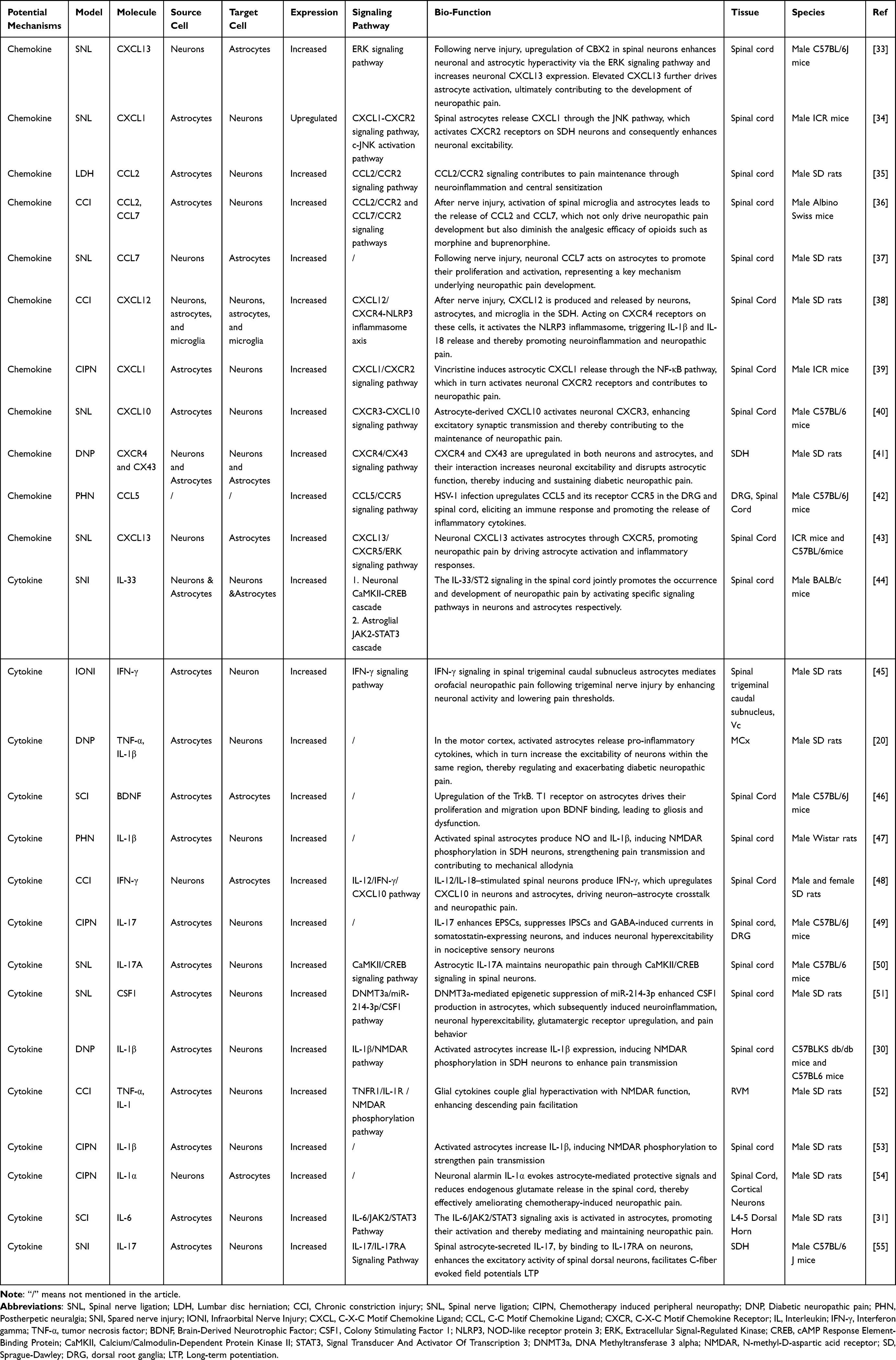 |
Table 1 Potential Cytokine and Chemokines Mechanisms in Different Neuropathic Pain Models |
Other inflammatory mediators also play a role. For instance, IL-17 enhances CaMKII/CREB activity in spinal neurons, modulating synaptic transmission and sustaining chronic pain induced by chemotherapy or peripheral nerve injury.50 IFN-γ activates astrocytes and increases neuronal activity in trigeminal nerve injury models.45 Nerve injury can epigenetically downregulate miR-214-3p in astrocytes, leading to upregulation of secreted colony-stimulating factor-1 (CSF-1), which enhances neuronal excitability and exacerbates neuropathic pain.51 Furthermore, the IL-33/ST2 signaling pathway in the spinal cord promotes neuropathic pain development through a dual mechanism involving neuronal CaMKII–CREB and astrocytic JAK2–STAT3 pathways.44
Similarly, astrocytes release chemokines, including CXCL1 and CCL2. These molecules act on neuronal receptors CXCR2 and CCR2, activating downstream signaling and enhancing neuronal excitability in pain pathways.56 For example, CXCL1 is upregulated and released from spinal astrocytes, and acts on CXCR2-expressing spinal neurons to activate ERK and CREB signaling, enhance neuronal excitability, and induce immediate early gene expression, thereby promoting central sensitization and persistent pain.34,57 Following inflammation or nerve injury, activated spinal astrocytes secrete CCL2, which binds to CCR2 expressed on neurons, modulating synaptic transmission and enhancing NMDA receptor function, thus facilitating central sensitization.58,59 In addition, CXCL10 expression is upregulated in spinal neurons and astrocytes after nerve injury. CXCL10 enhances spontaneous excitatory postsynaptic currents and potentiates NMDA- and AMPA-induced currents in spinal neurons, further amplifying neuronal responsiveness to pain stimuli.40 Collectively, Cytokines and chemokines mediate neuron-astrocyte signaling, forming a positive feedback loop that enhances neuronal excitability and sustains neuropathic pain.
Extracellular Vesicles
Extracellular vesicles(EVs), particularly exosomes, are key mediators of intercellular communication. Exosomes are nanoscale vesicles, approximately 30–150 nm in diameter, capable of carrying diverse bioactive cargos, including miRNAs, mRNAs, proteins, and lipids, which can be internalized by target cells via endocytosis to modulate gene expression and cellular functions. While studies of exosomes in neuropathic pain remain scarce, accumulating evidence highlights their roles in various neurodegenerative conditions.60
miRNAs packaged within astrocyte-derived exosomes have emerged as critical regulators of neuronal gene expression and synaptic plasticity. For instance, exosomes enriched in miR-26a-5p exert neuroprotective effects by inhibiting neuronal apoptosis and promoting dendritic development.61 Mechanistically, miR-26a-5p targets neural cell adhesion molecule and activates the AKT/GSK3-β/CRMP2 signaling pathway, thereby enhancing neuronal function.61 Similarly, miR-378a-5p carried by astrocyte-derived exosomes mitigates NLRP3-mediated pyroptosis and neuroinflammation, conferring neuroprotection in cerebral ischemia.62 Neurons also release exosomes to transfer miR-124-3p to microglia and astrocytes, suppressing M1 microglia and A1 astrocyte activation through modulation of MYH9 and the PI3K/AKT/NF-κB signaling axis, which facilitates functional recovery following spinal cord injury.63
Beyond miRNAs, exosomes transport specific proteins that critically regulate neuronal functions. Astrocytic processes release exosomes containing neuroglobins, contributing to noncanonical CNS signaling and selectively targeting neurons to exert protective effects.64 Following mild cortical spreading depolarization, stressed neurons release small HMGB1-enriched exosomes that are internalized by astrocytic processes, activating NF-κB p65 signaling and eliciting inflammatory responses.65 Interestingly, astrocyte-derived exosomes (ADEVs) exhibit stimulus-dependent protein cargos: ATP or IL-10 stimulation enriches proteins promoting neurite outgrowth, dendritic branching, synaptic transmission, and neuronal survival, whereas IL-1β-stimulated ADEVs are enriched in proteins that modulate peripheral immune responses and recruit immune cells into the CNS.66 Collectively, these findings highlight exosomes as key mediators of astrocyte-neuron communication and suggest their potential as therapeutic targets for chronic pain intervention.
Synapse-Associated Molecules
Astrocytes orchestrate neuronal plasticity via synapse-associated molecules, thereby influencing synaptic transmission and promoting neuropathic pain (Table 2). D-serine, a key NMDAR co-agonist released by astrocytes, enhances excitatory synaptic transmission.15,67,68 Members of the Hevin/SPARC protein family exert opposing effects on synaptic remodeling: Hevin promotes postsynaptic dendritic spine formation, potentially enhancing nociceptive signaling, whereas SPARC inhibits synaptic remodeling, reducing pain hypersensitivity.69 Astrocyte-derived Hevin has been shown to sustain neuropathic and inflammatory pain by upregulating spinal GluN2B expression.70 In addition, astrocytes secrete Chordin-like 1, which promotes synapse maturation and constrains plasticity by increasing GluA2 receptor expression at synapses.71
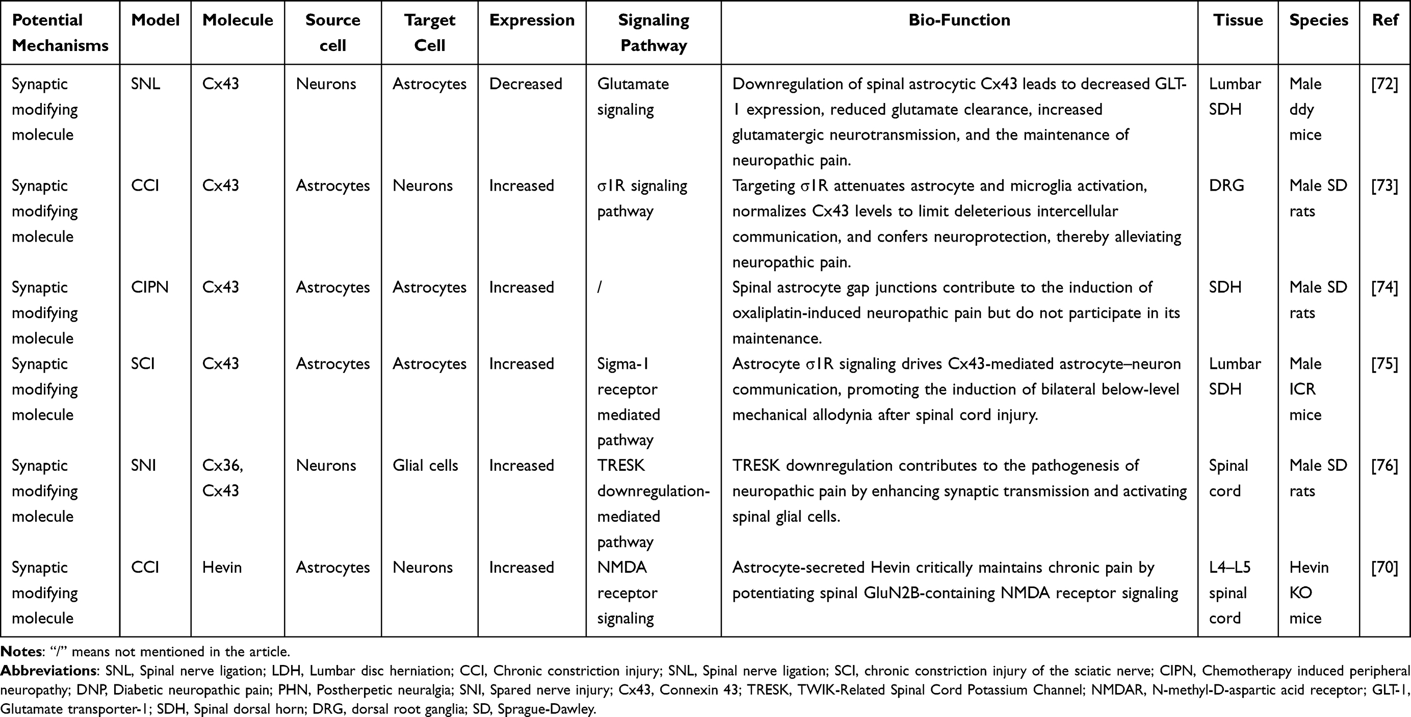 |
Table 2 Potential Synapse-Associated Molecules Mechanisms in Different Neuropathic Pain Models |
Matrix metalloproteinases (MMPs) are key regulators of the extracellular synaptic matrix and play pivotal roles in neuropathic pain. MMP-9 degrades chondroitin sulfate proteoglycans (CSPGs), influencing synaptic stability and facilitating synaptic remodeling.77 CSPGs form perisynaptic barriers that limit synaptic plasticity and maintain synaptic stability.78 Abnormal CSPG accumulation after nerve injury may impede repair, whereas its degradation promotes plasticity and modulates pain signal transmission.79 Furthermore, the Sema3A/Neuropilin-1 signaling pathway regulates synaptic plasticity, with astrocyte-derived Sema3A mediating axonal retraction and synaptic functional changes via Neuropilin-1 receptor.79
Connexins (Cxs) modulate astrocyte–neuron interactions and inter-astrocytic coupling, driving synaptic abnormalities and pain sensitization. Neuronal potassium channel activity downregulation leads to upregulation of neuronal Cx36 and astrocytic Cx43, enhancing synaptic transmission and glial activation.76 In chemotherapy-induced neuropathic pain, increased astrocytic Cx43 mediates aberrant intercellular communication, facilitating pain hypersensitivity.74 Following spinal cord injury, Sigma-1 Receptor activation further elevates astrocytic Cx43 expression and coupling, accelerating central sensitization and mechanical allodynia.75 Collectively, astrocytes influence neuronal synaptic plasticity and excitatory transmission through the secretion and regulation of multiple synapse-associated molecules.
Ion Homeostasis
Astrocytes engage in bidirectional interactions with neurons through diverse mechanisms of ionic homeostasis, which play crucial roles in the initiation and maintenance of neuropathic pain80 (Table 3). Astrocytes regulate extracellular K⁺ buffering via Kir4.1 potassium channels, thereby modulating neuronal excitability.81 In chronic constriction injury models, impaired K⁺ channel function in dorsal horn astrocytes increases astrocytic excitability, alters neuronal firing patterns, and enhances neuronal excitability.82 Aberrant Ca2⁺ signaling also represents a key factor in pain modulation. Astrocytic Ca2⁺ fluctuations directly influence synaptic function and neurotransmitter release, while Ca2⁺ signals mediated via P2Y receptor activation further potentiate pain signal transmission.83 Notably, electroacupuncture has been reported to modulate Ca2⁺ dynamics in rostral ventromedial medulla astrocytes, activating downstream signaling pathways to achieve analgesic effects.84
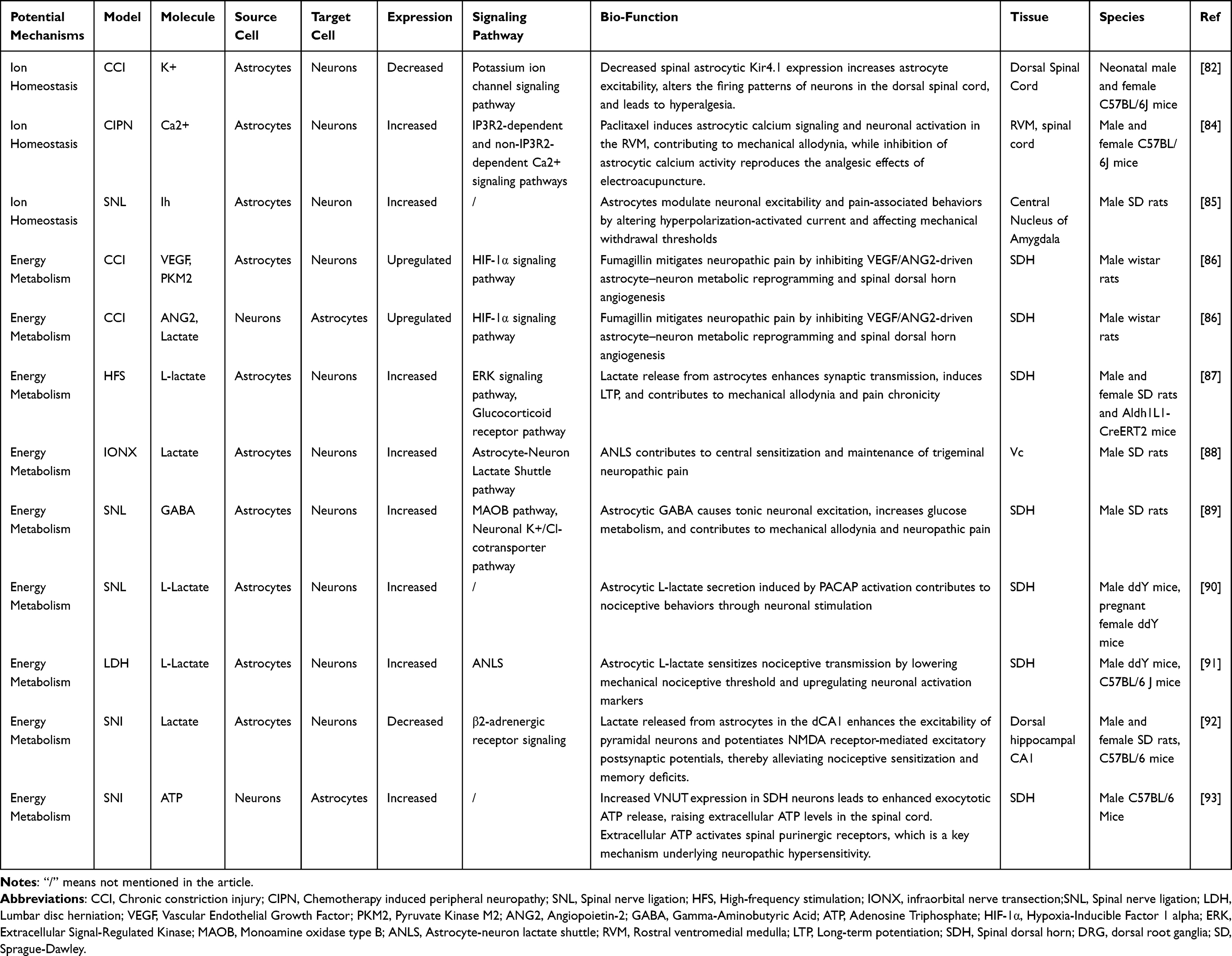 |
Table 3 Potential Ion Homeostasis and Energy Metabolism Mechanisms in Different Neuropathic Pain Models |
Astrocytes also modulate neuronal excitability through Na⁺/K⁺-ATPase. In migraine, Na⁺/K⁺-ATPase dysfunction leads to elevated extracellular Na⁺, concomitant with reduced perisynaptic glutamate transporter density, slowing glutamate clearance, and increasing cortical susceptibility to spreading depolarization, thereby enhancing nociceptive responses to migraine triggers.94 Astrocytic regulation of Cl− homeostasis further influences neuronal excitability; impaired Cl− efflux raises intracellular Cl− in neurons, weakening GABAergic inhibition and promoting hyperexcitability.95 Activation of astrocytic BDNF-TrkB signaling downregulates Cl− extrusion transporters while enhancing the release of pro-inflammatory cytokines and neurotrophic factors, establishing a positive feedback loop that sustains pain signaling.96 In spared nerve injury models, differential target multiplexed programming of spinal cord stimulation modulates a broader array of ion homeostasis-related proteins compared with low-frequency stimulation. Regulation of proteins controlling Ca2⁺, Na⁺, K⁺, and Cl− homeostasis leads to reduced intracellular Ca2⁺, enhanced GABAergic inhibition, and restored K⁺ and Cl− balance, thereby reversing ion dysregulation and neuronal hyperexcitability in pain states.97 Overall, astrocytes regulate neuronal excitability and synaptic transmission via ion homeostasis, critically contributing to the onset and maintenance of chronic pain.
Energy Metabolism
Reciprocal metabolic coupling, via astrocytic lactate and ATP release and mitochondrial transfer, shapes neuronal excitability and perpetuates pain signaling (Table 3). Among these mechanisms, lactate shuttling is particularly critical. During inflammatory pain, astrocytes generate lactate through glycogenolysis and transport it to neurons via monocarboxylate transporters. Lactate serves as an energy substrate, facilitating synaptic plasticity and heightened excitability in spinal neurons, thereby sustaining pain states.88,98 In the spinal cord, lactate shuttling enhances C-fiber-induced long-term potentiation, exacerbating mechanical hypersensitivity.87,99 In higher brain regions such as the anterior cingulate cortex and hippocampus, astrocyte-derived lactate modulates synaptic plasticity, augments pain sensitization, and influences pain-associated cognitive and emotional processes.100,101 ATP also regulates neuronal activity. Astrocyte activation triggers ATP release, which enhances neuronal excitability and pain hypersensitivity, effects that can be reversed upon blockade of ATP receptors.102 Conversely, dorsal horn neurons in neuropathic pain states release ATP via vesicular nucleotide transporter-mediated vesicular exocytosis, sustaining pain signaling.93
Mitochondrial transfer mediated by astrocytes has emerged as a novel form of intercellular communication, essential for maintaining neuronal function and metabolic homeostasis. Although its role in neuropathic pain remains unclear, extensive studies in various neurodegenerative and injury models provide insights. For instance, the Tak1 signaling pathway promotes mitochondrial transfer from astrocytes to hypothalamic proopiomelanocortin neurons, maintaining glucose and cholesterol homeostasis.103 In Rett Syndrome models, astrocytic mitochondrial dysfunction and excessive reactive oxygen species (ROS) production impair their neuronal support capacity.104 Acupuncture interventions in acute stroke models enhance astrocyte-to-neuron mitochondrial transfer, conferring neuroprotection.105 Recent studies also show that LRP1-mediated mitochondrial transfer from astrocytes mitigates cerebral ischemic injury through inhibition of ADP-ribosylation factor 1 myristoylation,106 and reactive astrocytes with abnormal mitochondrial dynamics are closely associated with neuronal damage in multiple pathological conditions.107 These findings suggest that astrocytic mitochondrial transfer can be protective or harmful depending on context and may modulate neuronal metabolism, synaptic plasticity, and ROS to influence pain pathways, warranting further study.
Neurotransmitter Regulation
Astrocytes regulate neurotransmitter signaling to sustain neuronal excitability and modulate pain (Table 4). In key pain-processing regions such as the spinal dorsal horn and hypothalamus, astrocytes maintain the balance of excitatory and inhibitory synaptic transmission by clearing excess glutamate from the synaptic cleft via GLT-1.28 When astrocytic function is impaired or GLT-1 expression is downregulated, glutamate clearance is compromised, leading to elevated extracellular glutamate levels, enhanced excitatory synaptic plasticity, and central sensitization, thereby exacerbating chronic pain phenotypes.108,109 Moreover, astrocytes can sense synaptic glutamate through metabotropic glutamate receptor 5, triggering intracellular Ca2⁺-dependent responses that further disrupt excitatory signaling.110
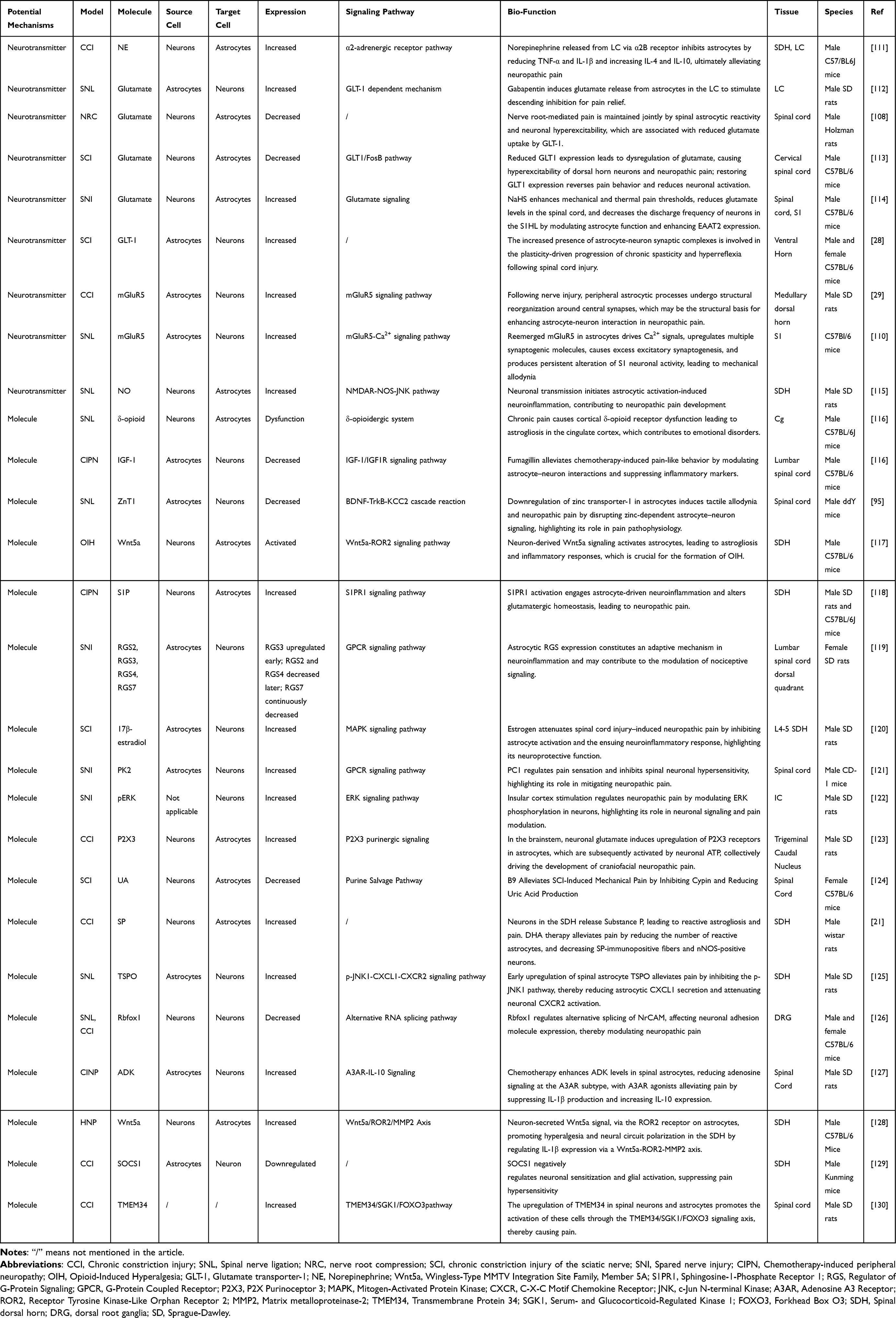 |
Table 4 Potential Neurotransmitter and Other Molecule Mechanisms in Different Neuropathic Pain Models |
Beyond glutamate, norepinephrine also participates in astrocyte–neuron cross talk. Norepinephrine released from locus coeruleus neurons in the midbrain acts on astrocytes to suppress pro-inflammatory activation, thereby reducing astrocyte-mediated neuroinflammation, limiting glutamate-driven neuronal hyperexcitability, and alleviating pain transmission.111 Pharmacological studies indicate that upregulating GLT-1 in astrocytes enhances glutamate clearance, suppresses neuronal hyperexcitability, and significantly improves pain behaviors in spinal cord injury or neuropathic pain models.131 Together, astrocytes orchestrate neurotransmitter signaling to modulate synaptic transmission and central sensitization.
Conclusions and Future Directions
In this review, we summarized the multifaceted mechanisms by which astrocyte-neuron interactions contribute to the initiation and maintenance of neuropathic pain. These interactions occur both through direct cell-cell contacts and via indirect pathways, including cytokine/chemokine signaling, extracellular vesicle release, synapse-associated molecules, ion homeostasis, energy metabolism, and neurotransmitter regulation (Figure 1). These mechanisms are highly interconnected, collectively shaping central sensitization and synaptic plasticity.
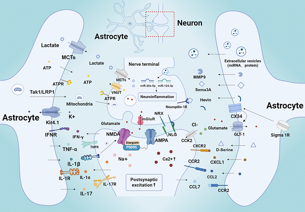 |
Figure 1 Astrocyte-Neuron Crosstalk Mechanisms and Molecular Mediators in Neuropathic Pain. Activated astrocytes regulate neuronal activity and synaptic plasticity via multiple interconnected mechanisms. These include direct structural modulation of the tripartite synapse, release of proinflammatory cytokines (eg, IL-1β, TNF-α, IL-6, IL-17, IFN-γ, IL-33, CSF-1) and chemokines (eg, CCL2, CXCL1, CXCL13), and secretion of gliotransmitters such as ATP, D-serine, and glutamate. In addition, astrocyte-derived factors, including Hevin and MMPs, orchestrate excitatory synapse formation, while exosomes and metabolic signals further contribute to central sensitization and the maintenance of neuropathic pain. The red box marks the tripartite synapse composed of astrocytes, presynaptic membrane and postsynaptic membrane, with its detailed magnified diagram shown inside the box. Black dashed arrows indicate the transport, paracrine secretion and transcellular signal regulatory pathways of various molecules and extracellular vesicles. The upward solid arrow denotes enhanced postsynaptic excitation. |
In the central nervous system, astrocytes are not merely supportive cells for neurons but are pivotal regulators of neural network function. Through the formation of tripartite synapses and the envelopment of presynaptic terminals and postsynaptic dendrites by PAPs, astrocytes are capable of sensing neuronal activity and modulating synaptic transmission, thereby contributing to the regulation of synaptic plasticity.23–25 Via gap junctions and an extensive astrocytic network, they facilitate lateral signal propagation, enabling local neuronal activity to be coordinated across broader neural circuits. In addition, astrocytes provide essential metabolic support, including the release of lactate and ATP as well as mitochondrial transfer, ensuring neuronal energy supply and the maintenance of activity-dependent plasticity.88,102,103 Astrocytes also play a central role in maintaining ion homeostasis, including Ca2⁺, K⁺, and Cl−, thereby regulating neuronal excitability and preventing aberrant firing.81,83,95 Under inflammatory or chronic pain conditions, astrocytes release gliotransmitters, cytokines, and chemokines, participating in bidirectional neuron-glia signaling that amplifies central sensitization and synaptic plasticity.31,56
Accumulating evidence suggests that astrocyte-neuron communication in neuropathic pain is mediated by an integrated signaling network rather than independent pathways. Following nerve injury, excessive neuronal activity and the release of neurotransmitters activate astrocytes and trigger multiple downstream responses, including the production of cytokines and chemokines, extracellular vesicles, synapse-associated molecules, and alterations in ion homeostasis and metabolic coupling.132 Importantly, these pathways exhibit substantial functional crosstalk. For example, astrocyte-derived TNF-α and IL-1β not only directly enhance neuronal excitability but also impair glutamate homeostasis and facilitate NMDA receptor activation, while IL-6 amplifies inflammatory signaling through the JAK2/STAT3 pathway.
These signaling pathways further interact to reinforce maladaptive neuron-astrocyte communication. Extracellular vesicles can propagate inflammatory signals by transferring regulatory miRNAs and proteins, whereas synapse-associated molecules such as Hevin and D-serine directly promote excitatory synaptic remodeling.69 Meanwhile, disturbances in ion homeostasis and energy metabolism further potentiate cytokine production and glutamatergic transmission, establishing a feed-forward loop that sustains neuronal hyperexcitability. Despite their molecular diversity, these mechanisms ultimately converge on common downstream events, including enhanced excitatory synaptic plasticity, central sensitization, and persistent neuropathic pain.
Despite significant progress in recent years, several challenges remain. First, most studies have been conducted in rodent models, with limited evidence from human patients. Although rodent models provide important mechanistic insights, species differences may limit the direct translatability of findings. Second, astrocyte functions are highly context- and region-dependent, and may exhibit distinct roles across different brain regions, disease stages, or physiological states.133,134 Moreover, astrocyte-neuron interactions involve multiple inflammatory mediators, metabolites, neurotrophic factors, and ion channels, which may act synergistically or antagonistically.9,17,135 Future studies combining single-cell sequencing, multi-omics analyses, and systems biology approaches, along with advanced neuroimaging techniques, are needed to comprehensively map these molecular networks and dynamically monitor astrocyte function in vivo.136–138 Astrocytes involved in neuropathic pain are increasingly recognized as a heterogeneous population with distinct functional phenotypes. Rather than representing a uniform cell type, astrocytes exhibit marked region-specific and state-dependent characteristics. In particular, homeostatic astrocytes preserve synaptic homeostasis by maintaining glutamate uptake, ion balance, and metabolic support, whereas reactive astrocytes acquire diverse phenotypes that differentially regulate neuroinflammation, synaptic remodeling, and neuronal excitability following nerve injury. Consequently, distinct astrocyte subpopulations are likely to contribute to different stages of neuropathic pain, including its initiation, maintenance, and resolution. Future studies integrating single-cell multi-omics and spatial transcriptomics are needed to define the molecular and functional characteristics of these astrocyte subsets and their dynamic interactions with neurons. Such advances will facilitate the development of precision therapies that selectively target pathogenic astrocyte populations while preserving their essential homeostatic functions. In addition, it is also worth noting that the bidirectional communication between microglia and astrocytes represents another critical regulatory axis in neuropathic pain and other CNS disorders.139,140 Microglia-derived factors such as complement component C1q, TNF-α, and IL-1α can drive the conversion of astrocytes into the neurotoxic A1 phenotype, thereby exacerbating neuronal dysfunction.141,142 However, this aspect is beyond the scope of the present discussion.
From a translational perspective, while glia-targeted therapeutic strategies have shown promising results in preclinical models, their clinical application remains challenging. Current approaches targeting astrocyte-neuron signaling primarily focus on restoring glutamate homeostasis, inhibiting astrocyte-derived synaptogenic factors, modulating astrocytic inflammatory signaling pathways, regulating connexin-mediated gliotransmission, and interfering with extracellular vesicle-mediated neuron-glia communication. These strategies aim to normalize aberrant synaptic transmission and attenuate central sensitization. A major obstacle is the selective modulation of astrocyte function without inadvertently affecting other glial populations, such as microglia or oligodendrocytes. Enhancing astrocytic glutamate clearance has emerged as a promising strategy for pain modulation. β-lactam antibiotics such as ceftriaxone and cefadroxil reduce extracellular glutamate, suppress neuronal hyperexcitability, and produce significant antinociceptive effects in neuropathic and inflammatory pain models.143 Similarly, clavulanic acid has been shown to alleviate neuropathic pain through GLT-1 dependent mechanisms.144 Increased transporter activity also dampens spinal astrocyte activation and synaptic hyperexcitability.108 However, clinical evidence directly demonstrating therapeutic efficacy remains limited. Furthermore, the intimate association of astrocytes with the blood-brain barrier necessitates that therapeutics are designed to efficiently penetrate this barrier while minimizing systemic side effects.145,146 With advances in nanomedicine, bioengineered materials, and gene editing technologies, more precise and effective intervention strategies may be developed, offering new therapeutic options for patients with neuropathic pain.147–149
While most studies have focused on the pronociceptive functions of reactive astrocytes after nerve injury, recent evidence suggests that homeostatic astrocytes are integral components of spinal non-neuronal pain-gating mechanisms. Activation of low-threshold Aβ afferents recruits spinal astrocytes to suppress nociceptive transmission through endogenous adenosine signaling and long-term depression of neurokinin-1 receptor-positive (NK1R⁺) projection neurons, thereby contributing to endogenous antinociception.150 This context-dependent functional transition may provide a more comprehensive framework for understanding astrocyte-neuron interactions in neuropathic pain. Under physiological conditions, astrocytes maintain synaptic homeostasis and limit nociceptive transmission, whereas following nerve injury they undergo reactive remodeling and acquire pronociceptive properties that facilitate central sensitization.
In summary, astrocytes play a critical role in the development and maintenance of neuropathic pain. Through inflammatory mediators, metabolic pathways, and synaptic regulation, they form a complex interactive network with neurons. Although key pathways have been partially elucidated, further investigation is required to understand their dynamic changes and molecular mechanisms under different pathological conditions. Targeting astrocytes may provide a novel strategy for the treatment of neuropathic pain and pave the way for more effective, individualized therapeutic approaches.
Acknowledgments
The authors express gratitude to Home for Researchers (https://www.home-for-researchers.com) for their English language editing assistance. The figure was created with BioRender.com (License No. DB29YM6SFN; https://app.biorender.com).
Author Contributions
All authors made a significant contribution to the work reported, whether that is in the conception, study design, execution, acquisition of data, analysis and interpretation, or in all these areas; took part in drafting, revising or critically reviewing the article; gave final approval of the version to be published; have agreed on the journal to which the article has been submitted; and agree to be accountable for all aspects of the work.
Funding
This work was supported by the National Natural Science Foundation of China (82171217) and the Natural Science Foundation of Beijing Municipality (7252025, 7252029, 7254332).
Disclosure
The authors declare no conflicts of interest in this work.
References
1. Dzung H, De Leon-Casasola OA. Neuropathic pain: an updated grading system for research and clinical practice. Essence Analg Analg. 2010;157:30–19. doi:10.1017/CBO9780511841378.007
2. Jensen TS, Finnerup NB. Allodynia and hyperalgesia in neuropathic pain: clinical manifestations and mechanisms. Lancet Neurol. 2014;13:924–935. doi:10.1016/S1474-4422(14)70102-4
3. Kaplan CM, Kelleher E, Irani A, Schrepf A, Clauw DJ, Harte SE. Deciphering nociplastic pain: clinical features, risk factors and potential mechanisms. Nat Rev Neurol. 2024;20:347–363. doi:10.1038/s41582-024-00966-8
4. Yamauchi N, Sato K, Sato K, et al. Chronic pain–induced neuronal plasticity in the bed nucleus of the stria terminalis causes maladaptive anxiety. Sci Adv. 2022:8. doi:10.1126/sciadv.abj5586
5. Borbjerg MK, Wegeberg AM, Nikontovic A, et al. Understanding the impact of diabetic peripheral neuropathy and neuropathic pain on quality of life and mental health in 6,960 people with diabetes. Diabetes Care. 2025;48:588–595. doi:10.2337/dc24-2287
6. Thouaye M, Yalcin I. Neuropathic pain: from actual pharmacological treatments to new therapeutic horizons. Pharmacol Ther. 2023;251:108546. doi:10.1016/j.pharmthera.2023.108546
7. Jayathilake NJ, Phan TT, Kim J, Lee KP, Park JM. Modulating neuroplasticity for chronic pain relief: noninvasive neuromodulation as a promising approach. Exp Mol Med. 2025;57:501–514. doi:10.1038/s12276-025-01409-0
8. Soliman N, Moisset X, Ferraro MC, et al. Pharmacotherapy and non-invasive neuromodulation for neuropathic pain: a systematic review and meta-analysis. Lancet Neurol. 2025;24:413–428. doi:10.1016/S1474-4422(25)00068-7
9. Milligan ED, Watkins LR. Pathological and protective roles of glia in chronic pain. Nat Rev Neurosci. 2009;10:23–36. doi:10.1038/nrn2533
10. Endo F, Kasai A, Soto JS, et al. Molecular basis of astrocyte diversity and morphology across the CNS in health and disease. Science. 2022;378:1–12. doi:10.1126/science.adc9020
11. Sharma H, Khan S, Lohani A, Chandra P, Sachan N. Anandamide as a therapeutic target for alleviating neuropathic pain and inflammation in rat models. Curr Neurovasc Res. 2026;22:167–181. doi:10.2174/0115672026391315250822063941
12. Panatier A, Vallée J, Haber M, Murai KK, Lacaille JC, Robitaille R. Astrocytes are endogenous regulators of basal transmission at central synapses. Cell. 2011;146:785–798. doi:10.1016/j.cell.2011.07.022
13. Baldwin KT, Eroglu C. Astrocytes “Chordinate” synapse maturation and plasticity. Neuron. 2018;100:1010–1012. doi:10.1016/j.neuron.2018.11.027
14. Clarke LE, Barres BA. Emerging roles of astrocytes in neural circuit development. Nat Rev Neurosci. 2013;14:311–321. doi:10.1038/nrn3484
15. Abreu DS, Gomes JI, Ribeiro FF, Diógenes MJ, Sebastião AM, Vaz SH. Astrocytes control hippocampal synaptic plasticity through the vesicular-dependent release of D-serine. Front Cell Neurosci. 2023;17. doi:10.3389/fncel.2023.1282841
16. Crosby ND, Winkelstein BA. Spinal astrocytic thrombospondin-4 induced by excitatory neuronal signaling mediates pain after facet capsule injury. Ann Biomed Eng. 2016;44:3215–3224. doi:10.1007/s10439-016-1639-x
17. Ji RR, Donnelly CR, Nedergaard M. Astrocytes in chronic pain and itch. Nat Rev Neurosci. 2019;20:667–685. doi:10.1038/s41583-019-0218-1
18. Zhou Y, Tian X, Tian Y, Mei W, Liu D, Ye D. Wnt signaling: a prospective therapeutic target for chronic pain. Pharmacol Ther. 2021;107984. doi:10.1016/j.pharmthera.2021.107984
19. Takeda I, Yoshihara K, Cheung DL, et al. Controlled activation of cortical astrocytes modulates neuropathic pain-like behaviour. Nat Commun. 2022;13:1–12. doi:10.1038/s41467-022-31773-8
20. Lu J, Yang L, Xu Y, et al. The modulatory effect of motor cortex astrocytes on diabetic neuropathic pain. J Neurosci. 2021;41:5287–5302. doi:10.1523/JNEUROSCI.2566-20.2021
21. Manzhulo IV, Ogurtsova OS, Kipryushina YO, et al. Neuron-astrocyte interactions in spinal cord dorsal horn in neuropathic pain development and docosahexaenoic acid therapy. J Neuroimmunol. 2016;298:90–97. doi:10.1016/j.jneuroim.2016.07.015
22. Iwata K, Katagiri A, Shinoda M. mechanisms underlying neuron-glial cell communication in the TG after orofacial inflammation and trigeminal nerve injury. J Oral Sci. 2017;59:173–175. doi:10.2334/josnusd.16-0858
23. Lalo U, Koh W, Lee CJ, Pankratov Y. The tripartite glutamatergic synapse. Neuropharmacology. 2021;199:108758. doi:10.1016/j.neuropharm.2021.108758
24. Perea G, Navarrete M, Araque A. Tripartite synapses: astrocytes process and control synaptic information. Trends Neurosci. 2009;32:421–431. doi:10.1016/j.tins.2009.05.001
25. Navarrete M, Araque A. Basal synaptic transmission: astrocytes rule! Cell. 2011;146:675–677. doi:10.1016/j.cell.2011.08.006
26. Tewari BP, Woo ALM, Prim CE, et al. Astrocytes require perineuronal nets to maintain synaptic homeostasis in mice. Nat Neurosci. 2024;27:1475–1488. doi:10.1038/s41593-024-01714-3
27. Baldwin KT, Tan CX, Strader ST, et al. HepaCAM controls astrocyte self-organization and coupling. Neuron. 2021;109:2427–2442.e10. doi:10.1016/j.neuron.2021.05.025
28. Benson CA, King JF, Kauer SD, Waxman SG, Tan AM. Increased astrocytic GLT-1 expression in tripartite synapses is associated with SCI-induced hyperreflexia. J Neurophysiol. 2023;130:1358–1366. doi:10.1152/JN.00234.2023
29. Cho YS, Kim DH, Bae JY, et al. Structural reorganization of medullary dorsal horn astrocytes in a rat model of neuropathic pain. Brain Struct Funct. 2024;229:1757–1768. doi:10.1007/s00429-024-02835-y
30. Liao YH, Zhang GH, Jia D, et al. Spinal astrocytic activation contributes to mechanical allodynia in a mouse model of type 2 diabetes. Brain Res. 2011;1368:324–335. doi:10.1016/j.brainres.2010.10.044
31. Lee JY, Park CS, Seo KJ, et al. IL-6/JAK2/STAT3 axis mediates neuropathic pain by regulating astrocyte and microglia activation after spinal cord injury. Exp Neurol. 2023;370:114576. doi:10.1016/j.expneurol.2023.114576
32. Dai X, Liu L, Song F, et al. Targeting the JAK2 / STAT3 signaling pathway for chronic pain. Aging Dis. 2023;14:1–15. doi:10.14336/AD.2022.0619
33. Han M, Zhang F, Wang Y, et al. Spinal CBX2 contributes to neuropathic pain by activating ERK signaling pathway in male mice. Neurosci Lett. 2023;812:137363. doi:10.1016/j.neulet.2023.137363
34. Zhang ZJ, Cao DL, Zhang X, Ji RR, Gao YJ. Chemokine contribution to neuropathic pain: respective induction of CXCL1 and CXCR2 in spinal cord astrocytes and neurons. Pain. 2013;154:2185–2197. doi:10.1016/j.pain.2013.07.002
35. Zhu X, Cao S, Zhu M, Liu JQ, Chen JJ, Gao YJ. Contribution of chemokine CCL2/CCR2 signaling in the dorsal root ganglion and spinal cord to the maintenance of neuropathic pain in a rat model of lumbar disc herniation. J Pain. 2014;15:516–526. doi:10.1016/j.jpain.2014.01.492
36. Kwiatkowski K, Popiolek-Barczyk K, Piotrowska A, et al. Chemokines CCL2 and CCL7, but not CCL12, play a significant role in the development of pain-related behavior and opioid-induced analgesia. Cytokine. 2019;119:202–213. doi:10.1016/j.cyto.2019.03.007
37. Ke BC, Huang XX, Li Y, et al. Neuronal-derived Ccl7 drives neuropathic pain by promoting astrocyte proliferation. Neuroreport. 2016;27:849–857. doi:10.1097/WNR.0000000000000625
38. Cheng KI, Chen SL, Hsu JH, et al. Loganin prevents CXCL12/CXCR4-regulated neuropathic pain via the NLRP3 inflammasome axis in nerve-injured rats. Phytomedicine. 2021;92:153734. doi:10.1016/j.phymed.2021.153734
39. Zhou L, Hu Y, Li C, et al. Levo-corydalmine alleviates vincristine-induced neuropathic pain in mice by inhibiting an NF-kappa B-dependent CXCL1/CXCR2 signaling pathway. Neuropharmacology. 2018;135:34–47. doi:10.1016/j.neuropharm.2018.03.004
40. Jiang BC, He LN, Wu XB, et al. Promoted interaction of C/EBPα with demethylated Cxcr3 gene promoter contributes to neuropathic pain in mice. J Neurosci. 2017;37:685–700. doi:10.1523/JNEUROSCI.2262-16.2016
41. Zhu D, Fan T, Chen Y, et al. CXCR4/CX43 regulate diabetic neuropathic pain via intercellular interactions between activated neurons and dysfunctional astrocytes during late phase of diabetes in rats and the effects of antioxidant N-Acetyl-L-Cysteine. Oxid Med Cell Longev. 2022;2022. doi:10.1155/2022/8547563
42. Wu S, Yang S, Li R, et al. HSV-1 infection-induced herpetic neuralgia involves a CCL5/CCR5-mediated inflammation mechanism. J Med Virol. 2023;95:1–16. doi:10.1002/jmv.28718
43. Jiang BC, Cao DL, Zhang X, et al. CXCL13 drives spinal astrocyte activation and neuropathic pain via CXCR5. J Clin Invest. 2016;126:745–761. doi:10.1172/JCI81950
44. Liu S, Mi W-L, Li Q, et al. Spinal IL-33/ST2 signaling contributes to neuropathic pain. Anesthesiology. 2016;123:1154–1169. doi:10.1097/ALN.0000000000000850
45. Asano S, Okada-Ogawa A, Kobayashi M, et al. Involvement of interferon gamma signaling in spinal trigeminal caudal subnucleus astrocyte in orofacial neuropathic pain in rats with infraorbital nerve injury. Mol Pain. 2023;19:1–13. doi:10.1177/17448069231222403
46. Matyas JJ, O’Driscoll CM, Yu L, et al. Truncated TrkB.T1-mediated astrocyte dysfunction contributes to impaired motor function and neuropathic pain after spinal cord injury. J Neurosci. 2017;37:3956–3971. doi:10.1523/JNEUROSCI.3353-16.2017
47. Zhang GH, Lv MM, Wang S, et al. Spinal astrocytic activation is involved in a virally-induced rat model of neuropathic pain. PLoS One. 2011:6e23059. doi:10.1371/journal.pone.0023059
48. Yang Y, Zhang S, Yang J, et al. The aqueous extract of armadillidium vulgare latreille alleviates neuropathic pain via inhibiting neuron-astrocyte crosstalk mediated by the IL-12-IFN-γ-IFNGR-CXCL10 pathway. J Ethnopharmacol. 2025;340:119173. doi:10.1016/j.jep.2024.119173
49. Luo H, Liu HZ, Zhang WW, et al. Interleukin-17 regulates neuron-glial communications, synaptic transmission, and neuropathic pain after chemotherapy. Cell Rep. 2019;29:2384–2397.e5. doi:10.1016/j.celrep.2019.10.085
50. Yao CY, Weng ZL, Zhang JC. Interleukin-17A acts to maintain neuropathic pain through activation of CaMKII/CREB signaling in spinal neurons. Mol Neurobiol. 2016;53:3914–3926. doi:10.1007/s12035-015-9322-z
51. Liu L, Xu D, Wang T, et al. Epigenetic reduction of miR-214-3p upregulates astrocytic colony-stimulating factor-1 and contributes to neuropathic pain induced by nerve injury. Pain. 2020;161. doi:10.1097/j.pain.0000000000001681
52. Wei F, Guo W, Zou S, Ren K, Dubner R. Supraspinal glial-neuronal interactions contribute to descending pain facilitation. J Neurosci. 2008;28:10482–10495. doi:10.1523/JNEUROSCI.3593-08.2008
53. Ji XT, Qian NS, Zhang T, et al. Spinal astrocytic activation contributes to mechanical allodynia in a rat chemotherapy-induced neuropathic pain model. PLoS One. 2013;8:2–13. doi:10.1371/journal.pone.0060733
54. Di Cesare Mannelli L, Micheli L, Cervetto C, et al. Neuronal alarmin IL-1α evokes astrocyte-mediated protective signals: effectiveness in chemotherapy-induced neuropathic pain. Neurobiol Dis. 2022;168:105716. doi:10.1016/j.nbd.2022.105716
55. Sun JL, Dai WJ, Shen XY, Lü N, Zhang YQ. Interleukin-17 is involved in neuropathic pain and spinal synapse plasticity on mice. J Neuroimmunol. 2023;377:578068. doi:10.1016/j.jneuroim.2023.578068
56. Jiang S, Liang J, Li W, et al. The role of CXCL1/CXCR2 axis in neurological diseases. Int Immunopharmacol. 2023;120:110330. doi:10.1016/j.intimp.2023.110330
57. Cao DL, Zhang ZJ, Xie RG, Jiang BC, Ji RR, Gao YJ. Chemokine CXCL1 enhances inflammatory pain and increases NMDA receptor activity and COX-2 expression in spinal cord neurons via activation of CXCR2. Exp Neurol. 2014;261:328–336. doi:10.1016/j.expneurol.2014.05.014
58. Xie RG, Gao YJ, Park CK, et al. Spinal CCL2 promotes central sensitization, long-term potentiation, and inflammatory pain via CCR2: further insights into molecular, synaptic, and cellular mechanisms. Neurosci Bull. 2018;34:13–21. doi:10.1007/s12264-017-0106-5
59. Zhang H, Bin MS, Gao YJ, et al. Spinal CCL2 promotes pain sensitization by rapid enhancement of NMDA-Induced currents through the ERK-GluN2B pathway in mouse lamina II neurons. Neurosci Bull. 2020;36:1344–1354. doi:10.1007/s12264-020-00557-9
60. Upadhya R, Zingg W, Shetty S, Shetty AK. Astrocyte-derived extracellular vesicles: neuroreparative properties and role in the pathogenesis of neurodegenerative disorders. J Control Release. 2020;323:225–239. doi:10.1016/j.jconrel.2020.04.017
61. Li J, Xu H, Zhang K, et al. Astrocyte-derived exosomes-transported miRNA-26a-5p ameliorates sevoflurane-induced cognitive dysfunction in aged mice. Transl Res. 2024;268:79–96. doi:10.1016/j.trsl.2024.01.007
62. Sun R, Liao W, Lang T, et al. Astrocyte-derived exosomal miR-378a-5p mitigates cerebral ischemic neuroinflammation by modulating NLRP3-mediated pyroptosis. Front Immunol. 2024;15:1–14. doi:10.3389/fimmu.2024.1454116
63. Jiang D, Gong F, Ge X, et al. Neuron-derived exosomes-transmitted miR-124-3p protect traumatically injured spinal cord by suppressing the activation of neurotoxic microglia and astrocytes. J Nanobiotechnology. 2020;18:1–20. doi:10.1186/s12951-020-00665-8
64. Venturini A, Passalacqua M, Pelassa S, et al. Exosomes from astrocyte processes: signaling to neurons. Front Pharmacol. 2019;10:1–11. doi:10.3389/fphar.2019.01452
65. Neuroinflammation D, Martinus RD. Review article role of mitochondrial stress protein HSP60 in 2020. 2020.
66. Datta chaudhuri A, Dasgheyb RM, DeVine LR, Bi H, Cole RN, Haughey NJ. Stimulus-dependent modifications in astrocyte-derived extracellular vesicle cargo regulate neuronal excitability. Glia. 2020;68:128–144. doi:10.1002/glia.23708
67. Choi SR, Roh DH, Yoon SY, et al. Astrocyte D-serine modulates the activation of neuronal NOS leading to the development of mechanical allodynia in peripheral neuropathy. Molecular Pain. 2019;15. doi:10.1177/1744806919843046
68. Masuoka T, Ikeda R, Konishi S. Persistent activation of histamine H 1 receptors in the hippocampal CA1 region enhances NMDA receptor-mediated synaptic excitation and long-term potentiation in astrocyte- and D-serine-dependent manner. Neuropharmacology. 2019;151:64–73. doi:10.1016/j.neuropharm.2019.03.036
69. Fan S, Gangwar SP, Machius M, Rudenko G. Interplay between hevin, SPARC, and MDGAs: modulators of neurexin-neuroligin transsynaptic bridges. Structure. 2021;29:664–678.e6. doi:10.1016/j.str.2021.01.003
70. Chen G, Xu J, Luo H, et al. Hevin/Sparcl1 drives pathological pain through spinal cord astrocyte and NMDA receptor signaling. JCI Insight. 2022;7. doi:10.1172/jci.insight.161028
71. Blanco-Suarez E, Liu TF, Kopelevich A, Allen NJ. Astrocyte-Secreted Chordin-like 1 drives synapse maturation and limits plasticity by increasing synaptic GluA2 AMPA receptors. Neuron. 2018;100:1116–1132.e13. doi:10.1016/j.neuron.2018.09.043
72. Morioka N, Zhang FF, Nakamura Y, Kitamura T, Hisaoka-Nakashima K, Nakata Y. Tumor necrosis factor-mediated downregulation of spinal astrocytic connexin43 leads to increased glutamatergic neurotransmission and neuropathic pain in mice. Brain Behav Immun. 2015;49:293–310. doi:10.1016/j.bbi.2015.06.015
73. Denaro S, D’Aprile S, Torrisi F, et al. Sigma-1 receptor targeting inhibits connexin 43 based intercellular communication in chronic neuropathic pain. Inflamm Res. 2024;73:1711–1726. doi:10.1007/s00011-024-01926-0
74. Yoon SY, Robinson CR, Zhang H, Dougherty PM. Spinal astrocyte gap junctions contribute to oxaliplatin-induced mechanical hypersensitivity. J Pain. 2013;14:205–214. doi:10.1016/j.jpain.2012.11.002
75. Choi SR, Roh DH, Yoon SY, et al. Astrocyte sigma-1 receptors modulate connexin 43 expression leading to the induction of below-level mechanical allodynia in spinal cord injured mice. Neuropharmacology. 2016;111:34–46. doi:10.1016/j.neuropharm.2016.08.027
76. Zhou J, Chen H, Yang C, Zhong J, He W, Xiong Q. Reversal of TRESK downregulation alleviates neuropathic pain by inhibiting activation of gliocytes in the spinal cord. Neurochem Res. 2017;42:1288–1298. doi:10.1007/s11064-016-2170-z
77. Crapser JD, Spangenberg EE, Barahona RA, Arreola MA, Hohsfield LA, Green KN. Microglia facilitate loss of perineuronal nets in the Alzheimer’s disease brain. EBioMedicine. 2020;58. doi:10.1016/j.ebiom.2020.102919
78. Emerson J, Delgado T, Girardi P, Johnson GVW. Deletion of transglutaminase 2 from mouse astrocytes significantly improves their ability to promote neurite outgrowth on an inhibitory matrix. Int J Mol Sci. 2023;24:6058. doi:10.3390/ijms24076058
79. Mascio G, Notartomaso S, Martinello K, et al. A progressive build-up of perineuronal nets in the somatosensory cortex is associated with the development of chronic pain in mice. J Neurosci. 2022;42:3037–3048. doi:10.1523/JNEUROSCI.1714-21.2022
80. Wu J, Hu H, Li X. Spinal neuron-glial crosstalk and ion channel dysregulation in diabetic neuropathic pain. Front Immunol. 2025;16:1–21. doi:10.3389/fimmu.2025.1480534
81. Ohno Y, Kinboshi M, Shimizu S. Inwardly rectifying potassium channel kir4.1 as a novel modulator of BDNF expression in astrocytes. Int J Mol Sci. 2018;19:1–18. doi:10.3390/ijms19113313
82. Ou M, Chen Y, Liu J, et al. Spinal astrocytic MeCP2 regulates Kir4.1 for the maintenance of chronic hyperalgesia in neuropathic pain. Prog Neurobiol. 2023;224:102436. doi:10.1016/j.pneurobio.2023.102436
83. Song S, Wang Q, Qu Y, et al. Pregabalin inhibits purinergic P2Y2 receptor and TRPV4 to suppress astrocyte activation and to relieve neuropathic pain. Eur J Pharmacol. 2023;960:176140. doi:10.1016/j.ejphar.2023.176140
84. Chen X, Mi W, Gao T, Ding F, Wang W. Astrocytes in the rostral ventromedial medulla mediate the analgesic effect of electroacupuncture in a rodent model of chemotherapy-induced peripheral neuropathic pain. Pain. 2025;166:916–926. doi:10.1097/j.pain.0000000000003433
85. Mazzitelli M, Ponomareva O, Presto P, John J, Neugebauer V. Impaired amygdala astrocytic signaling worsens neuropathic pain-associated neuronal functions and behaviors. Front Pharmacol. 2024;15:1–15. doi:10.3389/fphar.2024.1368634
86. Wen ZH, Wu ZS, Cheng HJ, et al. Intrathecal fumagillin alleviates chronic neuropathy-induced nociceptive sensitization and modulates spinal astrocyte-neuronal glycolytic and angiogenic proteins. Mol Neurobiol. 2025;62:246–263. doi:10.1007/s12035-024-04254-w
87. Huang R, Han S, Qiu Y, et al. Glucocorticoid regulation of lactate release from spinal astrocytes contributes to the induction of spinal LTP of C-fiber-evoked field potentials and the development of mechanical allodynia. Neuropharmacology. 2022;219:109253. doi:10.1016/j.neuropharm.2022.109253
88. Hu Y, Zou H, Zhong Z, et al. The role of astrocyte-neuron lactate shuttle in neuropathic orofacial pain. J Oral Rehabil. 2024;51:2513–2528. doi:10.1111/joor.13847
89. Ju YH, Cho J, Park JY, et al. Tonic excitation by astrocytic GABA causes neuropathic pain by augmenting neuronal activity and glucose metabolism. Exp Mol Med. 2024;56:1193–1205. doi:10.1038/s12276-024-01232-z
90. Kambe Y, Youkai M, Hashiguchi K, et al. Spinal astrocyte-neuron lactate shuttle contributes to the pituitary adenylate cyclase-activating polypeptide/pac1 receptor-induced nociceptive behaviors in mice. Biomolecules. 2022:12. doi:10.3390/biom12121859
91. Miyamoto K, Ishikura K, Kume K, Ohsawa M. Astrocyte-neuron lactate shuttle sensitizes nociceptive transmission in the spinal cord. Glia. 2019;67:27–36. doi:10.1002/glia.23474
92. Han S, Jiang B, Ren J, et al. Impaired lactate release in dorsal CA1 astrocytes contributed to nociceptive sensitization and comorbid memory deficits in rodents. Anesthesiology. 2024;140:538–57. doi:10.1097/ALN.0000000000004756
93. Masuda T, Ozono Y, Mikuriya S, et al. Dorsal horn neurons release extracellular ATP in a VNUT-dependent manner that underlies neuropathic pain. Nat Commun. 2016;7:1–11. doi:10.1038/ncomms12529
94. Romanos J, Romanos J, Benke D, et al. Astrocyte dysfunction increases cortical dendritic excitability and promotes cranial pain in familial migraine. Sci Adv. 2020:6. doi:10.1126/sciadv.aaz1584
95. Kitayama T, Morita K, Motoyama N, Dohi T. Down-regulation of zinc transporter-1 in astrocytes induces neuropathic pain via the brain-derived neurotrophic factor - K+-Cl− co-transporter-2 signaling pathway in the mouse spinal cord. Neurochem Int. 2016;101:120–131. doi:10.1016/j.neuint.2016.11.001
96. Zhao S, Wang F, Wang L, et al. Involvement of the BDNF–TrkB–KCC2 pathway in neuropathic pain after brachial plexus avulsion. Brain Behav. 2022;12:1–10. doi:10.1002/brb3.2464
97. Tilley DM, Cedeño DL, Vetri F, Platt DC, Vallejo R. Differential target multiplexed spinal cord stimulation programming modulates proteins involved in ion regulation in an animal model of neuropathic pain. Mol Pain. 2021;17:1–14. doi:10.1177/17448069211060181
98. Marty-Lombardi S, Lu S, Ambroziak W, et al. Neuron–astrocyte metabolic coupling facilitates spinal plasticity and maintenance of inflammatory pain. Nature Metabol. 2024;6:494–513. doi:10.1038/s42255-024-01001-2
99. Reid P, Scherer K, Halasz D, et al. Astrocyte neuronal metabolic coupling in the anterior cingulate cortex of mice with inflammatory pain. Brain Behav Immun. 2025;125:212–225. doi:10.1016/j.bbi.2024.12.025
100. Wang Y, Peng Y, Zhang C, Zhou X. Astrocyte-neuron lactate transport in the ACC contributes to the occurrence of long-lasting inflammatory pain in male mice. Neurosci Lett. 2021;764:136205. doi:10.1016/j.neulet.2021.136205
101. Wang J, Tu J, Cao B, et al. Astrocytic L-Lactate signaling facilitates amygdala-anterior cingulate cortex synchrony and decision making in rats. Cell Rep. 2017;21(9):2407–2418. doi:10.1016/j.celrep.2017.11.012
102. Nam Y, Kim J-H, Kim J-H, et al. Reversible induction of pain hypersensitivity following optogenetic stimulation of spinal astrocytes. Cell Rep. 2016;17(11):3049–3061. doi:10.1016/j.celrep.2016.11.043
103. Yin K, Zhang T, Lu X, et al. Tak1 licenses mitochondrial transfer from astrocytes to POMC neurons to maintain glucose and cholesterol homeostasis. Cell Rep. 2024;43(12):114983. doi:10.1016/j.celrep.2024.114983
104. López-Molina L, Pereda-Velarde A, Di Franco N, et al. Mitochondria from huntington´s disease striatal astrocytes are hypermetabolic and compromise neuronal branching. Cell Commun Signal. 2025;23(1). doi:10.1186/s12964-025-02341-6
105. Guo Y, Fu T, Cheng Y, et al. Mechanisms of electroacupuncture-induced neuroprotection in acute stroke rats: the role of astrocyte-mediated mitochondrial transfer. Cell Commun Signal. 2025;23(1). doi:10.1186/s12964-025-02287-9
106. Zhou J, Zhang L, Peng J, et al. Astrocytic LRP1 enables mitochondria transfer to neurons and mitigates brain ischemic stroke by suppressing ARF1 lactylation. Cell Metab. 2024;36(9):2054–2068.e14. doi:10.1016/j.cmet.2024.05.016
107. Cassina P, Miquel E, Martínez-Palma L, Cassina A. Mitochondria and astrocyte reactivity: key mechanism behind neuronal injury. Neuroscience. 2025;567:227–234. doi:10.1016/j.neuroscience.2024.12.058
108. Nicholson KJ, Gilliland TM, Winkelstein BA. Upregulation of GLT-1 by treatment with ceftriaxone alleviates radicular pain by reducing spinal astrocyte activation and neuronal hyperexcitability. J Neurosci Res. 2014;92(1):116–129. doi:10.1002/jnr.23295
109. Luo R, Hu X, Li X, et al. Dysfunctional astrocyte glutamate uptake in the hypothalamic paraventricular nucleus contributes to visceral pain and anxiety-like behavior in mice with chronic pancreatitis. Glia. 2024;72(11):2022–2037. doi:10.1002/glia.24595
110. Danjo Y, Shigetomi E, Hirayama YJ, et al. Transient astrocytic mGluR5 expression drives synaptic plasticity and subsequent chronic pain in mice. J Exp Med. 2022:219. doi:10.1084/jem.20210989
111. Li J, Wei Y, Zhou J, et al. Activation of locus coeruleus-spinal cord noradrenergic neurons alleviates neuropathic pain in mice via reducing neuroinflammation from astrocytes and microglia in spinal dorsal horn. J Neuroinflammation. 2022;19(1):1–19. doi:10.1186/s12974-022-02489-9
112. Suto T, Severino AL, Eisenach JC, Hayashida K-I. Gabapentin increases extracellular glutamatergic level in the locus coeruleus via astroglial glutamate transporter-dependent mechanisms. Neuropharmacology. 2014;81:95–100. doi:10.1016/j.neuropharm.2014.01.040
113. Falnikar A, Hala TJ, Poulsen DJ, Lepore AC. GLT 1 overexpression reverses established neuropathic pain-related behavior and attenuates chronic dorsal horn neuron activation following cervical spinal cord injury. Glia. 2016;64(3):396–406. doi:10.1002/glia.22936
114. Glaser EP, Kopper TJ, Bailey WM, et al. Cytosolic phospholipase A2 in infiltrating monocyte derived macrophages does not impair recovery after spinal cord injury in female mice. Sci Rep. 2025;15(1):1–13. doi:10.1038/s41598-024-84936-6
115. Wang W, Mei X-P, Wei -Y-Y, et al. Neuronal NR2B-containing NMDA receptor mediates spinal astrocytic c-Jun N-terminal kinase activation in a rat model of neuropathic pain. Brain Behav Immun. 2011;25(7):1355–1366. doi:10.1016/j.bbi.2011.04.002
116. Narita M, Kuzumaki N, Narita M, et al. Chronic pain-induced emotional dysfunction is associated with astrogliosis due to cortical δ-opioid receptor dysfunction. J Neurochem. 2006;97(5):1369–1378. doi:10.1111/j.1471-4159.2006.03824.x
117. Liu X, Bae C, Liu B, et al. Development of opioid-induced hyperalgesia depends on reactive astrocytes controlled by Wnt5a signaling. Mol Psychiatry. 2023;28(2):767–779. doi:10.1038/s41380-022-01815-0
118. Stockstill K, Doyle TM, Yan X, et al. Dysregulation of sphingolipid metabolism contributes to bortezomib-induced neuropathic pain. J Exp Med. 2018;215(5):1301–1313. doi:10.1084/jem.20170584
119. Doyen PJ, Vergouts M, Pochet A, et al. Inflammation-associated regulation of RGS in astrocytes and putative implication in neuropathic pain. J Neuroinflammation. 2017;14(1):1–11. doi:10.1186/s12974-017-0971-x
120. Lee JY, Choi HY, Ju B-G, Yune TY. Estrogen alleviates neuropathic pain induced after spinal cord injury by inhibiting microglia and astrocyte activation. Biochim Biophys Acta Mol Basis Dis. 2018;1864(7):2472–2480. doi:10.1016/j.bbadis.2018.04.006
121. Guida F, Lattanzi R, Boccella S, et al. PC1, a non-peptide PKR1-preferring antagonist, reduces pain behavior and spinal neuronal sensitization in neuropathic mice. Pharmacol Res. 2015;91:36–46. doi:10.1016/j.phrs.2014.11.004
122. Kim K, Nan G, Kim L, et al. Insular cortex stimulation alleviates neuropathic pain via ERK phosphorylation in neurons. CNS Neurosci Ther. 2023;29(6):1636–1648. doi:10.1111/cns.14126
123. Mah W, Lee SM, Lee J, et al. A role for the purinergic receptor P2X3 in astrocytes in the mechanism of craniofacial neuropathic pain. Sci Rep. 2017;7(1):1–11. doi:10.1038/s41598-017-13561-3
124. Singh NK, Gandu SR, Li L, et al. Cypin inhibition as a therapeutic approach to treat spinal cord injury–induced mechanical pain. ENeuro. 2024;11(2):1–12. doi:10.1523/ENEURO.0451-23.2024
125. Liu X, Liu H, Xu S, et al. Spinal translocator protein alleviates chronic neuropathic pain behavior and modulates spinal astrocyte-neuronal function in rats with L5 spinal nerve ligation model. Pain. 2016;157:103–116. doi:10.1097/j.pain.0000000000000339
126. He L, Guo H, Wang H, et al. Rbfox1 regulates alternative splicing of Nrcam in primary sensory neurons to mediate peripheral nerve injury-induced neuropathic pain. Neurotherapeutics. 2024:21. doi:10.1016/j.neurot.2023.e00309
127. Wahlman C, Doyle TM, Little JW, et al. Chemotherapy-induced pain is promoted by enhanced spinal adenosine kinase levels through astrocyte-dependent mechanisms. Pain. 2018;159(6):1025–1034. doi:10.1097/j.pain.0000000000001177
128. Liu X, Bae C, Gelman BB, Chung JM, Tang S-J. A neuron-to-astrocyte Wnt5a signal governs astrogliosis during HIV-associated pain pathogenesis. Brain. 2022;145(11):4108–4123. doi:10.1093/brain/awac015
129. Zhang T, Sun K, Shen W, Qi L, Yin W, Wang L-W. SOCS1 regulates neuropathic pain by inhibiting neuronal sensitization and glial activation in mouse spinal cord. Brain Res Bull. 2016;124:231–237. doi:10.1016/j.brainresbull.2016.05.012
130. Yao C-H, Zhang Z-M, Zhang S, et al. Berbamine alleviates neuropathic pain via suppressing spinal TMEM34/SGK1/FOXO3 axis. Phytomedicine. 2025;140:156619. doi:10.1016/j.phymed.2025.156619
131. Musolf AM, Haarman Annechien EG, Luben RN, et al. Rare variant analyses across multiethnic cohorts identify novel genes for refractive error. Commun Biol. 2023;6(1):6. doi:10.1038/s42003-022-04323-7
132. Zhang L, Zhou Y, Li J, et al. 5-HT 1F receptor agonist ameliorates mechanical allodynia in neuropathic pain via induction of mitochondrial biogenesis and suppression of neuroin fl ammation. Front Pharmacol. 2022;13:1–19. doi:10.3389/fphar.2022.834570
133. Baldwin KT, Murai KK, Khakh BS. Astrocyte morphology. Trends Cell Biol. 2024;34(7):547–565. doi:10.1016/j.tcb.2023.09.006
134. Liddelow SA, Olsen ML, Sofroniew MV. Reactive astrocytes and emerging roles in Central Nervous System (CNS) disorders. Cold Spring Harb Perspect Biol. 2024;16(7):a041356. doi:10.1101/cshperspect.a041356
135. Ikeda H, Kiritoshi T, Murase K. Contribution of microglia and astrocytes to the central sensitization, inflammatory and neuropathic pain in the juvenile rat. Mol Pain. 2012;8:1–10. doi:10.1186/1744-8069-8-43
136. Lee H-G, Rone JM, Li Z, et al. Disease-associated astrocyte epigenetic memory promotes CNS pathology. Nature. 2024;627(8005):865–872. doi:10.1038/s41586-024-07187-5
137. Qian Z, Qin J, Lai Y, Zhang C, Zhang X. Large-Scale integration of Single-Cell RNA-Seq data reveals astrocyte diversity and transcriptomic modules across six central nervous system disorders. Biomolecules. 2023;13(4):1–23. doi:10.3390/biom13040692
138. Gao M-Y, Wang J-Q, He J, Gao R, Zhang Y, Li X. Single-Cell RNA-Sequencing in astrocyte development, heterogeneity, and disease. Cell Mol Neurobiol. 2023;43(7):3449–3464. doi:10.1007/s10571-023-01397-7
139. Liu Y, Cai X, Shi B, et al. Mechanisms and therapeutic prospects of microglia-astrocyte interactions in neuropathic pain following spinal cord injury. Mol Neurobiol. 2025;62(4):4654–4676. doi:10.1007/s12035-024-04562-1
140. Sun M, Rong J, Zhou M, et al. Astrocyte-Microglia crosstalk: a novel target for the treatment of migraine. Aging Dis. 2024;15(3):1277–1288. doi:10.14336/AD.2023.0623
141. Liddelow SA, Guttenplan KA, Clarke LE, et al. Neurotoxic reactive astrocytes are induced by activated microglia. Nature. 2017;541(7638):481–487. doi:10.1038/nature21029
142. Lawrence JM, Schardien K, Wigdahl B, Nonnemacher MR. Roles of neuropathology-associated reactive astrocytes: a systematic review. Acta Neuropathol Commun. 2023;11(1):1–28. doi:10.1186/s40478-023-01526-9
143. Nozad A, Hamidi N, Amani M. The role of glutamate transporter-1 in firing activity of locus coeruleus neurons and nociception in rats. Exp Brain Res. 2021;239(4):1287–1294. doi:10.1007/s00221-021-06065-0
144. Kristensen PJ, Gegelashvili G, Munro G, Heegaard AM, Bjerrum OJ. The β-lactam clavulanic acid mediates glutamate transport-sensitive pain relief in a rat model of neuropathic pain. Eur J Pain. 2018;22:282–294. doi:10.1002/ejp.1117
145. Said N, Venketaraman V. Neuroinflammation, blood–brain barrier, and HIV reservoirs in the CNS: an in-depth exploration of latency mechanisms and emerging therapeutic strategies. Viruses. 2025;17(4):1–26. doi:10.3390/v17040572
146. Bao Y, Lu W. Targeting cerebral diseases with enhanced delivery of therapeutic proteins across the blood-brain barrier. Expert Opin Drug Deliv. 2023;20(12):1681–1698. doi:10.1080/17425247.2023.2193390
147. Qian D, Xu J, Zhang X, et al. Microenvironment self-adaptive nanomedicine promotes spinal cord repair by suppressing inflammation cascade and neural apoptosis. Adv Mater. 2024;36(50):1–19. doi:10.1002/adma.202307624
148. Dai D, He L, Chen Y, Zhang C. Astrocyte responses to nanomaterials: functional changes, pathological changes and potential applications. Acta Biomater. 2021;122:66–81. doi:10.1016/j.actbio.2020.12.013
149. Dill-Macky AS, Lee EN, Wertheim JA, Koss KM. Glia in tissue engineering: from biomaterial tools to transplantation. Acta Biomater. 2024;190:24–49. doi:10.1016/j.actbio.2024.10.017
150. Xu Q, Ford NC, He S, et al. Astrocytes contribute to pain gating in the spinal cord. Sci Adv. 2021;6287:1–14.
 © 2026 The Author(s). This work is published and licensed by Dove Medical Press Limited. The
full terms of this license are available at https://www.dovepress.com/terms
and incorporate the Creative Commons Attribution
- Non Commercial (unported, 4.0) License.
By accessing the work you hereby accept the Terms. Non-commercial uses of the work are permitted
without any further permission from Dove Medical Press Limited, provided the work is properly
attributed. For permission for commercial use of this work, please see paragraphs 4.2 and 5 of our Terms.
© 2026 The Author(s). This work is published and licensed by Dove Medical Press Limited. The
full terms of this license are available at https://www.dovepress.com/terms
and incorporate the Creative Commons Attribution
- Non Commercial (unported, 4.0) License.
By accessing the work you hereby accept the Terms. Non-commercial uses of the work are permitted
without any further permission from Dove Medical Press Limited, provided the work is properly
attributed. For permission for commercial use of this work, please see paragraphs 4.2 and 5 of our Terms.