Back to Journals » Infection and Drug Resistance » Volume 16
Molecular Characterization of Fasciola hepatica in Sheep Based on DNA Sequences of Ribosomal ITS-1
Authors Alsulami MN, Mohamed K, Wakid MH ![]() , Abdel-Gaber R
, Abdel-Gaber R ![]() , Timsah AG
, Timsah AG ![]() , Al-Megrin WAI, Khan A, Elkholy WA, Abdelaal KAA, Elshabrawy HA, El-Kady AM
, Al-Megrin WAI, Khan A, Elkholy WA, Abdelaal KAA, Elshabrawy HA, El-Kady AM ![]()
Received 13 May 2023
Accepted for publication 25 July 2023
Published 12 October 2023 Volume 2023:16 Pages 6661—6671
DOI https://doi.org/10.2147/IDR.S421206
Checked for plagiarism Yes
Review by Single anonymous peer review
Peer reviewer comments 2
Editor who approved publication: Prof. Dr. Héctor Mora-Montes
Muslimah N Alsulami,1 Khalil Mohamed,2 Majed H Wakid,3,4 Rewaida Abdel-Gaber,5 Ashraf G Timsah,6,7 Wafa Abdullah I Al-Megrin,8 Adil Khan,9 Walaa A Elkholy,10 Khaled AA Abdelaal,11 Hatem A Elshabrawy,12 Asmaa M El-Kady13
1Department of Biology, College of Science, University of Jeddah, Jeddah, Saudi Arabia; 2Department of Epidemiology, Faculty of Public Health and Health Informatics, Umm Al-Qura University, Mecca, Saudi Arabia; 3Department of Medical Laboratory Sciences, Faculty of Applied Medical Sciences, King Abdulaziz University, Jeddah, 21589, Saudi Arabia; 4Special Infectious Agents Unit, King Fahd Medical Research Center, King Abdulaziz University, Jeddah, Saudi Arabia; 5Department of Zoology, College of Science, King Saud University, Riyadh, Saudi Arabia; 6Department of Microbiology, Faculty of Medicine, Al-Baha University, Al Baha, Saudi Arabia; 7Department of Medical Parasitology, Faculty of Medicine Al-Azhar University, New Damietta City, Egypt; 8Department of Biology, College of Science, Princess Nourah bint Abdulrahman University, Riyadh, Saudi Arabia; 9Department of Zoology, Bacha Khan University, Charsadda, Pakistan; 10Department of Medical Parasitology, Faculty of Medicine, Al-Azhar University, Cairo, Egypt; 11EPCRS Excellence Center, Plant Pathology and Biotechnology Lab, Faculty of Agriculture, Kafrelsheikh University, Kafr El Sheikh, 33516, Egypt; 12Department of Molecular and Cellular Biology, College of Osteopathic Medicine, Sam Houston State University, Conroe, TX, 77304, USA; 13Department of Medical Parasitology, Faculty of Medicine, South Valley University, Qena, Egypt
Correspondence: Hatem A Elshabrawy; Khaled AA Abdelaal, Tel +201283557774, Fax +20473109524, Email [email protected]; [email protected]
Introduction: World Health Organization (WHO) considers Fascioliasis as a neglected tropical disease that requires global efforts for disease control. Data from the genetic characterization of Fasciola population shed light on the spread of infections among animals which could help in the development of effective parasite control. The aim of the present work was to genetically characterize Fasciola adult worms isolated from sheep in Saudi Arabia by sequence analysis of ITS-1 region.
Methods: A total of 12,653 slaughtered sheep in Jeddah city, Saudi Arabia were examined for the presence of Fasciola spp. adult worms. The ITS-1 region of all parasites was amplified and sequenced.
Results: Overall, 12 variants DNA sequences were obtained. The variance of isolates ranged from 0.00771 to 0.34405. BLAST search showed that all obtained sequences were Fasciola hepatica and had > 99.3% similarity with F. hepatica isolates from Spain and USA (from different hosts other than sheep). Phylogenetic analysis showed that Fasciola isolates were closely related to isolates from different countries.
Discussion: The current study showed that F. hepatica was the only spp. isolated from sheep in Jeddah. Further studies from different localities in Saudi Arabia are needed to help in the development of disease control.
Keywords: genetic diversity, characterization, Fasciola hepatica, parasitic diseases, sheep
Introduction
Fascioliasis is a zoonotic disease of public health significance and a worldwide distribution.1 The causative agents of Fascioliasis are F. hepatica (F. hepatica; temperate liver fluke) and Fasciola gigantica (tropical liver fluke).2,3 Although it has been recognized for centuries, the disease is currently expanding and has a serious impact on humans, animals, and livestock all over the world.4,5 It is well documented that fasciolosis, according to the World Health Organization (WHO), is a tropical zoonotic disease with public health concern which necessitate effective disease control and prevention measures.6 The primary definitive hosts of Fasciola species are sheep and cattle, with hundreds of millions of animals are estimated to be infected.7,8 A wide range of symptoms have been reported in animal fascioliasis. Within 8–10 weeks of infection, severe anemia, liver failure, and mortality may occur.9–12 The disease usually reduces the animal production of meat, milk, and wool and increases the likelihood of secondary bacterial infections.13 Animal fascioliasis results in an annual economic loss of more than 3.2 million dollars worldwide.14 Although fascioliasis is primarily a disease of ruminants, outbreaks of human infections have been documented during the past three decades. Fascioliasis is reportedly reemerging and emerging in some Middle Eastern, African, and Asian nations.15–19 Several European nations, including Portugal, Spain, and France, continue to record cases of locally acquired human fascioliasis.18,19 Infection is rare in North America, where most of the recorded cases are immigrants and visitors.20–23
Estimates indicated that 17 million individuals are infected and 180 million are at risk of infection.4 Children are the most commonly affected by fascioliasis, especially in low-income rural areas in South Asia, Africa, and Asia.12 Animal fascioliasis has been reported in Saudi Arabia where prevalence rates for sheep and cattle are 13.5% and 52.9%, respectively.13,24 In Saudi Arabia, fascioliasis is thought to be the primary cause of liver condemnation of 52.06% of cattle, causing an annual economic loss of $0.3 million dollars.13 Additionally, human cases of fascioliasis among foreign workers in Saudi Arabia have been also reported.25
It is often difficult to differentiate between Fasciola species in epidemiological studies on the basis of the morphological characteristics of the parasite.26 So, following morphological identification to genus level, molecular approaches are usually applied for the genotyping of the Fasciola parasite. DNA-based methods for amplification and sequencing of nuclear ribosomal internal transcribed spacers (ITS-1, ITS-2), 28S rRNA, mitochondrial NADH: Ubiquinone Oxidoreductase Core Subunit 1 (MT-ND1), and cytochrome c oxidase I (COI) genes are commonly used for genetic characterization.27–30 In both liver fluke taxonomy and epidemiology, these methods are often used.31,32 The distribution and spread of infection among animals are revealed by data from the genetic characterization of the Fasciola population, which may one day aid in the creation of efficient parasite control strategies and parasite elimination.31,32 Furthermore, genetic research is essential to determining the origin, toxicity, evolution, and development of parasites’ anthelmintic treatment resistance.33 There is not much data for the genotyping of Fasciola spp. in Saudi Arabia, despite the fact that extensive DNA sequencing for F. hepatica and F. gigantica is available from numerous nations. The goal of the current work was to use sequence analysis of the internal transcribed spacer (ITS-1) region to genetically characterize Fasciola spp. isolated from sheep in Jeddah, Saudi Arabia.
Materials and Methods
Study Area
During the first of June and the first of July of 2020, the current study was conducted in Jeddah, Saudi Arabia. A Saudi city called Jeddah is located in the middle of the Red Sea’s eastern shore. Jeddah has a warm climate in the winter compared with the other Saudi Arabian towns, with temperatures ranging from 15 °C in the morning to 28 °C in the afternoon. Summers are so hot and humid, especially in September. Jeddah experiences limited rainfall, with the most of it falling in November and December.
Sample Collection and Parasitological Analysis
A total of 12,653 slaughtered sheep in Jeddah’s central municipal abattoir were subjected to the present study. Animals included both males and females of two age groups; young (less than one year) and adults (more than one year). Livers and gall bladders of slaughtered animals were examined for the presence of adult worms of Fasciola spp. The collected worms were identified initially on the basis of morphological characteristics using a dissecting microscope,34 after which they were washed extensively in phosphate buffer saline (PBS) and kept in 80% ethanol at room temperature until further use for extraction of genomic DNA.
After complete evaporation of ethanol, each sample was washed in distilled water for 3 consecutive times then subjected to genomic DNA extraction. A small part of the parasite tissue was removed from the lateral zone of adult flukes (to avoid likely contamination by sperm or eggs present in the reproductive organs) and was used for DNA extraction and crushed into tiny particles.34 Total genomic DNA extraction was done using the Jena Bioscience Blood-Animal-Plant DNA Preparation – Columns Kit (250 preparations) (Catalog Number: PP-213) following the manufacturer’s protocols, and stored at −20°C until further use. Primers targeting the 680 bp internal transcribed spacer-1 (ITS-1) region of the ribosomal RNA (rRNA) genes were used for Polymerase Chain Reaction (PCR) (Table 1).
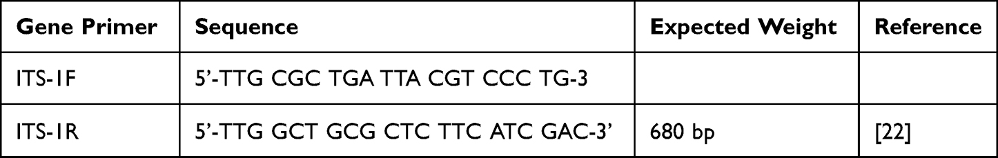 |
Table 1 Showing Primers Targeting Internal Transcribed Spacer-1 (ITS-1) Region of Fasciola Adult Worms |
PCR reaction with a total volume of 50 μL contains 5 μL of DNA solution, 25 μL liters of mastermix 2X (Thermo Fisher Scientific, USA), 1.0 μL primer (0.2 M), and 18.0 μL of distilled water was used. The following reaction conditions were used for PCR in a thermal cycler (Master-cycler Personal, Eppendorf): pre-denaturation for 10 min at 95 °C, then 25 PCR cycles of denaturation at 94 °C for 90 seconds, annealing at 58 °C for, and elongation at 72 °C for 90 seconds, followed by a final extension of at 72 °C for 10 min. Through electrophoresis on a 1% agarose gel with TAE buffer containing SYBR Safe (Invitrogen, SYBR SafeTM, Cat. No. S33102), PCR products were examined. For 90 minutes, electrophoresis was carried out at 90V. A GelDoc EZ Imager was used to report the size of the PCR products (Bio-Rad, GelDoc EZ Imager, Cat. No. 1708270).
Nucleotide Sequences of the ITS-1 Gene
To confirm the PCR results and identify the species of Fasciola adult flukes isolated from infected sheep in the present study, the obtained PCR products were used as templates for DNA sequence. Amplified ITS-1 fragments were sent to Macrogen (Seoul – South Korea) (https://dna.macrogen.com/) for sequencing of the ITS-1 region using the same primers used for PCR.
Sequence and Phylogenetic Analyses
NCBI BLAST search (with default settings) was used to retrieve related Fasciola sp. sequences from the GenBank. The obtained DNA sequences were aligned against DNA sequences of Fasciola species from different hosts and countries that had previously been deposited in the GenBank. Multiple alignments were done using the MUSCLE program of MEGA X software35. All sequences obtained from the current study (12 unique sequences) were deposited in the GenBank and as-signed the accession numbers from MZ429416.1 to MZ429427.1. Using the Tamura 3 parameter model of the MEGA X program and the Maximum Likelihood approach (ML), phylogenetic analysis and pairwise nucleotide changes of the ITS-1 region were performed. Fascioloides magna was used as outgroup (accession no. EF534991). Sequences of F. hepatica from different countries used for construction of the phylogenic tree together with sequences obtained from the present study are shown in Table 2.
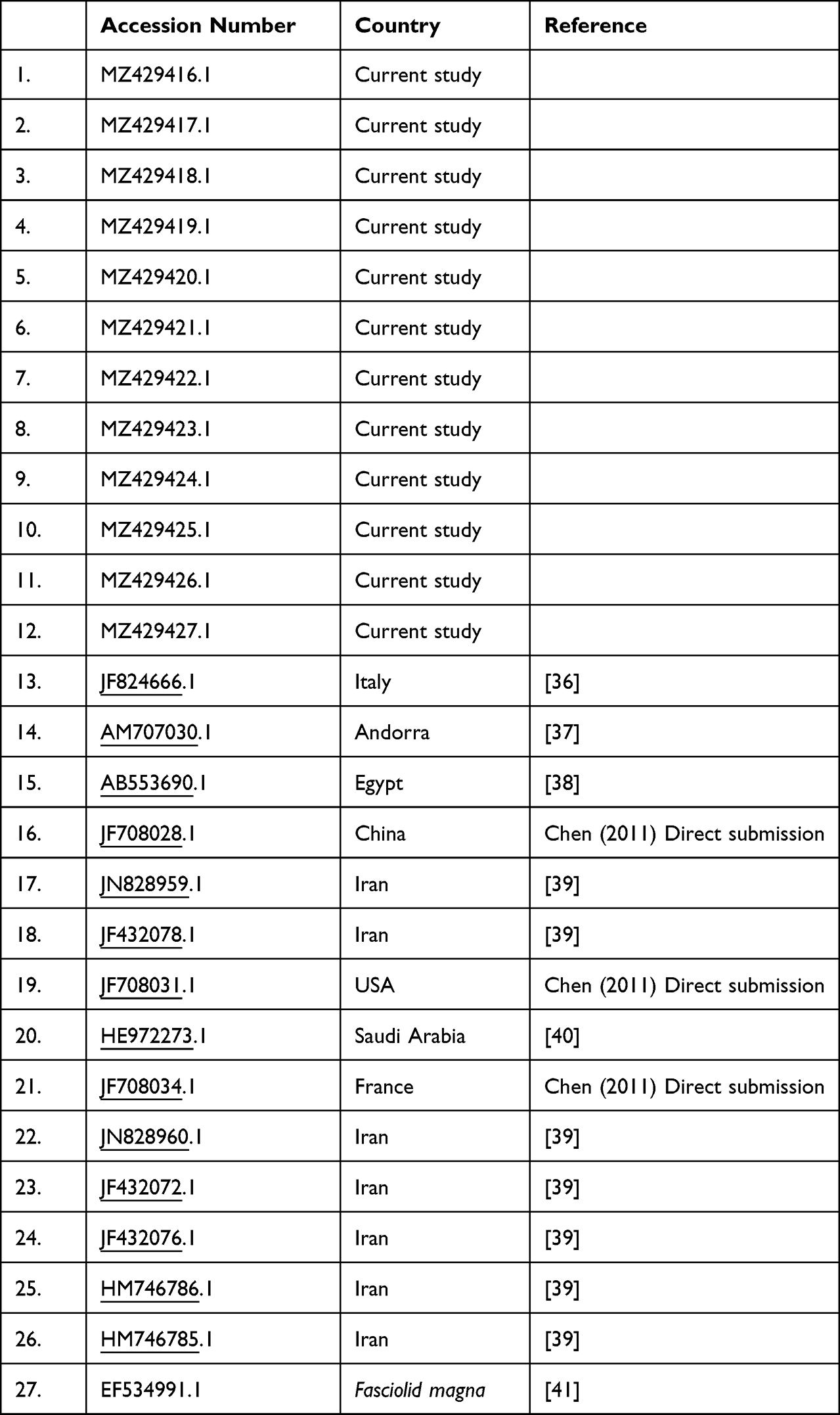 |
Table 2 Showing Accession Numbers of Fasciola spp. Used for Construction of the Phylogenetic Tree |
Statistical Analysis
The gathered data and outcomes were statistically analyzed using the SPSS version 22 program (Statistical Program for the Social Sciences).42 The Chi-square test was used to analyze the category variables. Statistical significance was defined as a P-value of 0.05 or lower.
Results
Prevalence of Fasciola Infection in Sheep
The present study included both local and imported Somalian animals (42.93% versus 57.07%). Examination of the liver and gall bladder of 12,653 slaughtered sheep revealed that 1043 (8.24%) were infected with Fasciola adult worm, 7.16% were local sheep and 9.6% were imported animals (Table 3).
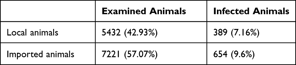 |
Table 3 Infection Rate Among Local and Imported Animals in the Present Study |
Significantly higher infection rate was reported among adults than young animals (14.27% vs 1.46%; P = 0.002). Moreover, female animals were recognized to have higher infection rates than males with no statistical significance (16.69% vs 2.6%, P = 0.279) (Table 4).
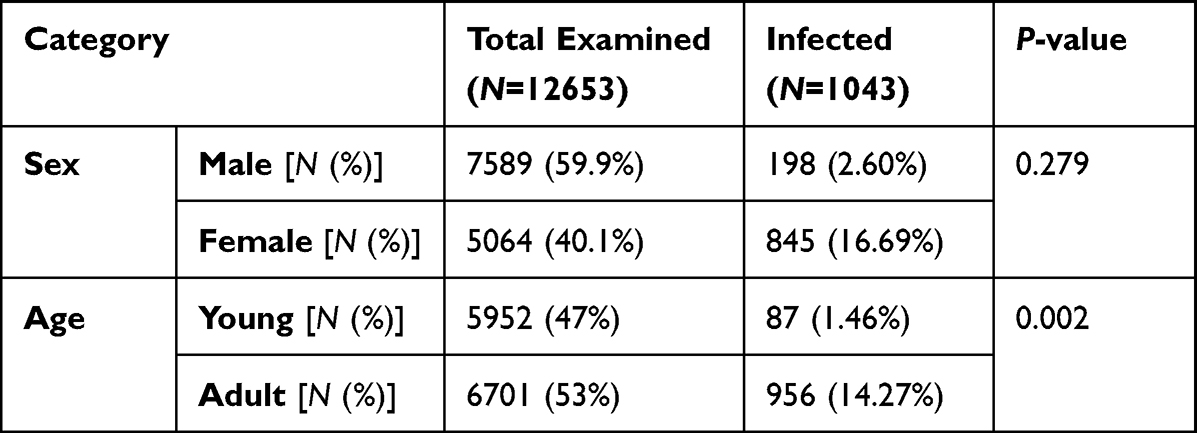 |
Table 4 Gender and Age Group of F. hepatica-Infected Sheep. The Numbers (N) of Infected Male, Female, Adult, and Young Sheep Were Determined and Percentages (%) of Total Infected Animals Were Calculated |
PCR Amplification of F. hepatica ITS-1 Gene
Fasciola spp. adult worms were collected from infected animals, then genomic DNA was extracted and successfully amplified from all examined adults (1043). PCR products of 680 bp were obtained (Figure 1).
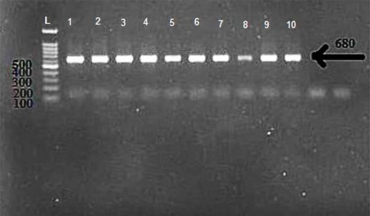 |
Figure 1 Agarose gel electrophoretic analysis of representative ITS-1 PCR products (680 bp) of F. hepatica adult worms. Lane of 1–10 representing samples. L: DNA ladder (100bp). |
Next, all PCR products (n = 1043) were used for DNA sequence of the ITS-1 region. Twelve different DNA sequences were obtained. The obtained nucleotide sequence data were deposited in the GenBank (Accession numbers MZ429416.1 - MZ429427.1). The variance of isolates ranged from 0. 00771 to 0.34405. Detailed results of nucleotide variation are shown in Table 5.
 |
Table 5 Genetic p-Distances Between Haplotypes Based on Sequence Analysis of Partial ITS1 Gene Analysis of F. hepatica Samples Obtained in the Present Study |
Using the BLAST 2 software, all sequences obtained from the present study were aligned with F. hepatica sequences deposited in the GenBank from different countries. DNA sequences of the present study shared >99.3% identity with F. hepatica sequences from Iran (MK377136, MF969010) and Egypt (LC076196).
The Maximum Likelihood method and Tamura-3 model were used to construct a phylogenetic tree to compare the DNA sequences of the present study with other F. hepatica Gen-Bank-accessible sequences. The phylogenetic tree supported the results of BLAST research as the isolates from the present work were closely related to F. hepatica isolates deposited in the GenBank. F. hepatica isolates were arranged in two clusters. Figure 2 displays a trustworthy grouping of the ITS-1 sequences of F. hepatica from the current investigation with those from Iran, Asia (China and Saudi Arabia), Europe (Italy and France), Africa (Egypt and Andorra), and the USA.
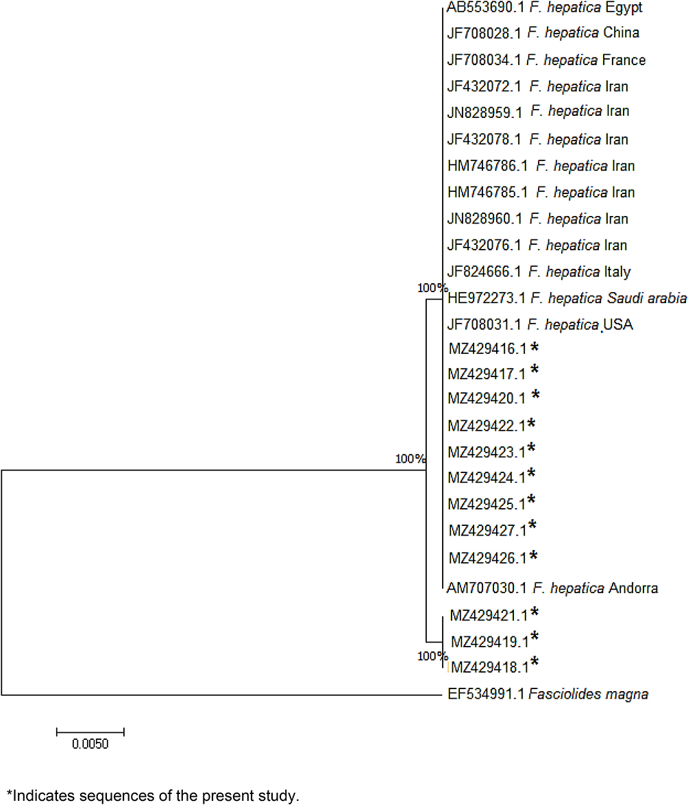 |
Figure 2 Phylogenetic relationship of F. hepatica isolated from Saudi Arabian sheep to other Fasciola sp. deposited in the GenBank based on the partial (ITS-1) gene nucleotide sequences, using Fascioloides magna as an out-group trematode. |
Discussion
Animal fascioliasis is usually a subclinical disease that is associated with economic losses, mostly due to a decline in animal productivity and liver condemnations.43,44 So, global efforts for disease control and prevention are needed to minimize the impact of the disease on animals and livestock. A deeper understanding of the dynamics of disease epidemiology and parasite taxonomy may be gained through advanced molecular and genetic studied and could aid in the development of effective control programs.31,32 In the present study, we examined 12,653 slaughtered sheep at the municipal abattoir of Jeddah, Saudi Arabia. We found that 8.2% of sheep were infected. Previous findings from different localities in Saudi Arabia reported a wide range of infection rates. Higher infection rate was reported in Riyadh by Magda and Al-Megrin in 2005 (21.9%)24 and by Degheidy et al in 2012 (16.9%).13 On the other hand, lower prevalence had been reported by Mgzoub and Kasim who examined different animals from different regions in Saudi Arabia and reported an infection rate in sheep ranging from 0.18% to 2.4%.45 Different infection rates from different localities may be explained by several factors including climate, ecological conditions, seasons, sources, and types of animals which may play a role in this issue.
In the present study, higher prevalence of fasciolosis was found among adults with statistical significance. In agreement with this finding, previous studies reported higher infection rates among adults than young animals.46,47 On the other hand, regarding gender, female sheep showed higher infection rate than males (with no statistical significance). This finding is supported by results of previous studies indicating the difference in susceptibility of both sexes to fasciolosis.46,48,49 This difference may be attributed to exposure of females to stress during pregnancy and parturition increasing their probability to infection.50
In the present study, PCR and DNA sequence were used to further identify and characterized Fasciola isolates. Several genetic loci including ITS regions (ITS-1 and ITS-2) have been used as a good target for diagnosis and genetic characterization of Fasciola spp.6,30,31,34,38,44,51–56 We used primers targeting 680 bp ITS-1 region. The obtained DNA sequence data confirmed that the isolated Fasciola adult worms are F. hepatica. This finding is in agreement with previous results reported by Alajmi, who studied Fasciola population isolated from sheep in Riyadh, Saudi Arabia.28 The author reported infection of sheep with both F. hepatica and F. gigantica with predominance of F. hepatica (80%).28 Furthermore, in the Al Taif region of Saudi Arabia, Shalaby et al reported identical infection rates for both F. hepatica and F. gigantica in imported sheep.40 Fasciola hepatica is the most widespread and common species in temperate regions, while F. gigantica is found in tropical countries of Africa, according to numerous studies based on both nuclear and mitochondrial sequences.57–61 Several studies particularly in Asian populations reported pure and mixed forms of Fasciola species in ruminants; Japan,31,62 China,63,64 Korea,31,57,65 Iran,66,67 Bangladesh,68 and Vietnam.69,70 The phylogenetic analysis of ITS-1 sequences using MEGA X application showed a rooted tree with Fasciolid magnua ITS-1 as an outgroup. The 12 Saudi Arabian F. hepatica isolates have been grouped into two clades with isolates from Iran, Africa (Egypt and Andorra), Asia (China and Saudi Arabia), Europe (Italy and France), and the USA.
Conclusion
Our research revealed that the only Fasciola species living in sheep included in this paper is F. hepatica. We think that by better comprehending the distribution and spread of Fasciola species among animals in Saudi Arabia, we can create more effective management strategies. It is well known that using molecular methods is the best way to distinguish between distinct Fasciola species based on several genetic loci. Even though there have been numerous reports of Fasciola infections in Saudi Arabia, there has only been a limited amount of molecular evidence to support any of these findings. To give information that will aid in the creation of fascioliasis management strategies and reduce economic losses, additional research utilizing a sizable number of samples from various locations in Saudi Arabia are required.
Ethics Statement
The present study was approved by the Institutional Review Board, College of Science, University of Jeddah, Saudi Arabia (protocol code UJ212430061).
Funding
This research was supported by Princess Nourah bint Abdulrahman University Researchers Supporting Project number (PNURSP2023R39), Princess Nourah bint Abdulrahman University, Riyadh, Saudi Arabia and by the Researchers Supporting Project number (RSP2023R25), King Saud University, Riyadh, Saudi Arabia.
Disclosure
The authors report no conflicts of interest in this work.
References
1. Mas-Coma S, Valero MA, Bargues MD. Fasciola, lymnaeids and human fascioliasis, with a global overview on disease transmission, epidemiology, evolutionary genetics, molecular epidemiology and control. Adv Parasitol. 2009;69:41–146 doi:10.1016/S0065-308X(09)69002-3.
2. Nyindo M, Lukambagire A-H. Fascioliasis: an ongoing zoonotic trematode infection. Biomed Res Int. 2015;2015:786195. doi:10.1155/2015/786195
3. Alemneh T, Ayelign M. Study on prevalence and economic importance of bovine fasciolosis in three districts of north-east Amhara Region, Ethiopia. J Infect Non Infect Dis. 2017;3:24.
4. Mas-Coma S, Bargues MD, Valero MA. Human fascioliasis infection sources, their diversity, incidence factors, analytical methods and prevention measures. Parasitology. 2018;145(13):1665–1699. doi:10.1017/S0031182018000914
5. Cwiklinski K, Dalton JP, Dufresne PJ, et al. The Fasciola hepatica genome: gene duplication and polymorphism reveals adaptation to the host environment and the capacity for rapid evolution. Genome Biol. 2015;16(1):71. doi:10.1186/s13059-015-0632-2
6. Javanmard E, Ohari Y, Sadeghi A, et al. Multigene typing and phylogenetic analysis of Fasciola from endemic foci in Iran. Infect Genet Evol. 2020;80:104202. doi:10.1016/j.meegid.2020.104202
7. Caravedo MA, Cabada MM. Human fascioliasis: current epidemiological status and strategies for diagnosis, treatment, and control. Res Rep Trop Med. 2020;11:149–158. doi:10.2147/RRTM.S237461
8. Valero MA, Varea MT, Marín R. Fasciola hepatica: lithogenic capacity in experimentally infested rats and chemical determination of the main stone components. Parasitol Res. 2000;86(7):558–562. doi:10.1007/s004360000201
9. Arias-Pacheco C, Lucas JR, Rodríguez A, Córdoba D, Lux-Hoppe EG. Economic impact of the liver condemnation of cattle infected with Fasciola hepatica in the Peruvian Andes. Trop Anim Health Prod. 2020;52(4):1927–1932. doi:10.1007/s11250-020-02211-y
10. Nyirenda SS, Sakala M, Moonde L, et al. Prevalence of bovine fascioliasis and economic impact associated with liver condemnation in abattoirs in Mongu district of Zambia. BMC Vet Res. 2019;15(1):33. doi:10.1186/s12917-019-1777-0
11. Zewde A, Bayu Y, Wondimu A. Prevalence of bovine fasciolosis and its economic loss due to liver condemnation at wolaita sodo municipal Abattair, Ethiopia. Vet Med Int. 2019;2019:9572373. doi:10.1155/2019/9572373
12. Mazeri S, Rydevik G, Handel I, Bronsvoort BMD, Sargison N. Estimation of the impact of Fasciola hepatica infection on time taken for UK beef cattle to reach slaughter weight. Sci Rep. 2017;7(1):7319. doi:10.1038/s41598-017-07396-1
13. Degheidy NS, Al-Malki JS. Epidemiological studies of fasciolosis in human and animals at Taif, Saudi Arabia. World Appl Sci J. 2012;19:1099–1104.
14. Mehmood K, Zhang H, Sabir AJ, et al. A review on epidemiology, global prevalence and economical losses of fasciolosis in ruminants. Microb Pathog. 2017;109:253–262. doi:10.1016/j.micpath.2017.06.006
15. Qureshi AW, Zeb A, Mansoor A, Hayat A, Mas-Coma S. Fasciola hepatica infection in children actively detected in a survey in rural areas of Mardan district, Khyber Pakhtunkhawa province, northern Pakistan. Parasitol Int. 2019;69:39–46. doi:10.1016/j.parint.2018.11.003
16. Sah R, Khadka S, Khadka M, et al. Human fascioliasis by Fasciola hepatica: the first case report in Nepal. BMC Res Notes. 2017;10(1):1–4. doi:10.1186/s13104-017-2761-z
17. Zoghi S, Emami M, Shahriarirad S, et al. Human fascioliasis in nomads: a population-based serosurvey in southwest Iran. Infez Med. 2019;27(1):68–72.
18. Temido H, Oliveira-Santos M, Parente F, Santos L. Fascioliasis—a rare cause of hepatic nodules. Case Rep. 2017;2017:bcr-2017–220363.
19. Espinel J, Goñi M. Obstructive jaundice of a parasitic etiology. Rev Esp Enferm Dig. 2019;11(2):165–166.
20. Fried B, Abruzzi A. Food-borne trematode infections of humans in the United States of America. Parasitol Res. 2010;106:1263–1280. doi:10.1007/s00436-010-1807-0
21. Micic D, Oto A, Charlton MR, Benoit J-L, Siegler M. Hiding in the water. N Engl J Med. 2020;382(19):1844–1849. doi:10.1056/NEJMcps1902741
22. Kain D, Mukkala AN, Boggild A. Prolonged antibiotic use leading to Clostridium difficile colitis in an ill returned traveller with acute fascioliasis. J Travel Med. 2018;25(1):tay012. doi:10.1093/jtm/tay012
23. Forbes AB. Parasites of Cattle and Sheep: A Practical Guide to Their Biology and Control. Wallingford UK: CAB International; 2021:149–200.
24. Sanad MM, Al-Megrin WA. Fascioliasis among local and imported sheep in Saudi Arabia: parasitological and serological diagnosis. J Egypt Soc Parasitol. 2005;35(3 Suppl):1121–1134.
25. El-Mathal EM, Fouad MA. Human fascioliasis among immigrant workers in Saudi Arabia. J Egypt Soc Parasitol. 2005;35(3 Suppl):1199–1207.
26. Aryaeipour M, Rouhani S, Bandehpour M, Mirahmadi H, Kazemi B, Rokni MB. Genotyping and phylogenetic analysis of Fasciola spp. Isolated from sheep and cattle using PCR-RFLP in Ardabil Province, Northwestern Iran. Iran J Public Health. 2014;43(10):1364–1371.
27. Galavani H, Gholizadeh S, Hazrati Tappeh K. Genetic characterization of Fasciola isolates from West Azerbaijan province Iran based on ITS1 and ITS2 Sequence of Ribosomal DNA. Iran J Parasitol. 2016;11(1):52–64.
28. Alajmi RA. Molecular characterization of Fasciola flukes using mitochondrial 28S rRNA gene in Naimi Saudi sheep. Saudi J Biol Sci. 2019;26(1):112–117. doi:10.1016/j.sjbs.2017.06.010
29. Itagaki T, Sakaguchi K, Terasaki K, Sasaki O, Yoshihara S, Van Dung T. Occurrence of spermic diploid and aspermic triploid forms of Fasciola in Vietnam and their molecular characterization based on nuclear and mitochondrial DNA. Parasitol Int. 2009;58(1):81–85. doi:10.1016/j.parint.2008.11.003
30. Marcilla A, Bargues MD, Mas-Coma S. A PCR-RFLP assay for the distinction between Fasciola hepatica and Fasciola gigantica. Mol Cell Probes. 2002;16(5):327–333. doi:10.1006/mcpr.2002.0429
31. Itagaki T, Kikawa M, Terasaki K, Shibahara T, Fukuda K. Molecular characterization of parthenogenic Fasciola sp. in Korea on the basis of DNA sequences of ribosomal ITS1 and mitochondrial NDI gene. J Vet Med Sci. 2005;67(11):1115–1118. doi:10.1292/jvms.67.1115
32. Sharifiyazdi H, Moazeni M, Rabbani F. Molecular characterization of human Fasciola samples in Gilan province, Northern Iran on the basis of DNA sequences of ribosomal and mitochondrial DNA genes. Comp Haematol Int. 2011;21:1–6.
33. Beesley NJ, Williams DJ, Paterson S, Hodgkinson J. Fasciola hepatica demonstrates high levels of genetic diversity, a lack of population structure and high gene flow: possible implications for drug resistance. Int J Parasitol. 2017;47(1):11–20. doi:10.1016/j.ijpara.2016.09.007
34. Anh DN, Anh LT, Tuan LQ, et al. Identification of Fasciola Species Isolates from Nghe An Province, Vietnam, Based on ITS1 Sequence of Ribosomal DNA Using a Simple PCR-RFLP Method. J Parasitol Res. 2018;2018:2958026. doi:10.1155/2018/2958026
35. Kumar S, Stecher G, Li M, Knyaz C, Tamura K. MEGA X: molecular Evolutionary Genetics Analysis across Computing Platforms. Mol Biol Evol. 2018;35(6):1547–1549. doi:10.1093/molbev/msy096
36. Farjallah S, Ben Slimane B, Piras CM, Amor N, Garippa G, Merella P. Molecular characterization of Fasciola hepatica from Sardinia based on sequence analysis of genomic and mitochondrial gene markers. Exp Parasitol. 2013;135(3):471–478. doi:10.1016/j.exppara.2013.08.006
37. Alasaad S, Huang CQ, Li QY, et al. Characterization of Fasciola samples from different host species and geographical localities in Spain by sequences of internal transcribed spacers of rDNA. Parasitol Res. 2007;101(5):1245–1250. doi:10.1007/s00436-007-0628-2
38. Amer S, Dar Y, Ichikawa M, et al. Identification of Fasciola species isolated from Egypt based on sequence analysis of genomic (ITS1 and ITS2) and mitochondrial (NDI and COI) gene markers. Parasitol Int. 2011;60(1):5–12. doi:10.1016/j.parint.2010.09.003
39. Mahami-Oskouei M, Dalimi A, Forouzandeh-Moghadam M, Rokni M. Molecular Identification and differentiation of Fasciola isolates using PCR- RFLP method based on internal transcribed spacer (ITS1, 5.8S rDNA, ITS2). Iran J Parasitol. 2011;6(3):35–42.
40. Shalaby I, Gherbawy Y, Banaja A. Molecular characterization of Fasciola species isolated from imported sheep in Taif region (Saudi Arabia). Trop Biomed. 2013;30(1):15–26.
41. Králová-Hromadová I, Bazsalovicsová E, Demiaszkiewicz AW. Molecular characterization of Fascioloides magna (Trematoda: fasciolidae) from south-western Poland based on mitochondrial markers. Acta Parasitol. 2015;60(3):544–547. doi:10.1515/ap-2015-0077
42. Mather LE, Austin KL. The Statistical Package for the Social Sciences (SPSS) as an adjunct to pharmacokinetic analysis. Biopharm Drug Dispos. 1983;4(2):157–172. doi:10.1002/bdd.2510040208
43. Martínez-Valladares M, Robles-Pérez D, Martínez-Pérez JM, et al. Prevalence of gastrointestinal nematodes and Fasciola hepatica in sheep in the northwest of Spain: relation to climatic conditions and/or man-made environmental modifications. Parasit Vectors. 2013;6(1):282. doi:10.1186/1756-3305-6-282
44. Olsen A, Frankena K, Bødker R, et al. Prevalence, risk factors and spatial analysis of liver fluke infections in Danish cattle herds. Parasit Vectors. 2015;8(1):160. doi:10.1186/s13071-015-0773-x
45. Magzoub M, Kasim AA. The prevalence of fascioliasis in Saudi Arabia. Trop Anim Health Prod. 1978;10(1):205–206. doi:10.1007/BF02235342
46. Khan MK, Sajid MS, Khan MN, Iqbal Z, Iqbal MU. Bovine fasciolosis: prevalence, effects of treatment on productivity and cost benefit analysis in five districts of Punjab, Pakistan. Res Vet Sci. 2009;87(1):70–75. doi:10.1016/j.rvsc.2008.12.013
47. Pfukenyi DM, Monrad J, Mukaratirwa S. Epidemiology and control of trematode infections in cattle in Zimbabwe: a review. J S Afr Vet Assoc. 2005;76(1):9–17. doi:10.4102/jsava.v76i1.387
48. Phiri AM, Phiri IK, Sikasunge CS, Monrad J. Prevalence of fasciolosis in Zambian cattle observed at selected abattoirs with emphasis on age, sex and origin. J Vet Med B Infect Dis Vet Public Health. 2005;52(9):414–416. doi:10.1111/j.1439-0450.2005.00872.x
49. Phiri AM, Phiri IK, Siziya S, Sikasunge CS, Chembensofu M, Monrad J. Seasonal pattern of bovine fasciolosis in the Kafue and Zambezi catchment areas of Zambia. Vet Parasitol. 2005;134(1–2):87–92. doi:10.1016/j.vetpar.2005.06.010
50. Spithill TW, Smooker PM, Copeman DB. ”Fasciola gigantica”: epidemiology, control, immunology and molecular biology. Fasciolosis. 1999;1999:465–525.
51. Nguyen S, Amer S, Ichikawa M, Itagaki T, Fukuda Y, Nakai Y. Molecular identification of Fasciola spp. (Digenea: platyhelminthes) in cattle from Vietnam. Parasite. 2012;19(1):85–89. doi:10.1051/parasite/2012191085
52. Relf V, Good B, Hanrahan J, McCarthy E, Forbes A, deWaal T. Temporal studies on Fasciola hepatica in Galba truncatula in the west of Ireland. Vet Parasitol. 2011;175:287–292. doi:10.1016/j.vetpar.2010.10.010
53. Chougar L, Mas-Coma S, Artigas P, et al. Genetically ‘pure’ Fasciola gigantica discovered in Algeria: DNA multimarker characterization, trans-Saharan introduction from a Sahel origin and spreading risk into north-western Maghreb countries. Transbound Emerg Dis. 2020. doi:10.1111/tbed.13572
54. Giovanoli Evack J, Schmidt RS, Boltryk SD, et al. Molecular confirmation of a fasciola gigantica × fasciola hepatica hybrid in a Chadian bovine. J Parasitol. 2020;106(2):316–322. doi:10.1645/19-66
55. Diyana JNA, Mahiza MIN, Latiffah H, et al. Occurrence, morphometric, and molecular investigation of cattle and buffalo liver adult fluke in Peninsular Malaysia main abattoirs. J Parasitol Res. 2020;2020:5436846. doi:10.1155/2020/5436846
56. Hasanpour H, Falak R, Naddaf SR, et al. Molecular Characterization of Fasciola spp. from Some Parts of Iran. Iran J Public Health. 2020;49(1):157–166.
57. Agatsuma T, Arakawa Y, Iwagami M, et al. Molecular evidence of natural hybridization between Fasciola hepatica and F. gigantica. Parasitol Int. 2000;49(3):231–238. doi:10.1016/S1383-5769(00)00051-9
58. Ai L, Chen M-X, Alasaad S, et al. Genetic characterization, species differentiation and detection of Fasciola spp. by molecular approaches. Parasit Vectors. 2011;4(1):101. doi:10.1186/1756-3305-4-101
59. Raina OK, Jacob SS, Sankar M, et al. Genetic characterization of Fasciola gigantica from different geographical regions of India by ribosomal DNA markers. J Parasit Dis. 2015;39(1):27–32. doi:10.1007/s12639-013-0276-7
60. Valero MA, Panova M, Mas-Coma S. Phenotypic analysis of adults and eggs of Fasciola hepatica by computer image analysis system. J Helminthol. 2005;79(3):217–225. doi:10.1079/JOH2005301
61. Walker S, Makundi A, Namuba F, et al. The distribution of Fasciola hepatica and Fasciola gigantica within southern Tanzania - Constraints associated with the intermediate host. Parasitology. 2008;135:495–503. doi:10.1017/S0031182007004076
62. Taira K, Saitoh Y. Estimated egg production of Fasciola gigantica (Japanese strain) in goats experimentally infected with 50 metacercariae. Helminthologia. 2010;47(4):199–203. doi:10.2478/s11687-010-0031-x
63. Chen JX, Chen MX, Ai L, et al. An Outbreak of Human Fascioliasis gigantica in Southwest China. PLoS One. 2013;8(8):e71520. doi:10.1371/journal.pone.0071520
64. Yuan W, Liu JM, Lu K, et al. Molecular identification and seasonal infections of species of Fasciola in ruminants from two provinces in China. J Helminthol. 2016;90(3):359–363. doi:10.1017/S0022149X15000383
65. Choe SE, Nguyen TT, Kang TG, Kweon CH, Kang SW. Genetic analysis of Fasciola isolates from cattle in Korea based on second internal transcribed spacer (ITS-2) sequence of nuclear ribosomal DNA. Parasitol Res. 2011;109(3):833–839. doi:10.1007/s00436-011-2323-6
66. Shafiei R, Sarkari B, Sadjjadi SM, Mowlavi GR, Moshfe A. Molecular and morphological characterization of Fasciola spp. Isolated from different host species in a newly emerging focus of human fascioliasis in Iran. Vet Med Int. 2014;2014:405740. doi:10.1155/2014/405740
67. Ashrafi K, Valero MA, Peixoto RV, Artigas P, Panova M, Mas-Coma S. Distribution of Fasciola hepatica and F. gigantica in the endemic area of Guilan, Iran: relationships between zonal overlap and phenotypic traits. Infect Genet Evol. 2015;31:95–109. doi:10.1016/j.meegid.2015.01.009
68. Rahman AKMA, Islam SKS, Talukder MH, Hassan MK, Dhand NK, Ward MP. Fascioliasis risk factors and space-time clusters in domestic ruminants in Bangladesh. Parasit Vectors. 2017;10(1):228. doi:10.1186/s13071-017-2168-7
69. Le TH, De NV, Agatsuma T, et al. Human fascioliasis and the presence of hybrid/introgressed forms of Fasciola hepatica and Fasciola gigantica in Vietnam. Int J Parasitol. 2008;38(6):725–730. doi:10.1016/j.ijpara.2007.10.003
70. Bui TD, Doanh PN, Saegerman C, Losson B. Current status of fasciolosis in Vietnam: an update and perspectives. J Helminthol. 2016;90(5):511–522. doi:10.1017/S0022149X15000929
 © 2023 The Author(s). This work is published and licensed by Dove Medical Press Limited. The
full terms of this license are available at https://www.dovepress.com/terms
and incorporate the Creative Commons Attribution
- Non Commercial (unported, 3.0) License.
By accessing the work you hereby accept the Terms. Non-commercial uses of the work are permitted
without any further permission from Dove Medical Press Limited, provided the work is properly
attributed. For permission for commercial use of this work, please see paragraphs 4.2 and 5 of our Terms.
© 2023 The Author(s). This work is published and licensed by Dove Medical Press Limited. The
full terms of this license are available at https://www.dovepress.com/terms
and incorporate the Creative Commons Attribution
- Non Commercial (unported, 3.0) License.
By accessing the work you hereby accept the Terms. Non-commercial uses of the work are permitted
without any further permission from Dove Medical Press Limited, provided the work is properly
attributed. For permission for commercial use of this work, please see paragraphs 4.2 and 5 of our Terms.