Back to Journals » Neuropsychiatric Disease and Treatment » Volume 19
miR-98-5p Prevents Hippocampal Neurons from Oxidative Stress and Apoptosis by Targeting STAT3 in Epilepsy in vitro
Received 12 May 2023
Accepted for publication 9 August 2023
Published 30 October 2023 Volume 2023:19 Pages 2319—2329
DOI https://doi.org/10.2147/NDT.S415597
Checked for plagiarism Yes
Review by Single anonymous peer review
Peer reviewer comments 2
Editor who approved publication: Dr Roger Pinder
Zhizhuan Guo,1,2 Wenwen Zhong,3 Zhengshou Zou4
1Department of Neurology, Shanxi Bethune Hospital, Shanxi Academy of Medical Sciences, Tongji Shanxi Hospital, Third Hospital of Shanxi Medical University, Taiyuan, 030032, People’s Republic of China; 2Tongji Hospital, Tongji Medical College, Huazhong University of Science and Technology, Wuhan, 430030, People’s Republic of China; 3Department of Rehabilitation Medicine, Huangshi Maternal and Child Health Hospital, Edong Medical Group, Huang Shi, Hubei, 435000, People’s Republic of China; 4Department of Neurology, Huangshi Central Hospital, Edong Medical Group, Huangshi, Hubei, 435000, People’s Republic of China
Correspondence: Zhengshou Zou, Department of Neurology, Huangshi Central Hospital, Edong Medical Group, 141 Tianjin Road, Huangshi, Hubei, 435000, People’s Republic of China, Email [email protected]
Purpose: Epilepsy is a serious mental disease, for which oxidative stress and hippocampal neuron death after seizure is crucial. Numerous miRNAs are involved in epilepsy. However, the function of miR-98-5p in oxidative stress and hippocampal neuron death after seizure is unclear, which is the purpose of current study.
Methods: Magnesium ion (Mg2+)-free solution was used to establish the in vitro epilepsy model in hippocampal neurons. Oxidative stress was exhibited by measuring malondialdehyde (MDA) level and superoxide Dismutase (SOD) activity using enzyme-linked immune sorbent assay (ELISA) kits. 3-(4,5-Dimethylthiazol-2-yl)-2,5-diphenyltetrazolium bromide (MTT) assay and flow cytometry were applied for the examination of neuron viability and apoptosis, respectively. Quantitative reverse‐transcription polymerase chain reaction (qRT-PCR) and Western blot were used to evaluate the mRNA and protein levels of miR-98-5p and signal transducer and activator of transcription (STAT3), respectively. The relationship between miR-98-5p and STAT3 was predicted by TargetScan 7.2, and identified by dual-luciferase reporter assay and RNA immunoprecipitation (RIP) assay.
Results: miR-98-5p was decreased in the in vitro epileptic model of hippocampal neurons induced by Mg2+-free solution, whose overexpression rescued oxidative stress and neuron apoptosis in epileptic model. Moreover, overexpression of STAT3, one downstream target of miR-98-5p, partially eliminated the effects of miR-98-5p mimic.
Conclusion: We shed lights on a pivotal mechanism of miR-98-5p in regulating neuron oxidative stress and apoptosis after seizures, providing potential biomarkers for the diagnosis of epilepsy and therapeutic targets for the treatment of epilepsy.
Keywords: epilepsy, oxidative stress, apoptosis, miR-98-5p, STAT3
Introduction
Epilepsy is a chronic non-communicable neurological disorder in the central nervous system (CNS).1 It has the characteristics of recurrent seizures and high synchronous discharge of brain neurons,2,3 and is accompanied with comorbidity, depression, mortality, and anxiety.4 At present, there are approximately 70 million epileptic patients worldwide, and the incidence is increasing year by year. Long-term recurrent seizures induce neuron death and cause brain damage, in which hippocampal neurons are the most severely damaged.5–7 Hippocampus is a brain structure with special structure and function, which is closely related to learning and memory,8 and is very sensitive to epileptic discharge in the brain. The damage of hippocampal neurons will further aggravate the course of epilepsy and cause cognitive impairment.9 Therefore, it is of great importance to investigate the mechanism of epilepsy-induced hippocampal neuron death.
Glutamatergic synapses are the main excitatory synapses in the brain, mediating the transmission of most excitatory synaptic in the CNS.10 Epilepsy is closely related to the hyperactivity of glutamatergic neurons, which are able to over-activate downstream neurons and produce excitatory neurotoxicity.11,12 N-methyl-D-aspartic acid receptor (NMDAR) is a kind of important ionic glutamate receptors which located in the glutamatergic postsynaptic membrane. When neurons are over-activated, NMDA receptor will mediate a large amount of extracellular calcium ion (Ca2+) influx, and activate cell death pathway.13,14 Numerous anti-convulsant properties are exerted by NMDA antagonists in animals with acute/chronic epilepsy.15
As acknowledged, the increase of intracellular Ca2+ leads to excessive accumulation of reactive oxygen species (ROS), which causes oxidative stress in neuron.16,17 Subsequently, excessive oxidative stress results in protein oxidation, lipid oxidation, DNA damage, other oxidative reactions, and eventually cell death.18,19
MicroRNA (miRNA) is a kind of endogenous small non-coding RNAs with 18–24 nucleotides, which can bind target genes, and form RNA-induced silencing complex (RISC) to inhibit the translation of target genes.20,21 Consequently, miRNAs participate in numerous pivotal physiological processes.22,23 Massive literatures have reported the relationship between miRNAs and epilepsy,24,25 eg, miR-101a-3p attenuates pilocarpine-induced epilepsy by repressing c-Fos expression.26 Moreover, massive miRNAs are involved in Mg2+-free condition induced in vitro epilepsy models in hippocampal neurons, for instance, miR-29a alleviates seizure-induced cell death and inflammation in hippocampal neurons by targeting HMGB1,27 miR-30b-5p protects against cell proliferation and attenuates apoptosis in hippocampal neurons by targeting GRIN2A.28 Specifically, in 2017, miR-98-5p was discovered to be down-regulated in the brain samples of post-traumatic epilepsy rats,29 however, the roles of miR-98-5p in epilepsy remains unclear.
Herein, miR-98-5p expression is downregulated in the Mg2+-free-induced hippocampal neurons; moreover, miR-98-5p prevents neurons from Mg2+-free-induced oxidative stress and apoptosis by targeting STAT3. Current study reveals a novel mechanism of miR-98-5p in regulating neuron survival in epilepsy, which provides an important scientific basis for exploring the mechanism of epilepsy caused neuron death.
Materials and Methods
Cell Culture and Epilepsy Model Establishment
All 2-day postnatal Sprague-Dawley rats were maintained and used. Ethical and legal approval was obtained from the Ethical Commission of Huangshi Central Hospital prior to the commencement of the study. All experiments were performed following guidelines and regulations of the Ethical Commission of Huangshi Central Hospital. Firstly, rats were sacrificed according to standard protocols (100 mg/kg intraperitoneal sodium pentobarbital). Thereafter, hippocampus was dissociated from rats, dissected out in ice-cold PBS, trypsinized (0.25%) and maintained with the whole culture medium (NeuroBasal-A medium, 2% B27 supplement, 0.5 mmol/L glutamine and 10% FBS), before being plated onto poly-D-lysine (50 µg/mL) coated glass coverslips. Cells (2 × 10⁵ cells/mL) were incubated in an incubator containing 5% CO2 at 37°C and maintained by replacing half medium with fresh culture medium every 4 days.
The well established in vitro epilepsy model in hippocampal neurons was conducted by referring to previous studies.30,31 In brief, at day 14, the culture medium was removed and harvested as conditioned medium, and primary neurons were appended with Mg2+-free solution (pH 7.3, without MgCl2): 2.5 mM KCl, 145 mM NaCl, 2 mM CaCl2, 10 mM HEPES, 10 mM D-glucose and 0.002 mM glycine, and cultured in an incubator at 37°C for 3 h; thereafter, neurons were restored to the physiological concentration of 1mM MgCl2 by rinse with 3× 1.5 mL of culture medium at 37°C, returned to maintenance feed and incubated at 37°C with 5% CO2. Hippocampal neurons in control group were treated with culture medium containing 1 mM MgCl2.
Antibodies and Reagents
The antibodies are as follows: rabbit anti-STAT3 primary antibody (Cell signaling technology, #12640), rabbit anti-GAPDH primary antibody (Cell signaling technology, #5174), goat anti-rabbit secondary antibody (Cell signaling technology, #7074).
The regents are as follows: poly-D-lysine (Sigma Aldrich), Neurobasal (Thermo Fisher Scientific), B27 (Thermo Fisher Scientific), GlutaMax (Thermo Fisher Scientific), Penicillin-Streptomycin (Thermo Fisher Scientific), Lipofectamine 2000 (Thermo Fisher Scientific), Trizol (Thermo Fisher Scientific), PrimeScript qRT-PCR Reagent kit (Takara), SYBR Green Mix (Roche), Dual Luciferase Assay System (Promega Corporation), MDA and SOD ELISA kits (Nanjing Jiancheng Bioengineering Institute), Flow cytometry apoptosis detection kit (BD Biosciences), protease inhibitor cocktail (Sigma-Aldrich), RIPA buffer (Roche), Annexin V-FITC (Thermo Fisher Scientific), Propidium iodide (Thermo Fisher Scientific), ECL Western blotting substrate (Pierce).
Cell Transfection
For transfection experiments, after replacing half medium at 14DIV, 50 nM miR-NC mimic (5’-UCGCUUGGUGCAGGUCGGG-3’, GenaPhama, Shanghai, China) or miR-98-5p mimic (5’-UGAGGUAGUAAGUUGUAUUGUU-3’, GenaPhama), and 2 µg pcDNA3.1 (GenaPhama) or pcDNA3.1-STAT3 (GenaPhama) was transfected into neurons by Lipofectamine 2000 (Thermo Fisher Scientific). At 4 h later, the culture medium was replaced by mixed conditioned medium (half conditioned culture medium and half fresh culture medium). At 48 h later, cells were harvested for the subsequent experiments.
qRT-PCR
Total RNA was extracted from cultured neurons by Trizol reagent, and reverse transcribed into cDNA by PrimeScript qRT-PCR Reagent kit. SYBR Green Mix kit was used for qRT-PCR experiments. The expression levels of miR-98-5p and STAT3 were analyzed by 2−∆∆Ct method, and normalized to the internal controls U6 and GAPDH, respectively.
Western Blot Analysis
Cultured neurons were rinsed by PBS, and lysed by protease inhibitor cocktail contained RIPA buffer. The protein samples were mixed with SDS loading buffer and loaded to SDS-PAGE gel for electrophoresis onto PVDF membranes. After blocking with 5% BSA in TBST for 1 h at room temperature, the PVDF membranes were incubated with primary antibodies overnight at 4°C, and washed thoroughly with TBST; then incubated with secondary antibodies for 1 h at room temperature. After wash thoroughly, the HRP signals were detected with ECL substrate. The intensity of positive protein band was measured by ImageJ software, and the average value of control group was normalized to 1.
MDA Level and SOD Activity Measurement
Neurons were washed with PBS, and harvested before being lysed by protease inhibitor cocktail contained RIPA buffer on ice for 30 min. Thereafter, neurons were centrifuged at 10,000g for 10 min at 4°C. At last, the supernatant was used for the determination of MDA level and SOD activity with MDA and SOD ELISA kits, respectively.
Flow Cytometric Assay
Cultured neurons were washed by PBS and trypsinized (0.25%) before being harvested from each well. Subsequently, neurons were suspended in 100 µL PBS and mixed with Annexin V-FITC and Propidium iodide, followed by incubation at dark room temperature for 15 min. After washing with PBS, each sample was mixed with SA-FLOUS and incubated at dark 4°C for 20 min. Finally, neurons were analyzed by flow cytometry, and Annexin V-FITC positive cells were considered as apoptotic neurons.
MTT Assay
At 14DIV, each group of neurons was incubated with 250 µL MTT solution and 250 µL fresh Neurobasal medium at 37°C for 3 h. Afterwards, MTT solution was replaced with MTT solvent. Neurons were shaken for 15 min before being subjected to the measurement of the absorbance at 570 nm.
Dual Luciferase Reporter Assays
The binding site between miR-98-5p and STAT3 was predicted by TargetScan 7.2 (http://www.targetscan.org/vert_72/). The 3’ untranslated region (3’ UTR) sequences of wild type (WT) and mutant (MUT) STAT3 were inserted into pGL3-luciferase reporter plasmids using KpnI and XhoI restriction sites, and the corresponding reconstructed plasmids were named as STAT3-WT and -MUT, respectively. Subsequently, STAT3-WT and -MUT were co-transfected with miR-98-5p mimic or NC mimic into cultured neurons using Lipofectamine 2000. At 48 h after transfection, cells were trypsinized and transferred to a 24-well plate. The luciferase reporter activities were measured by Dual Luciferase Assay System with Renilla luciferase activity as the reference control.
RNA Immunoprecipitation (RIP) Assay
Nuclei from cells were isolated, lysed and incubated with primary antibodies against anti-Ago2 (Abcam) and anti-IgG (Abcam) at 4°C overnight. Then, RNA immuno-precipitated with RBP was isolated immediately following the addition of protein A agarose (Abcam) and protein G agarose (Abcam). After wash, RNA was purified and reverse transcribed into cDNA. Gene expression was measured by RT-qPCR as aforementioned.
Statistical Analyses
All the data were stated as mean ± S.D and analyzed using SPSS version 17.0. Difference between two groups was measured by unpaired Student’s t test, and the differences among four groups were measured by one‐way analysis of variance analysis followed by Tukey's test. p<0.05 was considered as significant difference.
Results
Oxidative Stress and Cell Viability in an in vitro Epilepsy Model in Hippocampal Neurons
Herein, Mg2+-free solution was used to treat 14DIV cultured rat primary hippocampal neurons for 3 h to induce epilepsy in vitro.30,31 At 20 h later, ELISA and MTT were used to detect the oxidative stress and cell viability of the neurons, respectively.
Lipid peroxidation, due to oxidative stress is involved in some epilepsy types and seizure recurrence.32 The brain contains high concentration of PUFA which is more prone to lipid peroxidation, therefore, the assessment of MDA level is an important indicator of lipid peroxidation for various diseases. The superoxide dismutases play a crucial role in eliminating superoxide anion radicals (O2•−) generated from extracellular stimulants, including ionizing radiation and oxidative insults together with produced in mitochondrial matrix.33 Herein, ELISA exhibited significantly higher MDA level and lower SOD activity in Mg2+-free solution treated neurons compared to controls (p<0.01, Figure 1A and B).
In addition, MTT exerted significantly lower cell viability in Mg2+-free solution treated neurons compared to controls (p<0.01, Figure 1C).
The findings indicated that Mg2+-free solution treated neurons had stronger oxidative pressure and lower cell viability.
Expression of miR-98-5p and STAT3 in an in vitro Epilepsy Model in Hippocampal Neurons
Subsequently, we intended to explore how these aforementioned changes were induced. By searching literatures, it was found that the expression of miR-98-5p in the brain samples of post-traumatic epilepsy rats was decreased;29 in epileptic cell models, the expression of STAT3 was increased,34,35 which was closely related to oxidative stress and cell survival.36,37
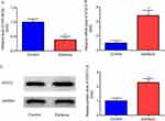
Therefore, qRT-PCR and Western blotting were performed to detect the expressions of miR-98-5p and STAT3 in Mg2+-free solution induced in vitro epilepsy model in hippocampal neurons. Compared with control group, the expression of miR-98-5p decreased significantly (p<0.01, Figure 2A), while the mRNA and protein levels of STAT3 increased significantly (p<0.01, Figure 2B–D), in Mg2+-free solution treated neurons.
Manipulating miR-98-5p and STAT3 Expression in an in vitro Epilepsy Model in Hippocampal Neurons
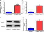
To make clear the roles of miR-98-5p and STAT3 in epilepsy, it was needed to achieve their ectopic expressions in hippocampal neurons by miR-98-5p mimic and pcDNA3.1-STAT3. As shown in Figure 3A–D (p<0.01), the expressions of miR-98-5p and STAT3 were increased by transfection of miR-98-5p mimic and pcDNA3.1-STAT3, respectively.
Thereafter, we were eager to know whether the expression of STAT3 could be regulated by miR-98-5p. In Figure 4A–C, miR-98-5p mimic (p<0.05) significantly attenuated Mg2+-free solution (p<0.01) induced upregulation of mRNA and protein levels of STAT3, which were partially eliminated by pcDNA3.1-STAT3 (p<0.05).
Collectively, these findings indicated that miR-98-5p was involved in epilepsy by regulating STAT3.
Effects of miR-98-5p and STAT3 on Oxidative Stress, Cell Viability and Apoptosis in an in vitro Epilepsy Model in Hippocampal Neurons
Afterwards, to identify the involvement of miR-98-5p as well as the relation between miR-98-5p and STAT3 in epilepsy induced increase of oxidative stress and decrease of cell viability, miR-98-5p mimic and/or pcDNA3.1-STAT3 were transfected into Mg2+-free solution treated neurons. In Figure 5A–C, ELISA and MTT exhibited that miR-98-5p mimic (p<0.05) significantly attenuated Mg2+-free solution (p<0.01) induced upregulation of MDA level, downregulation of SOD activity and cell viability, which were partially reversed by pcDNA3.1-STAT3 (p<0.05).
Cell apoptosis is an important cause of seizure induced neuron loss, and is closely related to oxidative stress and cell viability.38 Thereafter, we planned to investigate whether miR-98-5p and STAT3 affected Mg2+-free treatment induced cell apoptosis, miR-98-5p mimic and/or pcDNA3.1-STAT3 were transfected into Mg2+-free solution treated neurons. As shown in Figure 6A and B, flow cytometry exerted that, miR-98-5p mimic (p<0.01) significantly attenuated Mg2+-free solution (p<0.001) induced cell apoptosis, which was partially eliminated by pcDNA3.1-STAT3 (p<0.01).
In summary, overexpression of miR-98-5p prevented hippocampal neurons from oxidative stress, cell viability reduction and cell apoptosis, while STAT3 overexpression had the reverse effects.
STAT3 is Targeted by miR-98-5p
According to the aforementioned findings, it was found that the expression of STAT3 was regulated by miR-98-5p, and the roles of miR-98-5p and STAT3 in seizure induced oxidative stress and cell apoptosis were completely opposite, demonstrating that STAT3 might be targeted by miR-98-5p.
Consequently, we searched the TargetScan 7.2 website, and found that the 3’ UTR of STAT3 was potentially targeted by miR-98-5p (Figure 7A). Next, we designed WT and MUT sequences of STAT3 3’UTR to perform dual luciferase reporter assay, which exhibited that in STAT3-WT transfected neurons, the luciferase activity was significantly lowered by miR-98-5p mimic compared to miR-NC mimic (p<0.05); however, in STAT3-MUT transfected neurons, there was no significant difference in the luciferase activity between miR-NC mimic group and miR-98-5p mimic group (p>0.05, Figure 7B). Furthermore, RIP assay displayed that miR-98-5p and STAT3 were highly enriched in the Ago2 group compared to anti-IgG group (p<0.01, Figure 7C). Altogether, these results stated clearly that STAT3 was targeted by miR-98-5p.
Discussion
Herein, miR-98-5p expression was decreased, while STAT3 expression was increased in Mg2+-free induced in vitro epilepsy model in hippocampal neurons. In addition, overexpression of miR-98-5p attenuated Mg2+-free induced phenotypes of increased oxidative stress and cell apoptosis in epilepsy neurons, which was partially abolished by overexpression of STAT3. Altogether, miR-98-5p prevents hippocampal neurons from oxidative stress and apoptosis by targeting STAT3 in epilepsy.
In current study, one of the key findings was that miR-98-5p expression was decreased after Mg2+-free treatment, which was consistent with a previous study showing that miR-98-5p was down-regulated in brain samples of post-traumatic epilepsy rats.29 The result indicated that, on one hand, decreased expression of miR-98-5p was a co-occurrence phenomenon in epilepsy models, suggesting a biomarker potential of miR-98-5p for epilepsy diagnosis; on the other hand, decreased expression of miR-98-5p was regulated by some early unknown epilepsy-related mechanisms. However, overexpression of miR-98-5p alone did not have any significant effect on control neurons (data not shown), which suggests that miR-98-5p only function under Mg2+-free conditions.
Afterwards, STAT3 expression was discovered to be increased in Mg2+-free solution induced epilepsy model, which was in consistent with previous reports showing increased or overactivated STAT3 after seizure, eg, Choi et al found that nuclear STAT3 was rapidly and constantly activated in the rat hippocampus following kainic acid-induced seizures,34 Jiang et al reported that STAT3 was elevated in hippocampus of epileptic mice following lipopolysaccharide (LPS)-induced seizures.35
In addition, inhibiting the expression of STAT3 by overexpressing miR-98-5p could repress neuronal oxidative stress and apoptosis effectively, while overexpression of STAT3 could promote neuronal oxidative stress and apoptosis. These results indicated that miR-98-5p and STAT3 had opposite functions, STAT3 was a downstream target of miR-98-5p, and STAT3 expression was closely related to neuron viability after seizure. Thus, STAT3 might be a potential therapeutic target for epilepsy, which was further proved by a very recent report in 2023 Jul, where Tipton et al discovered that, in a mouse line with selective STAT3 knock-out in excitatory neurons, there was inhibited seizure progression, and demonstrated that targeting neuronal STAT3 may be an effective disease-modifying strategy for epilepsy.39
miR-98-5p was related to the antioxidant mechanism in various diseases, eg, oxygen-glucose deprivation/reoxygenation (OGD/R)-induced neuronal injury,40 and cerebral ischemia/reperfusion injury.41 Additionally, numerous studies have exhibited the facilitation effects of STAT3 in oxidative stress and cell apoptosis in other models, eg, middle cerebral artery occlusion (MCAO) model,42 and cerebral ischemia-reperfusion injury.43 However, there is no literature about the relation between miR-98-5p/STAT3 axis and epilepsy-induced oxidative stress, neuron apoptosis. Herein, we reported for the first time that miR-98-5p prevented neurons from oxidative stress and apoptosis by targeting STAT3 in Mg2+-free induced epilepsy model in hippocampal neuron. Moreover, there were several reports about the relationship between miR-98-5p/STAT3 axis and other diseases, for example, overexpression of miR-98 attenuates neuropathic pain by targeting STAT3 in chronic constriction injury rats,44 miR-98 inhibits cell proliferation and invasion by targeting STAT3 in nasopharyngeal carcinoma,45 miR-98-5p promotes apoptosis and inhibits migration by targeting STAT3 in A549 cells,46 which further strengthen the interaction between miR-98-5p and STAT3 in current study.
Altogether, current study identified that miR-98-5p overexpression prevents hippocampal neurons from Mg2+-free-induced cell viability reduction, oxidative stress and apoptosis by targeting STAT3.
Nevertheless, there remain two limitations in the present study: 1. In the in vitro model of epilepsy in cultured hippocampus neurons, the intact intra-hippocampal connectivity is lost, which will be improved in the future study by using the in vitro epilepsy models of whole hippocampus, whole brain, acute slice and organotypic slice, etc, 2. Epilepsy is characterized by recurrent seizures and seizure is quantified by behavior or electrophysiological recordings. However, no electrophysiological recording is displayed in the result part of this manuscript. As a result, more in vitro models of epilepsy will be conducted in the future study to demonstrate the electrophysiological recording of seizure-like events.
Conclusion
Current study reported that the expression of miR-98-5p was decreased, while STAT3 was increased in cultured hippocampal epilepsy neuron model in vitro. What’s more, the present study revealed a novel mechanism of miR-98-5p overexpression in preventing neurons from Mg2+-free-induced cell viability reduction, oxidative stress and apoptosis, which were partially abolished by STAT3. In sum, current study provided potential biomarkers for the diagnosis of epilepsy and therapeutic targets for the treatment of epilepsy.
Data Sharing Statement
The data and materials were available from the corresponding author.
Funding
There is no funding to report.
Disclosure
The authors report no conflicts of interest in this work.
References
1. Kobylarek D, Iwanowski P, Lewandowska Z, et al. Advances in the potential biomarkers of epilepsy. Front Neurol. 2019;10:685. doi:10.3389/fneur.2019.00685
2. De Kinderen RJA, Lambrechts DAJE, Wijnen BFM, et al. An economic evaluation of the ketogenic diet versus care as usual in children and adolescents with intractable epilepsy: an interim analysis. Epilepsia. 2016;57(1):41–50. doi:10.1111/epi.13254
3. Pitkanen A, Sutula TP. Is epilepsy a progressive disorder? Prospects for new therapeutic approaches in temporal-lobe epilepsy. Lancet Neurol. 2002;1(3):173–181. doi:10.1016/s1474-4422(02)00073-x
4. Berg AT, Berkovic SF, Brodie MJ, et al. Revised terminology and concepts for organization of seizures and epilepsies: report of the ILAE Commission on classification and terminology, 2005-2009. Epilepsia. 2010;51(4):676–685. doi:10.1111/j.1528-1167.2010.02522.x
5. Yoo JG, Jakabek D, Ljung H, et al. MRI morphology of the hippocampus in drug-resistant temporal lobe epilepsy: shape inflation of left hippocampus and correlation of right-sided hippocampal volume and shape with visuospatial function in patients with right-sided TLE. J Clin Neurosci. 2019;67:68–74. doi:10.1016/j.jocn.2019.06.019
6. Sloviter RS. Decreased hippocampal inhibition and a selective loss of interneurons in experimental epilepsy. Science. 1987;235(4784):73–76. doi:10.1126/science.2879352
7. Huberfeld G, Blauwblomme T, Miles R. Hippocampus and epilepsy: findings from human tissues. Rev Neurol (Paris). 2015;171(3):236–251. doi:10.1016/j.neurol.2015.01.563
8. Muller RU, Stead M, Pach J. The hippocampus as a cognitive graph. J Gen Physiol. 1996;107(6):663–694. doi:10.1085/jgp.107.6.663
9. De Lanerolle NC, Kim JH, Robbins RJ, Spencer DD. Hippocampal interneuron loss and plasticity in human temporal lobe epilepsy. Brain Res. 1989;495(2):387–395. doi:10.1016/0006-8993(89)90234-5
10. Kennedy MB. The postsynaptic density at glutamatergic synapses. Trends Neurosci. 1997;20(6):264–268. doi:10.1016/s0166-2236(96)01033-8
11. Barkerhaliski ML, White HS. Glutamatergic mechanisms associated with seizures and epilepsy. Cold Spring Harbor Perspect Med. 2015;5(8):a022863. doi:10.1101/cshperspect.a022863
12. Javitt DC. Glutamate as a therapeutic target in psychiatric disorders. Mol Psychiatry. 2004;9(11):984–997. doi:10.1038/sj.mp.4001551
13. Choi DW, Koh J, Peters S. Pharmacology of glutamate neurotoxicity in cortical cell culture: attenuation by NMDA antagonists. J Neuroscience. 1988;8(1):185–196. doi:10.1523/JNEUROSCI.08-01-00185.1988
14. Lau A, Tymianski M. Glutamate receptors, neurotoxicity and neurodegeneration. Pflügers Arch. 2010;460(2):525–542. doi:10.1007/s00424-010-0809-1
15. Chapman AG. Glutamate and epilepsy. J Nutr. 2000;130(4S Suppl):1043S–1045S. doi:10.1093/jn/130.4.1043S
16. Brookes PS, Yoon Y, Robotham JL, Anders MW, Sheu S. Calcium, ATP, and ROS: a mitochondrial love-hate triangle. Am J Physiol Cell Physiol. 2004;287(4):C817–C833. doi:10.1152/ajpcell.00139.2004
17. Gorlach A, Bertram K, Hudecova S, Krizanova O. Calcium and ROS: a mutual interplay. Redox Biol. 2015;6:260–271. doi:10.1016/j.redox.2015.08.010
18. Tan S, Sagara Y, Liu Y, Maher PA, Schubert D. The regulation of reactive oxygen species production during programmed cell death. J Cell Biol. 1998;141(6):1423–1432. doi:10.1083/jcb.141.6.1423
19. Aguiar CCT, Almeida AB, Araujo PVP, et al. Oxidative stress and epilepsy: literature review. Oxid Med Cell Longev. 2012;2012:795259. doi:10.1155/2012/795259
20. Jonas S, Izaurralde E. Towards a molecular understanding of microRNA-mediated gene silencing. Na Rev Genet. 2015;16(7):421–433. doi:10.1038/nrg3965
21. Gregory RI, Chendrimada TP, Shiekhattar R. MicroRNA biogenesis: isolation and characterization of the microprocessor complex. Methods Mol Biol. 2006;342:33–47. doi:10.1385/1-59745-123-1:33
22. Ambros VR. The functions of animal microRNAs. Nature. 2004;431(7006):350–355. doi:10.1038/nature02871
23. Bartel DP. MicroRNAs: target recognition and regulatory functions. Cell. 2009;136(2):215–233. doi:10.1016/j.cell.2009.01.002
24. Henshall DC. MicroRNA and epilepsy: profiling, functions and potential clinical applications. Curr Opin Neurol. 2014;27(2):199–205. doi:10.1097/WCO.0000000000000079
25. Reschke CR, Henshall DC. microRNA and epilepsy. Adv Exp Med Biol. 2015;888:41–70. doi:10.1007/978-3-319-22671-2_4
26. Geng J, Zhao H, Liu X, Geng J, Gao Y, He B. MiR-101a-3p attenuated pilocarpine-induced epilepsy by downregulating c-FOS. Neurochem Res. 2021;46(5):1119–1128. doi:10.1007/s11064-021-03245-w
27. Wu Y, Zhang Y, Zhu S, Tian C, Zhang Y. MiRNA-29a serves as a promising diagnostic biomarker in children with temporal lobe epilepsy and regulates seizure-induced cell death and inflammation in hippocampal neurons. Epileptic Disord. 2021;23(6):823–832. doi:10.1684/epd.2021.1331
28. Zheng H, Wu L, Yuan H. miR-30b-5p targeting GRIN2A inhibits hippocampal damage in epilepsy. Open Med. 2023;18(1):20230675. doi:10.1515/med-2023-0675
29. Li Z, Ma Y, Zhou F, et al. Identification of microRNA-potassium channel messenger RNA interactions in the brain of rats with post-traumatic epilepsy. Front Mol Neurosci. 2021;13:610090. doi:10.3389/fnmol.2020.610090
30. Sombati S, DeLorenzo RJ. Recurrent spontaneous seizure activity in hippocampal neuronal networks in culture. J Neurophysiol. 1995;73(4):1706–1711. doi:10.1152/jn.1995.73.4.1706
31. Blair RE, Deshpande LS, Sombati S, Elphick MR, Martin BR, DeLorenzo RJ. Prolonged exposure to WIN55,212-2 causes downregulation of the CB1 receptor and the development of tolerance to its anticonvulsant effects in the hippocampal neuronal culture model of acquired epilepsy. Neuropharmacology. 2009;57(3):208–218. doi:10.1016/j.neuropharm.2009.06.007
32. Wojciak RW, Mojs E, Stanislawska-Kubiak M, Samborski W. The serum zinc, copper, iron, and chromium concentrations in epileptic children. Epilepsy Res. 2013;104(1–2):40–44. doi:10.1016/j.eplepsyres.2012.09.009
33. Miao L, St. Clair DK. Regulation of superoxide dismutase genes: implications in disease. Free Radic Biol Med. 2009;47(4):344–356. doi:10.1016/j.freeradbiomed.2009.05.018
34. Choi JS, Kim SY, Park HJ, et al. Upregulation of gp130 and differential activation of STAT and p42/44 MAPK in the rat hippocampus following kainic acid-induced seizures. Brain Res Mol Brain Res. 2003;119(1):10–18. doi:10.1016/j.molbrainres.2003.08.010
35. Jiang Q, Tang G, Zhong XM, Ding DR, Wang H, Li JN. Role of Stat3 in NLRP3/caspase-1-mediated hippocampal neuronal pyroptosis in epileptic mice. Synapse. 2021;75(12):e22221. doi:10.1002/syn.22221
36. Yu H, Zhi J, Cui Y, et al. Role of the JAK-STAT pathway in protection of hydrogen peroxide preconditioning against apoptosis induced by oxidative stress in PC12 cells. Apoptosis. 2006;11(6):931–941. 1. doi:10.1007/s10495-006-6578-9
37. Yu H, Liu Z, Zhou H, et al. JAK-STAT pathway modulates the roles of iNOS and COX-2 in the cytoprotection of early phase of hydrogen peroxide preconditioning against apoptosis induced by oxidative stress. Neurosci Lett. 2012;529(2):166–171. doi:10.1016/j.neulet.2012.09.013
38. Henshall DC, Simon RP. Epilepsy and apoptosis pathways. J Cereb Blood Flow Metab. 2005;25(12):1557–1572. doi:10.1038/sj.jcbfm.9600149
39. Tipton AE, Cruz Del Angel Y, Hixson K, et al. Selective neuronal knockout of STAT3 function inhibits epilepsy progression, improves cognition, and restores dysregulated gene networks in a temporal lobe epilepsy model. Ann Neurol. 2023;94(1):106–122. doi:10.1002/ana.26644
40. Sun X, Li X, Ma S, Guo Y, Li Y. MicroRNA-98-5p ameliorates oxygen-glucose deprivation/reoxygenation (OGD/R)-induced neuronal injury by inhibiting Bach1 and promoting Nrf2/ARE signaling. Biochem Biophys Res Commun. 2018;507(1–4):114–121. doi:10.1016/j.bbrc.2018.10.182
41. Yu S, Zhai J, Yu J, Yang Q, Yang J. miR-98-5p protects against cerebral ischemia/reperfusion injury through anti-apoptosis and anti-oxidative stress in mice. J Biochem. 2021;169(2):195–206. doi:10.1093/jb/mvaa099
42. Sun Y, Cheng M, Liang X, Chen S, Wang M, Zhang X. JAK2/STAT3 involves oxidative stress-induced cell injury in N2a cells and a rat MCAO model. Int J Neurosci. 2020;130(11):1142–1150. doi:10.1080/00207454.2020.1730829
43. Yang B, Zang LE, Cui JW, Zhang MY, Ma X, Wei LL. Melatonin plays a protective role by regulating miR-26a-5p-NRSF and JAK2-STAT3 pathway to improve autophagy, inflammation and oxidative stress of cerebral ischemia-reperfusion injury. Drug Des Devel Ther. 2020;14:3177–3188. doi:10.2147/DDDT.S262121
44. Zhong L, Fu K, Xiao W, Wang F, Shen LL. Overexpression of miR-98 attenuates neuropathic pain development via targeting STAT3 in CCI rat models. J Cell Biochem. 2018. doi:10.1002/jcb.28076
45. Liu J, Chen W, Chen Z, et al. The effects of microRNA-98 inhibits cell proliferation and invasion by targeting STAT3 in nasopharyngeal carcinoma. Biomed Pharmacother. 2017;93:869–878. doi:10.1016/j.biopha.2017.06.094
46. Liu H, Wei M, Wang G. miR-98-5p promotes apoptosis and inhibits migration by reducing the level of STAT3 in A549 cells. Xi Bao Yu Fen Zi Mian Yi Xue Za Zhi. 2018;34(6):522–527.
 © 2023 The Author(s). This work is published and licensed by Dove Medical Press Limited. The full terms of this license are available at https://www.dovepress.com/terms.php and incorporate the Creative Commons Attribution - Non Commercial (unported, v3.0) License.
By accessing the work you hereby accept the Terms. Non-commercial uses of the work are permitted without any further permission from Dove Medical Press Limited, provided the work is properly attributed. For permission for commercial use of this work, please see paragraphs 4.2 and 5 of our Terms.


