")
Back to Journals » Drug Design, Development and Therapy » Volume 16
Intrathecal Injection of Ropivacaine Reduces Cervical Resistance in Late-Pregnant Rats
Authors Hu Y, Fan LJ, Jiang YM, Liu H, Yong H, Peng C
Received 3 December 2021
Accepted for publication 19 April 2022
Published 26 April 2022 Volume 2022:16 Pages 1183—1189
DOI https://doi.org/10.2147/DDDT.S352411
Checked for plagiarism Yes
Review by Single anonymous peer review
Peer reviewer comments 2
Editor who approved publication: Professor Manfred Ogris
Yu Hu,1,* Li-Jun Fan,2,3,* Yue-Ming Jiang,1 Hong Liu,4 Hui Yong,5 Chong Peng1
1Department of Anesthesiology, Xuzhou Maternity and Child Health Care Hospital, Xuzhou, Jiangsu, People’s Republic of China; 2Jiangsu Province Key Laboratory of Anesthesiology, Xuzhou Medical University, Xuzhou, Jiangsu, People’s Republic of China; 3Jiangsu Province Key Laboratory of Anesthesia and Analgesia Application Technology, Xuzhou Medical University, Department of Anesthesiology, Xuzhou, Jiangsu, People’s Republic of China; 4Heping Women and Children’s Hospital, Xuzhou, Jiangsu, People’s Republic of China; 5Department of Cardiology, The Affiliated Huaian No.1 People’s Hospital of Nanjing Medical University, Huaian, Jiangsu, People’s Republic of China
*These authors contributed equally to this work
Correspondence: Chong Peng, Department of Anesthesiology, Xuzhou Maternity and Child Health Care Hospital, No. 46 Heping Road Xuzhou, Xuzhou, Jiangsu, People’s Republic of China, 221010, Email [email protected]
Purpose: Neuraxial (spinal and epidural) anesthesia is the cornerstone of ensuring the satisfaction rate of painless delivery; however, whether it prolongs the first stage of labor remains controversial. Although current clinical research results tend to be negative, the conclusions are not convincing due to the lack of basic research. This study was conducted to provide a theoretical reference for this controversy through basic research.
Materials and Methods: A spinal anesthesia model was established by the intrathecal injection of 0.1% ropivacaine in late-pregnant rats (day 22). The cervical resistance test was used to measure the tension of different groups of isolated cervical tissues. Western blotting and cervical tissue cyclic AMP (cAMP) enzyme-linked immunosorbent assay were performed to clarify the possible related mechanisms.
Results: Cervical resistance experiments showed that the intrathecal injection of ropivacaine decreased the cervical resistance, and norepinephrine injection reversed this effect. Western blotting showed that α 2A adrenergic receptor (α2A-AR) levels gradually increased over time in pregnant rats. The cAMP enzyme-linked immunosorbent assay revealed that the intrathecal injection of norepinephrine reversed the increase in cervical tissue cAMP concentration caused by ropivacaine injection.
Conclusion: Ropivacaine relaxes the cervix. Further, α2-AR may be involved in the process of cervical contraction.
Keywords: neuraxial anesthesia, labor stage, cervical resistance, cAMP
Introduction
Neuraxial anesthesia is widely used for painless delivery because of its excellent analgesic effect,1,2 and the low concentration of local anesthetics (eg, 0.1% ropivacaine) guarantees safety. However, clinicians are always concerned whether neuraxial anesthesia would prolong the labor stage of the parturient, so that the anesthesiologist might have to wait until the cervix of the parturient opens to 3–4 cm before inducing anesthesia. Existing clinical studies report different conclusions on whether neuraxial anesthesia prolongs the labor stage.3–7 Therefore, obstetricians and the parturient undergoing painless delivery have the impression that the timing of intraspinal anesthesia intervention is “the later, the better.”
In fact, the first stage of labor depends on the tension of the cervix. More importantly, the tension of the cervix at this stage appears to be closely related to the function of the paracervical adrenergic nerves. During pregnancy, the uterus has characteristic but poorly understood innervation remodeling, which involves a deep denervation process.8–11 The degeneration of adrenergic nerves is most obvious in the uterine body and fundus, whereas the cervical adrenergic innervation remains intact or hardly affected.12–14As the adrenergic system plays a vital role in regulating the contractility of the uterus during pregnancy, we speculated that neuraxial anesthesia could relax the cervix by blocking the paracervical adrenergic nerves, thereby shortening the first stage of labor.
To confirm our speculation, we conducted this study to clarify the impact of neuraxial anesthesia on cervical tension at the two levels of cervical tension changes and molecular biological changes, which would help improve people’s correct understanding of painless delivery.
Materials and Methods
All experimental protocols and animal handling procedures were approved by the Animal Care and Use Committee of Xuzhou Medical University (No.202112A149). The protocols are consistent with the National institute of Health Guide for the Care and Use of Laboratory Animals and the International Association for the Study of Pain’s guidelines for pain research.
Animal Mating
Sprague–Dawley (SD) rats (Experimental Animal Center, Jinan, China) were maintained at ~21°C ± 0.5°C with alternating lights simulating day and night. Mature female (weighing 180–200 g) and male (weighing 240–260 g) SD rats were mated in a mating cage separated by a metal gate. Within approximately 4–5 h after mating, the presence of mating plugs or vaginal smears was used to confirm mating.
Intrathecal Cannulation
Intrathecal cannulation was performed as previously described.15 The rats were anesthetized using 5% isoflurane, and a 6-cm long PE-10 catheter (Becton Dickinson, Sparks, MD, USA) was implanted into the subarachnoid space at the L4–L5 level. Isoflurane was administered when necessary during the operation. Tail or hind-limb movements when unconscious were considered as signs of dura penetration. The catheter was then pushed further by 1.5 cm into the subarachnoid space, and the end of the catheter was heat sealed. On the following day, 10 μL of 2% lidocaine was injected to confirm the accuracy of the catheter position. The rats are allowed to recover for 3 days.
Administration of Study Drugs
The catheterized rats were anesthetized using isoflurane and placed into a transparent plexiglass box. Each group of rats (n = 6 per group) was treated with ropivacaine (AstraZeneca AB, Wilmington, US) (0.1%, 10 µL), norepinephrine (NE) (Slleck, Shanghai, China) (1 mM, 10 µL), or normal saline (10 μL). The latter two were used as controls.
Cervical Resistance Test
Cervical tissue was collected at 22 days of pregnancy. The cervix was suspended between the two hooks of the tension transducer (HV-4; Taimeng Software, Chengdu, China). The cervix was bathed in standard Krebs buffer (composition: 130 mM NaCl, 5 mM KCl, 2 mM CaCl2, 1.2 mM NaH2PO4, 0.56 mM MgCl2, 25 mM NaHCO3, and 5 mM glucose, pH = 7.4). The temperature of the buffer was maintained at 37°C by perfusing carbon (95% O2 + 5% CO2). The cervical tissue was balanced with 1 g of tension in the buffer for 2 h to ensure that the basic value of the data acquisition system (BL-420N; Thai Union Software, Chengdu, China) is zero. The buffer was changed every 15 min. The experimental parameters were set to record the load (g) mi1 for each stretch of 1 mm and the load mi2 after 30 min of standing at 1-mm stretch. The result of (m1i + m2j)/2(i,j = 1, 2 … 8) was taken as the load at that time. The total stretch was 8 mm. It was not necessary to convert into a stress–strain curve after plotting the load–elongation curve. The linear regression was obtained using the least square method, and the slope of the linear regression reflected the Young’s modulus.
Western Blotting
Cervical tissues from sham, 18-, 20-, and 22-day-pregnant animals were rapidly removed for evaluation. Briefly, 40 μg of protein was loaded and electrophoresed on a 10% sodium dodecyl sulfate–polyacrylamide gel at 100 V. The proteins were then subjected to wet transfer onto a polyvinylidene fluoride membrane at a constant current of 300 mA. Membranes were blocked with 3% bovine serum albumin at room temperature for 2 h. Rabbit anti- α2A adrenergic receptor (α2A-AR) antibody (1:500 dilution; ab85570, Abcam, UK) and rabbit anti- β-actin antibody (1:5000 dilution; ab8227, Abcam, UK) were used as primary antibodies. The membranes were incubated with primary antibodies overnight at 4°C. The membranes were then washed three times (5 min per wash) with Tris-buffered saline with Tween-20 (TBST) and subsequently incubated with horseradish peroxidase-labeled goat anti-rabbit Ig (1:5000) for 2 h at room temperature. Membranes were then rinsed six times with TBST (5 min per wash), followed by evaluation using the UVITEC gel camera. Protein band densities were determined and normalized to β-actin band density. The fold change observed in specimens from the control group was set to 1.0 for relative quantification.
Measurement of Cervical cAMP Accumulation
Cervical tissue cAMP concentration was measured using the cAMP ELISA Kit (R&D Systems, Minneapolis, MN, USA). The sample was weighed immediately after it was collected, homogenized in 10 volumes of ice-cold 5% trichloroacetic acid, and centrifuged at 12,000 rpm for 10 min. The supernatant was extracted with three volumes of water-saturated ether. After drying, the extracts were stored at −80°C until the cAMP assay. The tissue cAMP level was expressed in pmol/mg tissue.
Statistical Analysis
All experiments were conducted on at least six animals. All data were expressed as mean ± standard error of the mean. One-way ANOVA was used to evaluate the results; multiple group mean values were compared using the Bonferroni post hoc test. Algorithms in GraphPad Prism 5 (GraphPad Software, San Diego, CA, USA) were used for all analyses.
Results
Changes in Cervical Resistance in Different Groups
The animal study design and timeline are presented in Figure 1A. To confirm the effect of ropivacaine, which is commonly used for painless delivery, on cervical resistance, we first conducted an isolated cervical resistance test on rats in the control group. The obtained results were compared with those of the cervical resistance test on rats in the experimental group injected with intrathecal ropivacaine. The linear regression results of the load–elongation curve showed that the linear slope of the ropivacaine group was significantly lower than that of the control group (Figure 1B and D), indicating that the intrathecal injection of ropivacaine relaxes the cervix. To determine whether the cervical relaxation caused by ropivacaine is related to paracervical sympathetic nerve block, the cervical resistance of the ropivacaine + NE group was also measured. Results showed that the linear slope of the ropivacaine + NE group was significantly higher than that of the ropivacaine group (Figure 1C and D). This indicated that NE intrathecal injection had reversed the effect of ropivacaine in relaxing the cervix. The cervical resistance of each group is presented in Table 1.
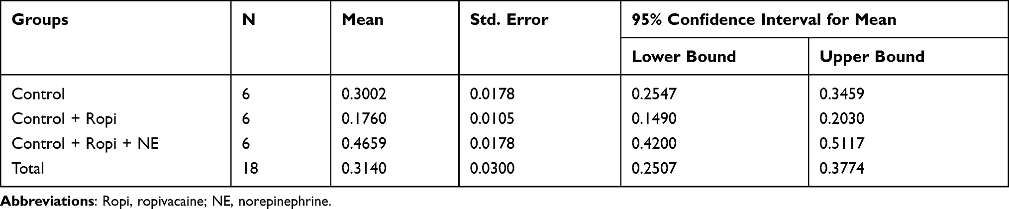 |
Table 1 Cervical Resistance in Each Group (g/mm) |
 |
Figure 1 Norepinephrine (NE) reverses the decrease in cervical tone caused by ropivacaine (Ropi). (A) Schematic diagram of the study timeline. (B and C) One of the load–elongation linear regression results of each group, gray represents the test value of the control group; green represents the control + ropivacaine group; black represents the control + ropivacaine + NE group. The slope of the regression line reflects cervical tension. (D) Compared with the control group, the cervical resistance in the control + ropivacaine group was significantly reduced (**P < 0.01); compared with the control + ropivacaine group, the cervical resistance in the control + ropivacaine + NE group was significantly enhanced (###P < 0.001) (n = 6 for each group). |
Western Blotting and Detection of Cervical cAMP
To investigate the possible mechanism underlying the effect of ropivacaine and NE on cervical tension, we evaluated the cervical α2A-AR levels in the late-pregnant rats. Western blotting showed that cervical α2A-AR levels gradually increased over time in pregnant rats, with the expression peaking on day 22 (Figure 2A and B). Subsequently, we assessed the concentration of cAMP in the cervical tissues of rats from the three groups (Figure 2C). Results showed that the intrathecal injection of ropivacaine increased the cAMP concentration in the cervical tissue, whereas NE could reverse this effect of ropivacaine. The cervical cAMP concentration in each group is presented in Table 2.
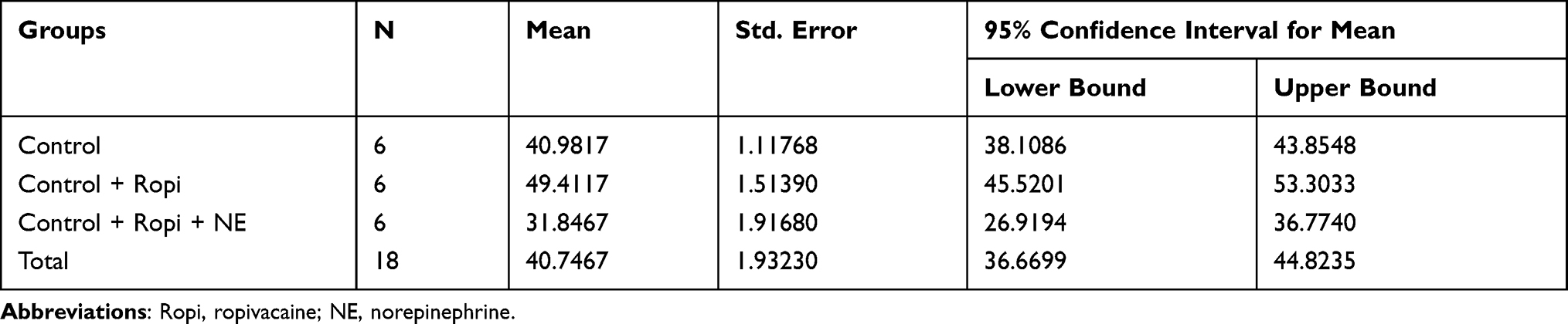 |
Table 2 Cervical cAMP Concentration in Each Group (pmol/mg Tissue) |
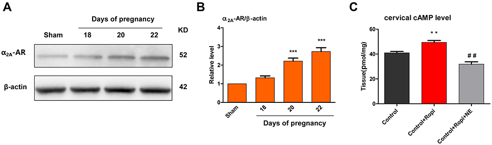 |
Figure 2 α2A adrenergic receptor (α2A-AR) levels and cAMP concentration in the cervical tissue. (A and B) Cervical α2A-AR levels gradually increased over time in pregnant rats, with the expression peaking on day 22 (***P < 0.001) (n = 4 for each group). (C) Compared with the control group, the cervical tissue cAMP concentration in the control + ropivacaine group was significantly increased (**P < 0.01); compared with the control + ropivacaine group, the cervical tissue cAMP concentration in the control + ropivacaine + NE group was significantly reduced (##P < 0.01) (n = 6 for each group). |
Discussion
An innovative finding of this study was that after establishing the neuraxial anesthesia model, the physical experiment of elastic modulus accurately reflected the cervical tension in vitro, and on this basis, the molecular biology experiment further explained the possible causes of cervical tension changes.
The physical experiment method of cervical tissue tension has been introduced in detail in some studies.16–18 Unlike these previous studies, when plotting the load–elongation curve, we averaged the initial load (m1i) and the load after 30 min of rest (m2j), which can better reflect the resistance of the cervical tissue. In fact, in the static stretching method, which is the classic method for Young’s modulus measurement, it is necessary to record the stretched length when the load increases and decreases sequentially.19 Furthermore, the method of linear regression can intuitively reflect the law of data distribution, and it is also one of the methods that can accurately calculate the elastic modulus of an object in addition to the method of successful difference.20–22 In this study, we found that the intrathecal injection of 0.1% ropivacaine significantly reduced the cervical tension in rats at 22 days of pregnancy. This result can indicate that ropivacaine, a commonly used drug for painless delivery, can promote the progression of the first stage of labor at a common concentration of 0.1%. This result is consistent with several clinical observations.1,2
In molecular biology experiments, we evaluated the cervical levels of α2A-AR, the main subtype of α2 adrenergic receptor. Western blotting showed that cervical α2A-AR levels gradually increased over time, with the expression peaking on day 22. We also measured cAMP concentration in the cervical tissue. The enzyme-linked immunosorbent assay results showed that the intrathecal injection of ropivacaine increased the cAMP concentration in the cervical tissue of rats at 22 days of pregnancy. It is well known that cAMP can relax the uterine smooth muscle by inhibiting the activity of myosin light-chain kinase (MLCK).23 In this study, NE reversed the effect of ropivacaine in increasing the cAMP concentration of the cervical tissue. In the cervical resistance test, NE also reversed the effect of ropivacaine in reducing cervical tension in late-pregnant rats. These results suggested that α2 adrenergic receptors are involved in the process of cervical contraction. The known adrenergic receptors are all G protein-coupled receptors. The activation of adrenergic receptors (which are approximately divided into α1, α2, and β receptors) requires the mediation of G protein to couple with the second messenger to produce a series of signal transduction and physiological effects.24,25 In contrast to the effect of β receptors in increasing the concentration of cAMP in smooth muscle cells by coupling to Gs, α2 receptors couple with Gi, which can inhibit the activity of adenylate cyclase and reduce the synthesis of cAMP, thereby reducing MLCK activity inhibition with the result of smooth muscle cell contraction.26–28 Some studies concerning the relationship between cervical tone and adrenergic receptors also support the abovementioned discussion.29–32
To summarize, ropivacaine, a commonly used drug for painless delivery, can significantly relax the cervical tension of late-pregnant rats by intrathecal injection at a common concentration of 0.1%. This finding provides meaningful theoretical support for the concept that neuraxial anesthesia shortens the first stage of labor. In other words, the intervention of neuraxial anesthesia for painless delivery need not wait until the cervical opening reaches 3–4 cm, but it should be provided as soon as possible when the parturient feels pain due to regular uterine contractions. Furthermore, α2 adrenergic receptors may play an important role in cervical contraction. Perhaps intrathecal injection of long-acting α2 receptor agonists could play a positive role in the treatment of preterm labor.
Of course, this study has several shortcomings. For instance, we did not analyze the effect of NE on the tension of isolated cervical tissue at different concentrations. Moreover, NE receptors were not investigated in detail, such as the contribution of α1 receptor-coupled Gq to smooth muscle cell contraction. Nonetheless, our study results provide theoretical support for the necessity of early intervention by anesthesiologists for labor analgesia, which has positive social significance in improving the understanding of labor.
Funding
This study was supported by the Xuzhou Medical University Affiliated Hospital Development Fund (XYFY2020025 to Chong Peng).
Disclosure
The authors declare that they have no competing interests in this work.
References
1. Grant EN, Tao W, Craig M, McIntire D, Leveno K. Neuraxial analgesia effects on labour progression: facts, fallacies, uncertainties and the future. BJOG. 2015;122(3):288–293. doi:10.1111/1471-0528.12966
2. Hawkins JL. Epidural analgesia for labor and delivery. N Engl J Med. 2010;362(16):1503–1510. doi:10.1056/NEJMct0909254
3. Poma S, Scudeller L, Verga C, et al. Effects of combined spinal-epidural analgesia on first stage of labor: a cohort study. J Matern Fetal Neonatal Med. 2019;32(21):3559–3565. doi:10.1080/14767058.2018.1467892
4. Wong CA, Scavone BM, Peaceman AM, et al. The risk of cesarean delivery with neuraxial analgesia given early versus late in labor. N Engl J Med. 2005;352(7):655–665. doi:10.1056/NEJMoa042573
5. Luo S, Chen Z, Wang X, Zhu C, Su S. Labor epidural analgesia versus without labor epidural analgesia for multiparous women: a retrospective case control study. BMC Anesthesiol. 2021;21(1):133. doi:10.1186/s12871-021-01355-0
6. Ren J, Wang T, Yang B, et al. Risk factors and safety analyses for intrapartum fever in pregnant women receiving epidural analgesia during labor. Med Sci Monit. 2021;27:e929283. doi:10.12659/MSM.929283
7. Wong CA, Scavone BM, Sullivan JT, McCarthy RJ. Early labor neuraxial analgesia: effects on the progress and outcome of labor. Anesthesiology. 2010;112(4):1053–1055. doi:10.1097/ALN.0b013e3181d403ad
8. Haase EB, Buchman J, Tietz AE, Schramm LP. Pregnancy-induced uterine neuronal degeneration in the rat. Cell Tissue Res. 1997;288(2):293–306. doi:10.1007/s004410050815
9. Thorbert G, Alm P, Björklund AB, Owman C, Sjöberg NO. Adrenergic innervation of the human uterus. Disappearance of the transmitter and transmitter-forming enzymes during pregnancy. Am J Obstet Gynecol. 1979;135(2):223–226. doi:10.1016/0002-9378(79
10. Hervonen A, Kanerva L, Lietzén R. Histochemically demonstrable catecholamines and cholinesterases of the rat uterus during estrus cycle, pregnancy and after estrogen treatment. Acta Physiol Scand. 1973;87(2):283–288. doi:10.1111/j.1748-1716.1973.tb05392.x
11. Moustafa FA. Changes in cholinergic and noradrenergic nerves in the pregnant and postpartum uterus of the albino rat and Guinea pig. Acta Anat. 1988;132(4):310–316. doi:10.1159/000146593
12. Bryman I, Norström A, Dahlström A, Lindblom B. Immunohistochemical evidence for preserved innervation of the human cervix during pregnancy. Gynecol Obstet Invest. 1987;24(2):73–79. doi:10.1159/000298782
13. Lundberg LM, Alm P, Carlén B. S-100-immunoreactive nerves in the Guinea-pig uterus with reference to ultrastructural correlations: effects of chemical sympathectomy and pregnancy. Cell Tissue Res. 1987;250(2):241–249. doi:10.1007/BF00219068
14. Norström A, Bryman I. Uptake of 3H-norepinephrine in different segments of the human non-pregnant and pregnant uterus. Gynecol Obstet Invest. 1989;27(1):26–28. doi:10.1159/000293610
15. Størkson RV, Kjørsvik A, Tjølsen A, Hole K. Lumbar catheterization of the spinal subarachnoid space in the rat. J Neurosci Methods. 1996;65(2):167–172. doi:10.1016/0165-0270(95)00164-6
16. Downing SJ, Sherwood OD. The physiological role of relaxin in the pregnant rat. III. The influence of relaxin on cervical extensibility. Endocrinology. 1985;116(3):1215–1220. doi:10.1210/endo-116-3-1215
17. Gáspár R, Kolarovszki-Sipiczki Z, Ducza E, et al. Terbutaline increases the cervical resistance of the pregnant rat in vitro. Naunyn Schmiedebergs Arch Pharmacol. 2005;371(1):61–71. doi:10.1007/s00210-004-1010-x
18. Wentz MJ, Shi SQ, Shi L, et al. Treatment with an inhibitor of catechol-O-methyltransferase activity reduces preterm birth and impedes cervical resistance to stretch in pregnant rats. Reproduction. 2007;134(6):831–839. doi:10.1530/REP-07-0245
19. Yang ZH, Yang JL, Miao QH, et al. Improvement of Young’s modulus measuring instrument by static stretching method. J Jinling Inst Technol. 2017;2:54.
20. Zhao XC, Xue-Mei LI, Xia XQ, et al. The application of MATLAB software for the Young’s modulus of the metal wire using the method of successive difference. Phys Exp Coll. 2014;4;43.
21. Evans DW, Scott AT, Teasdall RD, et al. Mechanical properties of lower limb dermis following static and cyclic compression. Biomed Sci Instrum. 2012;48:104–111.
22. Murase H, Koyama S, Honami N. Kalman filter charge simulation methods for the solution of inverse problems of two-or three-dimensional elasticity. Trans Jpn Soc Mech Eng. 1990;56(531):2310–2316.
23. Yuan W, López Bernal A. Cyclic AMP signalling pathways in the regulation of uterine relaxation. BMC Pregnancy Childbirth. 2007;7(1):S10. doi:10.1186/1471-2393-7-S1-S10
24. Liu N, Wang Y, Li T, Feng X. G-protein coupled receptors (GPCRs): signaling pathways, characterization, and functions in insect physiology and toxicology. Int J Mol Sci. 2021;22(10):5260. doi:10.3390/ijms22105260
25. Hilger D. The role of structural dynamics in GPCR-mediated signaling. FEBS J. 2021;288(8):2461–2489. doi:10.1111/febs.15841
26. Abdel-Latif AA. Cross talk between cyclic nucleotides and polyphosphoinositide hydrolysis, protein kinases, and contraction in smooth muscle. Exp Biol Med. 2001;226(3):153–163. doi:10.1177/153537020122600302
27. Pato MD, Kerc E. Characterization of the smooth muscle phosphatases and study of their function. Prog Clin Biol Res. 1987;245:207–218.
28. Pfitzer G. Invited review: regulation of myosin phosphorylation in smooth muscle. J Appl Physiol. 2001;91(1):497–503. doi:10.1152/jappl.2001.91.1.497
29. Bóta J, Hajagos-Tóth J, Ducza E, et al. The effects of female sexual hormones on the expression and function of α1A- and α1D-adrenoceptor subtypes in the late-pregnant rat myometrium. Eur J Pharmacol. 2015;769:177–184. doi:10.1016/j.ejphar.2015.11.015
30. Kolarovszki-Sipiczki Z, Gáspár R, Ducza E, et al. Effect of alpha-adrenoceptor subtype-selective inverse agonists on non-pregnant and late-pregnant cervical resistance in vitro in the rat. Clin Exp Pharmacol Physiol. 2007;34(1–2):42–47. doi:10.1111/j.1440-1681.2007.04529.x
31. Hajagos-Tóth J, Bóta J, Ducza E, et al. The effects of progesterone on the alpha2-adrenergic receptor subtypes in late-pregnant uterine contractions in vitro. Reprod Biol Endocrinol. 2016;14(1):33. doi:10.1186/s12958-016-0166-9
32. Gál A, Ducza E, Minorics R, et al. The roles of alpha2-adrenoceptor subtypes in the control of cervical resistance in the late-pregnant rat. Eur J Pharmacol. 2009;615(1–3):193–200. doi:10.1016/j.ejphar.2009.04.067
 © 2022 The Author(s). This work is published and licensed by Dove Medical Press Limited. The
full terms of this license are available at https://www.dovepress.com/terms.php
and incorporate the Creative Commons Attribution
- Non Commercial (unported, v3.0) License.
By accessing the work you hereby accept the Terms. Non-commercial uses of the work are permitted
without any further permission from Dove Medical Press Limited, provided the work is properly
attributed. For permission for commercial use of this work, please see paragraphs 4.2 and 5 of our Terms.
© 2022 The Author(s). This work is published and licensed by Dove Medical Press Limited. The
full terms of this license are available at https://www.dovepress.com/terms.php
and incorporate the Creative Commons Attribution
- Non Commercial (unported, v3.0) License.
By accessing the work you hereby accept the Terms. Non-commercial uses of the work are permitted
without any further permission from Dove Medical Press Limited, provided the work is properly
attributed. For permission for commercial use of this work, please see paragraphs 4.2 and 5 of our Terms.