Back to Archived Journals » Advances and Applications in Bioinformatics and Chemistry » Volume 17
In silico Molecular Docking Approach to Identify Potential Antihypertensive Compounds from Ajuga integrifolia Buch.-Ham. Ex D. Don (Armagusa)
Authors Tessema FB ![]() , Gonfa YH, Asfaw TB, Tadesse MG, Bachheti RK
, Gonfa YH, Asfaw TB, Tadesse MG, Bachheti RK ![]()
Received 29 November 2023
Accepted for publication 29 February 2024
Published 11 March 2024 Volume 2024:17 Pages 47—59
DOI https://doi.org/10.2147/AABC.S392878
Checked for plagiarism Yes
Review by Single anonymous peer review
Peer reviewer comments 3
Editor who approved publication: Dr Maria Miteva
Fekade Beshah Tessema,1,2 Yilma Hunde Gonfa,2,3 Tilahun Belayneh Asfaw,4 Mesfin Getachew Tadesse,2,5 Rakesh Kumar Bachheti2,6
1Department of Chemistry, College of Natural and Computational Science, Woldia University, Woldia, Ethiopia; 2Department of Industrial Chemistry, College of Natural and Applied Sciences, Addis Ababa Science and Technology University, Addis Ababa, Ethiopia; 3Department of Chemistry, College of Natural and Computational Science, Ambo University, Ambo, Ethiopia; 4Department of Chemistry, College of Natural and Computational Science, Gondar University, Gondar, Ethiopia; 5Centre of Excellence in Biotechnology and Bioprocess, Addis Ababa Science and Technology University, Addis Ababa, Ethiopia; 6Department of Allied Sciences, Graphic Era Hill University, Society Area, Clement Town, Dehradun, 248002, India
Correspondence: Fekade Beshah Tessema, Tel +251913141754, Email [email protected]
Background: Ajuga integrifolia (Armagusa) is used as a decoction to treat high blood pressure and diabetes, widely in Ethiopia. Specific compounds for anti-hypertension activity were not identified so far. This study aims to provide a scientific basis for the therapeutic use of A. integrifolia as an antihypertension agent.
Methods: In silico studies were used to evaluate the antihypertensive components of A. integrifolia. Flavonoids identified using HPLC analysis and iridoid glycosides isolated from A. integrifolia in this study and those isolated from synonyms (A. remota and A. bractosa) were considered in the molecular docking study. Interactions were studied by using Autodock vina (1.2) on PyRx 0.8 and visualizing in 2D and 3D using ligPlot+ and Discovery studio software. Activities like vasoprotection and druglikeness properties were predicted using online servers.
Results: Flavonoids such as quercetin, myricetin, and rutin were identified and quantified by HPLC analysis from different extracts of A. integrifolia. Reptoside and 8-O-acetylharpgide isolated from the aerial part of A. integrifolia. The binding energies of all 17 candidates considered in this study range from − 10.2 kcal/mol to − 7.5 kcal/mol and are lower than enalapril (reference drug: − 5.9 kcal/mol). The binding energies, in most case, constitute hydrogen bonding. Biological activity predicted using PASS test also showed that the flavonoids have more probability of activity than the iridoid glycosides. Druglikeness properties of the candidate molecules showed that most follow the Lipinski rule of five with few violations.
Conclusion: Lower binding energies involving hydrogen bonding and predicted activities concerning hypertension confirm the traditional use of the aerial part of the medicinal plant concerned. Flavonoids: rutin, myricetin, quercetin, and kaempferol take the leading role in the antihypertensive activity of the aerial part of A. integrifolia. The iridoid glycosides studied are almost similar in their effect on their antihypertensive activity and still better than the reference drug.
Keywords: A. integrifolia, antihypertension, flavonoids, iridoid glycosides
Introduction
Ethiopia is rich in biodiversity and traditional knowledge of medicinal potential herbs as a home of origins. About 5500 indigenous medicinal plants are known in Ethiopia.1 More than five thousand medicinal plants are known with the respective traditional practices against a more significant number of ailments.2 Ajuga is among the 260 genera of the family Lamiaceae. There are 40–50 species of the genus Ajuga with many variations. One of the medicinal herbs which belong to this genus is Ajuga integrifolia (syn: Ajuga remota, Ajuga bractosa) commonly known as Armagusa, Etselibawit, Medhanit, Tut astil, Anamuro.3 It belongs to the genus Ajuga and the family Lamiaceae. As a plant in the genus Ajuga, A. integrifolia is an evergreen flowering herb. It occurs in many parts of Ethiopia and east African countries like Djibouti, Eritrea, Kenya, Somalia, Sudan, Uganda and Tanzania. It also occurs in Yemen, Saudi Arabia, Afghanistan, and Eastern Asia.3
A. integrifolia is widely used in traditional medicine, and for detaching children from breastfeeding because of its bitter taste. Its medicinal uses are summarized in Table 1.
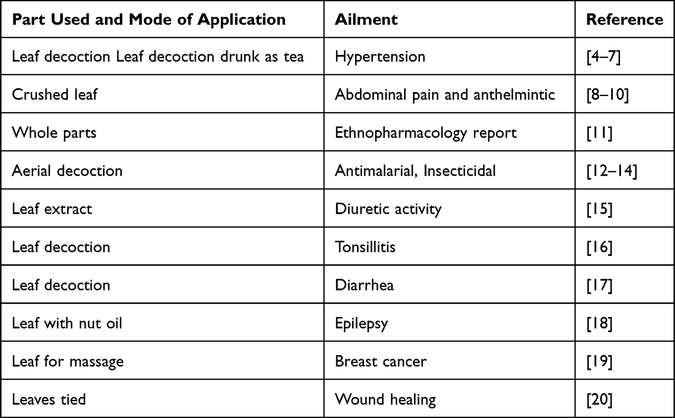 |
Table 1 Medicinal Uses of A. Integrifolia |
Hypertension is an alternative name for high blood pressure,4 a problem that affects one billion patients globally and kills nine million lives annually. Because of this, it is among the major global causes of early morbidity and death.21 Due to its chronic nature, hypertension is a worldwide public health concern that leads to many complications such as heart failure and stroke.4 More than the direct effect of hypertension in the health sector, even developing countries face challenges in dealing with hypertension complications. As a solution to these challenges, medicinal plants as alternative medicine are significant.22
The antihypertensive mechanism explored in the Chinese herbal formulations is reducing blood pressure variability (BPV), reducing the sympathetic nervous system’s activity, obstructing the renin–angiotensin system, enhancing endothelial function, avoiding target organ damage (TOD), enhancing insulin resistance, and enhancing the metabolism of glucose and lipids, calcium channel blocking, and improving blood rheological parameters, such as blood flow, viscosity, deformability and coagulation.22 An essential component of the renin–angiotensin system, angiotensin-converting enzyme (ACE) II, is important in counteracting the damaging effects of angiotensin II on the cardiovascular system. The ACE inhibitory action is present in a wide range of naturally occurring substances that are frequently used in ethnobotanics and, in certain cases, have a strong nutritional basis. Because synthetic molecules like enalapril were produced using a scaffold library of natural metabolites, bioproducts, including ACE inhibitors, are widely used. This demonstrates their potential as novel therapeutic sources; in some cases, natural chemicals can have lower IC50 values than synthetic ones, and they have fewer side effects overall. Natural products contain a variety of phytoconstituents known as ACE inhibitors, including flavonoids, xanthones, alkaloids, peptides, terpenes, and tannins.23 Recently, flavonoids b from several plant isolates have attracted a lot of attention as ACE (angiotensin converting enzyme) inhibitors.24 By preventing the conversion of angiotensin I to angiotensin II, quercetin and its glycosides help to control blood pressure.25 Using a test kit and strictly following the directions, the ACE inhibitory activity of Seseli pallasii essential oil was determined. The findings demonstrated dose-dependent suppression of ACE activity, as shown by IC50 = 0.33 mg/mL.26 With IC50 values of 2.51 µg/mL and 2.59 µg/mL, respectively, the Alchemilla viridiflora extract and miquelianin demonstrated dose-dependent in-vitro ACE inhibitory action. These findings indicated that flavonoids and other extract components, in addition to miquelian, may have also contributed to this activity.24 The two new peptide non-competitive ACE inhibitors, Thr-Thr-Trp (TTW) and Val-His-Trp (VHW), showed the strongest inhibitory activity, as shown by their respective IC50 values of 0.61 ± 0.12 and 0.91 ± 0.31 µM.27 Gene transfer can be used to overexpress this enzyme, which can help treat hypertension and cardiovascular disease.28
Prior investigations showed that species from the genus Ajuga are rich in flavonoids, terpenoids, anthocyanins, iridoid glycosides, polyphenols, and phytosterols.29–31 Flavonoid glycosides are recommended for the antihypertensive effect,4 and flavonoids like quercetin were found to reduce elevated blood pressure32 by improving endothelial function.33 Quercetin and myricetin, flavonoids found in diet, guard against oxidative stress and aging.34 Numerous diseases, such as lung cancer, cardiovascular disease, and osteoporosis, have shown promise improvements when treated with quercetin35 and responsible for antioxidant and antimicrobial activities.36 It is also reported as an agent that could be potentially useful to attenuate different effects of ethanol and as adjuvant pharmacotherapy for ethanol addiction.37 Myricetin is a flavonoid that demonstrates therapeutic actions in many central nervous system diseases.38 Myricetin and Quercetin were among 13 anti- AChE phytochemicals which were known with their activities against the acetylcholinesterase enzyme (AChE), which is a key enzyme responsible for the development of Alzheimer’s disease.39
Iridoids are characteristics of secondary metabolites of the species from the Ajuga genus with important information on chemotaxonomy.40 Ajugoside is a chemotaxonomic marker of the genus harpagide, and 8-O-acetylharpagide are for the family. Ajugoside, harpagide and 8-O-acetylharpagide are evidenced in the genus.41 Markers for different Lamiaceae species include ajugoside, reptoside, 8-O-acetylharpagide, and harpagide.30 Phytochemical investigations on the species of the genus Ajuga revealed that more than 13 iridoid glycosides have been isolated primarily from A. remota, A. ducmbens, and A. reptans. Iridoid glycosides from Ajuga spp are known for ethnopharmacological indications for many therapeutic effects including hypertension.29
Besides several ethnobotanical reports on the antihypertensive properties of A. integrifolia, there is a lack of information on pharmacological activities. Molecular docking studies are the best alternatives for planned, time and cost-effective pharmacological studies. The objective of this study is to evaluate the antihypertensive components of A. integrifolia using an in silico study. Total flavonoid and total phenol content values determined by Folin–Ciocalteu method and AlCl3 method, respectively, were significant among the medicinal plants studied. The antioxidant activity was also considered significant for further investigation. Local people used the boiled leaves decoction as an antihypertensive agent in the sample collection area. Aiming to provide a scientific basis for the therapeutic use of A. integrifolia as an antihypertension agent, iridoid glycosides as major components and flavonoids identified by HPLC analysis were examined for inhibiting the key renin–angiotensin system enzyme. Both groups of phytochemicals were considered in the molecular docking and ADME study. Iridoid glycosides isolated from A. integrifolia in this study and those isolated from synonyms like A. remota and A. bractosa are presented in Table 2.
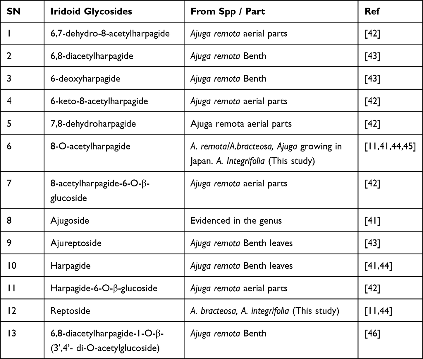 |
Table 2 Iridoid Glycosides Isolated from A. Integrifolia (This Study) and Synonyms: A. Remota and A. Bractosa |
Materials and Methods
Chemical and Reagents
An MQ (18.2) water purification system (Purelab flex 4 Elga) operating at 20.6 °C was used to distill and purify the water to wash plant materials and for HPLC analysis. All chemicals and reagents were HPLC grade (Merck India, Mumbai, and S.D. Fine-Chem, Mumbai, India). Flavonoid standards (purity ≥ 99.9%) were purchased from Sigma-China.
Plant Material Collection and Identification
The Addis Abeba Science and Technology University (AASTU) campus and the surrounding Koye Feche area were the sources of the aerial part of A. integrifolia and allowed to dry in the air (shade). The voucher specimen was identified by Mr. Melaku Wondafrash and deposited at the National Herbarium, College of Natural and Computational Sciences, Biology Department, Addis Ababa University. The sample was powdered using a coffee grinder and kept in a polyethylene bag till used.
Extraction and Isolation of Iridoid Glycosides from Aerial Sample
The powdered aerial part (500 g) was soaked at room temperature in 2 × 1 L of petroleum ether (AR) for 72 hours with occasional shaking. Filtering, combining, and drying under reduced pressure gave greenish-dark sticky material. A similar procedure was followed for chloroform (AR), ethyl acetate (AR), and methanol (AR). Fifteen grams of methanol extract adsorbed in 30 g of silica gel (70–220 mesh) and dried over a water bath at 40°C. Adsorbed and dried sample was applied on a column packed with 300 g of silica gel (70–220 mesh) in chloroform. The column equilibrated after loading the sample for 1 hour. Sixty fractions of each 150 mL collected by eluting with Chloroform, Chloroform:Methanol (9.5:0.5, 9:1, 8:2, 7:3, and 6:4) ratio. Fractions are grouped into 15 vials following the TLC profile. Vial 8 sample weight = 2.8 g was observed to have 1 red spot as major and other minor spots. To purify the 2.8 g sample, adsorbed on 6 g of silica gel was applied on a column packed with 125 g of silica gel in chloroform and eluted similarly as in the previous column. One hundred and twenty fractions each 20 mL collected and grouped into eight vials. The fourth vial (V4) 180 mg showed a single red spot and was sent to NMR and characterized as reptoside after comparison with literature data. The seventh vial also showed a single red-brown spot and minor spots. On purification, it gave 120 mg. It was identified as 8-O- acetyl harpagide, when characterized by NMR and compared with published literature data.
High-Performance Liquid Chromatography Analysis (Flavonoids)
Using ultra-high-performance liquid chromatography coupled with a diode array detector, flavonoids were quantified qualitatively (Ultimate −3000 UHPLC-DAD, Thermo Scientific Dionex, USA). The column was the reverse phase, measuring 4.6 × 250 mm and using Fortis 5mm C18. The mobile phase was methanol-acidified (1% acetic acid) ultra-pure water (60/40, v/v) flowing at a rate of 0.8 mL/min. The column and the autosampler were both set to 25°C and 35°C, respectively. A concentrated 10 μL portion of the material, dissolved in the mobile phase blend, was introduced into the column, achieving detection at 254 nm, 272 nm, 360 nm, and 372 nm. As an external reference standard, 2.5, 10, 20, 40, and 50 mg/mL of the standard mixture of quercetin, myricetin, and rutin (>99.9%, Sigma-China) was used.
In silico Study
Preparations of Receptor Protein and Ligands
Structures of the iridoid glycosides retrieved from PubChem (HTTP: //PubChem.ncbi.nlm.nih.gov) and some of which are also drawn using ChemDraw software (PerkinElmer ChemDraw Professional l8.2 and Chem3D Ultra 8.2). The structures used as ligands were prepared by minimizing all structures of component compounds using the steepest descent minimization in PyRx 0.8.
The 3D structure of the human Angiotensin-converting enzyme (native) was downloaded from the Protein Data Bank (http://www.rscb.org) PDB ID:1O8A.47 Using Pymol software, water molecules and ligands attached to them were removed during preparation. In addition to this, hydrogen atoms were added to the structure of the protein since most of the hydrogen atoms will be removed during X-ray diffraction.
Molecular Docking Analysis
The software used in the docking process was Autodock vina in Pyrex 1.2. Autodock vina is recommended to be fast and accurate for molecular docking activities.48 Molecular docking was performed to obtain the best inhibitor from the active compounds selected using Enalapril as a reference. Once the ligand and prepared receptor were chosen, the docking procedure began by setting the gird box on the active site receptor which was identified from the literature.49 The docking results were stored in PDB format, and the binding affinity (∆G) value was saved in Microsoft Excel format, expressed in kcal/mol. Discovery Studio was used to create the interactive 3D visualization, while LigPlot v.1.4.5 was used to visualize the docking results.
Pharmacokinetics Analysis
PreADMET online software (http://preadmet.bmdrc.org/) was used to estimate pharmacokinetic properties. The ability of each herb’s active components to be effectively absorbed by the human digestive system was assessed using the human intestinal absorption (HIA) test. The ligand structure data were input in mol file format and submitted, and the HIA test was carried out by accessing the preADMET software site. To further understand the pharmacokinetics of the active compounds, including if any of them would be able to enter the cell and interact with the target protein, the Lipinski rule of five test data was similarly retrieved from preADMET online program.
Prediction of Activity Spectra for Substances (PASS) Test
The Probability Activity (Pa) value, which represents a compound’s biological activity for vasoprotective and cardiac activities, was determined using PASS prediction, which was carried out using the PASS online web server with canonical smiles. The biological activity that aided in the treatment of hypertension was selected. Since it is a compound activity with potential for the wet-lab experiment, the Pa value that was employed was Pa >0.7.50 The PASS Online software (http://www.pharmaexpert.ru/passonline) was used to administer the PASS test. Using PubChem 070003–2 (http://pubchem.ncbi.nlm.nih.gov), SMILES was first searched for potential ligand compounds. The ligand compounds were then entered into the PASS program, which is used to predict activity (Get Prediction). Before performing lab testing, it was crucial to check the results of the biological activity test. If tests were conducted in a lab, the probability activity score—which forecasts the likelihood of success—would display the results.
Results
Following the extraction flowchart shown in Figure 1A the petroleum ether extract yield was 1.88%(w/w), chloroform extract yield was 2.13% (w/w), ethyl acetate extract was 0.61% (w/w), methanol extract 12.14% (w/w). Looking at the TLC profile of the polar component of the aerial part using chloroform: methanol (7:3) solvent system the presence of some four spots was observed for methanol extract as shown in Figure 1B. The investigation continued on methanol extract, and two iridoid glycosides isolated, and the NMR data obtained as shown below after comparing with literature data.51–54
 |
Figure 1 (A) Extraction flowchart for aerial part of A. integrifolia (B) TLC Comparison for the fraction from crude extraction. |
13CNMR Data for Reptoside
13C NMR (101 MHz, MeOD) δ 172.22(Ac CO), 141.42(C3), 107.86(C4), 98.14(C10), 92.59(C1), 88.72(C8), 76.68(C5’), 76.16(C3’), 73.07(C2’), 71.30(C4’), 70.15(C5), 61.32(C6’), 56.82(C9), 48.47(OMe), 37.50(C7), 36.66(C6), 21.08(C10), 20.54(OAc).
13CNMR Data for 8-O-Acetylharpagide
13C NMR (101 MHz, MeOD) δ 172.37(Ac CO), 143.62(C3), 104.14(C4), 98.67(C1’), 93.43(C1), 87.29(C8), 76.81(C6), 76.72(C3’), 76.06(C5’), 73.13(C2’), 72.68(C5), 70.28(C4’), 61.35(C6’), 53.69(C9), 44.70(C7), 21.28(C10), 21.1(Ac)
HPLC Analysis
Chromatograms were exported for qualitative determination of the flavonoid presence after HPLC analysis was completed utilizing the procedure outlined in the method section. The HPLC chromatogram (Figure 2A–C) revealed that the methanol extract contained myricetin (RT: 8.117 min) and quercetin (RT: 11.673 min), whereas the acetone dip immediate extract of the aerial part of A. integrifolia contained quercetin (RT: 12.600 min) and rutin (RT: 19.797 min).
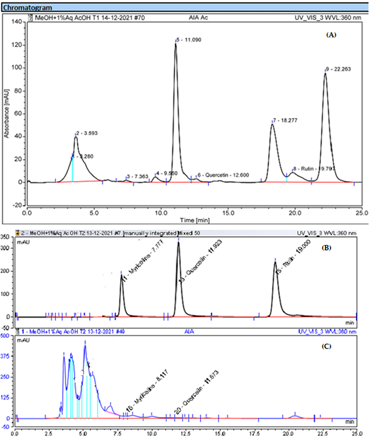 |
Figure 2 HPLC chromatograms for methanol and acetone extracts of aerial part of A. integrifolia. (A) AIA ac - acetone dip immediate extract (B) standards (C) AIA – methanol extract. |
Pharmacokinetic properties, including the drug-like properties, were studied using the Lipinski rule of five using data obtained from the swissADME online server and shown in Table 3 below.
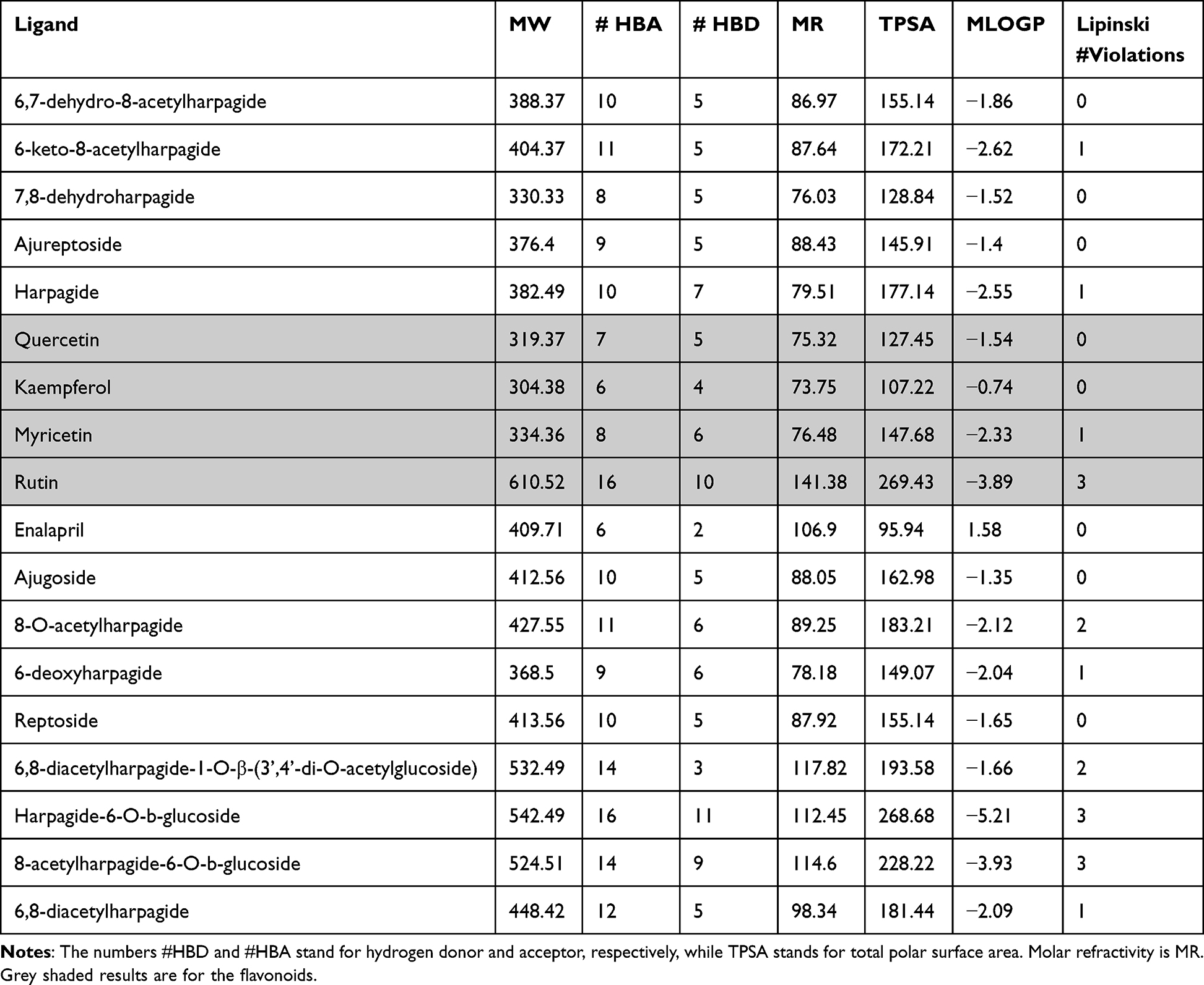 |
Table 3 Druglikeness Predictions of Compounds, Computed by SwissADME |
As shown in Table 4 flavonoids show both vasprotector activity as well as vasodilator activity in an acceptable range. Specifically, the second activity is the same as the reference drug used for comparison.
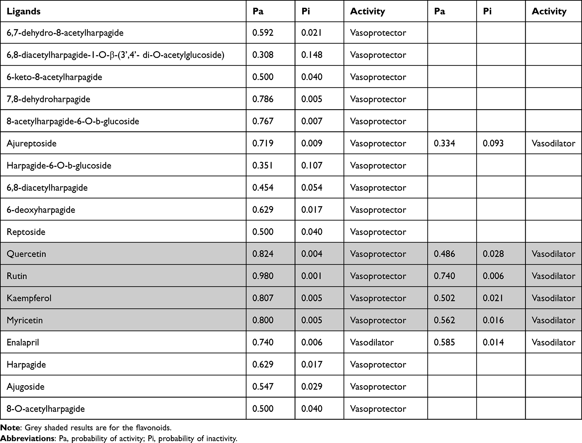 |
Table 4 PASS Activity Test Result Summary |
The LigPlot+ diagrams shown below (Figure 3A and B) depict similar information as the values of binding energies given in Table 5. Only representatives from the flavonoids (rutin) and iridoid glycosides (8-O-acetylharpagide) were shown for comparison with the reference drug (Enalapril). Both the hydrogen and hydrophobic interactions are considerably better than the reference drug.
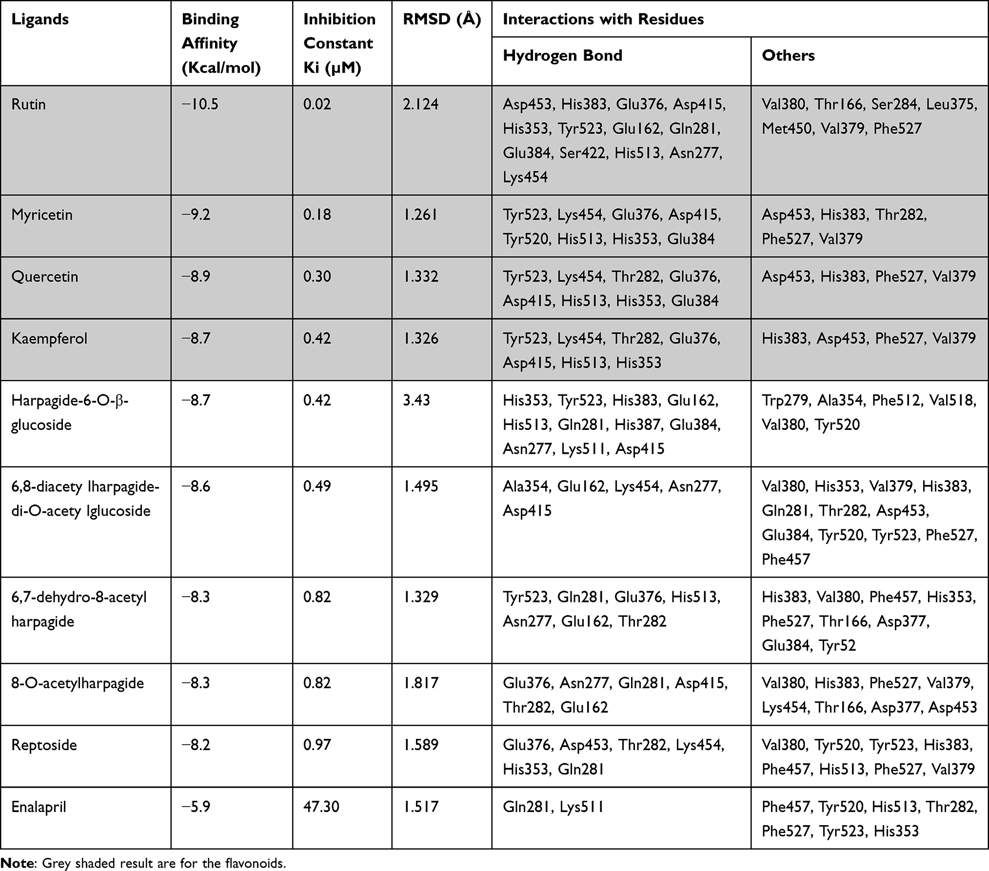 |
Table 5 Summary of Molecular Docking Result of Ligands with a Human Angiotensin-Converting Enzyme (PDB ID: 1O8A) |
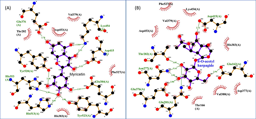 |
Figure 3 2D interactions diagrams using LigPlot+1.4.5: Human angiotensin-converting enzyme (PDB ID: 1O8A) complex with (A) Myricetin, (B) 8-O-acetylharpagide. Residue names in green color are those involved in hydrogen bonding. |
Discussion
Though iridoid glycosides were not reported from A. integrifolia, it is known as a chemotaxonomic identifier of Ajuga genus.41 We have isolated the expected identifier glycosides reptoside and 8-O-acetylharpagide and considered them in the study of antihypertensive activity. Both reptoside and 8-o-cetylharpagide were isolated from A. decumbens,55 A. chamaepitys,31 and A. reptans.56 The boiled decoction of the aerial part of the plant showed a TLC spot same as the compound identified as reptoside. The HPLC analysis showed us that the flavonoids quercetin, myricetin, and rutin are there in the respective extracts and so also in the aerial part of A. integrifolia.
Pharmacokinetic analysis shows that most of the ligands have lower gastrointestinal absorption. This will tell us to search for an alternative administration mode to be considered a drug. SwissADME analysis shows that not all of them inhibit Pgp substrate and CYPA inhibitor. All are bioavailable and synthetically accessible as the reference drug with no pain alerts. Most of the candidate compounds satisfy the rule of five for the druglikeness properties with no and at least one violation. This includes molecular mass less than 500 g/mol, less than or equal to ten hydrogen bond acceptors, less than or equal to five hydrogen bond donors, and log P ≤5 and molar refractivity in the range 70–110. The presence of glycoside moiety on rutin and iridoid di-glycosides reflects an effect on absorption. This results in poor absorption for those highly glycosylated components.
The active site residues were determined to be Gln281, His353, His513, Tyr520, Tyr523, Lys511, Glu384, His387, Tyr487 from pdbsum online server57 and PDB reference.49 Native ligands complexed with the receptor protein were used to locate the active site of the receptor protein. Residues like His353, Glu162, and Gln281 interact with most ligands via hydrogen bonding. This contributes to better binding affinities for the respective ligand compounds. Interactions involving hydrogen bonding are considerable towards the complexes’ stability and significantly affect inhibition.58 Most ligand’s interaction with the selected receptor protein involves hydrogen bonding to the active site residues. The different types of interaction contribute differently to the total binding energy. Hydrogen bonding contributes 16-fold to the hydrophobic interactions.48
According to a previous study, the smaller the Ki, the higher the binding affinity and the lower the dosage required to inhibit the target enzyme’s activity.59 Ki values of most interactions ranging from 0.02μM to 2.00μM are smaller, indicating lower inhibition concentrations for the candidate’s compounds (ligands). The Ki value for the reference drug enalapril is 47.30μM.59
Docked and original structures closely match, as indicated by the RMSD values obtained for the lowest-energy poses predicted and the interaction study values ranging from 1.261Å to 3.34 Å.59 The RMSD values closer to the reference drug determined that the conformation of the reference is deliberated as efficacious docking and it is close to crystallographic pose.60 The lower RMSD and more excellent hydrogen bond distribution are related to the stronger interaction between the ligands and the receptor protein. This contributes to the stability of the interactions61 as all are less than 0.3 nm. From the LigPlot+ diagram, one can see the possible hydrogen bonding interaction.62 Their distance as most is below 3 nm also indicates stronger binding interactions.58
Among the studied two groups of compounds, flavonoids were stronger in inhibiting the target receptor protein and can be considered to have better antihypertensive activity. Even the iridoid glycosides are better than the reference drug in binding interaction with the target receptor.
Vasoprotector PASS activities predicted for the flavonoids are more probable (>0.7) than the iridoid glycosides (0.3–0.7). The PASS test result is in line with the binding interaction study. Flavonoids also showed vasodilator activity in an acceptable range as that of the reference drug.
Conclusion
The extract of the aerial part of A. integrifoila exhibited an antihypertensive effect partly for the presence of flavonoids and iridoid glycosides. Lower binding energies involving hydrogen bonding and predicted activities to hypertension confirm the traditional use of the aerial part of the medicinal plant concerned. These results add credence to the long-standing use of A. integrifolia in the treatment of hypertension and offer valuable insights for optimizing its application in conventional medicine. The flavonoids studied showed a better inhibitory effect for the angiotensin-converting enzyme. Rutin, myricetin, and quercetin take the leading role in the antihypertensive activity of the aerial part of A. integrifolia. The iridoid glycosides studied are almost similar in their effect on their antihypertensive activity and still better than the reference drug. Further in vitro and in vivo research is necessary to identify and confirm the effect of these flavonoid and iridoid glycosides on blood pressure regulation. The study of the synergetic effects of these groups of compounds will also be a future concern.
Acknowledgment
The authors acknowledge Addis Ababa Science and Technology University for financial support and the Department of Industrial Chemistry (AASTU) for the chance given to do this investigation.
Funding
No external fund apart from PhD doctoral research support from AASTU.
Disclosure
All authors report no conflicts of interest in this work.
References
1. Dagne E. Natural database for Africa (NDA) Version 2.0; 2011.
2. Bekele E. Study on actual situation of medicinal plants in Ethiopia; 2007. Available from: http://www.endashaw.com.
3. Hedberg I. Flora of Ethiopia and Eritrea. Vol. 5, Gentianaceae to Cyclocheilaceae. Hedberg I, Kelbessa E, Edwards S, Demissew S, Persson E, eds. Ethiopia: The National Herbarium, Biology Department, Science Faculty, Addis Ababa University; 2006.
4. El-Hilaly J, Amarouch MY, Morel N, Lyoussi B, Quetin-Leclercq J. Ajuga iva water extract antihypertensive effect on stroke-prone spontaneously hypertensive rats, vasorelaxant effects ex vivo and in vitro activity of fractions. J Ethnopharmacol. 2021;270. doi:10.1016/j.jep.2021.113791
5. Suleman S, Alemu T. A survey on utilization of ethnomedicinal plants in Nekemte Town, East Wellega (Oromia), Ethiopia. J Herbs Spices Med Plants. 2012;18(1):37–41. doi:10.1080/10496475.2011.645188
6. Asres K, Bucar F, Kartnig T, Witvrouw M, Pannecouque C, Clercq E. Antiviral activity against human immunodeficiency virus type 1 (HIV-1) and type 2 (HIV-2) of ethnobotanically selected Ethiopian medicinal plants. Phytochem Res. 2001;15:62–69.
7. Fullas F. Ethiopian Traditional Medicine: Common Medicinal Plants in Perspective.
8. Teklay A, Abera B, Giday M. An ethnobotanical study of medicinal plants used in Kilte Awulaelo District, Tigray Region of Ethiopia. J Ethnobiol Ethnomed. 2013;9(1):1. doi:10.1186/1746-4269-9-65
9. Maryo M, Nemomissa S, Bekele T. An ethnobotanical study of medicinal plants of the Kembatta ethnic group in Enset-based agricultural landscape of Kembatta Tembaro (KT) Zone, Southern Ethiopia. Pelagia Res Libr Asian J Plant Sci Res. 2015;5(7):42–61. doi:10.13140/RG.2.2.11273.39523
10. Regassa R. Assessment of indigenous knowledge of medicinal plant practice and mode of service delivery in Hawassa city, southern Ethiopia. J Med Plants Res. 2013;7(9):517–535. doi:10.5897/JMPR012.1126
11. Israili ZH, Lyoussi B. Ethnopharmacology of the plants of genus Ajuga. Pak J Pharm Sci. 2009;22(4):425–462.
12. Asnake S, Teklehaymanot T, Hymete A, Erko B, Giday M. Survey of medicinal plants used to treat malaria by Sidama people of Boricha District, Sidama Zone, South Region of Ethiopia. Evid Based Complement Altern Med. 2016;1–6. doi:10.1155/2016/9690164
13. Bekele D, Asfaw Z, Petros B, Tekie H. Ethnobotanical study of plants used for protection against insect bite and for the treatment of livestock health problems in rural areas of Akaki District, Eastern Shewa, Ethiopia. Topclass J Herb Med. 2012;1(2):12–24.
14. Meragiaw M, Asfaw Z. Review of antimalarial, pesticidal and repellent plants in the Ethiopian traditional herbal medicine. Res Rev J Herb Sci. 2014;3(3):21–25.
15. Hailu W, Engidawork E. Evaluation of the Diuretic Activity of the Aqueous and 80 % Methanolic Extracts of the Leaves of Ajuga Remota B. (Lamiaceae) in Mice. Addis Ababa University; 2011.
16. Chekole G. Ethnobotanical study of medicinal plants used against human ailments in Gubalafto District, Northern Ethiopia. J Ethnobiol Ethnomed. 2017;13(55). doi:10.1186/s13002-017-0182-7
17. Gabriel T, Guji T. Ethnopharmacological survey of medicinal plants in Agaro District, Jimma Zone, South West Ethiopia. Int J Pharm Sci Res. 2014;5(8):3551–3559. doi:10.13040/IJPSR.0975-8232.5(8).3551-59
18. Abera B. Medicinal plants used in traditional medicine by Oromo people, Ghimbi District, Southwest Ethiopia. J Ethnobiol Ethnomed. 2014;10(40):1–15.
19. Tuasha N, Petros B, Asfaw Z. Medicinal plants used by traditional healers to treat malignancies and other human ailments in Dalle District, Sidama Zone, Ethiopia. J Ethnobiol Ethnomed. 2018;14(15):1–21. doi:10.1186/s13002-018-0213-z
20. Gebrehiwot M. An Ethnobotanical Study of Medicinal Plants in Seru Wereda, Arsi Zone of Oromia Region, Ethiopia. Addis Ababa University; 2010.
21. Sudharsanan N, Geldsetzer P. Impact of coming demographic changes on the number of adults in need of care for hypertension in Brazil, China, India, Indonesia, Mexico, and South Africa: a modeling study. Hypertension. 2019;73(4):770–776. doi:10.1161/HYPERTENSIONAHA.118.12337
22. Xiong X, Yang X, Liu Y, Zhang Y, Wang P, Wang J. Chinese herbal formulas for treating hypertension in traditional Chinese medicine: perspective of modern science. Hypertens Res. 2013;36(7):570–579. doi:10.1038/hr.2013.18
23. Rahman M, Islam R, Akash S, et al. In silico investigation and potential therapeutic approaches of natural products for COVID-19: computer-aided drug design perspective. Front Cell Infect Microbiol. 2022;12(929430):1–28. doi:10.3389/fcimb.2022.929430
24. Radović J, Suručić R, Niketić M, Kundakovic-Vasovic T. Angiotensin I-converting Enzyme (ACE) Inhibitory Activity and Chemical Composition of Alchemilla. Res Square. 2021;1–18. doi:10.21203/rs.3.rs-920698/v1
25. Muhammad SA, Fatima N. In silico analysis and molecular docking studies of potential angiotensin ‑ converting enzyme inhibitor using quercetin glycosides. Pharmacogn Mag. 2015;11(42):123–126. doi:10.4103/0973-1296.157712
26. Suručić R, Kundaković T, Lakušić B, Drakul D, Milovanović SR, Kovačević N. Variations in chemical composition, vasorelaxant and angiotensin I-converting enzyme inhibitory activities of essential oil from aerial parts of Seseli pallasii Besser (Apiaceae). Chem Biodivers. 2017;14(5):e1600407. doi:10.1002/cbdv.201600407
27. Xie J, Chen X, Wu J, et al. Antihypertensive effects, molecular docking study and isothermal titration calorimetry assay of the angiotensin I- converting enzyme inhibitory peptides from Chlorella vulgaris Antihypertensive effects, molecular docking study and isothermal titration ca. J Agric Food Chem. 2018. doi:10.1021/acs.jafc.7b04294
28. Hernández Prada JA, Ferreira AJ, Katovich MJ, et al. Structure-based identification of small-molecule angiotensin-converting enzyme 2 activators as novel antihypertensive agents. Hypertension. 2008;51(5):1312–1317. doi:10.1161/HYPERTENSIONAHA.107.108944
29. Toiu A, Mocan A, Vlase L, et al. Phytochemical composition, antioxidant, antimicrobial and in vivo anti-inflammatory activity of traditionally used Romanian Ajuga laxmannii (Murray) Benth. (“nobleman’s beard” - barba împăratului). Front Pharmacol. 2018;9:1–15. doi:10.3389/fphar.2018.00007
30. Toiu A, Mocan A, Vlase L, et al. Comparative phytochemical profile, antioxidant, antimicrobial and in vivo anti-inflammatory activity of different extracts of traditionally used Romanian Ajuga genevensis L. and A. Reptans L. (Lamiaceae). Molecules. 2019;24(8). doi:10.3390/molecules24081597
31. Venditti A, Frezza C, Maggi F, et al. Phytochemistry, micromorphology and bioactivities of Ajuga chamaepitys (L.) Schreb. (Lamiaceae, Ajugoideae): two new harpagide derivatives and an unusual iridoid glycosides pattern. Fitoterapia. 2016;113:35–43. doi:10.1016/j.fitote.2016.06.016
32. Duarte J, PeÂrez-Palencia R, Vargas F, et al. Antihypertensive effects of the flavonoid quercetin in spontaneously hypertensive rats. Br J Pharmacol. 2001;133(1):117–124.
33. Maaliki D, Shaito AA, Pintus G, El-Yazbi A, Eid AH. Flavonoids in hypertension: a brief review of the underlying mechanisms. Curr Opin Pharmacol. 2019;45:57–65. doi:10.1016/j.coph.2019.04.014
34. Deepika MPK. Health benefits of quercetin in age-related diseases. Molecules. 2022;27(8). doi:10.3390/molecules27082498
35. Shabir I, Pandey VK, Shams R, et al. Promising bioactive properties of quercetin for potential food applications and health benefits: a review. Front Nutr. 2022:9. doi:10.3389/fnut.2022.999752
36. Kanak S, Krzemińska B, Celiński R, Bakalczuk M, Dos Santos Szewczyk K. Phenolic composition and antioxidant activity of Alchemilla species. Plants. 2022;11(2709):1–29. doi:10.3390/plants11202709
37. Oruç Yunusoğlu. Evaluation of the effects of quercetin on the rewarding property of ethanol in mice doi. Neurosci Lett. 2022;768. doi:10.1016/j.neulet.2021.136383
38. Yunusoğlu O, Shahzadi A, Türel CA, Demirkol MH, Berköz M, Akkan AG. Investigation of the pharmacological potential of myricetin on alcohol addiction in mice. J Res Pharm. 2022;26(4):722–733.
39. Sarkar B, Alam S, Rajib TK, Islam SS, Araf Y, Ullah A. Identification of the most potent acetylcholinesterase inhibitors from plants for possible treatment of Alzheimer’ s disease: a computational approach. Egypt J Med Hum Genet. 2021;22:10.
40. Chen T, Diao QY, Yu HZ, Jiao CL, Ruan J. Phytochemical, cytotoxic and chemotaxonomic study on Ajuga forrestii Diels (Labiatae). Nat Prod Res. 2018;32(8):977–981. doi:10.1080/14786419.2017.1371161
41. Frezza C, Venditti A, Di Cecco M, Ciaschetti G, Serafini M, Bianco A. Iridoids and phenylethanoid glycosides from the aerial parts of Ajuga tenorei, an endemic Italian species. Nat Prod Res. 2017;31(2):218–223. doi:10.1080/14786419.2016.1218490
42. Manguro LOA, Ogur JA, Okora DM, Wagai SO, Lemmen P. Further flavonol and iridoid glycosides from Ajuga remota aerial parts. J Asian Nat Prod Res. 2007;9(7):617–629. doi:10.1080/10286020600979480
43. Tafesse TB, Hymete A, Mekonnen Y, Tadesse M. Antidiabetic activity and phytochemical screening of extracts of the leaves of Ajuga remota Benth on alloxan-induced diabetic mice. BMC Complement Altern Med. 2017;17(1):1–9. doi:10.1186/s12906-017-1757-5
44. Shimomura H, Sashida Y, Ogawa K. Iridoid glucosides and phenylpropanoid glycosides in Ajuga species of Japan. Phytochemistry. 1987;26(7):1981–1983. doi:10.1016/S0031-9422(00)81742-2
45. Cocquyt K, Cos P, Herdewijn P, Maes L, Van Den Steen PE, Laekeman G. Ajuga remota Benth: from ethnopharmacology to phytomedical perspective in the treatment of malaria. Phytomedicine. 2011;18(14):1229–1237. doi:10.1016/j.phymed.2011.08.063
46. Manguro LOA, Wagai SO, Lemmen P. Flavonol and iridoid glycosides of Ajuga remota aerial parts. Phytochemistry. 2006;67(8):830–837. doi:10.1016/j.phytochem.2006.01.005
47. Fang L, Geng M, Liu C, Wang J, Min W, Liu J. Structural and molecular basis of angiotensin-converting enzyme by computational modeling: insights into the mechanisms of different inhibitors. PLoS One. 2019;14(4):1–16. doi:10.1371/journal.pone.0215609
48. Trott O, Olson A. AutoDock Vina: improving the speed and accuracy of docking with a new scoring function, efficient optimization, and multithreading. J Comput Chem. 2012;31(2):455–461. doi:10.1002/jcc
49. Natesh R, Schwager SLU, Sturrock ED, Acharya KR. Crystal structure of the human enzyme – lisinopril complex. Nature. 2003;1429(1995):1427–1429.
50. Maharani MG, Lestari SR, Lukiati B. Molecular docking studies flavonoid (Quercetin, Isoquercetin, and Kaempferol) of single bulb garlic (Allium sativum) to inhibit lanosterol synthase as antihypercholesterol therapeutic strategies. AIP Conf Proc. 2020;2231. doi:10.1063/5.0002531
51. Shoji N, Umeyama A, Sunahara N, Arihara S. Ajureptoside, a novel C9 iridoid glycoside from Ajuga reptans. J Nat Prod. 1992;55(7):1004–1006.
52. Venditti A, Frezza C, Lorenzetti LM, Maggi F, Serafini M, Bianco A. Reassessment of the polar fraction of Stachys alopecuros (L.) Benth. subsp. divulsa (Ten.) grande (Lamiaceae) from the Monti Sibillini National Park: a potential source of bioactive compounds. J Intercult Ethnopharmacol. 2017;6(2):144–153. doi:10.5455/jice.20170327073801
53. Takeda Y, Tsuchida S, Fujita T. Four new iridoid glucoside p-coumaroyl esters from Ajuga decumbens. Phytochemistry. 1987;26(8):2303–2306. doi:10.1016/S0031-9422(00)84707-X
54. Gautam R, Jachak SM, Saklani A. Anti-inflammatory effect of Ajuga bracteosa Wall Ex Benth. mediated through cyclooxygenase (COX) inhibition. J Ethnopharmacol. 2011;133(2):928–930. doi:10.1016/j.jep.2010.11.003
55. Konoshima T, Takasaki M, Tokuda H, Nishino H. Cancer chemopreventive activity of an iridoid glycoside, 8-acetylharpagide, from Ajuga decumbens. Cancer Lett. 2000;157(1):87–92. doi:10.1016/S0304-3835(00)00479-1
56. Ono M, Furusawa C, Ozono T, et al. Four new iridoid glucosides from Ajuga reptans. Chem Pharm Bull. 2011;59(8):1065–1068. doi:10.1248/cpb.59.1065
57. Laskowski RA, Jabłońska J, Pravda L, Vařeková RS, Thornton JM. PDBsum: structural summaries of PDB entries. Protein Sci. 2018;27(1):129–134. doi:10.1002/pro.3289
58. Pitchai D, Roy A, Banu S. In vitro and in silico evaluation of NF-κB targeted costunolide action on estrogen receptor-negative breast cancer cells-A comparison with normal breast cells. Phyther Res. 2014;28(10):1499–1505. doi:10.1002/ptr.5155
59. Mulu A, Gajaa M, Woldekidan HB, Wmariam JF. The impact of curcumin derived polyphenols on the structure and flexibility COVID-19 main protease binding pocket: a molecular dynamics simulation study. PeerJ. 2021;9:1–16. doi:10.7717/peerj.11590
60. Utami W, Aziz HA, Fitriani N, Zikri AT, Mayasri A, Nasrudin D. In silico anti-inflammatory activity evaluation of some bioactive compound from ficus religiosa through molecular docking approach. J Phys. 2020. doi:10.1088/1742-6596/1563/1/012024
61. Tanuja J, Priyanka S, Tushar J, Subhash C. In silico screening of anti-inflammatory compounds from lichen by targeting cyclooxygenase-2. J Biomol Struct Dyn. 2019. doi:10.1080/07391102.2019.1664328
62. Laskowski RA, Swindells MB. LigPlot+: multiple ligand–protein interaction diagrams for drug discovery. J Chem Inf Model. 2011;51(10):2778–2786. doi:10.1021/ci200227u
 © 2024 The Author(s). This work is published and licensed by Dove Medical Press Limited. The
full terms of this license are available at https://www.dovepress.com/terms
and incorporate the Creative Commons Attribution
- Non Commercial (unported, 3.0) License.
By accessing the work you hereby accept the Terms. Non-commercial uses of the work are permitted
without any further permission from Dove Medical Press Limited, provided the work is properly
attributed. For permission for commercial use of this work, please see paragraphs 4.2 and 5 of our Terms.
© 2024 The Author(s). This work is published and licensed by Dove Medical Press Limited. The
full terms of this license are available at https://www.dovepress.com/terms
and incorporate the Creative Commons Attribution
- Non Commercial (unported, 3.0) License.
By accessing the work you hereby accept the Terms. Non-commercial uses of the work are permitted
without any further permission from Dove Medical Press Limited, provided the work is properly
attributed. For permission for commercial use of this work, please see paragraphs 4.2 and 5 of our Terms.