
Back to Journals » International Journal of Chronic Obstructive Pulmonary Disease » Volume 21

Improvement of Progressive Vanishing Lung Syndrome in COPD: A 7-Year Radiological Evolution Case Report and Literature Review
Authors Zhong X, Liu Q, Huang J, Liao H, Huang X 
Received 9 September 2025
Accepted for publication 8 January 2026
Published 14 January 2026 Volume 2026:21 566280
DOI https://doi.org/10.2147/COPD.S566280
Checked for plagiarism Yes
Review by Single anonymous peer review
Peer reviewer comments 3
Editor who approved publication: Prof. Dr. Richard Russell
Xinxin Zhong,1,2,* Qionghui Liu,1,* Jianqiang Huang,1 Huai Liao,1 Xinyan Huang1
1Department of Pulmonary and Critical Care Medicine, the First Affiliated Hospital of Sun Yat-Sen University, Guangzhou, Guangdong Province, People’s Republic of China; 2Department of Pulmonary and Critical Care Medicine, Red Cross Hospital of Yulin City, Yulin, Guangxi Province, People’s Republic of China
*These authors contributed equally to this work
Correspondence: Xinyan Huang, Department of Pulmonary and Critical Care Medicine, The First Affiliated Hospital of Sun Yat-sen University, 58# Zhongshan Er Road, Guangzhou, Guangdong Province, 510080, People’s Republic of China, Email [email protected]
Introduction: Vanishing lung syndrome (VLS) is a clinical condition that characterized by large bullae that compress the normal lung parenchyma. Longitudinal documentation of VLS disease progression and outcomes following endobronchial valve (EBV) treatment remains scarce, particularly for right middle lobe (RML) involvement.
Case Presentation: We report a 7-year longterm follow-up case of a 73-year-old female with chronic obstructive pulmonary disease(COPD) who presented with progressive dyspnea and declining pulmonary function. Serial computed tomography scans over 7 years documented the gradual expansion of emphysematous changes predominantly in the RML, with corresponding deterioration in pulmonary function tests (FEV1 decreased from 0.83L to 0.51L). Bronchoscopic lung volume reduction using a single endobronchial valve (EBV) was performed, leading to a significant improvement in dyspnea. Two months post-intervention, the patient demonstrated remarkable clinical improvement with a six-minute walking distance increase from 310 to 650 meters, FEV1 recovery to 97% of baseline (0.81L), and radiological evidence of complete RML atelectasis with adjacent lobe re-expansion.
Conclusion: This case underscores the insidious progression of VLS and highlights the utility of EBV therapy in the management of advanced cases.
Keywords: vanishing lung syndrome, VLS, bronchoscopic lung volume reduction, BLVR, endobronchial valve, EBV, chronic obstructive pulmonary disease, COPD
Introduction
Vanishing lung syndrome (VLS), also termed idiopathic giant bullous emphysema, is a rare clinical entity characterized by the presence of large bullae in the lungs that compress the adjacent normal lung parenchyma.1 These bullae develop a poorly understood pathogenesis involving alveolar wall destruction and formation of emphysematous spaces.2 We present the case of a 73-year-old female with a unique 7-year progression of right middle lobe (RML) dominant emphysema, offering novel insights into VLS evolution and management.
Case Presentation
A 73-year-old woman presented on July 6, 2021, with the chief complaint of recurrent cough and sputum production persisting for over 10 years, accompanied by exertional dyspnea that had worsened over the previous year. She was diagnosed with chronic obstructive pulmonary disease (COPD) in 2007 but only began regular treatment in 2010. Her medical history included hypertension, diabetes, and coronary artery disease, with no notable family history. She was an ex-smoker with a 20 pack-year history and denied exposure to occupational dust or illicit drugs.
Since 2020, the patient had reported progressively worsening exertional dyspnea, occasionally accompanied by bilateral lower extremity edema. A chest CT scan performed in July 2021 revealed uneven emphysema localized to the RML. Retrospective analysis of serial CT scans dating back to 2014 demonstrated progressive, heterogeneous emphysema in the RML (Figure 1A–D). This progression correlated with pulmonary functional deterioration: FEV1 dropped from 0.83L to 0.51L, while the RV/TLC ratio increased from 62% to 78%, inversely correlating with RML volume expansion (analyzed using Synapse 3D software, Fuji Film Co., Japan) (Figure 2A and B). Perfusion scanning with single-photon emission computed tomography (SPECT) revealed markedly reduced blood perfusion in the RML (Figure 3A).
 |
Figure 1 Serial chest CT findings during the follow-up and 2 months post-BLVR. (A–D) Shows insidious progression and heterogeneous of emphysema in the RML, with conronal and axial view showing the compression of the interlobar fissure and adjacent lobes. The axial view with LAA% showing the emphysema index with an attenuation value lower than −960 Hounsfield unit. (E) Shows the complete atelectasis of the RML and re-expansion of the adjacent lobes 2 months post-BLVR. Abbreviations: BLVR, bronchoscopic lung volume reduction; LAA%, low attenuation area/total lung volume. |
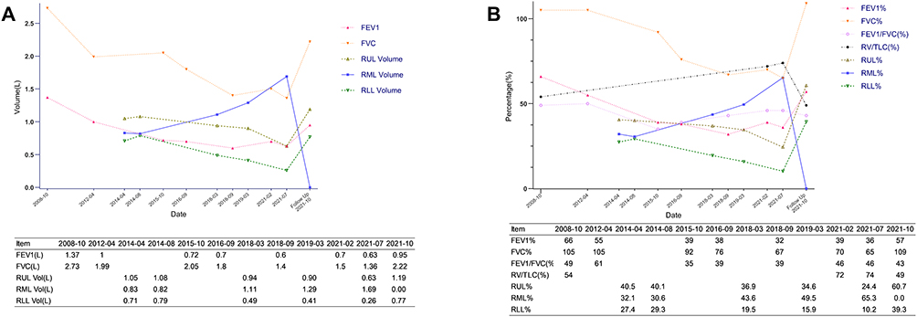 |
Figure 2 Serial pulmonary function tests during the follow-up and 2 months post-BLVR. (A) Shows a progressive decline in FEV1, FVC, RUL and RLL volume, while progressive expanding in the RML volume before BLVR. (B) Shows progressive decline FEV1%, FVC%, RV/TLC, RUL% and RLL%, and the negative change in RML%, while not apparent change in FEV1/FVC. Figure also demonstrated the reversion of pulmonary function test indices and the right lung lobe volumes 2 months post BLVR. Abbreviations: BLVR, bronchoscopic lung volume reduction; FEV1, forced expiratory volume in one second; FVC, forced vital capacity; FEV1/FVC, the ratio of FEV1 to FVC; RV, residual volume; TLC, total lung capacity; RV/TLC, the ratio of RV to TLC, FEV1%, the percentage of FEV1 to the predicted FEV1; FVC%, the percentage of FVC to the predicted FVC; RUL, right upperlobe; RML, RML; RLL, right lower lobe; RUL%, the percentage of RUL to the right lung; RML%, the percentage of RML to the right lung; RLL%, the percentage of RLL to the right lung. |
 |
Figure 3 Radiologic image related to the BLVR. (A) Shows a marked decrease of blood perfusion of the RML in single-photon emission computed tomography. (B) Shows the chest X ray taken on the first day post-BLVR. (C and D) Shows a chest CT scan obtained 2 months post-BLVR, confirming complete atelectasis of the RML and re-expansion of adjacent lobes. The red arrows in (B and C) point to atelectasis of the RML. The red circles in (C and D) show the location of the EBV. |
Bronchoscopy excluded significant stenosis or obstruction of the right middle bronchus. The absence of interlobar collateral ventilation was confirmed by using the Chartis system (PulmonX Corp., USA) (Figure 4A). In August 2021, the patient underwent bronchoscopic lung volume reduction (BLVR) via placement of a single endobronchial valve (EBV) (EBV-ts-5.5) into the RML bronchus (Figure 4B–D). This intervention resulted in a subjective improvement in dyspnea. Postoperative imaging showed a significant reduction in emphysematous changes in the RML, with partial re-expansion of the adjacent lobes (Figure 3B). The patient was discharged seven days later.
 |
Figure 4 Bronchoscopic image related to the BLVR. (A) Shows the measuring with the Chartis system. (B) Shows measuring of the right middle bronchus with flexible bronchoscopy. (C and D) Shows the EBV-ts-5.5 positioned in the right middle bronchus. |
At the two-month follow-up, the patient reported only mild dyspnea. Her exercise tolerance improved substantially, with six-minute walking distance increasing from 310 meters to 650 meters. Pulmonary function test(PFT) showed significant recovery in FEV1 and FVC, with FEV1 remarkably rebounding to 0.81L (97% of 2014 baseline) (Figure 2A and B). CT imaging confirmed complete atelectasis of the RML and re-expansion of the adjacent lobes (Figures 1E and 3C and D).
Discussion
VLS is a rare clinical syndrome that requires radiological imaging for diagnosis.3 The radiologic criteria were first proposed by Roberts et al, who defined it as the presence of giant bullae occupying at least one-third of a hemithorax. However, there is currently no universally accepted or standardized definition.3 It is most commonly associated with cigarette smoking, but may also be linked to alpha-1 antitrypsin deficiency and marijuana use.3,4 Although VLS secondary to COPD is proposed as progressive bullae enlargement and worsening dyspnea,5 the rate of progression remains unpredictable and has not been previously reported. A previous case report followed up five patients within one family for approximately 20 years, but none of these cases reported detailed disease progression before therapeutic intervention.6 Our case is among the first to provide detailed long-term follow-up data on disease progression prior to intervention.
The patient experienced a 7-year progression of VLS in the RML, with an inverse correlation between emphysematous volume and pulmonary function deterioration. Although the pathogenesis of VLS is not fully understood, it likely involves chronic alveolar wall destruction and potentially a check-valve mechanism, where air enters, but cannot exit the affected lobe.7,8 However, in this patient, bronchoscopy did not reveal significant bronchial stenosis or obstruction, which supports this hypothesis.
VLS management traditionally involves conservative measures or surgical interventions.1 Recently, bronchoscopic lung volume reduction (BLVR) using EBVs has emerged as a less invasive alternative for patients with poor pulmonary function.8,9 Although VLS was once defined as large bullae in one or both upper lobes,2 endobronchial valve therapy for RML-dominant VLS is relatively rare.
 |
Table 1 Characteristics of Cases Treated with BLVR in RML |
A review of published cases and our case revealed that, among the eight documented RML-targeted EBV interventions (Table 1), all patients were current or former smokers. Seven of these eight patients showed substantial improvements, with FEV1 increases ranging from 16% to 63% post-BLVR.8,10–13 Most patients required only one valve for treatment. Valve deployment (typically Zephyr EBVs 4.0–5.5) leads to bulla collapse and adjacent lung re-expansion, as seen in previous case reports where the residual volume decreased significantly post-BLVR.8 This aligns with the mechanistic rationale that EBVs promote atelectasis by blocking inspiratory airflow while allowing exhalation, thereby reducing hyperinflation and improving diaphragmatic mechanics.
The improvements in the 6-minute walk distance (6MWD) further validate the clinical relevance of these physiological changes, consistent with previous reports on enhanced exercise tolerance.10,12,13 It should be noted as a limitation that the long-term outcomes remain understudied, as most cases have follow-up periods of ≤12 months. However, some studies have demonstrated sustained benefits for more than one year,12 suggesting the durability of the response in suitable candidates.
Conclusion
This study presents a rare case of VLS secondary to COPD with detailed documentation of disease progression for 7 years prior to intervention. This case highlights the inverse relationship between RML emphysema volume and pulmonary function deterioration over time and underscores the utility of EBV therapy as an effective treatment option for advanced disease. Further studies are needed to evaluate long-term outcomes and optimize management strategies for this rare condition.
Data Sharing Statement
All data generated or analyzed during this study are included in this article. Further inquiries can be directed to the corresponding authors.
Statement of Ethics
Written informed consent was obtained from the patient for publication of the medical case details and any accompanying images. The patient was informed that all reasonable efforts would be made to preserve anonymity and that the report might be publicly accessible in academic and scientific domains. Institutional approval was required to publish this case, and ethical approval for the publication of this case report was granted by the Institutional Ethics Committee (IEC) for Clinical Research and Animal Trials of the First Affiliated Hospital of Sun Yat-sen University (Approval No. [2023]518). This approval includes the use and dissemination of clinical information and images for research and educational purposes.
Author Contributions
All authors made a significant contribution to the work reported, whether that is in the conception, study design, execution, acquisition of data, analysis and interpretation, or in all these areas; took part in drafting, revising or critically reviewing the article; gave final approval of the version to be published; have agreed on the journal to which the article has been submitted; and agree to be accountable for all aspects of the work.
Funding
This study was not supported by any sponsor or funding.
Disclosure
The authors have no conflicts of interest to declare in this work.
References
1. Yousaf MN, Chan NN, Janvier A. Vanishing Lung Syndrome: an Idiopathic Bullous Emphysema Mimicking Pneumothorax. Cureus. 2020;12(8):e9596. doi:10.7759/cureus.9596
2. Muhamad NI, Mohd Nawi SN, Yusoff BM, Ab Halim NA, Mohammad N, Wan Ghazali WS. Vanishing lung syndrome Masquerading as bilateral pneumothorax: a case report. Respir Med Case Rep. 2020;31:101276. doi:10.1016/j.rmcr.2020.101276
3. Salley JR, Kukkar V, Felde L. Vanishing lung syndrome: a consequence of mixed tobacco and marijuana use. BMJ Case Rep. 2021;14(5):e239255. doi:10.1136/bcr-2020-239255
4. Wiesel S, Siddiqui F, Khan T, Hossri S, El-Sayegh D. Vanishing Lung Syndrome: compound Effect of Tobacco and Marijuana Use on the Development of Bullous Lung Disease - A Joint Effort. Cureus. 2017;9(7):e1530. doi:10.7759/cureus.1530
5. Sharma N, Justaniah AM, Kanne JP, Gurney JW, Mohammed TLH. Vanishing Lung Syndrome (Giant Bullous Emphysema): CT Findings in 7 Patients and a Literature Review. J Thoracic Imag. 2009;24(3):227–6. doi:10.1097/RTI.0b013e31819b9f2a
6. Gao X, Wang H, Gou K, et al. Vanishing lung syndrome in one family: five cases with a 20-year follow-up. Mol Med Rep. 2015;11(1):567–570. doi:10.3892/mmr.2014.2673
7. Li Y, Su C, Yan Y, Wang Z, Wei X, Wang Z. A 75-Year-Old Man With Irregular Solid Components Within an Emphysematous Bulla. Chest. 2023;163(6):e265–e273. doi:10.1016/j.chest.2022.12.043
8. Gu LE, Rhee CK. Bronchoscopic lung volume reduction using an endobronchial valve to treat a huge emphysematous bullae: a case report. BMC Pulm Med. 2019;19(1):92. doi:10.1186/s12890-019-0849-z
9. Santini M, Fiorelli A, Vicidomini G, Di Crescenzo VG, Messina G, Laperuta P. Endobronchial treatment of giant emphysematous bullae with one-way valves: a new approach for surgically unfit patients. Eur J Cardiothorac Surg. 2011;40(6):1425–1431. doi:10.1016/j.ejcts.2011.03.046
10. Çetinkaya E, Özgül MA, Gül Ş, et al. Successful Treatment of Bulla with Endobronchial Valves. Case Rep Pulmonol. 2015;2015:947403. doi:10.1155/2015/947403
11. Tian Q, An Y, Xiao BB, Chen LA. Endobronchial valve to treat large bulla at right middle lobe in chronic obstructive pulmonary disease patients. J Thorac Dis. 2015;7(9):E374–E377. doi:10.3978/j.issn.2072-1439.2015.09.33
12. Hou G, Wang W, Wang Q, Kang J. Bronchoscopic bullectomy with a one-way endobronchial valve to treat a giant bulla in an emphysematic lung: a case report. Clin Respiratory J. 2016;10(5):657–660. doi:10.1111/crj.12257
13. Villeneuve T, Fumat R, Héluain V, Pascal P, Plat G, Guibert N. Response to exclusive right middle lobe treatment with endobronchial valves: a case report. Breathe. 2021;17(3):210108. doi:10.1183/20734735.0108-2021
 © 2026 The Author(s). This work is published and licensed by Dove Medical Press Limited. The
full terms of this license are available at https://www.dovepress.com/terms
and incorporate the Creative Commons Attribution
- Non Commercial (unported, 4.0) License.
By accessing the work you hereby accept the Terms. Non-commercial uses of the work are permitted
without any further permission from Dove Medical Press Limited, provided the work is properly
attributed. For permission for commercial use of this work, please see paragraphs 4.2 and 5 of our Terms.
© 2026 The Author(s). This work is published and licensed by Dove Medical Press Limited. The
full terms of this license are available at https://www.dovepress.com/terms
and incorporate the Creative Commons Attribution
- Non Commercial (unported, 4.0) License.
By accessing the work you hereby accept the Terms. Non-commercial uses of the work are permitted
without any further permission from Dove Medical Press Limited, provided the work is properly
attributed. For permission for commercial use of this work, please see paragraphs 4.2 and 5 of our Terms.
Recommended articles
Association Between Subsegmental Dosing Accuracy and Outcomes of Bronchoscopic Thermal Vapor Ablation: An Exploratory Retrospective Study in Severe Heterogeneous Emphysema
Yang H, Chen S, Zheng X, Cui N, Xie F, Hou H, Ye L, Herth FJF, Sun J
International Journal of Chronic Obstructive Pulmonary Disease 2026, 21:560457
Published Date: 23 January 2026