Back to Journals » International Journal of General Medicine » Volume 14
Identification of Brain Regions with Enhanced Functional Connectivity with the Cerebellum Region in Children with Attention Deficit Hyperactivity Disorder: A Resting-State fMRI Study
Received 22 January 2021
Accepted for publication 29 April 2021
Published 27 May 2021 Volume 2021:14 Pages 2109—2115
DOI https://doi.org/10.2147/IJGM.S303339
Checked for plagiarism Yes
Review by Single anonymous peer review
Peer reviewer comments 2
Editor who approved publication: Dr Scott Fraser
Li Ding,1 Gaofeng Pang2
1Department of Pediatrics, Changzhou Children’s Hospital of Nantong University, Changzhou, 213003, People’s Republic of China; 2Department of Pediatrics, The Third Affiliated Hospital of Soochow University, Changzhou, 213003, People’s Republic of China
Correspondence: Gaofeng Pang
Department of Pediatrics, The Third Affiliated Hospital of Soochow University, Changzhou, 213003, People’s Republic of China
Tel +86-13601503658
Email [email protected]
Background: To explore the brain regions with higher functional connectivity with the cerebellum at resting state and the brain functions related to cognitive function in children with attention-deficit hyperactivity disorder (ADHD).
Methods: Thirty children with ADHD and 33 typically developing children (TDC) were examined using resting-state functional magnetic resonance imaging (fMRI) scans. Seed-based functional connectivity (FC) analysis was performed.
Results: Four brain areas with higher FC values were identified in ADHD children. These four areas were the left middle frontal gyrus, right middle frontal gyrus, right superior temporal gyrus and left parahippocampal gyrus (P < 0.05). The results of the CPT show that the number of omission errors was significantly higher in the children with ADHD than in the TD group (5.13± 5.12 vs 2.18± 2.36, P = 0.000). The commission number in the ADHD group was also significantly higher than that of the TD group (4.03± 6.56 vs 2.00± 2.85, P = 0.002). However, no statistically significant difference was observed in the correct reaction time between the two groups (641.54± 146.79 ms vs 584.81± 145.82 ms, P = 0.835).
Conclusion: The dysfunction of cerebellar functional connectivity in specific brain regions may be one of the pathological and physiological causes of cognitive impairment of ADHD.
Keywords: attention deficit hyperactivity disorder, cerebellum, children, functional connectivity, resting-state functional magnetic resonance imaging
Introduction
Attention deficit hyperactivity disorder (ADHD) is a neurological disorder that occurs during childhood, and most of the patients continue to show the symptoms in adolescence and adulthood. The morbidity in children is about 5%.1 Complications of ADHD include learning disability, conduct disorder, and oppositional defiant disorder. Researchers have found multiple brain areas affected in ADHD including prefrontal lobe, inferior parietal lobule, anterior cingulate, cerebellum, and others.2 Especially, the important role of cerebellum in ADHD is increasingly recognized. Previously thought to be responsible for motor balance and coordination, cerebellum has been identified to have many other functions such as in the cognitive processing of emotions and negative stimulus, executive function, attention and language.3 Structural MRI analysis indicated that the volume of cerebellum in children with ADHD is smaller than that of normal children,4 especially the volume of gray matter in the left cerebellum is significantly smaller than that of the normal group.5 Moreover, the activation of the cerebellum in children with ADHD is lower than that of children recovered from ADHD and the normal control group.6 The human brain functions are often performed by the synergistic action of multiple brain regions. For example, Rhein et al showed that ADHD was associated with decreased functional connectivity between the salience and executive control networks, as well as with peripheral brain regions.7 However, there is still limited understanding of the correlations of cerebellum activity with other brain regions in ADHD patients.
Functional magnetic resonance imaging (fMRI) is a powerful brain imaging method for studying brain cognition.8 The method of functional connectivity (FC) based on fMRI is widely used in the study of brain networks. It refers to the correlation between time series of two different brain regions and is used to describe the relationship between different brain regions. The method of seed-based functional connectivity studies regions correlated with the activity in a seed region. In seed-based analysis, the cross-correlation is identified between the time-series of the seed and the rest of the brain.9 Zang et al found that the activation of bilateral cerebellum was decreased in ADHD group.10 Therefore, this study chooses the left cerebellum as the seed region to study the brain mechanism of cognitive representation of ADHD.
In this study, continuous performance test (CPT)11 was also used to analyze attention behavior of ADHD children. It is known that the brain area of cerebellum is associated with attention.3 Therefore, the correlation between the attention behavior and the cerebellar functional connectivity were analyzed. This study identified four brain regions showing higher functional connectivity with cerebellum in ADHD children, and their correlations with attention behavior were explored.
Materials and Methods
Participants
The ADHD group included 30 children (7–12 years old) who were diagnosed by the ADHD clinic in Changzhou Children’s Hospital from June 2015 to July 2017, according to the diagnostic criteria described by the Diagnostic and Statistical Manual of Mental Disorders, 5th Edition (DSM-V).12 There were 18 boys and 12 girls (9.6 ± 1.7 years old) (Table 1). All children met the following criteria: 1) no medical history related to the neural systems and mental health; 2) no history of psychiatric medication; and 3) sufficient cooperation to undergo MRI scan.
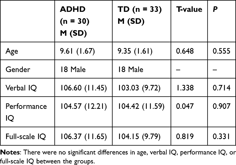 |
Table 1 Demographic Characteristics of the ADHD and TD Groups |
The typically developing controls (TDC) included 33 children from a local school. There were 18 boys and 15 girls (9.8 ± 1.6 years old). The age, gender and education were matched between the two groups (P > 0.05). Both ADHD and TDC children were right-handed and had intelligence quotient (IQ) scores of >80 (measured by the Wechsler Intelligence Scale for Chinese Children-Revised; WISCC-R). This study was approved by the Ethical Committee of Changzhou Children’s Hospital of Nantong University (No. 2014–012). Informed consent was obtained from the parent, and all the children agreed to participate. This study was conducted in accordance with the Declaration of Helsinki.
MRI Data Acquisition
A Siemens 1.5-Tesla MagnetomAvanto scanner was used to obtain the brain images. Children were asked to lay flat and not to do any motor activity. During fMRI scanning, participants closed eyes and remained in a calm and awake state. fMRI data were collected using the echo-planar imaging (EPI) sequence with the following parameters: axial slices=18, echo time (TE) = 40 ms, repetition time (TR) = 2000 ms, field of view (FOV) = 240×240 mm, 180 volumes (6 min), thickness/gap = 6.0/1.2 mm, matrix = 64 × 64, and flip angle = 90°. T1-weighted images were obtained using the following parameters: TE = 11 ms, TR = 414 ms, FOV = 240 mm × 240 mm, flip angle = 90°, thickness/gap = 5.0/1.5 mm, and in-plane resolution = 256×256.
Data Processing
The first 10 time series were discarded and the Data Processing Assistant for Resting-State fMRI (DPARSF) V2.3 was used to analyze the remaining data. The process of slice timing and head motion correction were performed first. Data would be discarded if head motion exceeded 3 mm. The functional scans were normalized to the standard template and re-sampled to 3×3×3 mm3. Subsequently, smoothing was conducted with a Gaussian kernel of 6 mm full width at half maximum. Further processes included removal of linear trends and temporal band-pass filtering (0.01~0.08 Hz). White matter and CSF would be regressed out.
The seed-based functional connectivity analysis was carried out using the Rest software with the left cerebellum (−49.5,-58.5,-18.5) as the seed region, which was shown to have the greatest difference between ADHD and TDC groups by Zang.10 The time series in the cerebellum were calculated and all voxels were averaged, followed by Pearson correlation analysis with the time series of other regions of the whole brain to obtain the FC brain map.
CPT Test
The continuous performance test (CPT) is an indicative Go/Nogo task. The stimulus content are the Arabic numerals 0~9. The number 1 is the cue (warning), the number 9 after 1 is the Go stimulus (target after warning), the other numbers after 1 are the Nogo stimulus (nontarget after warning). Thus, the following numbers after the presentation of the number 1 act as the distraction stimulus: 0, 2, 3, 4, 5, 6, 7, and 8. The stimulus consists of 500 numbers, among which the sequence of number 1 followed by the number 9 is 20%, the sequence of numbers 1-not-9 (the number after 1 is not 9) is 20%, the sequence of numbers not-1 to 9 (the number after not-1 is 9) is 20%, and the probability of other numbers appearing randomly. Children were asked to respond to the “9” button that appeared immediately after “1” and not to press any other number. The stimulus duration was 200 ms with a stimulus interval of 1300 ms. The stimulus appeared in the center of the cathode ray tube display, in black and white. The US e-prime software was used to control the presentation of stimuli and automatically record behavioral results.
Statistical Analysis
The two-sample t-tests were performed using Monte Carlo simulation implemented with REST software.13 The results of two-sample t-tests were overlaid on the Ch2 template.14 The correlations between the functional connection values and the CPT results were analyzed by DPABI (a toolbox for data processing and analysis for brain imaging). A corrected threshold of P< 0.05 corresponded to a combined threshold of P< 0.01 at individual voxel level and cluster size >40 mm3.
Results
Seed-based functional connectivity analysis was performed on a group of children diagnosed with ADHD and a matching control group. The time series in the cerebellum region of each participant were calculated and all voxels were averaged, followed by Pearson correlation analysis with the time series of other regions of the whole brain to obtain the FC brain map of the ADHD group and the control group. We identified four brain areas using cerebellum as the seed region in which the FC values of the ADHD group were obviously higher than those of controls. These four areas were the left middle frontal gyrus, right middle frontal gyrus, right superior temporal gyrus and left parahippocampal gyrus (P <0.05, after correction) (Table 2 and Figure 1). The Z scores of left middle frontal gyrus, right middle frontal gyrus, right superior temporal gyrus and left parahippocampal gyrus were 4.10, 4.38, 3.97, and 3.32, respectively, indicating that the FC values of the ADHD group were higher than that of the TDC group.
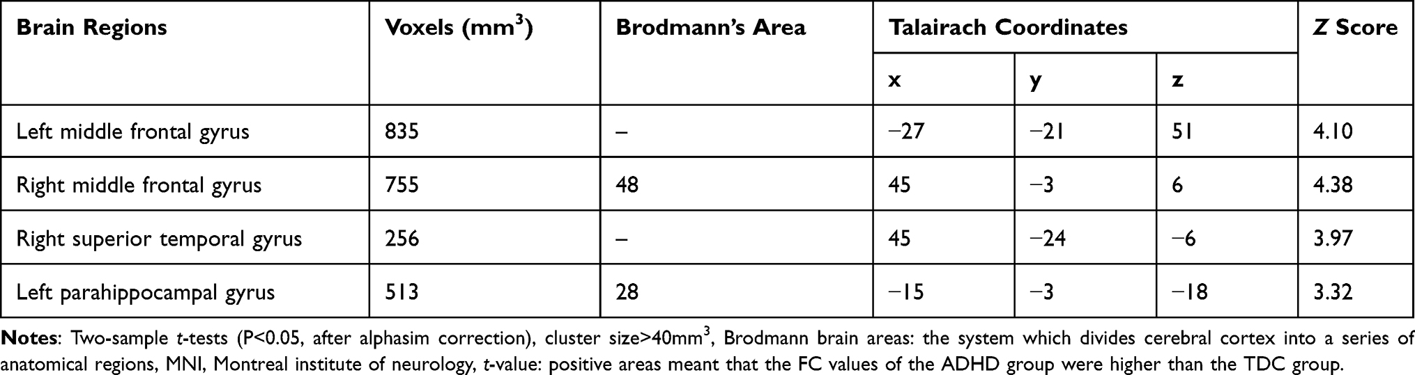 |
Table 2 The Comparison Between ADHD and Typically Developing Controls by Functional Connectivity |
 |
Figure 1 Comparison of the functional connectivity values in the ADHD and TDC groups. (P<0.05, after alphasim correction. The red areas indicate the brain regions in which the values of FC were higher in the ADHD group than in the TDC group). |
The CPT task was used to analyze the behavioral performance of the two groups separately and the results are presented in Table 3. Analysis of the reaction time demonstrated that there was no significance difference in the performance of the two groups through two-sample t-test analysis (P = 0.835). However, there was a tendency towards a slower response in the ADHD group (641.54±146.79) than in the TD group (584.81±145.82). Additionally, the number of omission errors and commission errors were significantly higher in the ADHD group than that of the TD group (P = 0.000 and P = 0.002). Furthermore, the hit number was significantly lower in the ADHD group than in the TD group (P = 0.000).
 |
Table 3 Results of the Continuous Performance Test the ADHD and TD Groups |
Discussion
In the present study, our results confirmed that children with ADHD exhibited lower scores than TD children in hit number, omission number and commission number in CPT tasks. These results are consistent with a deficit in attention and cognition in the ADHD group.15 Furthermore, our rs-fMRI findings reveal that these impairments could possibly be associated with inappropriate action in four specific brain areas, including the left middle frontal gyrus, right middle frontal gyrus, right superior temporal gyrus and left parahippocampal gyrus. These brain areas are thought to be involved in the process of executive control and cognition. Therefore, our study suggests that abnormal activation of cognitive brain areas leads to cognitive impairments in ADHD.
Frontal lobe is important in brain cognition and the frontal gyrus is involved in decision-making, which is related to emotion modularization and conflict resolution. The middle frontal gyrus has been recognized as the focal point of the cerebral cortex and is prominent in the storage of spatial information, which is part of the working memory in the human brain.16 In children with ADHD, the activation of the prefrontal cortex decreased, the activation of the left and right frontal gyrus decreased, and the activation of cerebellum increased.17 Unlike healthy children, ADHD children activate right middle frontal gyrus when they repeat the same thing over and over again.18 The ADHD children show no discrepant area for working memory and both the right middle frontal gyrus and the right precuneus are involved, while the normal children show more activity in the left middle frontal gyrus.19 This study showed that the enhanced FC between the cerebellum and the left and right middle frontal gyrus may cause the disorganized connection network, which led to attention deficit, impaired working memory and reduced decision-making function of children with ADHD.
The superior temporal gyrus is associated with the task of listening and participates in the normal development of the language-related cortex.20 At the same time, the superior temporal gyrus is involved in speech processing of high-level auditory processing, which is an important brain region for human auditory speech and speech expression.21 The abnormally reduced volume of the right superior temporal gyrus may be a neural structural marker for the abnormal assessment of social emotional information in school-age children.22 In the process of recessive emotion processing, both the superior temporal gyrus and medial prefrontal lobe are involved.23 The damage of the right superior temporal gyrus could lead to failure of visual search tasks.24 The enhanced FC between the cerebellum and the right superior temporal gyrus as revealed in our study could reduce the ability of original spatial perception, emotional attention and visual exploration, and impetuous processing of social emotions, which is a possible reason for the impulsiveness of children with ADHD. Meanwhile, spatial perception, auditory voice and visual search dysfunction may be the pathological mechanism of attention deficit in ADHD children. The left parahippocampal gyrus is part of the limbic lobe. Studies have shown that activation of the limbic lobe during sleep can promote the solidification of emotional memory and cognitive development. However, children with ADHD have low activation of the limbic lobe and the dysfunction of the frontal lobe-limbic circuit system, which leads to the weakening of memory solidification.25 Studies also have confirmed that the limbic lobe is related to motivation and emotional function.26 Our study found enhanced FC between the cerebellum and the left parahippocampal gyrus in ADHD children, which may result in abnormal emotional regulation mechanisms, poor emotional control and anger.
The attention network and default mode network (DMN) are two major cognitive network systems in human beings and they are antagonistic. One of the pathological bases of cognitive impairment in ADHD may be the interconnection of brain regions in the DMN or abnormal connections between the DMN and other brain regions.27 This study found that the connection between the cerebellum and the middle frontal gyrus is enhanced in children with ADHD, compared to the connectivity observed in a TD control group. As the left parahippocampal gyrus is a part of the DMN, enhanced functional connectivity between the cerebellum and the middle frontal gyrus and the left parahippocampal gyrus of the DMN may lead to abnormal antagonism between the DMN and the attention network in ADHD.
In this fMRI study, a greater functional separation was observed in a series of brain networks, including negative connections between the cerebellum and the frontal lobe, in ADHD. However, higher levels of internal changes and fairly strong positive connections in brain networks have been observed in ADHD.28 In this study, the functional connections between the cerebellum and the left and right middle frontal gyrus as well as the left parahippocampal gyrus were stronger in the ADHD group than in the TD group, with higher levels of internal changes observed in responses, which could lead to the decline of continuous attention ability.
Conclusions
Our study demonstrated that ADHD children showed stronger functional connections between cerebellum and the left, right middle frontal gyrus as well as the left parahippocampal gyrus, compared with the control group. The dysfunction of cerebellar functional connectivity in specific brain regions may be one of the pathological and physiological bases of cognitive impairment of ADHD.
Abbreviations
ADHD, attention deficit hyperactivity disorder; TDC, typically developing controls; fMRI, functional magnetic resonance imaging; FC, functional connectivity; CPT, continuous performance test; IQ, intelligence quotient; EPI, echo-planar imaging; TE, echo time; TR, repetition time; FOV, field of view; DPARSF, Data Processing Assistant for Resting-State fMRI.
Ethical Approval
This study was approved by the Ethical Committee of Changzhou Children’s Hospital of Nantong University (No. 2014-012). Informed consent was obtained from the parent, and all the children agreed to participate.
Author Contributions
All authors made a significant contribution to the work reported, whether that is in the conception, study design, execution, acquisition of data, analysis and interpretation, or in all these areas; took part in drafting, revising or critically reviewing the article; gave final approval of the version to be published; have agreed on the journal to which the article has been submitted; and agree to be accountable for all aspects of the work.
Funding
The study was supported by the science and technology program of basic applying of Changzhou (No. CJ20200081).
Disclosure
The authors report no financial or nonfinancial benefits have been received or will be received from any party related directly or indirectly to the subject of this article.
References
1. Bozhilova N, Michelini G, Kuntsi J, Asherson P. Mind wandering perspective on ADHD. Neurosci Biobehav Rev. 2018;92:464–476. doi:10.1016/j.neubiorev.2018.07.010
2. Wang XH, Jiao Y, Li L. Diagnostic model for attention-deficit hyperactivity disorder based on interregional morphological connectivity. Neurosci Lett. 2018;685:30–34. doi:10.1016/j.neulet.2018.07.029
3. Sokolov AA, Miall RC, Ivry RB. The cerebellum: adaptive prediction for movement and cognition. Trends Cogn Sci. 2017;21:313–332. doi:10.1016/j.tics.2017.02.005
4. Shaw P, Ishii-Takahashi A, Park MT, et al. A multicohort, longitudinal study of cerebellar development in attention deficit hyperactivity disorder. J Child Psychol Psychiatry. 2018;59:1114–1123. doi:10.1111/jcpp.12920
5. Kumar U, Arya A, Agarwal V. Neural alterations in ADHD children as indicated by voxel-based cortical thickness and morphometry analysis. Brain Dev. 2017;39:403–410. doi:10.1016/j.braindev.2016.12.002
6. Szekely E, Sudre GP, Sharp W, Leibenluft E, Shaw P. Defining the neural substrate of the adult outcome of childhood ADHD: a multimodal neuroimaging study of response inhibition. Am J Psychiatry. 2017;174:867–876. doi:10.1176/appi.ajp.2017.16111313
7. Rhein DV, Beckmann CF, Franke B, et al. Network-level assessment of reward-related activation in patients with ADHD and healthy individuals. Human Brain Mapping. 2017;38:2359–2369. doi:10.1002/hbm.23522
8. Parkes L, Fulcher B, Yücel M, Fornito A. An evaluation of the efficacy, reliability, and sensitivity of motion correction strategies for resting-state functional MRI. NeuroImage. 2018;171:415–436. doi:10.1016/j.neuroimage.2017.12.073
9. Lv H, Wang Z, Tong E, et al. Resting-state functional MRI: everything that nonexperts have always wanted to know. AJNR Am J Neuroradiol. 2018;39:1390–1399. doi:10.3174/ajnr.A5527
10. Zang YF, He Y, Zhu CZ, et al. Altered baseline brain activity in children with ADHD revealed by resting-state functional MRI. Brain Dev. 2007;29:83–91. doi:10.1016/j.braindev.2006.07.002
11. Eom H, Kim KK, Lee S, Hong YJ. Development of virtual reality continuous performance test utilizing social cues for children and adolescents with attention-deficit/hyperactivity disorder. Cyberpsychol Behav Soc Netw. 2019;22(3):198–204. doi:10.1089/cyber.2018.0377
12. Bastiaens L, Galus J. Comparison of the adult ADHD self report scale screener for DSM-IV and DSM-5 in a dually diagnosed correctional population. Psychiatr Q. 2018;89:505–510. doi:10.1007/s11126-017-9553-4
13. Mao D, Ding Z, Jia W, et al. Low-frequency fluctuations of the resting brain: high magnitude does not equal high reliability. PLoS One. 2015;10:e0128117. doi:10.1371/journal.pone.0128117
14. Li Z, Zang YF, Ding J, Wang Z. Assessing the mean strength and variations of the time-to-time fluctuations of resting-state brain activity. Med Biol Eng Comput. 2017;55:631–640. doi:10.1007/s11517-016-1544-3
15. Wang S, Yang Y, Xing W, et al. Altered neural circuits related to sustained attention and executive control in children with ADHD: an event-related fMRI study. Clin Neurophysiol. 2013;124:2181–2190. doi:10.1016/j.clinph.2013.05.008
16. Leung HC, Gore JC, Goldman-Rakic PS. Sustained mnemonic response in the human middle frontal gyrus during on-line storage of spatial memoranda. J Cognitive Neurosci. 2002;14:659–671. doi:10.1162/08989290260045882
17. Qian A, Wang X, Liu H, et al. Dopamine D4 receptor gene associated with the frontal-striatal-cerebellar loop in children with ADHD: a resting-state fMRI study. Neurosci Bull. 2018;34:497–506. doi:10.1007/s12264-018-0217-7
18. Epstein JN, Delbello MP, Adler CM, et al. Differential patterns of brain activation over time in adolescents with and without attention deficit hyperactivity disorder (ADHD) during performance of a sustained attention task. Neuropediatrics. 2009;40:1–5. doi:10.1055/s-0029-1220686
19. Fassbender C, Schweitzer JB, Cortes CR, et al. Working memory in attention deficit/hyperactivity disorder is characterized by a lack of specialization of brain function. PLoS One. 2011;6:e27240. doi:10.1371/journal.pone.0027240
20. Rajarethinam R, Venkatesh BK, Peethala R, Phan KL, Keshavan M. Reduced activation of superior temporal gyrus during auditory comprehension in young offspring of patients with schizophrenia. Schizophr Res. 2011;130:101–105. doi:10.1016/j.schres.2011.05.025
21. Mesgarani N, Cheung C, Johnson K, Chang EF. Phonetic feature encoding in human superior temporal gyrus. Science (New York). 2014;343:1006–1010. doi:10.1126/science.1245994
22. Pan LA, Ramos L, Segreti A, Brent DA, Phillips ML. Right superior temporal gyrus volume in adolescents with a history of suicide attempt. Bri J Psychiatry. 2015;206:339–340. doi:10.1192/bjp.bp.114.151316
23. Kana RK, Patriquin MA, Black BS, Channell MM, Wicker B. Altered medial frontal and superior temporal response to implicit processing of emotions in autism. Autism Res. 2016;9:55–66. doi:10.1002/aur.1496
24. Ellison A, Schindler I, Pattison LL, Milner AD. An exploration of the role of the superior temporal gyrus in visual search and spatial perception using TMS. Brain. 2004;127:2307–2315. doi:10.1093/brain/awh244
25. Prehnkristensen A, Munz M, Molzow I, Wilhelm I, Wiesner CD, Baving L. Sleep promotes consolidation of emotional memory in healthy children but not in children with attention-deficit hyperactivity disorder. PLoS One. 2013;8:e65098. doi:10.1371/journal.pone.0065098
26. Redlich R, Opel N, Bürger C, et al. The limbic system in youth depression: brain structural and functional alterations in adolescent in-patients with severe depression. Neuropsychopharmacology. 2018;43:546–554. doi:10.1038/npp.2017.246
27. Gronchi G, Giovannelli F. Dual process theory of thought and default mode network: a possible neural foundation of fast thinking. Front Psychol. 2018;9:1237. doi:10.3389/fpsyg.2018.01237
28. O’Halloran L, Cao Z, Ruddy K, et al. Neural circuitry underlying sustained attention in healthy adolescents and in ADHD symptomatology. NeuroImage. 2017;169:395–406. doi:10.1016/j.neuroimage.2017.12.030
 © 2021 The Author(s). This work is published and licensed by Dove Medical Press Limited. The
full terms of this license are available at https://www.dovepress.com/terms
and incorporate the Creative Commons Attribution
- Non Commercial (unported, 3.0) License.
By accessing the work you hereby accept the Terms. Non-commercial uses of the work are permitted
without any further permission from Dove Medical Press Limited, provided the work is properly
attributed. For permission for commercial use of this work, please see paragraphs 4.2 and 5 of our Terms.
© 2021 The Author(s). This work is published and licensed by Dove Medical Press Limited. The
full terms of this license are available at https://www.dovepress.com/terms
and incorporate the Creative Commons Attribution
- Non Commercial (unported, 3.0) License.
By accessing the work you hereby accept the Terms. Non-commercial uses of the work are permitted
without any further permission from Dove Medical Press Limited, provided the work is properly
attributed. For permission for commercial use of this work, please see paragraphs 4.2 and 5 of our Terms.