Back to Journals » International Journal of Nanomedicine » Volume 20
Hydrogel-Based Microspheres for Intra-Articular Osteoarthritis Therapy: Multifunctional Carriers for Drug Delivery and Cartilage Repair: A Comprehensive Review
Authors Zhao Z, Dong P, Wang H, Su J, Yu M, Shi H, Gao T, Zhang Q, Chen W, Yang J, Zhang H, Wei Y ![]()
Received 5 May 2025
Accepted for publication 6 August 2025
Published 4 September 2025 Volume 2025:20 Pages 10795—10813
DOI https://doi.org/10.2147/IJN.S538490
Checked for plagiarism Yes
Review by Single anonymous peer review
Peer reviewer comments 4
Editor who approved publication: Prof. Dr. Anderson Oliveira Lobo
Ze Zhao,1,* Peng Dong,1,* Haochen Wang,1 Jianbang Su,1 Minghao Yu,1 Haoyan Shi,1 Tianqi Gao,1 Qian Zhang,1 Wenzheng Chen,1 Jingyu Yang,2 Huijie Zhang,3 Yingliang Wei1
1Department of Orthopedics, Shengjing Hospital of China Medical University, Shenyang, People’s Republic of China; 2School of Business Administration, Shenyang Pharmaceutical University, Shenyang, 110016, People’s Republic of China; 3Department of Gynaecology and Obstetrics, Shengjing Hospital of China Medical University, Shenyang, Liaoning, People’s Republic of China
*These authors contributed equally to this work
Correspondence: Yingliang Wei, Department of Orthopedics, Shengjing Hospital of China Medical University, No. 36, Sanhao Street, Heping District, Shenyang, Liaoning, 110004, People’s Republic of China, Tel +86-024-96615-36311, Email [email protected] Huijie Zhang, Department of Gynaecology and Obstetrics, Shengjing Hospital of China Medical University, No. 36, Sanhao Street, Heping District, Shenyang, Liaoning, 110004, People’s Republic of China, Tel +86-024-96615-42211, Email [email protected]
Abstract: Intra-articular therapy has long been a key focus in the treatment of osteoarthritis (OA), with both intra-articular drug injection and cell therapy offering significant advantages over systemic administration due to their superior local therapeutic efficacy. However, several challenges remain, including difficulties in maintaining effective drug concentrations within the joint cavity, limited survival and functionality of injected cells, and the inability to accurately target drugs or cells to diseased tissues. To address these issues, hydrogel-based microsphere systems have recently been developed as carriers for drugs, cells, and bioactive factors, enabling more precise and efficient intra-articular therapies. This review summarizes recent advances in hydrogel microspheres for intra-articular injection and classifies them according to their functional properties. From the perspective of drug delivery, we discuss their roles in sustained release, cartilage penetration and targeting, stimulus-responsive release mechanisms, and inherent pharmacological activity. From the perspective of cell therapy, we explore their applications in cell delivery and cartilage tissue engineering, including their ability to enhance cell viability, maintain stemness, and enable the co-delivery of bioactive molecules. In conclusion, hydrogel microspheres represent a promising strategy for improving the efficacy of intra-articular treatments in osteoarthritis.
Keywords: osteoarthritis, microspheres, drug delivery, cartilage tissue engineering
Introduction
Osteoarthritis (OA) is a common joint disease, with risk factors including aging, obesity, and knee joint injuries.1 The primary pathological changes in OA involve the degradation of articular cartilage, synovial inflammation, osteophyte formation, and subchondral bone sclerosis,2,3 with cartilage degeneration being the hallmark of the disease. Due to the absence of vasculature, adult cartilage injuries are difficult to repair or regenerate.4 Factors such as local or systemic inflammation and mechanical loading can disrupt the metabolic balance of chondrocytes in the joint environment, leading to cartilage degradation and promoting the onset and progression of OA.5 In recent years, an increasing number of studies have highlighted the critical role of microenvironmental dysregulation in the progression of OA. Key factors involved include the release of inflammatory mediators, degradation of the cartilage extracellular matrix, and oxidative stress. Targeted therapies that modulate the joint microenvironment have shown potential in alleviating cartilage damage and slowing disease progression. Consequently, preventing cartilage damage and promoting repair after injury have become key areas of focus in OA treatment.
OA treatment encompasses non-pharmacological therapies such as exercise and self-management, as well as surgical interventions for joints with severe functional impairment, while pharmacological therapies are the most commonly used.6 Intra-articular drug injection is a common approach for the treatment of OA. Compared to systemic administration, it offers lower costs, reduced risk of systemic side effects, and higher local drug concentrations.7 However, conventional intra-articular drug delivery methods face several limitations, including short retention times in the joint leading to inefficacy, reduced drug permeability due to extracellular matrix (ECM) barriers, and poor drug delivery selectivity, which impedes targeted action.8,9 To address these issues, recent research has focused on using biomaterials such as hydrogels, nanoparticles, and microspheres as drug delivery systems for intra-articular therapy.10 These materials, when used as carriers for intra-articular therapeutic agents, can stabilize encapsulated drugs and enhance their efficacy.11 Microspheres, as a micrometer-sized sphere composed of single or multiple polymers—offer notable advantages in biocompatibility and drug release, depending on the materials used in their synthesis.12 Compared to hydrogels, which are also commonly employed as delivery vehicles, microspheres exhibit more uniform size and higher porosity. These features help overcome limitations associated with hydrogels, such as irregular shape and insufficient pore space, which can hinder both injection and controlled drug release.13 Consequently, microspheres demonstrate superior performance in drug loading capacity, encapsulation efficiency, stability preservation, and targeted delivery. Due to these advantageous properties, microspheres are a central focus of this review.
As the progression of OA continues, articular cartilage undergoes damage and degeneration, often failing to repair itself, ultimately leading to joint dysfunction. Therefore, for cases of more severe cartilage damage, the effectiveness of pharmacological treatments is limited, and there is often a need to integrate cartilage tissue engineering techniques.14 These techniques include the use of cartilage repair materials and stem cell therapies to promote cartilage regeneration and improve joint function.15 In stem cell therapy for OA, the primary goal is to promote the growth and differentiation of stem cells into chondrocytes at the damaged site.16 Natural material scaffolds and polymer-based hydrogel scaffolds used in cartilage tissue engineering can provide a supportive environment for cell adhesion, growth, and proliferation. However, traditional scaffolds often suffer from poor mechanical strength and uncontrollable degradation, which can compromise their structural support during tissue regeneration. In contrast, microsphere-based scaffolds offer improved mechanical properties and controllable degradation rates, making them more suitable for promoting cartilage regeneration. Furthermore, their ability to achieve controlled release of cells and growth factors during degradation significantly enhances the therapeutic outcomes in osteoarthritis treatment.17
This review aims to classify and critically evaluate the diverse roles of hydrogel-based microspheres in OA treatment, with a focus on drug delivery mechanisms and cartilage tissue engineering applications.
Microspheres as Drug Delivery Systems for Osteoarthritis Treatment
Pharmacological treatment has long been a primary focus in the management of OA. Intra-articular drug delivery, as an alternative to oral administration, offers the advantage of increasing local drug concentrations within the joint while addressing concerns related to systemic side effects and safety.10,18 However, intra-articular delivery is not without challenges. High concentrations of drugs injected into the joint cavity can lead to adverse reactions, and the initially elevated drug levels often decrease rapidly due to clearance mechanisms, thereby shortening the therapeutic duration.19 Furthermore, the ECM of cartilage, characterized by its highly dense and negatively charged structure, poses a significant barrier to drug penetration.20 This coupled with the inability to precisely target damaged areas, often results in suboptimal therapeutic outcomes. In some cases, the injected drugs may inadvertently harm healthy cells, exacerbating the risk of unintended side effects.9,10
In summary, intra-articular drug delivery for osteoarthritis requires optimization to address the limitations of conventional drug injection. Incorporating drug delivery systems as an adjunct to intra-articular injection offers a promising approach to overcoming these challenges. Microspheres, as a drug delivery vehicle, exhibit unique advantages over other carriers due to their distinctive properties. Previous studies have provided valuable insights into various microsphere-based systems for osteoarthritis treatment. In this section, we classify drug-loaded microspheres according to their specific roles in drug delivery. Furthermore, we discuss their therapeutic effects based on the distinct mechanisms through which they function in these roles.
Prolonging the Duration of Drug Action
As mentioned above, one of the major challenges in intra-articular drug therapy is the rapid clearance of drugs from the joint. In recent years, considerable research has focused on extending the duration of drug action within the joint. High drug-loaded microspheres that provide sustained release of the drug are a fundamental requirement for the treatment of osteoarthritis. However, the sustained release properties of microspheres vary depending on their composition and structure.21 Therefore, we classify microspheres based on their material composition and summarize the corresponding characteristics of various microsphere types, along with their drug release profiles (Figure 1).
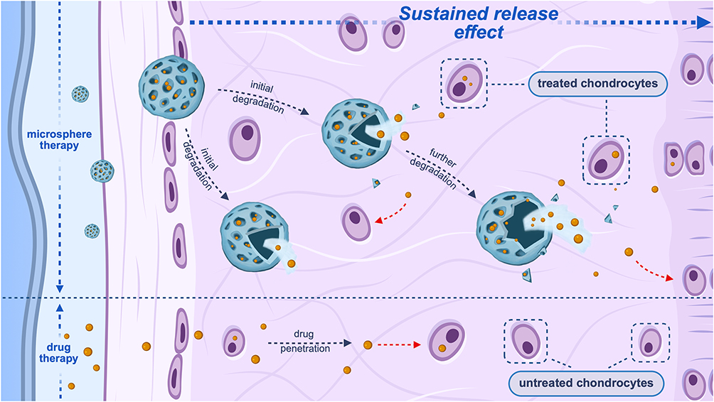 |
Figure 1 Role of microspheres in prolonging drug efficacy. Notes: This figure illustrates the process by which drug-loaded microspheres achieve the synchronized release of encapsulated drugs through gradual degradation. The sustained release property of microspheres ensures a stable and prolonged therapeutic effect of the drug compared to simple drug injections. This figure was created with BioRender (https://biorender.com/). |
Natural Polymer Microspheres
Natural polymer microspheres are spherical particles composed of biodegradable polymers derived from natural sources. Common examples include gelatin microspheres, chitosan microspheres, dextran microspheres, and hyaluronic acid microspheres.22 Due to the excellent biocompatibility of natural polymers, microspheres made from these materials generally exhibit minimal side effects and a lower risk of immune rejection during treatment. Additionally, the favorable biodegradability of natural polymer microspheres enhances their drug release capabilities, making them highly effective in therapeutic applications.
Gelatin is a water-soluble protein derivative obtained through the hydrolysis of tissues. As a natural protein, gelatin exhibits excellent biocompatibility.22 Moreover, it is biodegradable in vivo, with the degradation rate influenced by the preparation process and the concentration of degrading enzymes.7,23 As a result, drug-loaded gelatin microspheres can achieve a sustained-release function by gradually releasing their drug payload through slow degradation within the joint cavity.7 For example, a gelatin microsphere designed for drug delivery in osteoarthritis treatment can regulate its degradation rate by controlling the crosslinking density and responding to inflammation in chondrocytes, thereby facilitating the controlled release of the loaded drug and improving the therapeutic efficacy for osteoarthritis.23
Another natural polymer commonly used for microsphere preparation is dextran. Dextran-based microspheres, such as alkyl-modified dextran hydrogel microspheres loaded with ibuprofen, exhibit excellent biocompatibility and biodegradability. The three-dimensional network structure of dextran microspheres enhances drug retention time while slowing their degradation rate, thus enabling sustained release of the encapsulated drug.24
In addition, silk fibroin, known for its favorable physicochemical properties and biocompatibility, can also be used as a material for hydrogel microspheres.25 Microspheres formed through physical crosslinking tend to have higher rigidity and instability, whereas those obtained via chemical crosslinking overcome these limitations, offering better elasticity and stability. More importantly, chemically crosslinked silk hydrogel microspheres exhibit a more gradual degradation profile and slower degradation rate. Therefore, the development of silk hydrogel microspheres for drug delivery in osteoarthritis holds great potential.26
Synthetic Polymer Microspheres
Synthetic polymers, as another class of biodegradable materials, are also widely used in microsphere preparation. While synthetic polymers may not offer the same level of biocompatibility and biodegradability as natural polymers, their tunable properties can address some of the limitations of natural polymers, such as poor controllability and stability during fabrication. Additionally, the properties and functionality of synthetic polymer microspheres can be tailored to specific requirements, allowing for more precise and predictable drug release profiles. Common synthetic polymers used in microsphere formulations include polylactic acid (PLA), polyglycolic acid (PGA), and polylactic glycolic acid (PLGA).
PLGA is a widely used synthetic polymer material, and its favorable biodegradability and biocompatibility make PLGA microspheres highly effective for drug delivery.27 The excellent properties of PLGA microspheres arise primarily from the ability to adjust the monomer ratio of polylactic acid (LA) to glycolic acid (GA),27 as well as the molecular weight and concentration of PLGA, to meet specific requirements.28 By modifying the molecular weight and composition ratio of PLGA, the encapsulation efficiency, drug loading capacity, and sustained-release properties of PLGA microspheres can be enhanced, thereby improving their application in intra-articular drug delivery for osteoarthritis treatment.29
Continuous low-dose intra-articular injections of dexamethasone can alleviate joint inflammation by modulating catabolic and anabolic processes, as well as reduce pain associated with osteoarthritis.30 PLGA microspheres loaded with dexamethasone, due to their favorable biodegradability, can provide sustained release of dexamethasone in vitro to combat inflammation and protect engineered cartilage.31 Similarly, PLGA microspheres loaded with dexamethasone, prepared using the single emulsion/solvent extraction technique, exhibit prolonged and controllable release profiles, offering a novel approach to drug delivery for intra-articular treatment of osteoarthritis.32 Additionally, meloxicam-loaded PLGA microspheres, benefiting from good biodegradability, can also provide long-term anti-inflammatory effects.29
PLGA lyophilized microspheres loaded with tetramethylpyrazine (TMA) offer an extended drug release duration of up to 32 days and a joint retention time of 30 days. This exceptional sustained release, without a burst release phenomenon, enables TMA-PLGA microspheres to effectively alleviate joint inflammation and improve osteoarthritis-related symptoms.33
Triamcinolone acetonide (TAA) is commonly used to relieve knee joint inflammation and pain. However, due to the joint’s clearance mechanisms, the drug concentration and anti-inflammatory effects of intra-articular TAA injections rapidly decline within a short period. Therefore, PLGA microspheres loaded with TAA can stabilize its presence in the synovial fluid and extend its therapeutic duration, providing a sustained-release effect.34 However, some studies suggest that PLGA is not the ideal material for TAA delivery. Although TAA-loaded PLGA microspheres are more effective than free TAA in reducing osteoarthritis pain, the degradation rate and release profile of PLGA microspheres do not meet the clinical requirements for optimal sustained release. Consequently, replacing PLGA with another synthetic polymer, poly (ester amide) (PEA), in TAA-loaded microspheres significantly enhances the sustained-release capacity. PEA, as a carrier material, offers superior biocompatibility and safety, making TAA-PEA microspheres more effective in relieving osteoarthritis pain compared to TAA-PLGA microspheres.35 Therefore, PEA microspheres, as a carrier for intra-articular TAA injections in osteoarthritis treatment, demonstrate improved therapeutic efficacy over PLGA microspheres.36
Composite Microspheres
With the continued research into microspheres made from single materials, their limitations have gradually become evident. Microspheres prepared from a single material often suffer from certain defects that impair their performance. These issues include poor biocompatibility, inadequate degradation capacity,22 insufficient mechanical strength, and rapid clearance from the joint cavity,37 all of which ultimately reduce the sustained-release effectiveness of the drug loaded in the microspheres. To address these challenges, a promising solution is to replace the single material with two or more components, resulting in composite microspheres. Composite microspheres leverage the combination of different materials to compensate for the shortcomings of individual components, thereby enhancing their physicochemical properties and improving their overall performance.22
For example, when tripolyphosphate (TPP) is incorporated into the natural polymer chitosan, the interaction between the two can form a tightly structured chitosan/TPP composite microsphere. The porosity of these composite microspheres can be modulated by changes in the pH of the TPP solution. Under acidic conditions (low pH), these microspheres exhibit favorable drug release characteristics, enabling efficient and sustained drug delivery.38
Liposomes, as a common nano-carrier, offer excellent biocompatibility and encapsulation efficiency.39,40 However, their susceptibility to rapid clearance can lead to instability of the encapsulated drugs.41 To address this, the combination of liposomes with microspheres can enhance the retention time of the drug within the joint, improving the flexibility and stability of the composite microspheres. For example, composite microspheres formed by coupling methacrylate-modified chondroitin sulfate hydrogel with liposomes loaded with glycyrrhizin not only provide high drug loading and good encapsulation efficiency but also protect the liposomes and their encapsulated drugs from clearance,37 thereby maintaining the drug’s retention in the joint. The microspheres’ stable degradation rate further contributes to sustained drug release.10 Similarly, composite microspheres formed by photo-crosslinking liposomes containing arbutin with methacrylated gelatin also exhibit a slower in vitro degradation rate and effective sustained release of arbutin (ARB).42
In addition to liposomes, other nano-carriers such as polymeric nanoparticles, nano-micelles, and nanocapsules also offer protective effects for the encapsulated drugs. The unique particle size of nano-carriers enhances their therapeutic potential.43 For instance, embedding dexamethasone-loaded nanomicelles within GelMA microspheres creates an M1 macrophage-targeted micro/nano drug delivery system (GelMA@FPPD), which protects the drug-loaded nano-carriers from clearance and ensures sustained intra-articular release of dexamethasone, offering promising therapeutic benefits.44
In recent years, the incorporation of metal ions into microspheres has emerged as a key area of research. Metal-organic frameworks (MOFs), which possess a large surface area and optimal pore size, are considered promising drug carriers.45,46 One such MOF, the zinc imidazole framework (ZIF), exhibits high drug loading capacity and good biocompatibility.47 When ZIF-8 nanoparticles, loaded with esculetin, are incorporated into a hydrogel matrix and crosslinked via photo-activation, they form composite microspheres that not only tightly encapsulate IA-ZIF-8 but also protect it from premature clearance, enabling sustained drug release.48 Another approach involves the creation of a 3D metal-phenolic network (MPN) by combining metal ions with the chelating sites of tannic acid. This MPN serves as an alternative means of introducing metal ions into microspheres and exhibits both antioxidant and chondroprotective properties.49,50 When strontium (Sr²+), which has a beneficial effect on the cartilage matrix,51 is combined with tannic acid, composite microspheres made from gelatin/poly-lactic acid (PLA) incorporating TA/Sr²+-MPN not only possess antioxidant activity but also demonstrate more stable degradation rates, enhancing their sustained release properties.52
In conclusion, achieving sustained drug release is a fundamental requirement for drug-loaded microspheres used in the treatment of osteoarthritis. However, it is not enough to simply control the release rate; equally important is ensuring that the drug reaches the targeted lesion site accurately and is released in a timely and controlled manner, according to the therapeutic needs9 (Table 1).
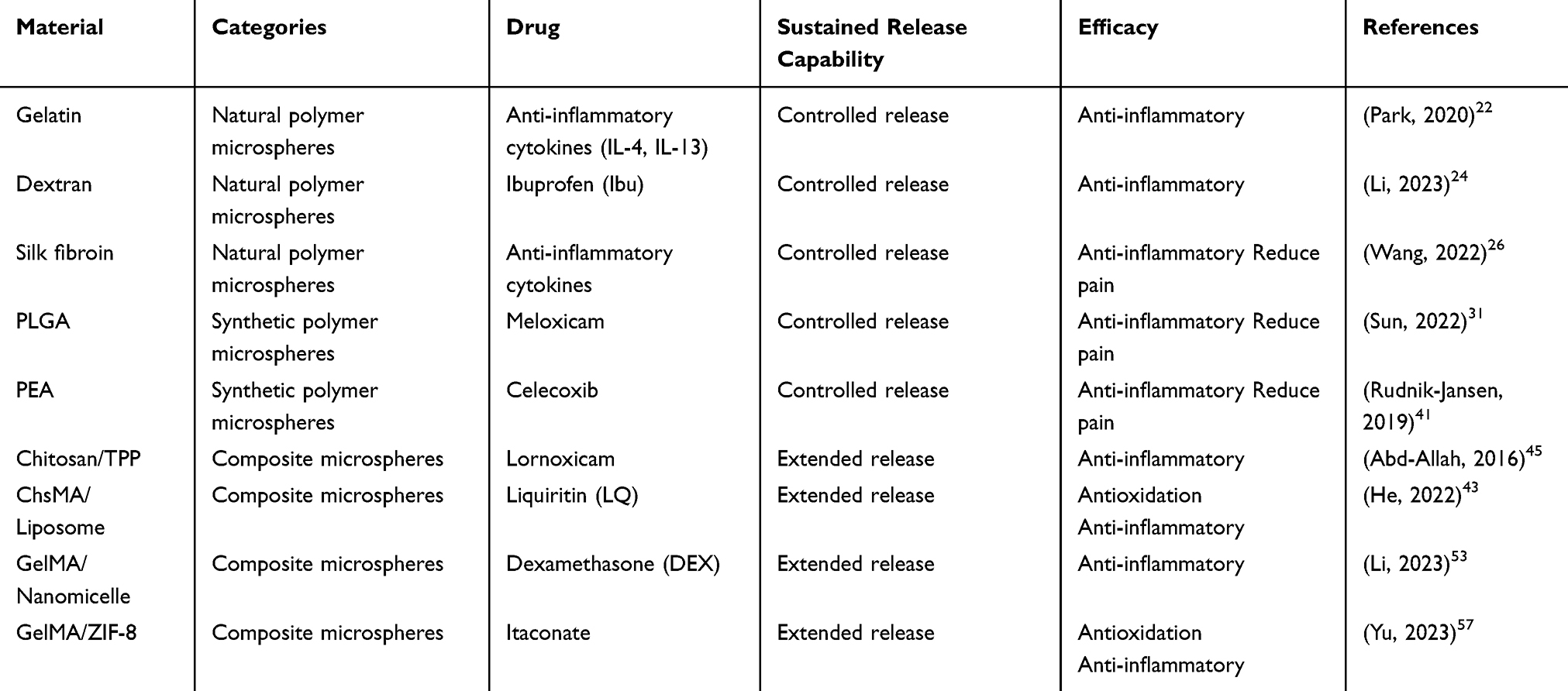 |
Table 1 The Sustained-Release Capabilities of Different Microspheres |
Enhancing Drug Permeation and Targeted Drug Delivery
The penetration capability required by microspheres during drug delivery arises from the complex structure of cartilage tissue. Most chondrocytes are located deep within the tissue.53 Cartilage is structurally and functionally divided into four distinct layers: superficial, middle, deep, and calcified. The superficial layer is characterized by densely packed collagen fibers and serves as a protective barrier. For microspheres to effectively reach and act within the cartilage, they must first penetrate this surface layer. The middle layer, located just beneath the surface, contains more loosely arranged collagen fibers and is rich in chondrocytes, making it a common target site for therapeutic microspheres. In contrast, the deep and calcified layers lie farther within the cartilage and are more difficult for drug-loaded microspheres to access due to their dense structure and limited permeability54 (Figure 2).
 |
Figure 2 The barrier-Penetration and cartilage-targeting functions of microspheres. Notes: This figure illustrates the process by which drug-loaded microspheres utilize their intrinsic charge interactions to penetrate the barrier formed by the cartilage extracellular matrix and subsequently target cartilage cells to exert their therapeutic effects, and the process by which microspheres containing type II collagen-targeting peptides can target collagen to achieve cartilage targeting by therapeutic microspheres. This figure was created with BioRender (https://biorender.com/). |
In addition to the anatomical location of chondrocytes, a major factor limiting the precise delivery of drug-loaded microspheres is the ECM that surrounds them, providing structural support and a conducive environment for cell survival. The ECM is composed of collagen fibers, highly negatively charged proteoglycans, and other macromolecules,55 forming a dense and complex network. This negatively charged matrix poses a significant barrier to the penetration of large macromolecules and negatively charged substances, thereby hindering the delivery of therapeutic agents to chondrocytes.56,57 Traditional drug delivery strategies often fail to overcome the ECM barrier, preventing effective targeting of chondrocytes and lesion sites. Moreover, such non-specific approaches may damage healthy tissue and cause undesirable side effects.58 Therefore, drug-loaded microspheres must be designed to penetrate the dense, negatively charged ECM and achieve targeted delivery to diseased cartilage regions, enabling precise and effective intra-articular therapy58,59 (Figure 2).
To effectively target chondrocytes, drug-loaded microspheres must first penetrate the various layers of cartilage. Upon intra-articular injection, microspheres encounter the superficial cartilage as the initial physical barrier. While small-sized microspheres can diffuse through the dense fibrous structure of the superficial cartilage due to their size and osmotic properties, their movement within the highly negatively charged extracellular ECM is often restricted.58 To overcome this challenge, surface modification with positively charged materials or loading the microspheres with positively charged drugs can enhance ECM penetration through electrostatic interactions between cations and the negatively charged ECM components.60
For instance, cationic liposomes—a class of positively charged nanocarriers—exhibit excellent biocompatibility and drug encapsulation efficiency. Their positive charge facilitates adhesion to the ECM, followed by gradual inward diffusion via electrostatic attraction.61 One example is GM-Lipo@ARB, a microsphere system composed of arbutin-loaded cationic liposomes encapsulated within methacryloyl gelatin.62 The cationic liposome coating enables the microspheres to effectively traverse the ECM and target cartilage tissue, while also protecting the liposomes from degradation, thereby allowing arbutin to exert its therapeutic effect in chondrocytes.42 Another system combines kartogenin (KGN)-loaded poly(amidoamine) (PAMAM) dendrimers with hyaluronic acid methacrylate (HAMA) to form drug delivery microspheres. These microspheres are further surface-modified with polylysine (PLL), creating a dual-positively charged system. The enhanced surface charge promotes targeted interaction with the ECM, facilitating gradual accumulation at the lesion site and contributing to effective osteoarthritis therapy through electrostatic targeting58 (Figure 2).
Once drug-loaded microspheres successfully penetrate into the middle or deeper layers of cartilage through particle size advantages and surface charge interactions, targeting chondrocytes to enhance therapeutic efficacy becomes a critical step. To achieve this, hydrogel microspheres can be functionalized with the cartilage-targeting peptide WYRGRL,55,63 which specifically binds to type II collagen—a major component of the cartilage matrix. These WYRGRL-modified microspheres exhibit selective binding to chondrocytes, enabling site-specific delivery of therapeutic agents such as dexamethasone (Dex) and kartogenin (KGN), thereby enhancing treatment efficacy at diseased cartilage sites.64 In early-stage osteoarthritis, damaged cartilage expresses type I collagen (Col1) differently from healthy tissue. Based on antigen-antibody interactions, hydrogel microspheres modified with anti-Col1 antibodies can more precisely target damaged chondrocytes, offering a promising strategy for localized cartilage repair65 (Figure 2).
While targeted microspheres in osteoarthritis therapy are commonly designed to deliver anti-inflammatory drugs to chondrocytes, they can also be engineered to target immune cells such as macrophages.66,67 For example, embedding dexamethasone-loaded folic acid–polyethylene glycol–polypropylene sulfide (FA–PEG–PPS) block copolymers into gelatin methacryloyl (GelMA) microspheres results in a system (GelMA@FPPD) capable of selectively targeting M1 macrophages. This approach enables sustained release of dexamethasone, suppresses local inflammatory responses, and ultimately delays the progression of osteoarthritis by modulating the joint microenvironment.44
Stimuli-Responsive Drug Release
The targeted microspheres discussed above enable site-specific drug delivery; however, optimizing therapeutic efficacy requires not only precise localization but also appropriate timing of drug release. Stimuli-responsive microspheres fulfill both requirements by responding to environmental cues—such as mechanical stress, inflammation, or hypoxia—at disease sites,68 triggering structural changes that lead to controlled drug release. This mechanism enables both spatially targeted and temporally regulated drug delivery.69
Stimuli-responsive hydrogel microspheres have been widely applied in drug delivery across various diseases,70 with diverse triggering stimuli. In OA, the inflammatory microenvironment plays a key role in disease progression, primarily through the upregulation of matrix-degrading enzymes such as matrix metalloproteinases (MMPs). Microspheres incorporating enzyme-sensitive peptides can respond to this enzymatic activity; the peptide linkers are cleaved in the inflammatory environment, releasing the encapsulated drugs71,72 (Figure 3).
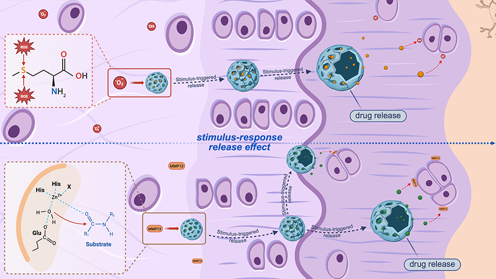 |
Figure 3 Stimuli-responsive drug release by microspheres. Notes: This figure contains stimulus-responsive microspheres responding to the presence of ROS as well as MMP13 in the OA microenvironment, interacting with the corresponding stimuli through their own specific targets after exposure to these triggers. As the microspheres degrade, they facilitate the sustained release of their encapsulated drug, ensuring localized therapeutic effects and improved treatment outcomes. This figure was created with BioRender (https://biorender.com/). |
For example, gelatin microspheres crosslinked with genipin degrade in response to catabolic enzymes present in inflamed joints. Similarly, gelatin microspheres exposed to collagenase in vitro mimic this enzymatic degradation.23 To specifically target MMP13—a key enzyme in OA pathology—bilayer hydrogel microspheres incorporating MMP13-sensitive peptides were developed. These microspheres release celecoxib-loaded liposomes from the outer layer in response to inflammatory degradation, while the inner core gradually releases chondroitin sulfate, thereby providing both anti-inflammatory and cartilage-repair effects in a sequential manner.73 Oxidative stress is another critical component of the OA inflammatory environment.74 Reactive oxygen species (ROS)-responsive microspheres have been developed to release antioxidant drugs in response to elevated ROS levels.74 For instance, KGN and dexamethasone were electrostatically loaded into a ROS-responsive copolymer integrated with GelMA hydrogel. The ROS-sensitive bonds within the system are cleaved by excess ROS, triggering the release of the therapeutic agents to reduce oxidative damage and inflammation.64 Furthermore, dual-responsive microspheres that combine MMP- and ROS-sensitivity have been designed to release dihydromyricetin (DMY) in a stimulus-specific and time-sequenced manner, enhancing therapeutic potential through synergistic response mechanisms75 (Figure 3).
It is important to note that while endogenous stimuli-responsive microspheres can autonomously respond to specific microenvironmental cues in OA cartilage, their drug release kinetics heavily depend on local pathological conditions, which are difficult to control. In contrast, exogenous stimuli such as near-infrared (NIR) light can offer greater control over drug release timing.76 By incorporating photothermal conversion agents into hydrogel microspheres, NIR irradiation can induce localized heating, modulating microsphere volume and triggering controlled drug release. These photothermally responsive systems allow precise spatial and temporal control over drug delivery, offering advantages over conventional stimuli-responsive strategies.76,77
Self-Therapeutic Effects of Microspheres
In research related to osteoarthritis treatment, microspheres are primarily used as drug delivery carriers, facilitating controlled release, targeting, and other auxiliary functions for the encapsulated drugs.9 As research progresses, it has become evident that microsphere systems can also exert therapeutic effects beyond their role as carriers for loaded drugs. As microspheres degrade in vivo, their constituent materials can exert therapeutic effects alongside the encapsulated drugs. In some cases, microspheres can provide therapeutic benefits even in the absence of loaded drugs.78 These intrinsic effects may arise not only from the pharmacological properties of the microsphere components but also from their physical characteristics and degradation products. For example, during the drug delivery process, certain microspheres can exert a lubricating effect, contributing to joint protection and symptom relief79 (Figure 4).
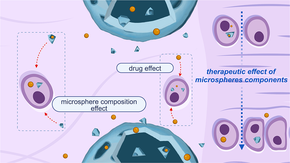 |
Figure 4 Therapeutic Effects of Microsphere Components. Notes: This figure illustrates how the intrinsic components of microspheres, upon degradation, can exert therapeutic effects through their inherent drug properties. This figure was created with BioRender (https://biorender.com/). |
Therapeutic Effects of Microsphere Components
Excessive ROS produced during OA can induce oxidative stress damage to the cartilage matrix through mechanisms such as the activation of inflammatory signaling pathways, protein modifications, and DNA damage, thus exacerbating OA progression.80,81 As a result, increasing evidence suggests that ROS can serve as a therapeutic target for osteoarthritis treatment.82 In microsphere systems designed to alleviate OA-related oxidative stress, antioxidant drugs are typically loaded to counteract ROS, while the ROS-responsive properties of these microspheres can enhance the therapeutic efficacy of the loaded drugs.82 However, a novel approach proposes that microspheres can exert therapeutic effects independent of any drug loading. Instead, these microspheres utilize their inherent material properties to combat ROS-induced oxidative stress and thereby treat osteoarthritis.
Studies have demonstrated that unloaded polyphenylene sulfide microspheres (PPS-MS), utilizing their intrinsic PPS component, can scavenge ROS and provide antioxidant effects, protecting chondrocytes from toxicity.78 When applied in an OA model, PPS-MS not only counteract high levels of ROS but also reduce matrix metalloproteinase (MMP) activity, thereby achieving notable anti-inflammatory and antioxidant effects.83 Similarly, another study developed drug-loaded chondroitin sulfate microspheres, which harness antioxidant and anti-inflammatory effects to treat OA.84,85 In this research, chondroitin sulfate, a natural compound with both anti-inflammatory and antioxidant properties, was used as the base material for the drug delivery microspheres.86 Upon degradation in the joint cavity, the released chondroitin sulfate molecules reduce ROS levels and inhibit ROS-related inflammatory pathways, thereby exerting antioxidant effects.37 Additionally, the microsphere’s incorporation of glycyrrhizin enables a dual antioxidant action, ultimately providing a therapeutic effect against OA.37,87 Thus, microspheres that rely on their own components for therapeutic action not only support intra-articular drug delivery but can also serve as an independent treatment method for alleviating osteoarthritis83 (Figure 4).
Treatment of OA Through Self-Lubricating Action
Pain relief remains a central focus in the treatment of OA,88 with the primary cause of pain being the increased friction resulting from the damage to articular cartilage, which disrupts the joint’s water-based lubrication system.89 Consequently, developing effective lubricants to restore joint lubrication is a key therapeutic approach.90 Drug delivery microspheres, with their inherent lubricating properties, can help alleviate OA symptoms and offer a promising treatment strategy.91
The lipid layer on the cartilage surface can interact synergistically with hyaluronic acid (HA) to form a hydrated lubrication layer.92 However, as OA progresses, the lipid layer may be damaged, and a lack of lipids in the synovial fluid can impair the formation of this hydrated lubrication layer.93 To address this, liposomes injected into the joint can serve as supplements to the lipid layer, effectively enhancing lubrication. One study incorporated rapamycin (RAPA)-loaded liposomes into HA microspheres (RAPA@Lipo@HM). These microspheres can continuously release liposomes through a rolling mechanism, forming a self-renewing hydrated lubrication layer on the surface of the HA-based microspheres, providing stable lubrication.94 Additionally, the cationic liposomes’ targeting ability and the chondroprotective effect of rapamycin improve the therapeutic efficacy of RAPA@Lipo@HMs for OA.95,96 Furthermore, hybrid exosomes, formed through membrane fusion between exosomes and liposomes, can be combined with HA-based hydrogel microspheres. These hybrid exosomes, along with the lubrication properties of HA microspheres, can offer enhanced joint lubrication.97
In addition to liposome-containing microspheres, those made from other materials can also exhibit lubrication effects similar to liposomes. For example, silk fibroin (silk) hydrogel microspheres, prepared using diethylene glycol dimethyl ether (BDDE), possess excellent hydrophilicity and a smooth surface.26 Upon injection into the joint cavity, they can reduce OA-related pain by providing a lubricating effect through ball-bearing and cartilage surface interactions.98,99 Additionally, PLGA porous microspheres containing nano-fat (NF) have a smooth, uniform surface and form a biological bearing by combining with oil, reducing friction between the joint surfaces and enhancing lubrication.100
Microspheres in Cartilage Tissue Engineering for OA Treatment
Articular cartilage has limited self-repair capacity due to its avascular and aneural nature, as well as the inherently low regenerative ability of chondrocytes.101 Cartilage damage caused by inflammation or trauma cannot be effectively treated with specific pharmacological interventions.102 While joint replacement surgery remains a treatment option for end-stage osteoarthritis, it is often unacceptable to patients seeking a higher quality of life.103 In recent years, tissue engineering has made significant advancements in addressing cartilage repair, a key therapeutic target in osteoarthritis management.104,105
Cartilage tissue engineering typically involves three components: stem cells, scaffolds for cell growth, and growth-stimulating factors.106 The primary strategy involves combining autologous chondrocytes or mesenchymal stem cells (MSCs) with growth scaffolds to generate cartilage tissue for repairing damaged areas.107 Microspheres play a versatile role in this process, functioning as scaffolds for cell growth and differentiation, as well as carriers for bioactive materials such as cells and growth factors. As a result, they have become a critical component of cartilage tissue engineering.102
Microspheres as Cell Carriers and Growth Scaffolds
Microspheres as Cell Carriers
Stem cell therapy has been a major focus in cartilage repair for OA patients,108 typically involving the intra-articular injection of MSCs to alleviate pain and slow down cartilage degeneration.109,110 However, the shear forces encountered during the injection process, as well as the complex joint environment, can lead to MSC damage or even cell death, reducing the number of viable cells and ultimately compromising the therapeutic efficacy.111 Therefore, a carrier is needed that can both protect MSCs and facilitate high cell loading.112
Hydrogel microspheres can effectively encapsulate cells, shielding them from shear forces and preventing exposure to unfavorable conditions, thereby protecting the cells.113 Due to their high porosity and large surface area, microspheres provide abundant space for cell attachment, enabling high cell loading. Porous microspheres made from a combination of chitosan and PLGA, for example, can enhance cell adhesion through their inherent electrostatic properties. These microspheres not only allow for high MSC loading but also offer good mechanical properties to protect the cells from damage114 (Figure 5).
 |
Figure 5 The Role of Microspheres as Cell Carriers and Scaffolds for Cell Growth and Differentiation. Notes: This figure illustrates how microspheres function as both cell carriers and scaffolds for cell growth, providing a suitable environment for chondrocyte differentiation both in vitro and in vivo. This figure was created with BioRender. |
Moreover, microspheres fabricated with specialized techniques can improve cell proliferation and migration while enhancing cell delivery capacity.114 Gelatin methacryloyl (GelMA) hydrogel microspheres, for instance, possess a porous structure that provides an optimal environment for cell adhesion and proliferation, thus improving the survival and engraftment rates of loaded cells.115 Additionally, microspheres made from freeze-gelation techniques, which impart mechanical stability, ensure that the microspheres remain compressible during injection, thus preserving the viability of the cells they carry.116
Microspheres as Cell Growth Scaffolds
After the successful injection of cells into the joint cavity, it is essential to ensure that the cells are safely and accurately delivered to the damaged cartilage area. Janus microspheres, which contain iron oxide magnetic nanoparticles (IONPs) MSCs, not only encapsulate the cells but also utilize the IONPs for targeted cartilage repair through electromagnetic guidance.117 Achieving optimal therapeutic outcomes requires more than just delivering the cells; it is crucial to provide a suitable environment for cell growth and differentiation once they reach the site of injury.118 Thus, microspheres used for cell delivery also serve as essential cell growth scaffolds.119
In certain cases, cells intended for cartilage tissue engineering need to proliferate in vitro before implantation, but during this culture process, dedifferentiation may occur.120 To address this issue, injectable cryogel microspheres (ICMs) provide a three-dimensional (3D) environment resembling the extracellular matrix (ECM) and incorporate gelatin RGD sequences for cell adhesion. This structure offers a supportive scaffold that maintains the chondrocyte phenotype, facilitating the growth and differentiation of cartilage cells.116
In other instances, the cells need to proliferate after implantation. Once the microspheres carrying the cells reach the damaged area, they act as scaffolds that support the cells, which is equally critical for achieving therapeutic success.121 Alginate-gelatin microspheres, combining the ECM-like properties of alginate with the cell-adhesive and proliferative qualities of gelatin, provide an enhanced scaffold for adipose-derived stem cells at the injury site. This dual-functional scaffold also addresses the challenges of cell loss due to contact inhibition and dedifferentiation.122
As growth scaffolds, microspheres not only simulate the ECM to support cell growth but also promote cartilage repair by recruiting cells and enhancing their adhesion to the microsphere surface.114 For example, hydrogel microspheres modified with acetaldehyde and carrying platelet-derived growth factor-BB (PDGF-BB) can adhere to the cartilage surface, where they recruit surrounding stem cells and Sn-chondrocytes. These microspheres further mimic the ECM in situ, providing a supportive environment for cell growth and promoting cartilage regeneration123 (Figure 5).
Microspheres Providing Active Factors to Promote Chondrocyte Differentiation
Recent studies have increasingly focused on the combination of stem cells, growth factors, and scaffolds in cartilage tissue engineering.124 Microspheres, serving as both cell carriers and scaffolds, play a critical role in this field. In some cases, the mere addition of stem cells is insufficient for cartilage regeneration; it is essential to induce the differentiation of stem cells into chondrocytes for effective cartilage repair.125 Therefore, incorporating bioactive factors that promote cell differentiation into the microsphere-based delivery systems becomes crucial.
Transforming growth factor-beta (TGF-β) has long been recognized as a key cytokine involved in cartilage differentiation.126 By developing microspheres that continuously release TGF-β3, an optimal environment for MSC differentiation into chondrocytes can be provided, thereby enhancing tissue repair.127 Additionally, PLGA-P188-PLGA-based microspheres not only provide a sustained release of TGF-β3 but also leverage their inherent properties to optimize TGF-β3 release, thereby stimulating cartilage regeneration more effectively.127 KGN as a small-molecule compound, can promote cartilage formation by activating the SOX9 transcription factor, mimicking the effects of TGF-β.127 Microspheres loaded with KGN and MSCs release the cells in response to the OA environment. As the microspheres degrade, KGN is concurrently released, inducing MSCs to differentiate into chondrocytes and promoting tissue regeneration.114
In cartilage tissue engineering, directly adding growth factors is the primary method for inducing chondrocyte differentiation. However, another approach involves indirectly regulating the expression of active factors by altering the environmental conditions.128 Hypoxic conditions, through hypoxia-inducible factors (HIFs), can influence stem cell differentiation and chondrocyte maturation.129 For example, pre-treating gelatin methacrylate microspheres loaded with chondroprogenitor cells under hypoxic conditions results in superior cartilage formation compared to those cultured under normoxic conditions.130 Moreover, dimethyloxalylglycine (DMOG), which inhibits HIF-1α degradation, can be used to simulate a hypoxic environment.131 By combining DMOG with parathyroid hormone-related protein (PTHrP), which regulates chondrocyte differentiation, and loading them into PLGA microspheres, a promising three-dimensional culture system is created. This system not only enhances chondrocyte differentiation but also stabilizes their phenotype, offering a viable strategy for cartilage regeneration.132
Conclusion and Perspectives
In conclusion, this review systematically summarizes the multifunctional applications of hydrogel-based microspheres in OA therapy, highlighting their capacity for sustained drug release, targeted delivery, and regenerative cartilage repair. In the treatment of OA, pharmacological therapy remains the primary approach, with intra-articular drug delivery gaining attention due to its superior safety profile and therapeutic efficacy.7 However, to optimize intra-articular drug delivery, it is essential to ensure that the drug reaches the damaged site accurately and has an extended retention time within the joint. Hydrogel microspheres stand out among the various intra-articular drug delivery systems due to their excellent performance. These microspheres not only offer the advantages of hydrogels, such as good drug protection and biocompatibility, but also feature higher porosity and greater structural stability.64
This review highlights the synthesis of multifunctional microspheres as drug carriers for osteoarthritis treatment, with a focus on their roles in controlled drug release, targeted delivery to cartilage, and intrinsic therapeutic effects. Due to their structural characteristics, microspheres have a higher drug-loading capacity and can maintain a high drug concentration. More importantly, microspheres can control drug release by altering their structure, thus addressing the issue of rapid drug release and ensuring prolonged drug concentrations. This sustained-release capability is a fundamental requirement for drug delivery systems. In this paper, microspheres for drug delivery are categorized based on their materials, and their effectiveness in drug release is analyzed. By comparison, composite microspheres—due to their complex composition—demonstrate superior sustained-release effects over single-material microspheres. In addition to the basic function of sustained release, microspheres can enhance drug penetration during delivery by leveraging electrostatic interactions, overcoming the dense structure and charge barriers around cartilage. Moreover, microspheres functionalized with specific peptides, such as those targeting type II collagen, can enable cartilage-targeted delivery. Furthermore, responsive microspheres can undergo structural changes in response to inflammatory or hypoxic conditions, thereby achieving controlled drug release based on environmental stimuli. Beyond drug delivery, microspheres can also contribute to therapeutic effects through their material composition. Some microspheres, composed of special materials, can create lubricating layers or act as biological bearings within the joint, providing joint lubrication and alleviating OA-related pain.
In addition to their applications in drug delivery, this review also summarizes the use of microspheres as carriers and scaffolds in cartilage tissue engineering. While drug delivery has been the primary application of microspheres in OA therapy, the irreversible destruction of articular cartilage in advanced OA cannot be reversed by drugs alone. Thus, microspheres designed for cartilage repair have emerged as promising alternatives. Stem cell therapy, a core component of cartilage tissue engineering, relies on microspheres to protect and deliver cells to the damaged areas, where they provide an optimal environment for cell growth. Moreover, when stem cells need to proliferate before implantation, microspheres can offer support during in vitro culture. However, in some cases, transplanted stem cells may fail to differentiate into chondrocytes and complete the repair. To address this, microspheres can carry bioactive factors that promote stem cell differentiation into chondrocytes, enhancing the regenerative process.
Current research indicates that microspheres are highly promising materials for OA treatment. Building on hydrogel technology, microspheres have been further optimized to improve their drug delivery and release capabilities. However, several challenges remain. Despite the advantages of sustained release, some microsphere materials still exhibit poor stability, leading to insufficient control over drug release rates. Both rapid and slow-release rates can negatively impact the therapeutic effect. Additionally, biocompatibility remains a concern, as certain microsphere materials may trigger foreign body reactions, affecting patient outcomes. Although an increasing number of stimuli-responsive and targeted microspheres for osteoarthritis treatment have been developed, there remains substantial room for improvement in their functionality. Future research should prioritize integrating environment-responsive delivery with cartilage-targeting mechanisms to improve therapeutic precision and durability in vivo.
Research on microspheres for OA treatment is ongoing, and their applications extend beyond drug delivery and cartilage tissue engineering. Recent studies have demonstrated that stress-electrically coupled hydrogel microspheres can alleviate cartilage damage and promote regeneration by reducing stress-electrical conversion losses under mechanical stimulation.133,134 Additionally, fluorescently labeled microspheres have been developed to integrate imaging and therapy, enabling real-time monitoring of cartilage repair through fluorescence signals. However, large-scale production of microspheres remains a significant challenge, leading to high manufacturing costs and limiting their clinical application. Moreover, few clinical trials or translational studies have been conducted to date.135 Therefore, further research is needed to improve the functionality, scalability, and clinical relevance of microsphere systems, ultimately enhancing their therapeutic potential and translational value in osteoarthritis treatment.
Acknowledgments
We sincerely thank all research participants for their outstanding contributions and collaboration in this study.
Author Contributions
All authors made a significant contribution to the work reported, whether that is in the conception, study design, execution, acquisition of data, analysis and interpretation, or in all these areas; took part in drafting, revising or critically reviewing the article; gave final approval of the version to be published; have agreed on the journal to which the article has been submitted; and agree to be accountable for all aspects of the work.
Funding
This work was supported by the Joint Fund Project General Support Program Project of Liaoning Provincial Department of Science and Technology (2023-MSLH-386) awarded to Yingliang Wei.
Disclosure
The authors declare that there are no potential conflicts of interest in this study.
References
1. Silverwood V, Blagojevic-Bucknall M, Jinks C, Jordan JL, Protheroe J, Jordan KP. Current evidence on risk factors for knee osteoarthritis in older adults: a systematic review and meta-analysis. Osteoarthritis Cartilage. 2015;23(4):507–515. doi:10.1016/j.joca.2014.11.019
2. Prieto-Alhambra D, Judge A, Javaid MK, Cooper C, Diez-Perez A, Arden NK. Incidence and risk factors for clinically diagnosed knee, Hip and hand osteoarthritis: influences of age, gender and osteoarthritis affecting other joints. Ann Rheum Dis. 2014;73(9):1659–1664. doi:10.1136/annrheumdis-2013-203355
3. Martel-Pelletier J, Barr AJ, Cicuttini FM, et al. Osteoarthritis. Nat Rev Dis Primers. 2016;2(1). doi:10.1038/nrdp.2016.72
4. Motta F, Barone E, Sica A, Selmi C. Inflammaging and Osteoarthritis. Clin Rev Allergy Immunol. 2023;64:222–238.
5. Fujii Y, Liu L, Yagasaki L, Inotsume M, Chiba T, Asahara H. Cartilage Homeostasis and Osteoarthritis. Int J Mol Sci. 2022;23(11):6316. doi:10.3390/ijms23116316
6. Hunter DJ, Bierma-Zeinstra S. Osteoarthritis. Lancet. 2019;393(10182):1745–1759. doi:10.1016/S0140-6736(19)30417-9
7. Rahimi M, Charmi G, Matyjaszewski K, Banquy X, Pietrasik J. Recent developments in natural and synthetic polymeric drug delivery systems used for the treatment of osteoarthritis. Acta Biomater. 2021;123:31–50. doi:10.1016/j.actbio.2021.01.003
8. Owen SG, Francis HW, Roberts MS. Disappearance kinetics of solutes from synovial fluid after intra-articular injection. Br J Clin Pharmacol. 1994;38(4):349–355. doi:10.1111/j.1365-2125.1994.tb04365.x
9. Zhou D, Zhou F, Sheng S, Wei Y, Chen X, Su J. TSC1 controls macrophage polarization to prevent inflammatory disease. Drug Discov Today. 2023;28:4696.
10. Ma L, Zheng X, Lin R, et al. Knee Osteoarthritis Therapy: recent Advances in Intra-Articular Drug Delivery Systems. Drug Des Devel Ther. 2022;16:1311–1347. doi:10.2147/DDDT.S357386
11. Mao L, Wu W, Wang M, et al. Targeted treatment for osteoarthritis: drugs and delivery system. Drug Deliv. 2021;28(1):1861–1876. doi:10.1080/10717544.2021.1971798
12. Ruan L, Su M, Qin X, et al. Progress in the application of sustained-release drug microspheres in tissue engineering. Mater Today Bio. 2022;16:100394.
13. He Z, Wang B, Hu C, Zhao J. An overview of hydrogel-based intra-articular drug delivery for the treatment of osteoarthritis. Colloids Surf B Biointerfaces. 2017;154:33–39. doi:10.1016/j.colsurfb.2017.03.003
14. Su J, Yu M, Wang H, Wei Y. Natural anti-inflammatory products for osteoarthritis: from molecular mechanism to drug delivery systems and clinical trials. Phytother Res. 2023;37(10):4321–4352. doi:10.1002/ptr.7935
15. Copp G, Robb KP, Viswanathan S. Culture-expanded mesenchymal stromal cell therapy: does it work in knee osteoarthritis? A pathway to clinical success. Cell Mol Immunol. 2023;20(6):626–650. doi:10.1038/s41423-023-01020-1
16. Xu X, Xu L, Xia J, Wen C, Liang Y, Zhang Y. Harnessing knee joint resident mesenchymal stem cells in cartilage tissue engineering. Acta Biomater. 2023;168:372–387. doi:10.1016/j.actbio.2023.07.024
17. Bai L, Zhang X, Han Z, Yang X, Hao Y. Injectable porous microspheres for articular cartilage regeneration through in situ stem cell recruitment and macrophage polarization. Acta Biomater. 2024;185:429–440. doi:10.1016/j.actbio.2024.07.007
18. Katz JN, Arant KR, Loeser RF. Diagnosis and Treatment of Hip and Knee Osteoarthritis: a Review. JAMA. 2021;325:568–578.
19. Maudens P, Jordan O, Allémann E. Recent advances in intra-articular drug delivery systems for osteoarthritis therapy. Drug Discov Today. 2018;23(10):1761–1775. doi:10.1016/j.drudis.2018.05.023
20. Jiang Q, Zhang S. Stimulus-Responsive Drug Delivery Nanoplatforms for Osteoarthritis Therapy. Small. 2023;19(23). doi:10.1002/smll.202206929
21. Kang ML, Im GI. Drug delivery systems for intra-articular treatment of osteoarthritis. Expert Opin Drug Deliv. 2014;11(2):269–282. doi:10.1517/17425247.2014.867325
22. Li Q, Chang B, Dong H, Liu X. Functional microspheres for tissue regeneration. Bioact Mater. 2023;25:485–499.
23. Park E, Hart ML, Rolauffs B, Stegemann JP, R TA. Bioresponsive microspheres for on-demand delivery of anti-inflammatory cytokines for articular cartilage repair. J Biomed Mater Res A. 2020;108(3):722–733. doi:10.1002/jbm.a.36852
24. Li Z, Feng X, Luo S, et al. High drug loading hydrophobic cross-linked dextran microspheres as novel drug delivery systems for the treatment of osteoarthritis. Asian J Pharm Sci. 2023;18:100830.
25. Rockwood DN, Preda RC, Yücel T, Wang X, Lovett ML, Kaplan DL. Materials fabrication from Bombyx mori silk fibroin. Nat Protoc. 2011;6(10):1612–1631. doi:10.1038/nprot.2011.379
26. Wang T, Li Y, Liu J, et al. Intraarticularly injectable silk hydrogel microspheres with enhanced mechanical and structural stability to attenuate osteoarthritis. Biomaterials. 2022;286:121611. doi:10.1016/j.biomaterials.2022.121611
27. Su Y, Zhang B, Sun R, et al. PLGA-based biodegradable microspheres in drug delivery: recent advances in research and application. Drug Deliv. 2021;28(1):1397–1418. doi:10.1080/10717544.2021.1938756
28. Li X, Wei Y, Wen K, Han Q, Ogino K, Ma G. Novel insights on the encapsulation mechanism of PLGA terminal groups on ropivacaine. Eur J Pharm Biopharm. 2021;160:143–151. doi:10.1016/j.ejpb.2021.01.015
29. Sun Z, Gu X, Hao T, et al. Intra-articular injection PLGA blends sustained-release microspheres loaded with meloxicam: preparation, optimization, evaluation in vitro and in vivo. Drug Deliv. 2022;29(1):3317–3327. doi:10.1080/10717544.2022.2144545
30. Lu YC, Evans CH, Grodzinsky AJ. Effects of short-term glucocorticoid treatment on changes in cartilage matrix degradation and chondrocyte gene expression induced by mechanical injury and inflammatory cytokines. Arthritis Res Ther. 2011;13(5). doi:10.1186/ar3456
31. Roach BL, Kelmendi-Doko A, Balutis EC, Marra KG, Ateshian GA, Hung CT. Dexamethasone Release from Within Engineered Cartilage as a Chondroprotective Strategy Against Interleukin-1α. Tissue Eng Part A. 2016;22(7–8):621–632. doi:10.1089/ten.tea.2016.0018
32. Stefani RM, Lee AJ, Tan AR, et al. Sustained low-dose dexamethasone delivery via a PLGA microsphere-embedded agarose implant for enhanced osteochondral repair. Acta Biomater. 2020;102:326–340. doi:10.1016/j.actbio.2019.11.052
33. Zhang X, Shi Y, Zhang Z, Yang Z, Huang G. Intra-articular delivery of tetramethylpyrazine microspheres with enhanced articular cavity retention for treating osteoarthritis. Asian J Pharm Sci. 2018;13:229–238.
34. Ho MJ, Jeong HT, Im SH, et al. Design and In Vivo Pharmacokinetic Evaluation of Triamcinolone Acetonide Microcrystals-Loaded PLGA Microsphere for Increased Drug Retention in Knees after Intra-Articular Injection. Pharmaceutics. 2019;11(8):419. doi:10.3390/pharmaceutics11080419
35. Janssen M, Timur UT, Woike N, et al. Celecoxib-loaded PEA microspheres as an auto regulatory drug-delivery system after intra-articular injection. J Control Release. 2016;244:30–40. doi:10.1016/j.jconrel.2016.11.003
36. Rudnik-Jansen I, Schrijver K, Woike N, et al. Intra-articular injection of triamcinolone acetonide releasing biomaterial microspheres inhibits pain and inflammation in an acute arthritis model. Drug Deliv. 2019;26(1):226–236. doi:10.1080/10717544.2019.1568625
37. He Y, Sun M, Wang J, et al. Chondroitin sulfate microspheres anchored with drug-loaded liposomes play a dual antioxidant role in the treatment of osteoarthritis. Acta Biomater. 2022;151:512–527. doi:10.1016/j.actbio.2022.07.052
38. Abd-Allah H, Kamel AO, Sammour OA. Injectable long acting chitosan/tripolyphosphate microspheres for the intra-articular delivery of lornoxicam: optimization and in vivo evaluationY. Fujii. Carbohydr Polym. 2016;149:263–273. doi:10.1016/j.carbpol.2016.04.096
39. Guimarães D, Cavaco-Paulo A, Nogueira E. Design of liposomes as drug delivery system for therapeutic applications. Int J Pharm. 2021;601:120571. doi:10.1016/j.ijpharm.2021.120571
40. Antimisiaris SG, Marazioti A, Kannavou M, et al. Overcoming barriers by local drug delivery with liposomes. Adv Drug Deliv Rev. 2021;174:53–86. doi:10.1016/j.addr.2021.01.019
41. Jeong ES, Son HA, Kim MK, et al. Fabrication of monodisperse liposomes-in-microgel hybrid microparticles in capillary-based microfluidic devices. Colloids Surf B Biointerfaces. 2014;123:339–344. doi:10.1016/j.colsurfb.2014.09.039
42. Jin J, Liu Y, Jiang C, et al. Arbutin-modified microspheres prevent osteoarthritis progression by mobilizing local anti-inflammatory and antioxidant responses. Mater Today Bio. 2022;16:100370.
43. Chen W, Schilperoort M, Cao Y, Shi J, Tabas I, Tao W. Macrophage-targeted nanomedicine for the diagnosis and treatment of atherosclerosis. Nat Rev Cardiol. 2022;19(4):228–249. doi:10.1038/s41569-021-00629-x
44. Li X, Li X, Yang J, et al. In Situ Sustained Macrophage-Targeted Nanomicelle-Hydrogel Microspheres for Inhibiting Osteoarthritis. Research. 2023;6:131.
45. He L, Liu Y, Liu J, et al. Core-shell noble-metal@metal-organic-framework nanoparticles with highly selective sensing property. Angew Chem Int Ed Engl. 2013;52(13):3741–3745. doi:10.1002/anie.201209903
46. He L, Pang K, Liu W, et al. Core–shell noble-metal@zeolitic-imidazolate-framework nanocarriers with high cancer treatment efficiency in vitro. J Mater Chem B. 2019;7(7):1050–1055. doi:10.1039/C8TB03318H
47. Cai W, Wang J, Chu C, Chen W, Wu C, Liu G. Metal-Organic Framework-Based Stimuli-Responsive Systems for Drug Delivery. Adv Sci (Weinh). 2019;6:1801526.
48. Yu H, Ren P, Pan X, et al. Intracellular Delivery of Itaconate by Metal-Organic Framework-Anchored Hydrogel Microspheres for Osteoarthritis Therapy. Pharmaceutics. 2023;15(3):724. doi:10.3390/pharmaceutics15030724
49. Larrañaga A, Isa ILM, Patil V, et al. Antioxidant functionalized polymer capsules to prevent oxidative stress. Acta Biomater. 2018;67:21–31. doi:10.1016/j.actbio.2017.12.014
50. Natarajan V, Madhan B, Tiku ML. Intra-Articular Injections of Polyphenols Protect Articular Cartilage from Inflammation-Induced Degradation: suggesting a Potential Role in Cartilage Therapeutics. PLoS One. 2015;10(6):e0127165. doi:10.1371/journal.pone.0127165
51. Zhao F, Lei B, Li X, et al. Promoting in vivo early angiogenesis with sub-micrometer strontium-contained bioactive microspheres through modulating macrophage phenotypes. Biomaterials. 2018;178:36–47. doi:10.1016/j.biomaterials.2018.06.004
52. Chen Y, Xu W, Shafiq M, et al. Injectable nanofiber microspheres modified with metal phenolic networks for effective osteoarthritis treatment. Acta Biomater. 2023;157:593–608. doi:10.1016/j.actbio.2022.11.040
53. Nasiri N, Hosseini S, Alini M, Khademhosseini A, Eslaminejad MB. Targeted cell delivery for articular cartilage regeneration and osteoarthritis treatment. Drug Discov Today. 2019;24(11):2212–2224. doi:10.1016/j.drudis.2019.07.010
54. Krishnan Y, Grodzinsky AJ. Cartilage diseases. Matrix Biol. 2018;71–72:51–69. doi:10.1016/j.matbio.2018.05.005
55. Hynes RO. The extracellular matrix: not just pretty fibrils. Science. 2009;326(5957):1216–1219. doi:10.1126/science.1176009
56. Theocharis AD, Skandalis SS, Gialeli C, Karamanos NK. Extracellular matrix structure. Adv Drug Deliv Rev. 2016;97:4–27.
57. Armiento AR, Stoddart MJ, Alini M, Eglin D. Biomaterials for articular cartilage tissue engineering: learning from biology. Acta Biomater. 2018;65:1–20.
58. Lin J, Chen L, Yang J, et al. Injectable Double Positively Charged Hydrogel Microspheres for Targeting-Penetration-Phagocytosis. Small. 2022;18(40). doi:10.1002/smll.202202156
59. Chen H, Qin Z, Zhao J, et al. Cartilage-targeting and dual MMP-13/pH responsive theranostic nanoprobes for osteoarthritis imaging and precision therapy. Biomaterials. 2019;225:119520. doi:10.1016/j.biomaterials.2019.119520
60. Geiger BC, Wang S, Padera Jr RF, Grodzinsky AJ, Hammond PT. Cartilage-penetrating nanocarriers improve delivery and efficacy of growth factor treatment of osteoarthritis. Sci Transl Med. 2018;10(469). doi:10.1126/scitranslmed.aat8800
61. Chen CH, Kuo SM, Tien YC, Shen PC, Kuo YW, Huang HH. Steady Augmentation of Anti-Osteoarthritic Actions of Rapamycin by Liposome-Encapsulation in Collaboration with Low-Intensity Pulsed Ultrasound. Int J Nanomed. 2020;1:3771–3790.
62. Kim YB, Zhao KT, Thompson DB, Liu DR. An anionic human protein mediates cationic liposome delivery of genome editing proteins into mammalian cells. Nat Commun. 2019;10:2905.
63. Xue S, Zhou X, Sang W, et al. Cartilage-targeting peptide-modified dual-drug delivery nanoplatform with NIR laser response for osteoarthritis therapy. Bioact Mater. 2021;6:2372–2389.
64. Yu H, Huang C, Kong X, et al. Nanoarchitectonics of Cartilage-Targeting Hydrogel Microspheres with Reactive Oxygen Species Responsiveness for the Repair of Osteoarthritis. ACS Appl Mater Interfaces. 2022;14:40711–40723.
65. He X, He S, Xiang G, et al. Precise Lubrication and Protection of Cartilage Damage by Targeting Hydrogel Microsphere. Adv Mater. 2024;36:2405943.
66. Kerneur C, Cano CE, Olive D. Major pathways involved in macrophage polarization in cancer. Front Immunol. 2022;13. doi:10.3389/fimmu.2022.1026954
67. Zhu L, Yang T, Li L, et al. TSC1 controls macrophage polarization to prevent inflammatory disease. Nat Commun. 2014;5(1). doi:10.1038/ncomms5696
68. Yu H, Gao R, Liu Y, Fu L, Zhou J, Li L. Stimulus-Responsive Hydrogels as Drug Delivery Systems for Inflammation Targeted Therapy. Adv Sci. 2024;11:2306152.
69. Yao SY, Yue YX, Ying AK, et al. An Antitumor Dual-Responsive Host-Guest Supramolecular Polymer Based on Hypoxia-Cleavable Azocalix[4]arene. Angew Chem Int Ed Engl. 2023;62(2). doi:10.1002/anie.202213578
70. Li S, Ma R, Hu XY, et al. Drug in Drug: a Host-Guest Formulation of Azocalixarene with Hydroxychloroquine for Synergistic Anti-Inflammation. Adv Mater. 2022;34:e202213578.
71. Ribeiro JS, Bordini EAF, Ferreira JA, et al. Injectable MMP-Responsive Nanotube-Modified Gelatin Hydrogel for Dental Infection Ablation. ACS Appl Mater Interfaces. 2020;12(14):16006–16017. doi:10.1021/acsami.9b22964
72. Song L, Chi J, Li Z, et al. An inflammation-responsive double-layer microneedle patch for recurrent atopic dermatitis therapy. Int J Pharm. 2023;643:123215. doi:10.1016/j.ijpharm.2023.123215
73. Miao K, Zhou Y, He X, et al. Microenvironment-responsive bilayer hydrogel microspheres with gelatin-shell for osteoarthritis treatment. Int J Biol Macromol. 2024;261:129862. doi:10.1016/j.ijbiomac.2024.129862
74. Riegger J, Schoppa A, Ruths L, Haffner-Luntzer M, Ignatius A. Oxidative stress as a key modulator of cell fate decision in osteoarthritis and osteoporosis: a narrative review. Cell Mol Biol Lett. 2023;28:76.
75. Xia X, Liu Y, Lu Y, et al. Retuning Mitochondrial Apoptosis/Mitophagy Balance via SIRT3-Energized and Microenvironment-Modulated Hydrogel Microspheres to Impede Osteoarthritis. Adv Healthc Mater. 2023;12:2302475.
76. Gong Z, Chen L, Zhou X, et al. MXene-Based Photothermal-Responsive Injectable Hydrogel Microsphere Modulates Physicochemical Microenvironment to Alleviate Osteoarthritis. Smart Med. 2025;4(2). doi:10.1002/smmd.70006
77. Chen F, Wang W, Zhao H, et al. MXene-Based Cartilage-Adhesive Microspheres for Photothermal-Controlled Hydrophobic Drug Release and Mesenchymal Stem Cell Delivery in Osteoarthritis. ACS Nano. 2025;19:1.
78. Gupta MK, Martin JR, Werfel TA, Shen T, Page JM, Duvall CL. Cell protective, ABC triblock polymer-based thermoresponsive hydrogels with ROS-triggered degradation and drug release. J Am Chem Soc. 2014;136(42):14896–14902. doi:10.1021/ja507626y
79. Yao Y, Wei G, Deng L, Cui W. Visualizable and Lubricating Hydrogel Microspheres Via NanoPOSS for Cartilage Regeneration. Adv Sci. 2023;10:2207438.
80. Henrotin Y, Kurz B, Aigner T. Oxygen and reactive oxygen species in cartilage degradation. Osteoarthritis Cartilage. 2005;13(8):643–654. doi:10.1016/j.joca.2005.04.002
81. Yudoh K, v TN, Nakamura H, Hongo-Masuko K, Kato T, Nishioka K. Potential involvement of oxidative stress in cartilage senescence and development of osteoarthritis: oxidative stress induces chondrocyte telomere instability and downregulation of chondrocyte function. Arthritis Res Ther. 2005;7(2). doi:10.1186/ar1499
82. Arra M, Swarnkar G, Ke K, et al. LDHA-mediated ROS generation in chondrocytes is a potential therapeutic target for osteoarthritis. Nat Commun. 2020;11(1). doi:10.1038/s41467-020-17242-0
83. O’Grady KP, Kavanaugh TE, Cho H, et al. Drug-Free ROS Sponge Polymeric Microspheres Reduce Tissue Damage from Ischemic and Mechanical Injury. ACS Biomater Sci Eng. 2018;4(4):1251–1264. doi:10.1021/acsbiomaterials.6b00804
84. Corradetti B, Taraballi F, Minardi S, et al. Chondroitin Sulfate Immobilized on a Biomimetic Scaffold Modulates Inflammation While Driving Chondrogenesis. Stem Cells Transl Med. 2016;5(5):670–682. doi:10.5966/sctm.2015-0233
85. Lee JY, Lee SH, Kim HJ, et al. The preventive inhibition of chondroitin sulfate against the CCl4-induced oxidative stress of subcellular level. Arch Pharm Res. 2004;27(3):340–345. doi:10.1007/BF02980070
86. Cömert Kılıç S. Does glucosamine, chondroitin sulfate, and methylsulfonylmethane supplementation improve the outcome of temporomandibular joint osteoarthritis management with arthrocentesis plus intraarticular hyaluronic acid injection. A randomized clinical trial. J Craniomaxillofac Surg. 2021;49(8):711–718. doi:10.1016/j.jcms.2021.02.012
87. Zhai KF, Duan H, Cui CY, et al. Liquiritin from Glycyrrhiza uralensis Attenuating Rheumatoid Arthritis via Reducing Inflammation, Suppressing Angiogenesis, and Inhibiting MAPK Signaling Pathway. J Agric Food Chem. 2019;67(10):2856–2864. doi:10.1021/acs.jafc.9b00185
88. Sakata R, Reddi AH. Platelet-Rich Plasma Modulates Actions on Articular Cartilage Lubrication and Regeneration. Tissue Eng Part B Rev. 2016;22(5):408–419. doi:10.1089/ten.teb.2015.0534
89. Cooper BG, Catalina B, Nazarian A, Snyder BD, Grinstaff MW. Active agents, biomaterials, and technologies to improve biolubrication and strengthen soft tissues. Biomaterials. 2018;181:210–226. doi:10.1016/j.biomaterials.2018.07.040
90. Morgese G, Benetti EM, Zenobi-Wong M. Molecularly Engineered Biolubricants for Articular Cartilage. Adv Healthc Mater. 2018;7(16). doi:10.1002/adhm.201701463
91. Yuan H, Mears LLE, Wang Y, et al. Lubricants for osteoarthritis treatment: from natural to bioinspired and alternative strategies. Adv Colloid Interface Sci. 2023;311:102814. doi:10.1016/j.cis.2022.102814
92. Seror J, Zhu L, Goldberg R, Day AJ, Klein J. Supramolecular synergy in the boundary lubrication of synovial joints. Nat Commun. 2015;6(1). doi:10.1038/ncomms7497
93. Sorkin R, Kampf N, Zhu L, Klein J. Hydration lubrication and shear-induced self-healing of lipid bilayer boundary lubricants in phosphatidylcholine dispersions. Soft Matter. 2016;12(10):2773–2784. doi:10.1039/C5SM02475G
94. Lei Y, Wang Y, Shen J, et al. Injectable hydrogel microspheres with self-renewable hydration layers alleviate osteoarthritis. Sci Adv. 2022;8(5). doi:10.1126/sciadv.abl6449
95. Dhanabalan KM, Gupta VK, Agarwal R. Rapamycin-PLGA microparticles prevent senescence, sustain cartilage matrix production under stress and exhibit prolonged retention in mouse joints. Biomater Sci. 2020;8(15):4308–4321. doi:10.1039/D0BM00596G
96. Brown SB, Wang L, Jungels RR, Sharma B. Effects of cartilage-targeting moieties on nanoparticle biodistribution in healthy and osteoarthritic joints. Acta Biomater. 2020;101:469–483. doi:10.1016/j.actbio.2019.10.003
97. Chen M, Lu Y, Liu Y, et al. Injectable Microgels with Hybrid Exosomes of Chondrocyte-Targeted FGF18 Gene-Editing and Self-Renewable Lubrication for Osteoarthritis Therapy. Adv Mater. 2024;36:2312559.
98. Lin W, Kluzek M, Iuster N, et al. Cartilage-inspired, lipid-based boundary-lubricated hydrogels. Science. 2020;370(6514):335–338. doi:10.1126/science.aay8276
99. Yang J, Han Y, Lin J, et al. Ball-Bearing-Inspired Polyampholyte-Modified Microspheres as Bio-Lubricants Attenuate Osteoarthritis. Small. 2020;16:2004519.
100. Han Z, Bai L, Zhou J, et al. Nanofat functionalized injectable super-lubricating microfluidic microspheres for treatment of osteoarthritis. Biomaterials. 2022;285:121545. doi:10.1016/j.biomaterials.2022.121545
101. Huey DJ, Hu JC, Athanasiou KA. Unlike bone, cartilage regeneration remains elusive. Science. 2012;338(6109):917–921. doi:10.1126/science.1222454
102. Jiang W, Xiang X, Song M, et al. An all-silk-derived bilayer hydrogel for osteochondral tissue engineering. Mater Today Bio. 2022;17:100485.
103. Luyten FP, Vanlauwe J. Tissue engineering approaches for osteoarthritis. Bone. 2012;51(2):289–296. doi:10.1016/j.bone.2011.10.007
104. Marcacci M, Filardo G, Kon E. Treatment of cartilage lesions: what works and why? Injury. 2013;44(1):S11–S15. doi:10.1016/S0020-1383(13)70004-4
105. Sulaiman SB, Idrus RBH, Hwei NM. Gelatin Microsphere for Cartilage Tissue Engineering. Polymers. 2020;12(10):2404. doi:10.3390/polym12102404
106. Mollon B, Kandel R, Chahal J, Theodoropoulos J. The clinical status of cartilage tissue regeneration in humans. Osteoarthritis Cartilage. 2013;21(12):1824–1833. doi:10.1016/j.joca.2013.08.024
107. Makris EA, Gomoll AH, Malizos KN, Hu JC, Athanasiou KA. Repair and tissue engineering techniques for articular cartilage. Nat Rev Rheumatol. 2015;11(1):21–34. doi:10.1038/nrrheum.2014.157
108. Chahal J, Gómez-Aristizábal A, Shestopaloff K, et al. Bone Marrow Mesenchymal Stromal Cell Treatment in Patients with Osteoarthritis Results in Overall Improvement in Pain and Symptoms and Reduces Synovial Inflammation. Stem Cells Transl Med. 2019;8(8):746–757. doi:10.1002/sctm.18-0183
109. Chen K, Man C, Zhang B, Hu J, Zhu SS. Effect of in vitro chondrogenic differentiation of autologous mesenchymal stem cells on cartilage and subchondral cancellous bone repair in osteoarthritis of temporomandibular joint. Int J Oral Maxillofac Surg. 2013;42(2):240–248. doi:10.1016/j.ijom.2012.05.030
110. McGonagle D, Baboolal TG, Jones E. Native joint-resident mesenchymal stem cells for cartilage repair in osteoarthritis. Nat Rev Rheumatol. 2017;13(12):719–730. doi:10.1038/nrrheum.2017.182
111. Yan X, Yang B, Chen Y, et al. Anti-Friction MSCs Delivery System Improves the Therapy for Severe Osteoarthritis. Adv Mater. 2021;33(52). doi:10.1002/adma.202104758
112. Ying H, Shen C, Pan R, Li X, Chen Y. Strategy insight: mechanical properties of biomaterials’ influence on hydrogel-mesenchymal stromal cell combination for osteoarthritis therapy. Front Pharmacol. 2023;14. doi:10.3389/fphar.2023.1152612
113. Yuan Z, Yuan X, Zhao Y, et al. Injectable GelMA Cryogel Microspheres for Modularized Cell Delivery and Potential Vascularized Bone Regeneration. Small. 2021;17(11). doi:10.1002/smll.202006596
114. Bai L, Han Q, Han Z, et al. Stem Cells Expansion Vector via Bioadhesive Porous Microspheres for Accelerating Articular Cartilage Regeneration. Adv Healthc Mater. 2024;13:2302327.
115. Li X, Li X, Yang J, et al. Living and Injectable Porous Hydrogel Microsphere with Paracrine Activity for Cartilage Regeneration. Small. 2023;19:2207211.
116. Huang YH, Chen HA, Chen CH, Liao HT, Kuo CY, Chen JP. Injectable gelatin/glucosamine cryogel microbeads as scaffolds for chondrocyte delivery in cartilage tissue engineering. Int J Biol Macromol. 2023;253:126528. doi:10.1016/j.ijbiomac.2023.126528
117. Thomas RG, Unnithan AR, Moon MJ, et al. Electromagnetic manipulation enabled calcium alginate Janus microsphere for targeted delivery of mesenchymal stem cells. Int J Biol Macromol. 2018;110:465–471. doi:10.1016/j.ijbiomac.2018.01.003
118. Hu X, Zhang W, Li X, et al. Strategies to Modulate the Redifferentiation of Chondrocytes. Front Bioeng Biotechnol. 2021;9. doi:10.3389/fbioe.2021.764193
119. Hong Y, Gao C, Xie Y, Gong Y, Shen J. Collagen-coated polylactide microspheres as chondrocyte microcarriers. Biomaterials. 2005;26(32):6305–6313. doi:10.1016/j.biomaterials.2005.03.038
120. Huang BJ, Hu JC, Athanasiou KA. Effects of passage number and post-expansion aggregate culture on tissue engineered, self-assembled neocartilage. Acta Biomater. 2016;43:150–159. doi:10.1016/j.actbio.2016.07.044
121. Chen XY, Chen JY, Tong XM, Mei JG, Chen YF, Mou XZ. Recent advances in the use of microcarriers for cell cultures and their ex vivo and in vivo applications. Biotechnol Lett. 2020;42(1):1–10. doi:10.1007/s10529-019-02738-7
122. Liao S, Meng H, Zhao J, et al. Injectable adipose-derived stem cells-embedded alginate-gelatin microspheres prepared by electrospray for cartilage tissue regeneration. J Orthop Translat. 2022;33:174–185. doi:10.1016/j.jot.2022.03.007
123. Xiong W, Han Z, Ding SL, et al. In Situ Remodeling of Efferocytosis via Lesion-Localized Microspheres to Reverse Cartilage Senescence. Adv Sci. 2024;11:2400345.
124. Curran JM, Tang Z, Hunt JA. PLGA doping of PCL affects the plastic potential of human mesenchymal stem cells, both in the presence and absence of biological stimuli. J Biomed Mater Res A. 2009;89:1–2.
125. Vinatier C, Mrugala D, Jorgensen C, Guicheux J, Noël D. Cartilage engineering: a crucial combination of cells, biomaterials and biofactors. Trends Biotechnol. 2009;27(5):307–314. doi:10.1016/j.tibtech.2009.02.005
126. Koga A, Tokuhashi Y, Ohkawa A, Nishimura T, Takayama K, Ryu J. Effects of fibronectin on osteoinductive capability of fresh iliac bone marrow aspirate in posterolateral spinal fusion in rabbits. Spine. 2008;33(12):1318–1323. doi:10.1097/BRS.0b013e3181732a5d
127. Bouffi C, Thomas O, Bony C, et al. The role of pharmacologically active microcarriers releasing TGF-beta3 in cartilage formation in vivo by mesenchymal stem cells. Biomaterials. 2010;31(25):6485–6493. doi:10.1016/j.biomaterials.2010.05.013
128. Wei W, Dai H. Articular cartilage and osteochondral tissue engineering techniques: recent advances and challenges. Bioact Mater. 2021;6:4830–4855.
129. Fu L, Zhang L, Zhang X, Chen L, Cai Q, Yang X. Roles of oxygen level and hypoxia-inducible factor signaling pathway in cartilage, bone and osteochondral tissue engineering. Biomed Mater. 2021;16(2):022006. doi:10.1088/1748-605X/abdb73
130. Feng K, Yu Y, Chen Z, et al. Injectable hypoxia-preconditioned cartilage progenitor cells-laden GelMA microspheres system for enhanced osteoarthritis treatment. Mater Today Bio. 2023;20:100637.
131. Nowak-Stępniowska A, Osuchowska PN, Fiedorowicz H, Trafny EA. Insight in Hypoxia-Mimetic Agents as Potential Tools for Mesenchymal Stem Cell Priming in Regenerative Medicine. Stem Cells Int. 2022;2022:1–24. doi:10.1155/2022/8775591
132. Chen L, Huang X, Chen H, et al. Hypoxia-mimicking scaffolds with controlled release of DMOG and PTHrP to promote cartilage regeneration via the HIF-1α/YAP signaling pathway. Int J Biol Macromol. 2023;226:716–729. doi:10.1016/j.ijbiomac.2022.12.094
133. Wang F, Han X, Han Z, et al. Slide-Ring Structured Stress-Electric Coupling Hydrogel Microspheres for Low-Loss Transduction Between Tissues. Adv Mater. 2025;37:2413156.
134. Han Z, Wang F, Xiong W, et al. Precise Cell Type Electrical Stimulation Therapy Via Force-electric Hydrogel Microspheres for Cartilage Healing. Adv Mater. 2025;37(7). doi:10.1002/adma.202570055
135. Little MW, Harrison R, MacGill S, et al. Genicular Artery Embolisation in Patients with Osteoarthritis of the Knee (GENESIS 2): protocol for a Double-Blind Randomised Sham-Controlled Trial. Cardiovasc Intervent Radiol. 2023;46(9):1276–1282. doi:10.1007/s00270-023-03477-z
 © 2025 The Author(s). This work is published and licensed by Dove Medical Press Limited. The
full terms of this license are available at https://www.dovepress.com/terms
and incorporate the Creative Commons Attribution
- Non Commercial (unported, 4.0) License.
By accessing the work you hereby accept the Terms. Non-commercial uses of the work are permitted
without any further permission from Dove Medical Press Limited, provided the work is properly
attributed. For permission for commercial use of this work, please see paragraphs 4.2 and 5 of our Terms.
© 2025 The Author(s). This work is published and licensed by Dove Medical Press Limited. The
full terms of this license are available at https://www.dovepress.com/terms
and incorporate the Creative Commons Attribution
- Non Commercial (unported, 4.0) License.
By accessing the work you hereby accept the Terms. Non-commercial uses of the work are permitted
without any further permission from Dove Medical Press Limited, provided the work is properly
attributed. For permission for commercial use of this work, please see paragraphs 4.2 and 5 of our Terms.
Recommended articles
Network Analysis of Osteoarthritis Progression Using a Steiner Minimal Tree Algorithm
Xie Y, Shao F, Ji Y, Feng D, Wang L, Huang Z, Wu S, Sun F, Jiang H, Miyamoto A, Wang H, Zhang C
Journal of Inflammation Research 2024, 17:3201-3209
Published Date: 18 May 2024
Nanotechnology in Orthopedic Care: Advances in Drug Delivery, Implants, and Biocompatibility Considerations
Xia L, Zhou C, Li Q, Liu L, Jiang C, Dai H, Zhang H, Zhao J, Liang W
International Journal of Nanomedicine 2025, 20:9251-9274
Published Date: 21 July 2025
Self-Assembled Carrier Free Oligomeric Proanthocyanidin/Tetrandrine Nanoparticles Ameliorate Osteoarthritis via Anti-Inflammatory and Anti-Ferroptotic Pathways
Xing C, Wu Y, Huang Y, Li W, Wang X, Chen H
International Journal of Nanomedicine 2025, 20:13165-13181
Published Date: 30 October 2025
Diabetes-Related Metabolic Osteoarthritis: Advanced Glycation–Collagen Axis, Cartilage Stiffening, and Biomaterials-Based Therapeutic Strategies
Li J, Cao B, Yu S, Li C, Xue M, Xie Q, Luo C, Duan C, Li Z, Jin C
International Journal of Nanomedicine 2026, 21:610406
Published Date: 29 May 2026