Back to Journals » International Journal of Chronic Obstructive Pulmonary Disease » Volume 19
High Blood Eosinophil Count at Stable State is Not Associated with Airway Microbiota Distinct Profile in COPD
Authors Perotin JM , Muggeo A, Lecomte-Thenot Q , Brisebarre A, Dury S, Launois C, Ancel J , Dormoy V , Guillard T , Deslee G
Received 8 January 2024
Accepted for publication 21 February 2024
Published 18 March 2024 Volume 2024:19 Pages 765—771
DOI https://doi.org/10.2147/COPD.S453526
Checked for plagiarism Yes
Review by Single anonymous peer review
Peer reviewer comments 2
Editor who approved publication: Dr Richard Russell
Jeanne-Marie Perotin,1,2 Anaëlle Muggeo,3 Quentin Lecomte-Thenot,3 Audrey Brisebarre,1 Sandra Dury,2 Claire Launois,1,2 Julien Ancel,1,2 Valérian Dormoy,1 Thomas Guillard,3 Gaëtan Deslee1,2
1Université de Reims Champagne-Ardenne, INSERM UMR-S 1250, P3Cell, Reims, France; 2CHU de Reims, Service des Maladies Respiratoires, Reims, France; 3Université de Reims Champagne-Ardenne, INSERM UMR-S 1250, P3Cell, CHU de Reims, Laboratoire de bactériologie-Virologie-Hygiène hospitalière-Parasitologie-Mycologie, Reims, France
Correspondence: Jeanne-Marie Perotin, Service des Maladies Respiratoires, CHU Maison Blanche, Inserm UMR-S 1250, 45 Rue Cognacq-Jay, Reims cedex, 51092, France, Email [email protected]
Purpose: The heterogeneity of clinical features in COPD at stable state has been associated with airway microbiota. Blood eosinophil count (BEC) represents a biomarker for a pejorative evolution of COPD, including exacerbations and accelerated FEV1 decline. We aimed to analyse the associations between BEC and airway microbiota in COPD at stable state.
Patients and Methods: Adult COPD patients at stable state (RINNOPARI cohort) were included and characterised for clinical, functional, biological and morphological features. BEC at inclusion defined 2 groups of patients with low BEC < 300/mm3 and high BEC ≥ 300/mm3. Sputa were collected and an extended microbiological culture was performed for the identification of viable airway microbiota.
Results: Fifty-nine subjects were included. When compared with the low BEC (n=40, 67.8%), the high BEC group (n=19, 32.2%) had more frequent exacerbations (p< 0.001) and more pronounced cough and sputum (p< 0.05). The global composition, the number of bacteria per sample and the α-diversity of the microbiota did not differ between groups, as well as the predominant phyla (Firmicutes), or the gender repartition.
Conclusion: In our study, high BEC in COPD at stable state was associated with a clinical phenotype including frequent exacerbation, but no distinct profile of viable airway microbiota compared with low BEC.
Keywords: COPD, eosinophil, sputum, microbiota
Introduction
Chronic obstructive pulmonary disease (COPD) is characterized by chronic respiratory symptoms including dyspnea, cough, sputum production, and exacerbations, due to abnormalities of the airways and/or alveoli that cause persistent, often progressive airflow limitation.1 COPD is a heterogeneous disease regarding clinical features, with high morbidity and mortality. The pathogenesis of COPD is still not completely elucidated, including structural and inflammatory changes.1 Blood eosinophil count (BEC) has been recently identified as a biomarker of interest in stable COPD. Greater BEC levels have been associated with greater FEV1 decline in the absence of inhaled corticosteroid (ICS) treatment, and with the risk of exacerbation.1,2 It is currently recommended to use BEC, not as a standalone biomarker but together with exacerbation history, to identify patients with the greatest likelihood of ICS treatment benefit.1,3
Airway microbiota at stable state might participate in the heterogeneity of COPD clinical presentation. Previous studies performed in a stable state identified associations between the sputum microbiome and COPD phenotypes.4–6 Associations with COPD inflammatory phenotypes including eosinophils have been recently investigated, suggesting that different profiles of bacteria phyla might be associated with different profiles of inflammation: low sputum eosinophil count in the presence of potential pathogenic microorganism (PPM),7,8 more diverse respiratory microbiome in subjects with BEC ≥2%.9 However, the relationships between BEC and airway microbiota and its clinical implications remain to be determined.
The most frequently used techniques for airway microbiota analyses (PCR amplification, sequencing of the bacterial 16S ribosomal RNA gene) do not involve bacterial culture. We previously described an extended conventional culture-based approach, allowing the identification of bacteria restricted to viable strains.6,10 In this study, we applied this culture technique and compared viable airway microbiota in COPD patients at a stable state with high and low BEC.
Methods
Patients
Patients with mild to severe COPD were included prospectively in the RINNOPARI cohort (Recherche et INNOvation en PAthologie Respiratoire Inflammatoire; University Hospital of Reims, France; NCT02924818) as described previously6 The regional ethics committee approved the study (Comité de Protection des Personnes—Dijon EST I, no. 2016-A00242-49). All patients provided written consent. Exclusion criteria were other pulmonary diseases, including asthma, bronchiectasis, cystic fibrosis, bronchopulmonary allergic aspergillosis, (CF), or pulmonary fibrosis. Stable state was defined as 4 weeks or more from the last exacerbation (defined as an acute worsening of respiratory symptoms resulting in additional therapy).6,11 COPD was defined by postbronchodilator FEV1/FVC <70%. The severity of COPD was determined by spirometric classification (GOLD 1: FEV1 ≥ 80%; GOLD 2: 50% ≤ FEV1 < 80%; GOLD 3: 30% ≤ FEV1 < 50%; GOLD 4: FEV1 < 30%), and ABE classification depending on exacerbations, dyspnea and CAT score.1 Emphysema was visually assessed and quantified from CT scan images as previously described.12,13 BEC was performed at inclusion, defining 2 groups of patients with low BEC <300/mm3 and high BEC ≥300/mm3, using the cut-off proposed by GOLD 2023.1
Sputum Analysis
Sputa were collected, and an extended microbiological culture was performed, as previously described.6 After liquefaction by N-acetylcysteine, serial dilutions (from 1/1000 to 1/100,000) of the sputum were performed and cultured at 37°C for 48 h (aerobic cultures), or 5% CO2 for 5 days (anaerobic cultures) on several agar media including Columbia blood, chocolate, Schaedler, and Pseudomonas-selective cetrimide (Thermo Fisher Scientific, USA) We next quantified all morphologically distinct colonies as colony-forming unit (CFU) per milliliter. Colonies were then identified using MALDI-TOF mass spectrometry (MALDI Biotyper®, Bruker Daltonics, Germany). We estimated the viable airway microbiota α-diversity using the Shannon index.
Statistical Analysis
The descriptive data are expressed as numbers (percentages), median [25th-75th quartiles], or mean values ± standard deviation, when appropriate. Qualitative variables were compared by the chi-square test or Fisher exact test. Quantitative variables were compared using the t-test or Mann–Whitney test. A p-value <0.05 was considered significant. The dissimilarities in bacterial communities between low BEC <300/mm3 and high BEC ≥300/mm3 groups were visualised in a low-dimensional Euclidean space, using unsupervised principal component analysis (PCA), that was plotted along the first two principal components (the two explaining most of the variance).
Results
Patients
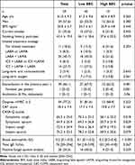
Fifty-nine subjects were included in the study, 40 in the low BEC group (BEC<300/mm3, 67.8%) and 19 in the high BEC group (BEC ≥ 300/mm3, 32.2%). Subjects’ characteristics are detailed in Table 1 and Supplementary Table 1. Briefly, they were predominantly men (57.6%), mean age 61 ± 9 years. COPD was severe to very severe in 57.6%, 66.1% had one or more exacerbations in the last year, with a median of 2 exacerbations. Two patients received long-term oral corticosteroids at inclusion, including one in low BEC group, and one in high BEC group, with no significant differences between groups (Table 1 and Table S1). When compared with the low BEC group, the high BEC group was characterised by a higher number of exacerbations per patient (median 2 [0–4] vs 1 [0–3], p<0.001), more pronounced symptoms of cough and sputum (p<0.05, CASA-Q, Table 1) and a trend for lower smoking exposure (p=0.06) and more frequent antibiotic course (p=0.06). We did not find any differences in terms of inhaled treatment, lung function, or emphysema score.
 |
Table 1 Patients’ Characteristics |
Microbiology
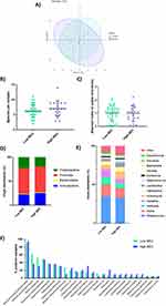
The extended culture method was applied to 59 sputa (1 sample per patient) to determine the viable airway microbiota. We identified 386 bacteria, from 71 different species, representing a mean of 6.6 bacteria per sample (Table S2). We compared the viable microbiota of the low and high BEC groups by PCA analysis and we found no difference in the global composition of the microbiota of the 2 groups (Figure 1A). The number of bacteria per sample and the α-diversity of the microbiota did not differ between low and high BEC groups (Figure 1B and C). The repartition of the bacterial phyla was similar in the 2 groups, with a predominance of Firmicutes (Figure 1D). We observed the same repartition of different genera: Streptococcus, Rothia, Veillonella, Neisseria, and Actinomyces were predominant and represented more than 65% of the bacteria identified (Figure 1E). The prevalence of the different species in the 2 groups was analysed and no difference was found between the low and high BEC groups (Figure 1F). The most common bacteria in both groups were Streptococcus oralis/mitis/pneumoniae, identified in more than 90% of samples, followed by Veillonella parvula/dispar/atypica found in more than 50% of samples. Bacterial quantifications of the viable microbiota ranged from 102 CFU/mL to 109 CFU/mL, with a median of 106 CFU/mL and no difference between the 2 groups (Table S2).
In this cohort of COPD patients at stable state, some PPM were detected: Staphylococcus aureus (n = 8, 13.6%), Haemophilus influenzae (n = 6, 10.2%), Moraxella catarrhalis (n = 5, 8.5%) and Pseudomonas aeruginosa (n = 3, 5.1%), with the same prevalence in the 2 groups (n=16; 40.0% in the low BEC group vs n=7, 36.8% in the high BEC group) (Table S2).
Discussion
In this cross-sectional analysis, focusing on COPD patients at stable state and using an original extended conventional culture-based approach that allows the identification of bacteria restricted to viable strains, we identified that high BEC is not associated with a distinct profile of viable airway microbiota, despite a BEC-associated clinical phenotype.
In our study, the high BEC group was characterised by a clinical phenotype including more frequent exacerbations in the last year, a trend for a more frequent antibiotic course, and more pronounced cough and sputum symptoms (CASA-Q). Elevated BEC at a stable state has been previously identified as a biomarker for exacerbation risk in COPD, with an incidence rate ratio of exacerbation of 1.32 for BEC ≥300/mm3 in the COPDgene study.2 An association between sputum inflammatory cells and symptoms at stable state has been shown, with neutrophilic inflammation being associated with cough, and eosinophilic inflammation with dyspnea.14,15 A recent post-hoc analysis suggested that COPD patients with both high eosinophil levels in sputum (≥3%) and chronic bronchitis might present a distinct profile of gene expression, characterised by the overexpression of T2- and phosphodiesterase-4-inhibitors-related genes.16 The high BEC group in our study was further characterised by a trend for lower smoking exposure, but a similarly altered lung function. This might indirectly reflect the previously observed accelerated lung function decline in COPD subjects with high eosinophil counts.17
We did not identify a distinct profile of viable airway microbiota in the high BEC group when compared with the low BEC group. Firmicutes and Streptococcus were the predominant bacteria phylum and species respectively, in line with previous analyses performed in COPD at a stable state.5,18 The previously reported decrease in Proteobacteria abundance and increase in Firmicutes phyla in subjects with high BEC,19 or the more diverse microbiome in subjects with BEC ≥2%9 were not found in our study. It must be pointed out that these previous studies used gene sequencing techniques (16S rRNA), which are not able to discriminate between viable and non-viable strains. Previous studies described the presence of PPM in subjects with low sputum eosinophil count.7,8 However, these studies used qPCR detection restricted to 3 species (H. influenzae, M. catarrhalis, and S. pneumoniae), therefore also including both viable and non-viable strains. Our study did not confirm those results.
Our study suffers from several limitations. Although monocentric, the patient’s recruitment was prospective and all benefited from an in-depth phenotypic characterisation. Exacerbation frequency was estimated retrospectively in the last year before inclusion. We performed only a one-point BEC assessment at inclusion, while BEC is known to vary over time in COPD.20 However, the clinical phenotype of the high BEC group including more frequent exacerbation matches with previous studies.2 Inhaled treatment was heterogeneous with 32% of the patients using ICS. ICS treatment can alter the microbiome in the small airways of patients with COPD and might have an impact on our results.21 However, inhaled treatment strategies including ICS in our study did not significantly differ between high and low BEC groups. Finally, our extended conventional culture-based approach may have lower sensitivity than a metagenomic approach for a more in-depth characterisation of the lung microbiota.
Conclusion
In our study using an original extended conventional culture-based approach, high BEC in COPD at a stable state was associated with a clinical phenotype including frequent exacerbation and more cough and sputum symptoms, but no distinct profile of viable airway microbiota compared with low BEC.
Data Sharing Statement
The data that support the findings of this study are available from the corresponding author upon reasonable request.
Ethics Approval and Informed Consent
The study was approved by the regional ethics committee (Comité de Protection des Personnes—Dijon EST I, no. 2016-A00242-49). Informed consent was obtained from all the patients. Our Study complies with the Declaration of Helsinki.
Author Contributions
All authors made a significant contribution to the work reported, whether that is in the conception, study design, execution, acquisition of data, analysis and interpretation, or in all these areas; took part in drafting, revising or critically reviewing the article; gave final approval of the version to be published; have agreed on the journal to which the article has been submitted; and agree to be accountable for all aspects of the work.
Funding
There is no funding to report.
Disclosure
J.M. Perotin reports lecture honoraria from AstraZeneca, and support for attending meetings from AstraZeneca and Chiesi; outside the submitted work. C. Launois reports support for attending meeting from Chiesi; outside the submitted work. V. Dormoy reports lecture honoraria from Chiesi and AstraZeneca; outside the submitted work. G. Deslée reports lecture honoraria from Chiesi, AstraZeneca, Boehringer Ingelheim, Sanofi and GlaxoSmithKline; outside the submitted work. The authors report no other conflicts of interest in this work.
References
1. Global strategy for the diagnosis, management, and prevention of chronic obstructive pulmonary disease (2023 report). Available from: https://goldcopd.org.
2. Yun JH, Lamb A, Chase R, et al. Blood eosinophil count thresholds and exacerbations in patients with chronic obstructive pulmonary disease. J Allergy Clin Immunol. 2018;141(6):2037–47.e10. doi:10.1016/j.jaci.2018.04.010
3. Singh D, Agusti A, Martinez FJ, et al. Blood eosinophils and chronic obstructive pulmonary disease: a global initiative for chronic obstructive lung disease science committee 2022 review. Am J Respir Crit Care Med. 2022;206(1):17–24. doi:10.1164/rccm.202201-0209PP
4. Galiana A, Aguirre E, Rodriguez JC, et al. Sputum microbiota in moderate versus severe patients with COPD. Eur Respir J. 2014;43(6):1787–1790. doi:10.1183/09031936.00191513
5. Garcia-Nuñez M, Millares L, Pomares X, et al. Severity-related changes of bronchial microbiome in chronic obstructive pulmonary disease. J Clin Microbiol. 2014;52(12):4217–4223. doi:10.1128/JCM.01967-14
6. Muggeo A, Perotin JM, Brisebarre A, et al. Extended bacteria culture-based clustering identifies a phenotype associating increased cough and Enterobacterales in stable chronic obstructive pulmonary disease. Front Microbiol. 2021;12:781797. doi:10.3389/fmicb.2021.781797
7. Beech AS, Lea S, Kolsum U, et al. Bacteria and sputum inflammatory cell counts; a COPD cohort analysis. Respir Res. 2020;21(1):289. doi:10.1186/s12931-020-01552-4
8. Kolsum U, Donaldson GC, Singh R, et al. Blood and sputum eosinophils in COPD; relationship with bacterial load. Respir Res. 2017;18(1):88. doi:10.1186/s12931-017-0570-5
9. Millares L, Pascual S, Montón C, et al. Relationship between the respiratory microbiome and the severity of airflow limitation, history of exacerbations and circulating eosinophils in COPD patients. BMC Pulm Med. 2019;19(1):112. doi:10.1186/s12890-019-0867-x
10. Dingle TC, Butler-Wu SM. Maldi-tof mass spectrometry for microorganism identification. Clin Lab Med. 2013;33(3):589–609. doi:10.1016/j.cll.2013.03.001
11. Global strategy for the diagnosis, management, and prevention of chronic obstructive pulmonary disease (2022 report). Available from: https://goldcopd.org.
12. Perotin JM, Adam D, Vella-Boucaud J, et al. Delay of airway epithelial wound repair in COPD is associated with airflow obstruction severity. Respir Res. 2014;15:151. doi:10.1186/s12931-014-0151-9
13. Washko GR, Criner GJ, Mohsenifar Z, et al. Computed tomographic-based quantification of emphysema and correlation to pulmonary function and mechanics. COPD. 2008;5(3):177–186. doi:10.1080/15412550802093025
14. Zysman M, Deslee G, Caillaud D, et al. Relationship between blood eosinophils, clinical characteristics, and mortality in patients with COPD. Int J Chron Obstruct Pulmon Dis. 2017;12:1819–1824. doi:10.2147/COPD.S129787
15. Contoli M, Baraldo S, Conti V, et al. Airway inflammatory profile is correlated with symptoms in stable COPD: a longitudinal proof-of-concept cohort study. Respirology. 2020;25(1):80–88. doi:10.1111/resp.13607
16. Singh D, Bassi M, Balzano D, et al. COPD patients with chronic bronchitis and higher sputum eosinophil counts show increased type-2 and PDE4 gene expression in sputum. J Cell Mol Med. 2021;25(2):905–918. doi:10.1111/jcmm.16146
17. Tan WC, Bourbeau J, Nadeau G, et al. High eosinophil counts predict decline in FEV 1: results from the CanCOLD study. Eur Respir J. 2021;57(5):2000838. doi:10.1183/13993003.00838-2020
18. Tangedal S, Nielsen R, Aanerud M, et al. Sputum microbiota and inflammation at stable state and during exacerbations in a cohort of chronic obstructive pulmonary disease (COPD) patients. PLoS One. 2019;14(9):e0222449. doi:10.1371/journal.pone.0222449
19. Dicker AJ, Huang JTJ, Lonergan M, et al. The sputum microbiome, airway inflammation, and mortality in chronic obstructive pulmonary disease. J Allergy Clin Immunol. 2021;147(1):158–167. doi:10.1016/j.jaci.2020.02.040
20. Schumann DM, Tamm M, Kostikas K, Stolz D. Stability of the blood eosinophilic phenotype in stable and exacerbated COPD. Chest. 2019;156(3):456–465. doi:10.1016/j.chest.2019.04.012
21. Yip W, Li X, Koelwyn GJ, et al. Inhaled corticosteroids selectively alter the microbiome and host transcriptome in the small airways of patients with chronic obstructive pulmonary disease. Biomedicines. 2022;10(5):1110. doi:10.3390/biomedicines10051110
 © 2024 The Author(s). This work is published and licensed by Dove Medical Press Limited. The full terms of this license are available at https://www.dovepress.com/terms.php and incorporate the Creative Commons Attribution - Non Commercial (unported, v3.0) License.
By accessing the work you hereby accept the Terms. Non-commercial uses of the work are permitted without any further permission from Dove Medical Press Limited, provided the work is properly attributed. For permission for commercial use of this work, please see paragraphs 4.2 and 5 of our Terms.

