
Back to Journals » International Journal of Nanomedicine » Volume 21
Hafnium-Oxide Based Nanoplatform with Tumor Microenvironment Responsive Drug Release for Enhanced Radio-Chemotherapy
Authors Liu H, Chen L  , Liu M, Liu Y, Jia R, Liu H, Liang J, Li X, Fauzia RP, Yu X, Bai Y, Wang M
Received 2 April 2026
Accepted for publication 19 June 2026
Published 8 July 2026 Volume 2026:21 612052
DOI https://doi.org/10.2147/IJN.S612052
Checked for plagiarism Yes
Review by Single anonymous peer review
Peer reviewer comments 2
Editor who approved publication: Professor Eng San Thian
Huanhuan Liu1,*, Lijuan Chen1,*, Mingbo Liu2,*, Yanqin Liu1, Rufeng Jia1, Hongming Liu1, Junting Liang3, Xiaochen Li4, Retna Putri Fauzia5, Xuan Yu1, Yan Bai1, Meiyun Wang1,6
1Department of Radiology, Henan Provincial People’s Hospital & the People’s Hospital of Zhengzhou University, Zhengzhou, Henan Province, People’s Republic of China; 2Department of Radiation Oncology, Henan Provincial People’s Hospital & the People’s Hospital of Zhengzhou University, Zhengzhou, Henan Province, People’s Republic of China; 3Clinical Bioinformatics Experimental Center, Henan Provincial People’s Hospital, People’s Hospital of Zhengzhou University, Zhengzhou, Henan Province, People’s Republic of China; 4Department of Nuclear Medicine, Henan Provincial People’s Hospital & the People’s Hospital of Zhengzhou University, Zhengzhou, Henan Province, People’s Republic of China; 5Department of Chemistry, Faculty of Mathematics and Natural Sciences, Universitas Padjadjaran, Sumedang, Indonesia; 6Biomedical Research Institute, Henan Academy of Sciences, Zhengzhou, Henan Province, People’s Republic of China
*These authors contributed equally to this work
Correspondence: Meiyun Wang, Department of Radiology, Henan Provincial People’s Hospital & the People’s Hospital of Zhengzhou University, Zhengzhou, Henan Province, People’s Republic of China, Email [email protected]
Purpose: Hafnium oxide nanoparticles have been established as effective radiosensitizers, however, tumor cells often develop resistance to single-modality radiotherapy, and the tumor microenvironment (TME) poses additional limitations to treatment efficacy. To address these challenges, we fabricated a doxorubicin and manganese oxide co-loaded hafnium oxide (MD-Hf) nanoplatform for synergistic radio-chemotherapy and evaluated its antitumor performance in cellular and animal models.
Results: In MD-Hf nanoplatform, the HfO2 nanocrystal functions as the radiosensitizer and carrier, the Dox works for chemotherapy, while the manganese oxide coating layer are capable of modulating the TME by depleting glutathione (GSH) and converting H2O2 in to ·OH radicals. Moreover, the MnOx coating also allows the nanoplatform possessing TME-responsive Dox release. Upon exposure to X-rays, the MD-Hf exhibited evident toxicity to Panc 02 tumor cells, only 63.9±10.7% cell remain alive after irradiated with 2Gy X-ray, which is much lower than 86.7±6.33% of the group administrated with pure HfO2 NPs. In vivo studies further demonstrated superior therapeutic outcomes with the MD-Hf nanoplatform, as evidenced by markedly reduced tumor size and weight compared to treatment with HfO2 nanoparticles alone. RNA-seq analysis reveals the Dox can potentiate organelle damage, and the MnOx can even activate immune response, which further corroborates the multifunctionality of the integrated nanoplatform.
Conclusion: The newly developed doxorubicin and manganese oxide co-loaded HfO2 nanoplatform significantly enhance radio-chemotherapeutic efficacy against pancreatic tumor cells, offering a promising strategy that may guide the future clinical development of HfO2-based radiotherapy.
Keywords: radiosensitizer, tumor microenvironment, drug release, radicals, nanoplatform
Introduction
Radiotherapy is one of the most widely applied treatment modalities for malignant tumors. The primary limitation of radiotherapy is the side-effect to healthy tissue. In clinic, the radiosensitizers, such as Olaparib and Bevacizumab, are frequently employed to enhance the radiosensitivity of tumor tissue.1,2 However, these radiosensitizers can only influence tumor cells rather than improve the utilization efficiency of ionizing radiation. To enhance local energy deposition, one promising approach is to introduce high atomic number (high-Z) elements such as gold, hafnium, and platinum at tumor sites.3 Among them, hafnium-based nanoparticles have emerged as particularly promising candidates.4 A well-known example is NBTXR3 (Nanobiotix),5–7 which has shown outstanding therapeutic effects in lung cancer, pancreatic cancer, head and neck cancer and so on.8 The clinical translation of NBTXR3 is benefited from its inert chemical properties and the optimal size. However, relying solely on a single therapeutic modality often results in limited efficacy in the eradication of tumor cells. Therefore, integration of multiple functionalities into a single platform can substantially improve treatment outcomes.9
Radiotherapy is frequently administered in conjunction with chemotherapy to address long-term resistance associated with monotherapy.10,11 The synergistic effect of combined radio-chemotherapy can improve the tumor sensitivity to ionizing radiation and exhibit impaired DNA repair capacity following ionizing radiation exposure.12 Sherstiuk developed a Dox loaded HfO2 nanoplatform for the treatment of various cell lines. The incorporation of HfO2 significantly reduced the IC50 of Dox compared to Dox alone, and the combined system can lead to evident cytotoxicity under 2Gy of X-ray.13 The drug-loading strategy is crucial to fabricate the excellent drug delivery system. Physical loading methods primarily depend on electrostatic interactions between drug molecules and the vesicles, often achieving high loading efficiency in a short period. However, electrostatic adsorption tends to lack sufficient stability in vivo.14,15 In contrast, chemical grafting offers improved stability but is generally more time-consuming, costly, and may introduce unintended toxicity due to the chemicals involved in the synthesis.16 Alternatively, coating nanoparticle with stimuli-responsive materials provides another promising avenue for developing smart drug delivery systems.17,18
The distinct characteristics of the tumor microenvironment (TME) can be used to design smart drug delivery system or enhance treatment outcome. For instance, the overexpress of GSH in TME can serve as a trigger for drug release. Lu et al developed a self-assembled nanoparticles and link with Paclitaxel (PTX) via -S-S- linker, upon uptake by tumor cells, the high concentration of GSH cleaves the disulfide linkers, thereby inducing drug release16 Moreover, therapeutic strategy that actively modulate the TME can further enhance cancer treatment efficacy. In previous research, we developed a MnO2/Pt nanocomposite system for enhanced radiotherapy.19 Within this system, MnO2 serves to deplete GSH in the tumor, while Pt acts as a radiosensitizer that amplifies reactive oxygen species (ROS) generation upon X-ray irradiation. Since GSH is a known ROS scavenger, its depletion synergistically enhances Pt-mediated radiotherapy. Additionally, manganese ions are capable of modulating immune pathways within tumors and thus influence the treatment. For instance, Hu et al demonstrated that Mn2⁺ ions can activate the cGAS-STING immune signaling pathway, contributing to sustained tumor growth suppression.20
In this work, we synthesized HfO2 nanoparticles and employed them as carriers for doxorubicin (DOX). Following drug loading, manganese oxide (MnOx) was deposited on the surface of the HfO2 nanoparticles to function as a gatekeeper (Scheme 1). Within this integrated system, HfO2 serves to enhance X-ray energy deposition at tumor tissue, while DOX acts as the chemotherapeutic agent. Concurrently, MnOx coating interacts with the TME by depleting GSH and H2O, thereby addressing key mechanisms of treatment resistance and facilitating the release of DOX. Meanwhile, the Mn can also act as the trigger for immune-response. By constructing such a multifunctional drug delivery platform, we aim to reduce the side-effects and treatment resistance associated with single therapy and achieve synergistic therapeutic outcomes.
 |
Scheme 1 Schematic illustration of the synthesis of HfO2-based nanoplatform and its application in enhanced radiotherapy. |
Material and Method
Materials
Hafnium chloride (≥99.9%, HfCl4), Potassium hydroxide, Doxorubicin, 4-(2-Hydroxyethyl)-1-piperazineethanesulfonic acid (HEPES), 5.5’-Dithio bis-(2-nitrobenzoic acid) (DTNB) were purchased from Macklin (Shanghai, China), Potassium permanganate (KMnO4) was purchased from Yantai Luoyang Chemical Reagent Factory, Bovine Serum Albumin (BSA) was obtained from BioFroxx, Glucose was purchased from Kermel Co., Ltd, (Tianjin, China). Fetal Bovine Serum (FBS) was purchased from Cell-box, Changsha, China. PBS buffer, RPMI 1640 Medium, CCK-8 assay kits were purchased from ServiceBio, Wuhan, China. All chemical reagents were used as received.
Synthesis
HfO2 Nanoparticles
HfO2 nanoparticles were synthesized via hydrothermal method. Briefly, 0.05 M HfCl4 aqueous solution was prepared by adding 0.4 g of HfCl4 powder to 25 mL of DI water and stirred under 80°C for 30 min. Then different volume of 0.2 M KOH solution was added to 6 mL HfCl4 solution under stirring for 10 min, followed by adding DI water to reach a fixed volume of 15 mL. A pH meter was applied to monitor the real-time pH value of the final solution (Table S1). The obtained milky mixture was then transferred to Teflon autoclave (25 mL) and heated to 180°C for 8 hours. The resultant solution was collected and washed with deionized water for three times. pH values, temperature and reaction duration were varied to optimizing the synthesis parameters.
Dox-Loaded HfO2 Nanoparticles
BSA was used for modifying the surface of HfO2 nanoparticles. Typically, 4 mL of HfO2 NPs solution (3 mg/mL) was mixed with 1 mL of BSA solution (1 mg/mL) by stirring for 4 hours. The resulting BSA@Hf NPs was then collected by centrifugation (10000 rpm, 10 min) and washed with DI water for three times. The obtained BSA@Hf NPs were re-suspended in HEPES buffer (20 mM, pH = 7.1). Dox aqueous solution with a concentration of 1 mg/mL was added to BSA@Hf NPs solution and stirred for another 4 hours. Then the un-loaded Dox was removed by centrifugation (4000 rpm, 15 min). The obtained samples were marked as D-Hf.
Manganese Oxide Coated HfO2 Nanoparticles
Briefly, 1 mL of KMnO4 aqueous solution with different concentrations of (5 mM, 1.76 mM and 0.5 mM) was added to 4 mL of D-Hf nanoparticles slowly under sonication. After 10 mins, the obtained samples were centrifugated and washed with DI water 3 times to remove the unreacted Mn precursors. The resulted sample was remarked as MD-Hf nanoparticles. The M-Hf sample was prepared by replacing the D-Hf with BSA@Hf nanoparticles.
Characterization
The phase of the HfO2 NPs was determined by X-ray diffraction (XRD) on the D/max 2500 equipment (Rigaku) operated at 40 kV and 30 mA, with a scanning rate of 10 °/min. Fourier transform infrared spectra (FT-IR) were recorded by an FT-IR spectrometer (Shimadzu-IR Tracer 100, Japan). The morphology and compositional distribution of the obtained samples was observed by transmission electron microscopy (TEM, JEOL-JEM 2100 F) with X-ray spectroscopy (EDS). Florescence spectra were determined by using a spectrofluorometer (Fluorolog®-3TCSPC, HORIBA, France). X-ray photoelectron spectroscopy (XPS) spectra were performed on an XPS spectroscope (Thermo SCIENTIFIC K-ALPHA). The UV-vis absorption spectrum/fluorescence intensity of the obtained samples were measured by a multimode a microplate reader (Synergy H1, Biotek). The hydrodynamic diameter and Zeta potential of the samples were determined by a zeta sizer (nano-ZS, Malvern). The content of elements was detected by an ICP-MS (Agilent 7850).
MnOx Coating Amount
The mass of the MnOₓ coating, specifically the Mn element, was determined using ICP-MS. To separate the MnOₓ coating from the MD-Hf nanoparticles, the MD-Hf samples were mixed with 10 mM GSH solution at a 1:1 volume ratio. After sonication for 30 minutes, the mixture was centrifuged, and the supernatant was collected for measurement.
Drug Release
The Dox release profile was detected by mixing 3 mL of D-Hf/MD-Hf with 3 mL of PBS buffer (pH value 5.8). For the samples treat with GSH, the GSH PBS solution (10 mM, pH = 5.8) was applied. 0.3 mL solution was collected at different time points and centrifugated at 10,000 rpm for 5 min, the upper solution was collected for measurement. The fluorescence signal of Dox was detected by the multifunctional plate-reader.
Catalase-Like Activity of M-Hf NPs
H2O2 Consumption
The interaction between Mn-Hf NPs and H2O2 was evaluated by Methylene Blue (MB) decomposition. MB solution (30 μM) was mixed with 0.1 M H2O2 solution with a volume ratio of 1:1, then 0.2 mL of M-Hf NPs was added to 1 mL MB and H2O2 mixture and reacted for 20 minutes. The mixed solution was centrifugated at 10000 rpm for 5 min, then collected 200 μL of the upper solution and measured the UV-vis absorbance of MB.
GSH Consumption
The GSH consumption ability of Mn-Hf was measured by DTNB essay. Briefly, 200 μL of GSH solution (10mM) was mixed with 200 μL of Mn-Hf solution, and incubated for 20 minutes. Then, we centrifugated the mixture at 10000 rpm for 5 min, collected 50 μL upper solution and added it in to the 96 well-plates containing 150 μL of DTNB solution (PBS buffer, 2 mM) in each vial. 20 min later, UV-vis absorption intensity at 412 nm was measured by the plate-reader.
ROS Detection
Electron paramagnetic resonance (EPR) was applied to detect the ROS generated by the reaction between Mn-Hf NPs and H2O2. Briefly, 1 mL of DMPO solution (100 mM) was added to 0.5 mL of M-Hf NPs samples, which was followed by adding 0.5 mL of 30% H2O2 to the mixture and react for 5 min. Then the samples were transferred to an EPR tube for measurement.
Cell Uptake and Cellular Distribution
The Panc 02 murine pancreatic cell line and Hep 1–6 murine hepatoma cell line (Servicebio Technology Co., Ltd. Wuhan, China) were cultured in RPMI 1640 supplemented with 10% FBS under a humidified atmosphere containing 5% CO2 at 37°C.
To observe the cellular distribution, HfO2 NPs was modified with Ce6 via EDC/NHS method, which allows the activated Ce6 molecules to interact with BSA.21 Confocal laser scanning microscope (CLSM, FV4000, Olympus) was used to determine the cellular uptake of Ce6-HfO2 NPs in Panc 02 and Hep 1–6 cell lines. Cells (1 × 10⁴) were seeded in a cell culture dish specific for CLMS (Biosharp, China) and cultured overnight. Then 20 µL Ce6-HfO2 NPs solution was added and incubated for 16 hours. The cells were washed three times with PBS and fixed with 4% paraformaldehyde solution for 20 minutes. After fixation, the cells were washed three times with PBS and stored in the refrigerator. Prior to imaging, Hoechst 33342 (ServiceBio) was added to the cells. Laser excitation was performed at 405 nm, with emission detected at 465 nm for Hoechst 33342 and 660 nm for Ce6. The incubation time of Ce6-HfO2 NPs was varied to investigate the time-dependent cell uptake.
Cellular GSH Consumption Detection
Panc 02 cells were plated on 6-well plates with a cell density of 1*105 cells/well (2 mL/well) and kept overnight. Then PBS (100 µL, pH 7.4), M-Hf samples (50 µL, 100 µL) were added to each well and incubated for another 18 h. Then the cells were collected and analyzed the cellular GSH concentration by a GSH kit (S0053, Beyotime).
Cell Viability Test
Panc 02/Hep 1–6 cells were plated on 96-well plates with a cell density of 5000 cells/well (100 μL/well) and incubated overnight. Then 10 μL of the samples were added to each well and incubated for another 18 h, which was followed by being irradiated with a 320 keV X-ray source (PXI-X RAD320, Precision X-ray Inc, US) to 2 Gy, 4Gy, 6Gy and 8Gy. After 24 hours, the CCK-8 assay was carried out to evaluate the cell viability. The absorbance at 450 nm was measured by the plate-reader (Synergy H1, Biotek). The cells treated with PBS buffer were recognized as the control group.
DNA Damage
The DNA damage after treatment was evaluated by a γ-H2AX immunofluorescence test kit (Beyotime). Panc 02/Hep 1–6 cells were plated on 6-well plates with a cell density of 10000 cells/well (2.5 mL/well) and kept overnight. Then 200 μL of the PBS buffer, HfO2 nanoparticles and Mn-Hf nanoparticles were added to each well and incubated for another 18 h, followed by being irradiated with X-ray to 4 Gy. After 4 hours, the cells were fixed for 15 min, and consequently being stained with γ-H2AX antibody for 1 hours and Anti-rabbit IgG-AF488 for another 1 hours. Finally, the nucleus was stained with DAPI for 10 minutes at room temperature. An inverted fluorescence microscope (FV4000, Olympus) was applied to detect the signals.
Colony Formation Assay
Panc 02/Hep 1–6 cells were plated on 6-well plates with a cell density of 3000 cells/well (2.5 mL/well) and kept overnight. Then 250 μL of the samples were added to each well and incubated for another 18 h, which was followed by being irradiated with a 320 keV X-ray source (PXI-X RAD320, Precision X-ray Inc, US) to 4 Gy. After being incubated for another 8 days, cells were washed with PBS, fixed with paraformaldehyde and stained with crystal violet staining solution (0.5%, Kemiou Chemical Reagent).
Therapeutic Study in Tumor-Bearing Mouse Model
The male C57BL/6J mice aged 5–6 weeks were procured from Beijing Vital River Laboratory Animal Technology Co., Ltd. The mice were housed at the animal research facility of Henan Shuangyun Biotechnology Co., Ltd, where the temperature was maintained at 21◦C–22◦C and the light/dark cycle was 12 h (06:00 to18:00). After acclimatization for one-week, C57LB mice were subcutaneously injected with the Panc 02 (1.5 × 10⁶ cells) cells on the right leg. When the tumor diameter reached approximately 40 mm3, the tumor-bearing mice were grouped into eight groups (PBS group, D-Hf group, MD-Hf group, RT+PBS group, RT+ HfO2 group, RT+D-Hf group, RT+M-Hf group, RT+MD-Hf group. 30 μL of samples was intertumoral injected to the tumor, after 6 hours, the mice in RT+ groups were anesthetized with isoflurane (RWD Life Science Co., Ltd.) and irradiated with X-rays to 6 Gy. The size of tumor was measured until the test completed and the body weight of the mice was recorded. The tumor volume was calculated using the equation V = ab2/2 (a = length, b = width). At the 14th day (the endpoint of experiment), the mice were euthanized cervical dislocation, and the tumor tissues were harvested for tumor immunohistochemistry. All animal care and experimentation were conducted in accordance with Henan Shuangyun Animal Care and Use Committee-approved protocols (SYLS2025069). Hematoxylin and eosin (H&E), Ki67, γ-H2AX, Tunel, PTGS2 staining for evaluating the tumor proliferation and apoptotic index, the stained specimens were observed with Case Viewer (Wuhan Servicebio Technology Co., LTD).
Statistical Analysis
Data are presented as mean±standard deviation based on at least 3 independent replicates. Student’s t test was applied for the comparison among the obtained results. P values: ns, p>0.05;*p≤0.05, **p≤0.01, ***p≤0.001.
Result and Analysis
HfO2 nanocrystals were synthesized via the hydrothermal method,22 experimental parameters including pH value, reaction period and temperature were varied to optimize the composition and morphology of HfO2 nanoparticles. The XRD spectra in Figure 1a–c reveal that all resultants share an identical crystal phase (PDF # 00-006-0318). The three strongest peaks in the XRD pattern located at 28.3°, 31.7°, and 34.6° can be indexed to the (−111), (111), and (002) planes of monoclinic hafnium oxide.23 The results in Figure 1a indicate that the pH values have relatively minor influence on the crystallinity of HfO2, monoclinic products with well-crystal structure were synthesized in a broad pH range, from 1.28–10.8, after being heating at 180°C for 8 hours. The morphology of samples synthesized at pH 0.85, 3.45, and 5.58 was observed by TEM. All three samples exhibit typical spindle-like microstructure (Figure 1d–f). High-resolution TEM imaging of HfO2 shows clear lattice fringes with a spacing of 0.284 nm (Figure S1), which match the (111) plane of the monoclinic hafnium oxide.24 Moreover, the size distribution and zeta potential results in Figure 1g and h indicate that higher pH values lead to the formation of large HfO2 particles with a lower surface charge.
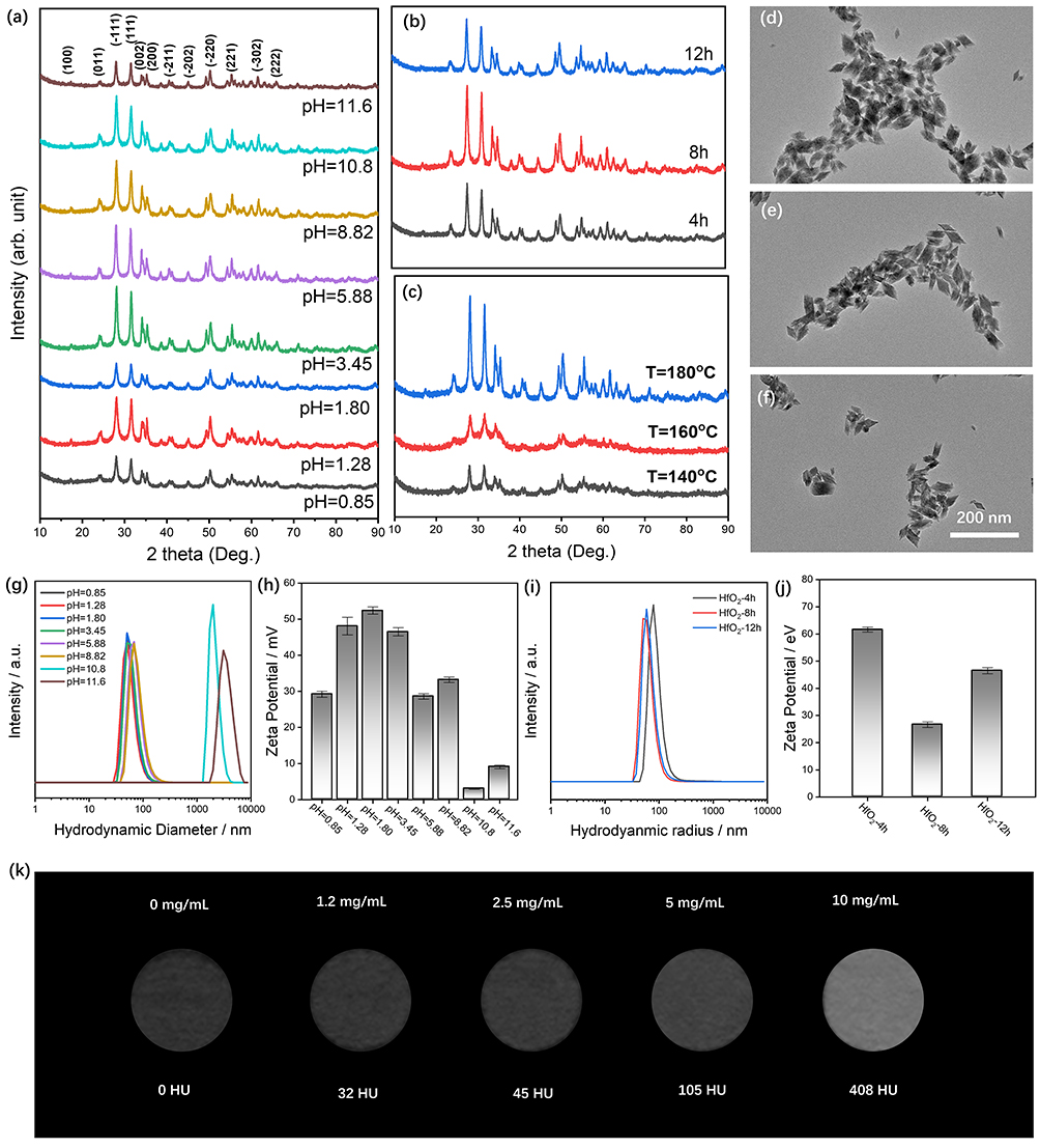 |
Figure 1 Typical XRD patterns of HfO2 resultants obtained via adjusting the (a) pH values, (b) reaction duration and (c) reaction temperature. TEM figures for typical HfO2 samples produced under (d) pH = 0.85, 180°C, 8 h, (e) pH = 3.45, 180°C, 8 h and (f) pH = 5.88, 180°C, 8 h; the (g) size distribution and (h) zeta potential of HfO2 samples obtained via adjusting pH values (the reaction temperature is 180°C, reaction period is 8 hours); the (i) size distribution and (j) zeta potential of HfO2 samples obtained via adjusting reaction period (the pH value is 3.45, the reaction temperature is 180°C); (k) CT images at room temperature as a function of HfO2 concentration. |
The reaction duration shows a negligible influence on both phase and size of the final HfO2 resultants (Figure 1i and j). However, the reacting temperature evidently affects the nucleation process of HfO2, though the samples synthesized under 160°C and 140°C also show the distinct peak of monoclinic hafnium oxide, their fluctuating XRD patterns suggest low crystallinity (Figure 1c), which is in accordance with previous publication.22 XPS technique was used to analyze the Hf and O elements of the obtained HfO2 NPs (Figure S2). Compared to the samples prepared under 180°C, 8h, the ones synthesized under shorter time or lower temperature show defected O element in the composition (Table S2), further confirming the crystallization process is incomplete. Given that hafnium oxide is composed of the high-atomic-number element, we further evaluated its potential for contrast enhancement in CT imaging. As shown in Figure 1k, signals from HfO2 at various concentrations can be readily detected using a clinically available CT scanner (Naeotom Alpha, Siemens), demonstrating its feasibility for tracing biodistribution in vivo.25
The typical HfO2 sample (synthesized under pH 3.45, 180°C, 8h) was selected for further functionalization. First, BSA decorated HfO2 nanoplatform (denoted as BSA@Hf) was synthesized by simply mixing their aqueous solutions for 4 hours.26 As for Dox loading, we first investigated the influence of solvent types on the Dox-loading process. As shown in Figure S3, Dox failed to interact with BSA@Hf when using DI water as solvent, while both HEPES and PBS buffers enabled successful Dox loading, which may due to the negative surface charge (Figure S4). Though using PBS buffer can achieve higher Dox loading efficiency (~12.4%), the sample dispersed in PBS showed noticeable aggregation after several hours. Therefore, HEPES buffer was chosen as the solvent for Dox-loading. As for MnOx coating, the ones dispersed in HEPES and PBS show immediate aggregates once the KMnO4 solution was added. Consequently, the DI water was selected as the solvent. As shown in Figure S5, directly loading MnOx onto the bare HfO2 nanoparticles is not possible, the MnO4− will not be reduced in the absence of BSA. Furthermore, the concentration of KMnO4 solution also plays a crucial role in coating process. At concentration of 5 mM and 1.76 mM, excess MnOx coating caused significant aggregation, whereas 0.5 mM KMnO4 solutions lead to the formation of M-Hf with good dispersity. The photos of different HfO2-based samples were shown in Figure S6.
The size distribution and surface charge were determined. As shown in Figure 2a and b), the bare HfO2 NPs possess a mean hydrodynamic diameter of 44 nm and +25.9 mV as the surface charge. The BSA layer on the HfO2 did not significantly alter the hydrodynamic diameter and surface charge of the HfO2 nanoparticles. In contrast, the introduction of either Dox or MnOx led to a noticeable increase in the size of the nanoplatform. Furthermore, the deposition of MnOx shifted the zeta potential from positive to negative. The stability of MD-Hf nanoparticles was tested under different conditions. As shown in Figure S7a and b), the size of MD-Hf NPs remained stable in both PBS buffer (pH 7.4) and 10 mM glutathione (GSH) solution after 5 hours, despite exhibiting differences in zeta potential (Figure S7c). In contrast, when dispersed in PBS buffer at pH 5.4, the nanoparticles immediately aggregated (Figure S8). The loading of Dox resulted in a slight increase in UV absorbance around 480 nm (Figure 2c). According to the UV-vis spectra, the loading efficiency of Dox was approximately 8.5%. Figure 2d displays the FT-IR spectra of the samples. The spectrum of unmodified HfO2 nanoparticles exhibits characteristic Hf–O vibrational peaks at 776 cm−1, 672 cm−1, and 520 cm−1. After BSA modification, the appearance of amide I and amide II peaks at 1662 cm−1 and 1538 cm−1 confirm the successful conjugation of BSA onto the HfO2 surface.24 In the drug-loaded sample (D-Hf), characteristic N–H bond peaks are observed at 2856 cm−1 and 1280 cm−1, indicating the successful loading of Dox onto the HfO2 nanoparticles.27
 |
Figure 2 The (a) size distribution, (b) zeta potential, (c) UV-vis and (d) FT-IR of HfO2-based samples, XPS spectra of M-Hf sample: (e) survey scan, (f) Hf4f, (g) O1s and (h) Mn2p. TEM-EDS mapping of MD-Hf sample: (i) HADDF, (j) Hf distribution, (k) Mn distribution and (l) C distribution. |
The chemical bonding and surface composition of the M-Hf sample were characterized by XPS. The survey scan (Figure 2e) confirms the presence of Hf, O, and Mn in the sample. High-resolution spectra for each element were subsequently analyzed. The Hf 4f spectrum (Figure 2f) can be deconvoluted into two peaks at 16.5 and 18.2 eV, corresponding to Hf 4f7/2 and Hf 4f5/2, respectively, which verifies the formation of HfO2.28 The O 1s spectrum (Figure 2g) is fitted with two peaks at 529.7 eV and 531.3 eV, assignable to Hf–O and O–H bonds, respectively.25 The Mn 2p spectrum (Figure 2h) is more complex than those of Hf 4f and O 1s, exhibiting three peaks at 641.5, 645.3, and 653.0 eV. The first two peaks correspond to Mn2⁺ and its satellite feature, while the peak at 653.0 eV indicates the presence of Mn4⁺ ions.29 Based on quantitative analysis from ICP-MS, the mass of Mn element in the M-Hf samples was estimated to be approximately 8.57±0.17 μg/mL. Elemental mapping by TEM-EDS of MD-Hf further revealed a uniform distribution of Mn on the surface of the HfO2 nanoparticles (Figure 2i and l)). However, because the copper grid used for TEM also has a carbon film, the carbon signal from Dox was not very reliable at high magnification due to interference from the grid background. Therefore, we switched to a larger field of view to analyze the composition of MD-Hf. As shown in Figure S9, besides Hf, signals of C and Mn were clearly detected, confirming the successful loading of Dox and MnOx.
The tumor microenvironment (TME) modulation capability of the MnOx-coated samples was evaluated. Hydrogen peroxide (H2O2) and glutathione (GSH) play key roles in TME. To assess the interaction between Mn-Hf and H2O2, methylene blue (MB) which could be degraded by ·OH radicals was used as the indicator.30 As shown in Figure 3a, the combination of H2O2 and M-Hf resulted in evident decrease in MB concentration, indicating that M-Hf can react with H2O2 and generate OH radicals.19 Moreover, the reaction between M-Hf and H2O2 produced a characteristic EPR signal pattern (Figure 3b) corresponding to ·OH radicals.31 GSH consumption was evaluated using the DTNB assay, in which DTNB reacts with GSH to form a yellow product with an absorption peak at 412 nm.32 The results in Figure 3c show that the addition of M-Hf caused a rapid decrease in GSH within 10 minutes, and the reaction continued progressively over 90 minutes. The cellular GSH consumption ability of M-Hf was also evaluated. As shown in Figure S10, there are 3.82±0.41 mM of GSH in the Panc 02 cells that treated with PBS, while addition of 100 μL of Mn-Hf can decrease the GSH concentration to 2.56±0.28 mM.
 |
Figure 3 (a) MB degradation by the reactive oxygen species generated due to Mn mediated fenton-like reaction; (b) EPR spectra of the mixture of Mn-Hf and H2O2, DMPO is applied as the radical probe, 30% H2O2 is applied; (c) the depletion of GSH by reacting with M-Hf as a matter of time; (d) the release of Dox from different samples; (e) the uptake of BSA@Hf by Panc 02 cell line as a function of incubation time. |
Figure 3d shows the Dox release profiles of different samples dispersed in PBS buffer (pH 5.6). D-Hf exhibited a burst release of Dox upon dispersion in a slightly acidic environment, whereas the MnOx-coated sample (MD-Hf) showed sustained, slow Dox release over 45 hours. The addition of GSH further accelerated Dox release from MD-Hf compared to the GSH-free condition. Moreover, we used TEM to evaluate GSH-regulated DOX release. As shown in Figure S11a and b, the MD-Hf NPs were surrounded by a layer after reacted with GSH solution for 30 min, and both Hf, Mn and C elements are evenly distributed on the surface of MD-Hf (Figure S11c–f). TEM-EDS results were used to compare the composition of MD-Hf before and after reaction with GSH, and the data are presented in Table S3. The mass ratios of C and Mn in the GSH-treated group decreased to 6.8% and 0.43%, respectively, compared to 7.44% (C) and 0.55% (Mn) in DM-Hf samples without GSH exposure. These results further confirm that GSH can break down the MnOx coating and induce DOX release from MD-Hf NPs.
Cellular uptake of BSA@Hf nanoparticles was observed using CLSM. For tracking purposes, Ce6 (a fluorescent molecule) was conjugated to the surface of BSA@Hf via EDC/NHS chemistry. As shown in Figure 3e, HfO2 nanoparticles showed neglect internalization into Pan02 cells within 30 minutes, and Ce6 fluorescence was clearly detected after 6 hours of incubation. Moreover, the signals of Ce6 show even stronger after 18 hours’ incubation, indicating that the HfO2 nanoparticles can stay quite long in cells. Figure 3d indicates that approximately 25% of loaded Dox was released from the Hf-based platform within 30 minutes, but the CLSM results show that Hf-based NPs are unlikely to be uptake by cells in 30 minutes, thus it is necessary to coat the Dox-loaded Hf NPs by MnOx layer to prevent the unwanted Dox release before being uptake. Moreover, HfO2 nanoparticles could also be uptake by Hep 1–6 cells after being incubated for 24 hours (Figure S12).
We further evaluated the biological properties of the as-prepared nanoplatform using Panc 02 and Hep 1–6 cell lines. According to the CCK-8 assay results shown in Figure 4a and b), HfO2 and M-Hf exhibited slightly higher cytotoxicity than the control group under X-ray irradiation. In contrast, D-Hf showed significant toxicity even in the absence of X-ray, indicating that the loaded Dox amount was sufficient to kill tumor cells and burst Dox release can lead to acute toxicity to tumor cells. MD-Hf caused negligible harm to Panc 02 cells without X-ray exposure. However, upon irradiation with X-ray doses, even as low as 2 Gy, the MD-Hf treated group shows that nearly 40% of Panc 02 cells were killed, and cell viability further decreased to 46.7 ± 10.5% under 8 Gy irradiation. Comparably, the HfO2-based radiosensitizers did not significantly enhance radiotherapy of Hep 1–6 cells, which are inherently more sensitive to X-ray irradiation. Colony formation assay was performed to evaluate the long-term effects of the HfO2-based radiosensitizers. As shown in Figure 4c, the applied HfO2-based samples showed negligible growth inhibition on Panc 02 cells without X-ray irradiation. However, clear growth inhibition was observed in X-ray irradiated groups. The presence of HfO2 nanoparticles led to fewer colony formation compared to the control group under 4 Gy irradiation. The addition of MD-Hf resulted in the least colony formation than the HfO2-treated group. Though the Hf-based radiosensitizer showed neglect short-term cytotoxicity on Hep 1–6 cells, the addition of the them could lead to the less formation of colonies, indicating the long-term growth inhibition of them (Figure S13).
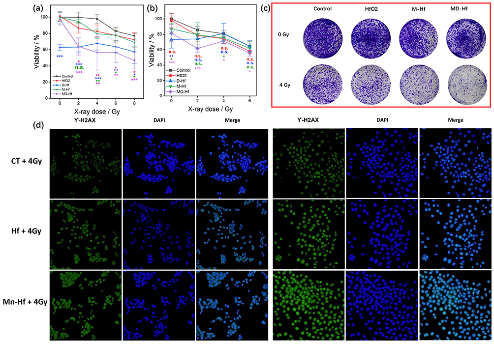 |
Figure 4 The cell toxicity of the as-prepared samples on (a) Panc 02 and (b) Hep1-6 cell line under X-ray exposure; (c) the colony formation of Panc 02 cell line after different treatments; (d) γ-H2AX staining images of Panc 02 and Hep 1–6 cell lines after different treatments. (ns, p>0.05;*p≤0.05, **p≤0.01, ***p≤0.001.). |
Fluorescence microscopy was also used to visualize DNA damage in Panc 02 and Hep 1–6 cells following X-ray exposure, with γ-H2AX staining serving as an indicator of DNA damage. As shown in Figure 4d, green fluorescence signals were observed in both Hep 1–6 and Panc 02 cells after irradiation with 4 Gy X-rays, confirming DNA damage. In Hep 1–6 cells, the addition of HfO2 or Mn-Hf samples resulted in a slightly increased FL intensity, revealing the HfO2 based NPs is capable to enhancing the radiotherapy by DNA damage. In terms of Panc 02 cells, the groups treated with HfO2 showed pronounced DNA damage after 4 Gy X-ray irradiation. The M-Hf treated Panc 02 cells exhibited the strongest green fluorescence signals than those treated with HfO2 alone, indicating that MnOx coating somehow enhances radiation-induced DNA damage compared to the bare HfO2 nanoparticles.
We further evaluated the antitumor efficacy of the HfO2-based nanoplatform using Panc 02 tumor-bearing mice. Different HfO2-based formulations were administered via intratumoral injection to the mice, while the control group received an equal volume (30 μL) of PBS buffer. Six hours post-injection, the mice were anesthetized and irradiated with 6 Gy X-rays using a clinical linear accelerator (Clinac 23EX, VARIAN, USA). Without irradiation, both D-Hf and MD-Hf samples showed similar growth tendency as the control group (Figure S14), indicating that chemotherapy mediated by D-Hf and MD-Hf alone did not significantly inhibit Panc 02 tumor progression within two weeks. As shown in Figure 5a, X-ray irradiation alone did not significantly suppress tumor growth compared to the control group, revealing that Panc 02 tumors exhibit considerable radiation-resistance. In contrast, combining Hf-containing samples with X-ray irradiation led to significant growth inhibition. Notably, the group treated with RT + MD-Hf displayed the slowest tumor growth (Figure 5b), the average tumor size is 114.8±13.4 mm3 on day 14th, while that for RT-HfO2 and RT+PBS group is 154.5±61.9 mm3 and 297.9±160.10 mm3. As shown in Figure 5c, mice in all groups maintained slightly increased body weight, suggesting that the administered doses did not cause systemic toxicity.
 |
Figure 5 (a) Illustration of the treatment schedule of Panc 02 bearing mice; the time-dependent curves of the tumor volume (b) and weight (c) of different treatment groups; (d) pictures of tumor excised from each group; (e) the histogram of tumor weight (1: PBS; 2: RT+PBS; 3. RT+ HfO2, 4. RT+D-Hf, 5. RT+M-Hf, 6. RT+MD-Hf, n = 4); (f) CT scans of tumor sites in tumor-bearing mice following intratumoral injection of HfO2 (the tumor site is highlighted with yellow box); (g) representative H&E, Ki67, TUNEL, γ-H2AX and PTGS2 immunohistochemical stained images of the different treatment groups. (**p≤0.01, ***p≤0.001.). |
Tumor photographs and weights are presented in Figure 5d and e. Groups that did not receive X-ray irradiation exhibited comparable tumor sizes and weights, consistent with the growth curve data. In contrast, groups exposed to X-ray irradiation showed markedly smaller tumors. Among these, the group treated with MD-Hf displayed the smallest tumor size and weight, demonstrating that the nanoplatform combining chemotherapy, TME modulation, and radiotherapy achieves superior therapeutic outcomes. Moreover, biodistribution of BSA@Hf nanoparticles was visualized using micro-CT (NEMO, PINGSENG Healthcare). As indicated in Figure 5f, HfO2 nanoparticles remained localized at the tumor site over one week, thereby minimizing potential off-target radiation damage from systemic distribution of HfO2 NPs.
As shown in Figure 5g, H&E staining of the control group showed abundant mitotic figures, indicating a high tumor proliferation rate and a high-grade malignant phenotype.19 In contrast, the RT+MD-Hf group exhibited marked nuclear pyknosis and interstitial fibrosis in H&E-stained sections, along with the lowest Ki-67 index among all groups. Both H&E and Ki-67 staining results suggested the most severe necrosis in RT+MD-Hf–treated cells.33 Moreover, TUNEL staining demonstrated widespread apoptosis, further supporting the enhanced radiotherapeutic effect mediated by MD-Hf. γ‑H2AX and PTGS2 staining indicated that the RT+MD-Hf group had more extensive DNA damage and more pronounced ferroptosis compared with the other groups.20 The H&E staining of main organs from PBS treated group and MD-Hf treated group are shown in Figure S15, no obvious organ damage or pathological abnormalities could be observed, indicating that intratumorally injected MD-Hf NPs are unlikely to affect healthy organs.
RNA sequencing was performed to investigate the mechanisms underlying the enhanced radiotherapy mediated by Hf-based nanoparticles. A comprehensive analysis was conducted to identify both unique and shared differentially expressed genes among the X-ray exposed groups that treated with HfO2, D-Hf, MD-Hf, and MD-Hf. A total of 30065 differentially expressed genes (DEGs) were identified in the comparison between HfO2-enhanced radiation and X-ray alone. The addition of HfO2 led to the upregulation of 326 genes and the downregulation of 299 genes (Figure S16a). Gene Ontology (GO) enrichment analysis was performed to compare the upregulated biological processes (BPs) across different treatment groups (Figure 6a–d). As shown in Figure 6a, the addition of HfO2 NPs during radiation can upregulate the cell differentiation process, which associated with a reduced malignancy. Moreover, we also analyzed the DEGs among HfO2 treated group with D-Hf, M-Hf and MD-Hf (Figure S16b–d), compared their upregulated BPs, and aimed to elucidate the functional contribution of each component within this HfO2-based nanoplatform.
 |
Figure 6 (a–d) GO analysis of the difference between the tumor cells received different treatments. Typical picture of the (e) TUNEL staining and (f) γ-H2AX staining of MD-Hf + RT treated tumor tissue. (g) The quantification of the positive cell number in Ki 67 staining of different treatment groups (n = 5). (ns, p>0.05, **p≤0.01, ***p≤0.001). |
The addition of Dox led to upregulation of several biological processes (BPs), including the ribose phosphate metabolic process, ribonucleotide biosynthetic process, and ATP biosynthetic process (Figure 6b). These processes are closely related to cellular metabolic function and energy consumption, suggesting that the inclusion of Dox may cause more severe Organelle damage and consequently activate the associated repair mechanisms.34 GO analysis further revealed that immune-related BPs were markedly affected in the M-Hf group (Figure 6c), indicating that the presence of MnOx is capable of triggering immune responses, which is consistent with previous reports.35,36 In the MD-Hf group, BPs related to cellular structure (eg., cell junction assembly, extracellular matrix organization) and immune activation (eg., leukocyte proliferation, positive regulation of leukocyte activation) were more prominently altered than in the HfO2 group, suggesting that the enhanced radiotherapy effectively induces structural damage and immune responses.35,37,38 These findings were further supported by the presence of cellular debris observed in TUNEL and γ-H2AX staining (Figure 6e and f). Moreover, the quantitative analysis of Ki67-positive cells also demonstrated that the MD-Hf group exhibited the fewest proliferating cells among these groups irradiated by 6 Gy X-ray (Figure 6g).
Discussion
Particle size plays a crucial role in nanoparticle-dominated therapy: larger particles are not easily uptake by cells, while excessively small ones are readily excreted by cells. Our results reveal that HfO2 samples with appropriate size for radiotherapy can be synthesized over a wide precursor pH range (0.85 to 8.82), alkaline conditions with higher pH value lead to the formation of over-sized particles. Both short reaction time and low reaction temperatures result in defective crystal structures, which may affect the radiosensitization performance of oxide NPs.39,40 Dox is applied as the chemotherapeutic agents, its pKa is about 8. In our experiment conditions, saying PBS, HEPES and DI water, the Dox would be positive charges. As shown in Figure S4, the BSA@Hf show strong negative charge in PBS buffer and weak negative charge in HEPEs buffer, which would be positive for Dox loading. Considering the requirement of final morphology, we chose HEPES as the Dox loading solvent. In terms of MnOx coating, the reduction rate of KMnO4 affects the morphology of the final product, saying an excessively fast reduction process leads to uneven coating, thereby inducing NP aggregation.
The MnOx coating is essential for this newly-developed nanoplatform. As shown Figure 3d, the Dox release profile could be slow down due to the MnOx coating, and the presence of GSH can induce rapid release of Dox by destroying the MnOx layer. The strong electronic attraction between Dox and BSA@Hf under PBS (pH 7.4) can prevent the unwanted Dox release in normal physiological environment. The MnOx layer can further guarantee the stability of Dox loading. The CLSM results indicated that monolayer Panc 02 cells require certain time to uptake the HfO2 NPs, the neutral pH and MnOx coating can guarantee the loading stability of Dox before entering cells. Once swallowed by tumor, the GSH and H2O2 in TME can lead to the decomposition of MnOx, the slightly acid condition of TME can consequently induce Dox release, which in turn leads to higher intratumoral Dox accumulation and subsequent increased organelles damage. Moreover, the MnOx layer is able to interact with GSH and H2O2, lower their concentration in TME. As a well-known scavenger of ROS, the depletion of GSH is beneficial to the indirect effect of radiation dominated by ROS.
As a well-known nano-scaled radiosensitizer, the HfO2 NPs are widely applied as the carrier of other functional cargos to construct multifunctional nanoplatform. For example, Sherstiuk et al developed a pH-sensitive Dox-loaded HfO2 nanoplatform that exhibited significant Dox release at pH 4.9.13 In comparison, we introduced a MnOx layer to further ensure the stability of Dox loading. Additionally, the presence of MnOx can achieve TME modulation by consuming GSH and H2O2 in tumor cells, and even trigger immune-related process such as T cell activation. Cao et al demonstrated that MnO2 can activate the cGAS-STING signaling pathway, which can cooperate with HfO2 NP-mediated radiotherapy to enhance therapeutic outcomes.20 The MD-Hf nanoplatform is not only a simple combination of radiotherapy, chemotherapy, and immunotherapy, it is a TME responsive smart system with rationally integrated functionalities. First, the HfO2 NPs working as a platform that ensures efficient drug delivery. Then, the pH-sensitive property, together with the MnOx coating, enables TME-triggered release of Dox, thereby enhancing its intracellular accumulation while minimizing systemic chemotherapeutic side effects. Furthermore, the MnOx layer depletes intracellular GSH, which will reduce ROS scavenging capacity and consequently potentiate HfO2-mediated radiotherapy. Besides, the presence of MnOx can also activate immune response, which will further improve the therapeutic outcome of MD-Hf.
Though we successfully synthesized Dox and MnOx co-loaded HfO2 (DM-Hf) NPs and show its therapeutic effect with both cell and animal models. There are still several disadvantages need to be addressed. 1. The synthesis of HfO2 by hydrothermal method is quite facile, but the DOX loading and MnOx coating processes still need be careful, which hinders its large-scale production. 2. The loading efficiency of Dox and MnOx is not very high. Currently, we used BSA which can interact with Dox in a physical manner and reduce the MnO4− to form MnOx coating to modify the surface of HfO2. It would be worthwhile to explore other surface-modifying agents, such as chitosan which is expected to exhibit stronger interactions with Dox.28 Besides, engineering hafnium-containing carriers with mesoporous structures with high specific surface area and abundant reactive sites may be a promising strategy to improve cargo loading efficiency.9 3. Hf-based NPs evidently enhanced radiotherapy in Panc 02 cells, which is not seen for Hep1-6 cells, indicating the function of MD-Hf is highly dependent on cell types, more experiments should be carried out to explore the proper cell lines. 4. Though mRNA analysis points out the presence of MnOx indeed affects immune-related genes and the Dox may induce organelle damage, RNA sequencing data only summarize differentially expressed genes, which cannot directly draw the conclusion. More bio-assay need to be done to study the cellular-level mechanism of the MD-Hf and understand the synergic effect between these therapies, but it is currently out of the scope of our work.5. The current Hf-based nanoplatform can lower the GSH level which efficiently benefits radiotherapy, but other factors, such as hypoxia, also strongly hinder the therapeutic outcome,41,42 substances that can relief hypoxia could be integrated into the Hf-based system in the future.
Conclusion
In summary, we have fabricated a HfO2 based multifunctional nanoplatform, which is sensitive to tumor microenvironment for combined therapy. The electronic attraction situation and MnOx coating making the loaded Dox keep stable in normal physiological environment, once uptake by cells, the GSH and H2O2 can decompose the MnOx layer and the slightly acid condition can consequently induce Dox release, which enables increased drug accumulation at tumor sites while avoiding off-target toxicity of Dox to other organs. The depletion of GSH and H2O2 can benefit radiotherapy in turn, while the Mn2+ ions can even trigger immune reactions. The effect of Dox and MnOx co-loaded HfO2 nanoparticles have been proven in solvent, cell and animal level. Comprehensive experiments were conducted to optimize the synthesis of HfO2 NPs with appropriate morphology, Dox loading, and MnOx coating, which would guide the engineering of HfO2 based NPs. In short, this work not only presents a smart, TME-responsive nanoplatform with excellent radiosensitization and controlled drug release properties, but also provides a versatile strategy for designing combinatorial cancer therapeutics that integrate imaging, therapy, and immune modulation into a single system.
Data Sharing Statement
The data are available from the corresponding author upon reasonable request.
Ethics Declarations
All animal experimental protocols in this study were approved by the Animal Use and Care Committee of Henan Shuangyun Biotechnology Co., Ltd. (Approval No. 2025-069) and were conducted in accordance with the Laboratory Animal Environment and Facilities (GB 14925-2010).
Acknowledgment
This study is financially supported by the Natural Science Foundation of Henan Province (252300423743), Ministerial Jointly Constructed Project on Radiation Medicine of Jiangsu Province (GZK12025049), National Key R&D Program of China (2023YFC2414200), Special Training Program for Clinical Research Physicians under the “Three 100” lnitiative of Henan Province (HNCRD202406), Henan Provincial Science and Technology Research Program (252102310395), Provincial Medical Science and Technology Research Joint Venture Project (LHGJ20250042).
Author Contributions
All authors made a significant contribution to the work reported, whether that is in the conception, study design, execution, acquisition of data, analysis and interpretation, or in all these areas; took part in drafting, revising or critically reviewing the article; gave final approval of the version to be published; have agreed on the journal to which the article has been submitted; and agree to be accountable for all aspects of the work.
Disclosure
The authors declare no competing interests in this work.
References
1. Qin C, Ji Z, Zhai E. et al. PARP inhibitor olaparib enhances the efficacy of radiotherapy on XRCC2-deficient colorectal cancer cells. Cell Death Dis. 2022;13(5):505. doi:10.1038/s41419-022-04967-7
2. Zhu M, Liu Z, Chen S, et al. Sintilimab plus bevacizumab combined with radiotherapy as first-line treatment for hepatocellular carcinoma with portal vein tumor thrombus: a multicenter, single-arm, Phase 2 study. Hepatology. 2024;80(4):807–17. doi:10.1097/HEP.0000000000000776
3. Gerken LRH, Gerdes ME, Pruschy M, Herrmann KI. Prospects of nanoparticle-based radioenhancement for radiotherapy. Mater Horiz. 2023;10(10):4059–4082. doi:10.1039/D3MH00265A
4. Ding S, Chen L, Liao J, et al. Harnessing hafnium-based nanomaterials for cancer diagnosis and therapy. Small. 2023;19(32):2300341. doi:10.1002/smll.202300341
5. Le Tourneau C, Liem X, Nguyen F, et al. Intratumoral Radioenhancer Nanoparticle NBTXR3 Followed by Radiotherapy in Head and Neck Cancer: a Phase 1 Dose-Expansion Nonrandomized Clinical Trial. JAMA Otolaryngol– Head Neck Surg. 2026;152(3):284–293. doi:10.1001/jamaoto.2025.4939
6. Huang CW, Hu HM, Hsu WH, et al. Results of phase Ib/II trial of PEP503 (NBTXR3, radioenhancer) with chemoradiotherapy in patients with rectal cancer. Nanomed. 2025;20(9):929–941. doi:10.1080/17435889.2025.2487411
7. Shen C, Sukari A, Stokes WA, et al. Phase I Dose Escalation/Dose Expansion Trial of NBTXR3/SBRT in Combination with Nivolumab or Pembrolizumab for the Treatment of anti-PD-1 Naïve or Resistant Patients with Recurrent/Metastatic Head and Neck Squamous Cell Carcinoma. Int J Radiat Oncol Biol Phys. 2025;123(1):S106–7. doi:10.1016/j.ijrobp.2025.06.1156
8. Bienassis C, Vivar OI, Hu Y, et al. The radioenhancer NBTXR3: bridging physics and biology to improve radiotherapy outcomes and prime anti-tumor immunity. J Exp Clin Cancer Res. 2025;44(1):320. doi:10.1186/s13046-025-03579-1
9. Wang X, Wang D, Liao Y, et al. Hafnium oxide-based sensitizer with radiation-triggered cuproptosis for radiotherapy. Nano Today. 2025;61:102626. doi:10.1016/j.nantod.2024.102626
10. Li J, Lv Z, Guo Y, et al. Hafnium (Hf)-chelating porphyrin-decorated Gold nanosensitizers for enhanced radio–radiodynamic therapy of coloncarcinoma. ACS Nano. 2023;17(24):25147–25156. doi:10.1021/acsnano.3c08068
11. Wen L, Chen L, Zheng S, et al. Ultrasmall Biocompatible WO3-x Nanodots for Multi-modality Imaging and Combined Therapy of Cancers. Adv Mater. 2016;28(25):5072–5079. doi:10.1002/adma.201506428
12. Maier P, Hartmann L, Wenz F, Herskind C. Cellular pathways in response to ionizing radiation and their targetability for tumor radiosensitization. Int J Mol Sci. 2016;17(1):102. doi:10.3390/ijms17010102
13. Sherstiuk AA, Tsymbal SA, Fakhardo AF, et al. Hafnium oxide-based nanoplatform for combined chemoradiotherapy. ACS Biomater Sci Eng. 2021;7(12):5633–5641. doi:10.1021/acsbiomaterials.1c00973
14. Rivera-Escobedo LA, Sánchez-Orozco JL, García-Cerda LA, et al. Hafnium-doped nano-magnetite/poly(N-vinylcaprolactam) composites for doxorubicin release. Mater Chem Phys. 2023;301:127670. doi:10.1016/j.matchemphys.2023.127670
15. He L, Anbardan ZJ, Habibovic P, van Rijt S. Doxorubicin and selenium-incorporated mesoporous silica nanoparticles as a combination therapy for osteosarcoma. ACS Appl Nano Mater. 2024;7(22):25400–25411. doi:10.1021/acsanm.4c04294
16. Lu S, Zhou S, Xiang X, et al. Paclitaxel prodrug nanoparticles boost antitumor efficacy via hitchhiking of human serum albumin. J Colloid Interface Sci. 2025;679:144–154. doi:10.1016/j.jcis.2024.10.075
17. Miao Y, Zhou X, Bai J, Zhao W, Zhao X. Hollow polydopamine spheres with removable manganese oxide nanoparticle caps for tumor microenvironment-responsive drug delivery. Chem Eng J. 2022;430:133089. doi:10.1016/j.cej.2021.133089
18. Luo J, Li X, Dong S, et al. Layer-by-layer coated hybrid nanoparticles with pH-sensitivity for drug delivery to treat acute lung infection. Drug Deliv. 2021;28(1):2460–2468. doi:10.1080/10717544.2021.2000676
19. Chen L, Liu M, Wang Y, et al. TME-activated MnO2/Pt nanoplatform of hydroxyl radical and oxygen generation to synergistically promote radiotherapy and MR imaging of glioblastoma. Int J Nanomed. 2024;19:11055–11070. doi:10.2147/IJN.S474098
20. Hu H, Zheng S, He C, et al. Radiotherapy-sensitized cancer immunotherapy via cGAS-STING immune pathway by activatable nanocascade reaction. J Nanobiotechnol. 2024;22(1):234. doi:10.1186/s12951-024-02502-8
21. Liu H, Xu B, Chen L, et al. Preparation and characterization of Chlorin e6-conjugated Au nanoparticles as the radiosensitizer for enhanced radiotherapy. ACS Omega. 2025;11(1):439–448. doi:10.1021/acsomega.5c04914
22. Wan Y, Zhou X. Formation mechanism of hafnium oxide nanoparticles by a hydrothermal route. RSC Adv. 2017;7(13):7763–7773. doi:10.1039/C6RA26663K
23. Ramadoss A, Kim SJ. Synthesis and characterization of HfO2 nanoparticles by sonochemical approach. J Alloys Compd. 2012;544:115–119. doi:10.1016/j.jallcom.2012.08.005
24. Li Y, Qi Y, Zhang H, et al. Gram-scale synthesis of highly biocompatible and intravenous injectable hafnium oxide nanocrystal with enhanced radiotherapy efficacy for cancer theranostic. Biomater. 2020;226:119538. doi:10.1016/j.biomaterials.2019.119538
25. Cui M, Tang Z, Ahmad Z, et al. Facile synthesis of manganese-hafnium nanocomposites for multimodal MRI/CT imaging and in vitro photodynamic therapy of colon cancer. Colloids Surf B Biointerfaces. 2024;237:113834. doi:10.1016/j.colsurfb.2024.113834
26. Nairi V, Medda S, Piludu M, et al. Interactions between bovine serum albumin and mesoporous silica nanoparticles functionalized with biopolymers. Chem Eng J. 2018;340:42–50. doi:10.1016/j.cej.2018.01.011
27. Sun N, Lei R, Xu J, et al. Fabricated porous silk fibroin particles for pH-responsive drug delivery and targeting of tumor cells. J Mater Sci. 2019;54(4):3319–3330. doi:10.1007/s10853-018-3022-9
28. Gupta A, Sood A, Bhardwaj D, et al. Functionalized chitosan decorated hafnium oxide@gold core–shell nanoparticles for multimodal cancer therapy. Adv Ther. 2024;7(2):2300165. doi:10.1002/adtp.202300165
29. Bao B, Liu J, Xu H, Liu B, Zhang K, Jin Z. Insight into a high temperature selective oxidation of HP40 alloy under a H2–H2O environment. RSC Adv. 2017;7(14):8589–8597. doi:10.1039/C6RA25933B
30. Vo QV, Thao LTT, Manh TD, et al. Reaction of methylene blue with OH radicals in the aqueous environment: mechanism, kinetics, products and risk assessment. RSC Adv. 2024;14(37):27265–27273. doi:10.1039/D4RA05437G
31. Liu D, Cao F, Xu Z, et al. Selective organ-targeting hafnium oxide nanoparticles with multienzyme-mimetic activities attenuate radiation-induced tissue damage. Adv Mater. 2024;36(19):2308098. doi:10.1002/adma.202308098
32. Dienes-Nagy Á, Vuichard F, Belcher S, Blackford M, Rösti J, Lorenzini F. Advantages and limitations of a high-throughput analytical method using an automated enzymatic assay to quantify the glutathione in grape juice. J Food Compos Anal. 2023;122:105440. doi:10.1016/j.jfca.2023.105440
33. Cao Y, Ding S, Hu Y, et al. An immunocompetent hafnium oxide-based STING nanoagonist for cancer radio-immunotherapy. ACS Nano. 2024;18(5):4189–4204. doi:10.1021/acsnano.3c09293
34. Song J, Shen Y, Wu Z, et al. Quantitative proteome and phosphoproteome profiling across three cell lines revealed potential proteins relevant to nasopharyngeal carcinoma metastasis. J Proteome Res. 2025;24(3):1356–1372. doi:10.1021/acs.jproteome.4c01002
35. Tang S, Zhou L, He H, et al. MnO2-melittin nanoparticles serve as an effective anti-tumor immunotherapy by enhancing systemic immune response. Biomater. 2022;288:121706. doi:10.1016/j.biomaterials.2022.121706
36. Peng J, Yang Q, Lei R, Wang Y, Liu G, Qian Z. Preferential activation of type I interferon-mediated antitumor inflammatory signaling by CuS/MnO2/diAMP nanoparticles enhances anti-PD-1 therapy for sporadic colorectal cancer. J Nanobiotechnol. 2024;22(1):699. doi:10.1186/s12951-024-02970-y
37. Guo Q. Changes in mitochondrial function during EMT induced by TGFbeta-1 in pancreatic cancer. Oncol Lett. 2017;13(3):1575–1580. doi:10.3892/ol.2017.5613
38. Cho J, Bejaoui M, Isoda H. Regulation of keratinocyte proliferation and differentiation by secoiridoid oleacein in monoculture and fibroblast co-culture models. Biomed Pharmacother. 2025;185:117985. doi:10.1016/j.biopha.2025.117985
39. Kotomin EA, Popov AI. Radiation-induced point defects in simple oxides. Nucl Instrum Methods Phys Res B. 1998;141(1–4):1–15. doi:10.1016/S0168-583X(98)00079-2
40. Petrik NG, Alexandrov AB, Vall AI. Interfacial energy transfer during gamma radiolysis of water on the surface of ZrO2 and some other oxides. J Phys Chem B. 2001;105(25):5935–5944. doi:10.1021/jp004440o
41. Song C, Tang C, Xu W, et al. Hypoxia-targeting multifunctional nanoparticles for sensitized chemotherapy and phototherapy in head and neck squamous cell carcinoma. Int J Nanomed. 2020;20:347–361. doi:10.2147/IJN.S233294
42. Song C, Xu W, Wei Z, et al. Anti-LDLR modified TPZ@ Ce6-PEG complexes for tumor hypoxia-targeting chemo-/radio-/photodynamic/photothermal therapy. J Mater Chem B. 2020;8(4):648–654. doi:10.1039/C9TB02248A
 © 2026 The Author(s). This work is published and licensed by Dove Medical Press Limited. The
full terms of this license are available at https://www.dovepress.com/terms
and incorporate the Creative Commons Attribution
- Non Commercial (unported, 4.0) License.
By accessing the work you hereby accept the Terms. Non-commercial uses of the work are permitted
without any further permission from Dove Medical Press Limited, provided the work is properly
attributed. For permission for commercial use of this work, please see paragraphs 4.2 and 5 of our Terms.
© 2026 The Author(s). This work is published and licensed by Dove Medical Press Limited. The
full terms of this license are available at https://www.dovepress.com/terms
and incorporate the Creative Commons Attribution
- Non Commercial (unported, 4.0) License.
By accessing the work you hereby accept the Terms. Non-commercial uses of the work are permitted
without any further permission from Dove Medical Press Limited, provided the work is properly
attributed. For permission for commercial use of this work, please see paragraphs 4.2 and 5 of our Terms.
Recommended articles
Biomimetic M1 Macrophage Membrane-Camouflaged Nanoplatform Remodels Tumor Microenvironment for Enhanced Antitumor Immunity
Bai X, Han X, Wang W, Wang N, Li L, Hu J, Zhang Q, Qian X
International Journal of Nanomedicine 2026, 21:604156
Published Date: 14 May 2026