
Back to Journals » International Journal of Nanomedicine » Volume 21
Biomimetic M1 Macrophage Membrane-Camouflaged Nanoplatform Remodels Tumor Microenvironment for Enhanced Antitumor Immunity
Authors Bai X  , Han X , Wang W, Wang N, Li L, Hu J, Zhang Q , Qian X
Received 19 February 2026
Accepted for publication 8 May 2026
Published 14 May 2026 Volume 2026:21 604156
DOI https://doi.org/10.2147/IJN.S604156
Checked for plagiarism Yes
Review by Single anonymous peer review
Peer reviewer comments 2
Editor who approved publication: Professor Eng San Thian
Xueying Bai,1,* Xingzhi Han,2,3,* Wenjing Wang,1 Ning Wang,1 Li Li,3 Jing Hu,3 Qun Zhang,3 Xiaoping Qian1
1Nanjing Drum Tower Hospital Clinical College of Traditional Chinese and Western Medicine, Nanjing University of Chinese Medicine, Nanjing, People’s Republic of China; 2The Second Affiliated Hospital of Nanjing University of Chinese Medicine, Nanjing, People’s Republic of China; 3Comprehensive Cancer Centre of Nanjing Drum Tower Hospital, Medical School of Nanjing University, Clinical Cancer Institute of Nanjing University, Nanjing, People’s Republic of China
*These authors contributed equally to this work
Correspondence: Qun Zhang, Comprehensive Cancer Centre of Nanjing Drum Tower Hospital, Medical School of Nanjing University, Clinical Cancer Institute of Nanjing University, Nanjing, People’s Republic of China, Email [email protected] Xiaoping Qian, Nanjing Drum Tower Hospital Clinical College of Traditional Chinese and Western Medicine, Nanjing University of Chinese Medicine, Nanjing, People’s Republic of China, Email [email protected]
Purpose: Immunotherapy has attracted increasing attention in cancer treatment, but its efficacy is greatly limited due to the low immunogenicity of tumors and immunosuppressive tumor microenvironment (TME). To address this, we constructed a biomimetic M1 macrophage membrane-Camouflaged nanoplatform (M1@CTP) for the co-delivery of the natural antitumor compound Tanshinone IIA (Tan IIA) and the immunogenic cell death (ICD) inducer Copper-diethyldithiocarbamate (CuET) to enhance antitumor immunity.
Methods: CuET/Tan IIA/PLGA (CTP) nanoparticles were synthesized using a previously reported two-step emulsification method. Subsequently, these nanoparticles were then coated with induced M1 macrophage membranes to obtain M1@CTP. We systematically characterized their morphology, physicochemical properties, and environmental stability. In vitro studies assessed cytotoxicity, immune activation, and tumor-targeting capability. Subsequently, the antitumor efficacy and modulation of the TME were assessed in vivo. Finally, the biosafety of the nanoplatform was evaluated via histopathological and biochemical analyses.
Results: Endowed by M1 macrophage membran coating, M1@CTP enables immune evasion and tumor homing, thereby prolonging systemic circulation time and achieving efficient tumor accumulation. Our study demonstrates that M1@CTP synergistically induces potent ICD, promotes dendritic cell maturation, and remodels the TME, leading to the infiltration of cytotoxic T lymphocytes. This process effectively converts “cold” tumors into “hot” ones and elicits a robust systemic antitumor immune response with favorable safety profiles. In addition, M1@CTP significantly enhanced the efficacy of immune checkpoint inhibitors in cold tumor models.
Conclusion: This study provides an innovative and precise immunotherapy nanoplatform that coordinately modulates the TME and induces robust antitumor immunity, offering a promising strategy to overcome current limitations in immunotherapy.
Keywords: cell membrane camouflaged, tumor microenvironment, anti-tumor immunotherapy, immunogenic cell death, nanoplatform
Introduction
Immunotherapy has brought a new era to tumor treatment by enhancing the immune system’s ability to recognize and eliminate tumor cells.1 However, the application of immunotherapy represented by immune checkpoint inhibitor (ICI) is restricted to immune-active tumor types. For “cold” tumors such as colorectal cancer (CRC), the efficacy of ICI remains limited.2,3 This may be attributed to their low immunogenicity, a strongly immunosuppressive tumor microenvironment (TME), and a relatively small number of tumor-infiltrating lymphocytes (TILs).4–6 Immunogenic cell death (ICD) is a key process that drives a systemic anti-tumor immune response. ICD induces dying tumor cells to release tumor-associated antigens and damage-associated molecular patterns (DAMPs), which are recognized by dendritic cells (DCs), promoting their maturation and antigen presentation. Subsequently, a large number of cytotoxic T lymphocytes (CTLs) infiltrate the tumor tissue, thereby reversing the TME and triggering anti-tumor immunogenicity.7–9 Therefore, identifying efficient and low-toxicity ICD inducers is crucial for developing novel strategies in tumor immunotherapy.
Copper-diethyldithiocarbamate (CuET) is formed by chelating disulfiram (DSF) with Cu2+ and acts as a copper ionophore that transports copper into mitochondria.10,11 Copper overload disrupts the lipoylation of key enzymes in the tricarboxylic acid cycle, leading to proteotoxic stress and tumor cell death, thereby triggering ICD.12,13 Multiple studies have demonstrated that CuET effectively enhances immunotherapy as an ICD inducer.10,14–16 Although CuET exhibits potent antitumor activity along with favorable biosafety, its poor water solubility, short half-life, and poor tumor selectivity pose challenges for copper accumulation at the tumor site, thus limiting its therapeutic potential.16–18 Therefore, there is a need to develop a tumor-targeted drug delivery system to improve the stability of CuET during systemic circulation, enhance ICD, and ultimately provide effective antitumor therapy.
The development of nanotechnology and biomedicine have broadened the application scope of drug targeted delivery. Nanoparticles (NPs) possess high drug loading capacity, adjustable physical and chemical properties, and flexible modifiability, thereby improving the solubility, stability, and in vivo behavior, making them ideal carriers for drug encapsulation.16 Building on this, biomimetic NPs camouflaged with biological membranes possess natural self-recognition properties, which allow them to prolong circulation time in the blood and provide specific targeting, thereby enhancing efficacy while reducing adverse reaction.19–21 Macrophages are one of the most abundant cells in the TME and have long circulation and high tumor-targeting specificity, making them promising candidates for biomimetic NPs.22,23 Furthermore, as functionally diverse immune cells, macrophages can polarize into anti-tumor M1 or pro-tumor M2 phenotype in response to different signals in the environment.24,25 Different research teams have confirmed that nanoparticles coated with M1 macrophage membranes retain their inherent proteins and receptors, enabling targeted interactions with tumor cells and promoting immune activation.26–28
Upon reaching the tumor, it is important to consider the anti-cancer efficacy of the loaded drugs. Combination therapy is an important strategy. As cancer treatment research progresses, the application of natural bioactive compounds in synergistic therapy has received increasing attention.29,30 Tanshinone IIA (Tan IIA), an active component derived from the traditional Chinese medicinal herb Salvia miltiorrhiza (Danshen), exhibits potent antitumor effects across various cancers through multiple pharmacological mechanisms.31–34 Additionally, studies have found that Tan IIA enhances the sensitivity of tumors to chemotherapy and ICI treatment.35,36 In this study, we constructed a biomimetic PLGA nanoparticle coated with M1 macrophage membranes for the co-delivery of the ICD inducer CuET and the natural antitumor compound Tan IIA (namely M1@CTP). We evaluated its physicochemical properties in terms of morphology, particle size, and drug release profile, and verified its notable immune evasion and targeting abilities, which effectively prolong systemic circulation and promote accumulation in tumor tissue. We evaluated the therapeutic efficacy of M1@CTP in a CT26 subcutaneous tumor model, intravenous administration of M1@CTP effectively induced ICD, promoted DC maturation and migration to draining lymph nodes, and increased the infiltration of CTLs into the TME. In addition, it reduced immunosuppressive components such as M2 macrophages and regulatory T cells (Tregs), thereby remodeling the TME and eliciting a systemic antitumor immune response and immune memory while demonstrating favorable biosafety. Moreover, these NPs significantly enhanced the efficacy of ICI. Our findings support M1@CTP as a tumor-targeting, safe, and potent biomimetic nanoplatform that promotes effective combination therapy, providing a new direction for tumor immunotherapy (Figure 1A and B).
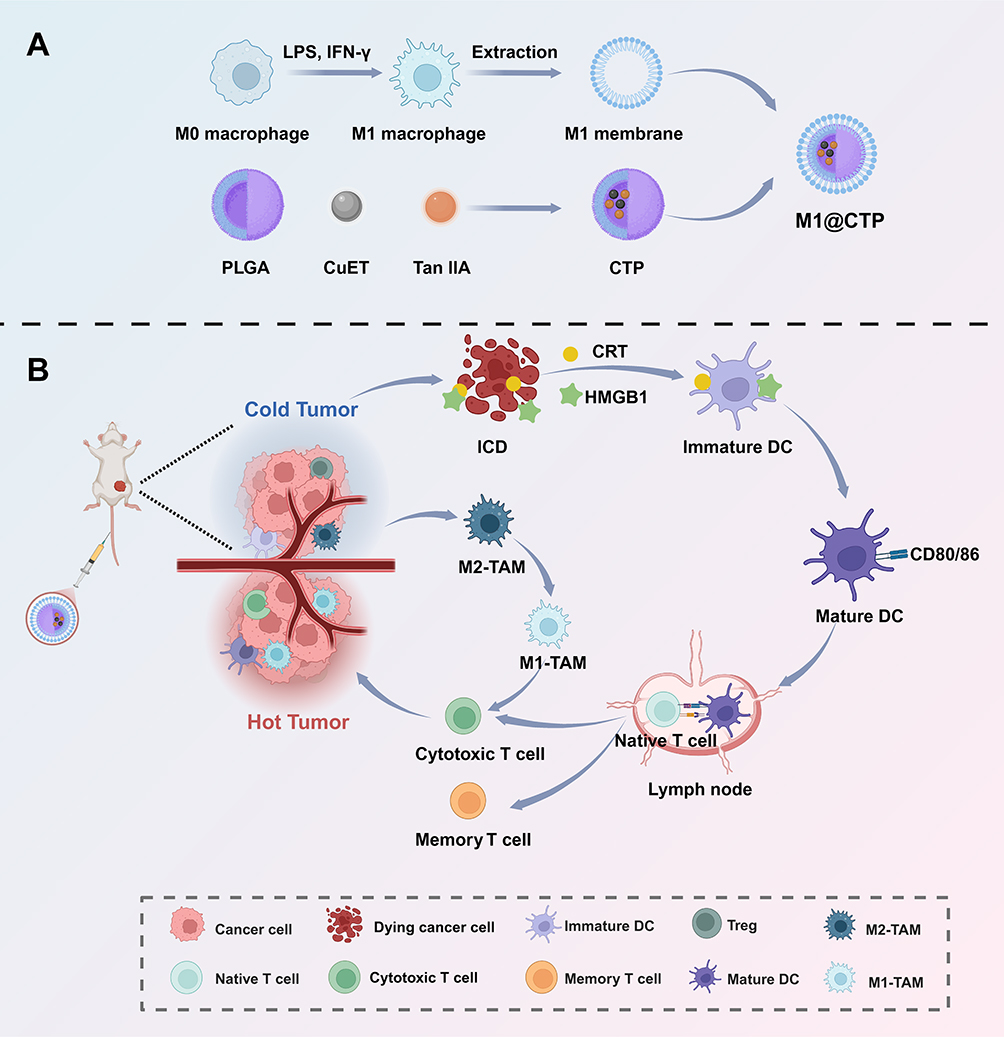 |
Figure 1 Schematic diagram of the preparation of M1@CTP nanoplatform and its antitumor mechanism. (A) Synthesis of M1@CTP nanoparticles. (B) M1@CTP induces immunogenic cell death (ICD) in tumor cells, triggering DAMP release and dendritic cell (DC) maturation. Mature DC activates CD8⁺ T cell and memory responses, while M1@CTP reduces immunosuppressive Tregs and M2 macrophages. These synergistic actions collectively convert immunologically “cold” tumors into “hot” ones, eliciting potent antitumor immunity. |
Materials and Methods
Materials
Tan IIA (purity ≥ 97%), CuET, PLGA, and polyvinyl alcohol (PVA) were purchased from Macklin Biochemical Technology Co., Ltd. (Shanghai, China). Dichloromethane (DCM) was purchased from Aladdin Biochemical Technology Co., Ltd (Shanghai, China). Roswell Park Memorial Institute (RPMI)-1640 medium, Dulbecco’s Modified Eagle Medium (DMEM), and fetal bovine serum (FBS) were obtained from Thermo Fisher Scientific, Inc (Massachusetts, USA). The Annexin V-Fluos staining kit was procured from Vazyme Technology Co., Ltd. (Nanjing, China). Recombinant mouse IFN-γ and GM-CSF were purchased from PeproTech, Inc ((New Jersey, USA). PE anti-mouse CD11c antibody, FITC anti-mouse CD86 antibody, APC anti-mouse CD80 antibody, APC anti-mouse CD3 antibody, Brilliant Violet 421 anti-mouse CD8a antibody, PerCP/Cyanine5.5 anti-mouse CD8a antibody, PE/Cyanine7 anti-mouse CD279 (PD-1) antibody, KO525 anti-mouse CD11b antibody, PE anti-mouse F4/80 antibody, APC anti-mouse CD206, FITC anti-mouse CD4 antibody, PE anti-mouse CD62L antibody, APC/FireTM 750 anti-mouse CD44 antibody, and Brilliant Violet 421 anti-mouse FOXP-3 antibody were purchased from BioLegend, Inc (California, USA). HMGB1 Antibody, Calreticulin Rabbit Monoclonal Antibody were purchased from Cell Signaling technology. Anti-mouse PD-1 monoclonal antibody was purchased from BioXcell (West Lebanon, USA).
Cell Culture and Mice
Mouse CRC cell line CT26 and the mouse macrophage line RAW264.7 were purchased from the Cell Bank of the Chinese Academy of Sciences (Shanghai, China). CT26 cells were cultured in RPMI-1640 medium, while RAW264.7 cells were cultured in DMEM. Both media were supplemented with 10% FBS and 1% penicillin‒streptomycin solution (Beyotime, Shanghai, China). All cell lines used in this study were authenticated by short tandem repeat (STR) profiling and confirmed to be free of mycoplasma contamination prior to experimentation.
Five- to six-week-old BALB/c and C57BL/6 female mice were purchased from GemPharmatech (Nanjing, China) and maintained in the specific pathogen-free (SPF) Laboratory of the Animal Center of the Affiliated Nanjing Drum Tower Hospital of Nanjing University Medical School (Nanjing, China), and housed under specific pathogen-free conditions (23 °C, 55 ± 5% humidity, 12 h light/dark cycle) with ad libitum access to food and water. All the animal experiments were conducted in accordance with the ARRIVE guidelines and approved by the Animal Ethics Committee of Drum Tower Hospital (2025AE01061).
Synthesis of CuET/Tan IIA/PLGA Nanoparticles
CuET/Tan IIA/PLGA (CTP) nanoparticles were synthesized using a previously reported two-step emulsification method.37 Briefly, 5 mg Tan IIA, 0.5 mg CuET, and 10 mg PLGA were dissolved in 0.5 mL of DCM to form the oil phase. This solution was then slowly added to 1.5 mL pre-cool 2% (w/v) PVA solution and sonicated under an ice bath for 10 min to form colostrum. Subsequently, 2.5 mL of a 0.5% (w/v) PVA solution was added, and ultrasound was continued for another 10 min to obtain stable double emulsion. The DCM solvent was removed by rotary evaporation at 37°C and 100 rpm for 30 min. The resulting nanoparticle suspension was subjected to centrifugation at 3500 rpm for 15 min using a 50 kDa cut-off molecular weight centrifugal filter (Sartorius, Germany) and repeated washing three times to remove free reagents. The purified CTP nanoparticles were finally lyophilized and stored at 4°C for further use.
Separation of M1 Macrophage Membrane
The M1-polarized macrophages were generated by using a previously reported method.38 In brief, RAW264.7 cells were stimulated with LPS (100 ng/mL) and IFN-γ (50ng/mL) for 24 h. The changes of morphology were observed under a microscope, and the expression levels of M1 markers CD80 and CD86 were detected by flow cytometry to verify the polarization effect.
For membrane extraction, the polarized macrophages were collected, washed with ice-cold PBS, and lysed in RIPA buffer supplemented with protease inhibitors. After centrifugation (12,000 rpm, 4°C, 10 min), the supernatant was extruded 21 times through a 400 nm polycarbonate membrane to ensure complete lysis. The homogenized suspension was subsequently centrifuged at 700 g for 10 min to remove debris. A final centrifugation at 14,000 g (4°C, 30 min) pelleted the purified M1 macrophage membranes. The M1 membrane was resuspended in PBS and stored at −80°C.
Synthesis and Characterization of M1@CTP NPs
For coating, the M1 membrane was mixed with CTP NPs at a mass ratio of 1:5 and co-extruded for 11 cycles to produce M1@CTP NPs, which were stored in PBS at 4°C.
The morphology of the nanoparticles was visualized by transmission electron microscopy (TEM) and further analyzed by confocal laser scanning microscopy (CLSM, Olympus, Japan). Fourier-transform infrared spectroscopy (FTIR, Thermo Fisher Scientific, USA) was employed to probe the chemical interactions between Tan IIA, CuET and PLGA NPs. The hydrodynamic properties, including average particle size, PDI, and size distribution, were measured by dynamic light scattering (DLS) using a Malvern Zetasizer Nano ZS (Malvern Panalytical, UK). Successful coating with the M1 macrophage membrane was verified by sodium dodecyl sulfate-polyacrylamide gel electrophoresis (SDS-PAGE), which confirmed the presence of characteristic membrane proteins. Finally, the drug release kinetics were studied by placing samples in dialysis bags with a molecular weight cutoff of 10 kDa and monitoring Tan IIA release into PBS at 37°C under varying pH conditions over 7 days.
Cytotoxicity Assay
The cytotoxicity of NPs and free Tan IIA against CT26 cells was evaluated using the Cell Counting Kit-8 (CCK-8) assay. Briefly, cells were seeded in 96-well plates and cultured overnight. Subsequently, the culture medium was replaced with fresh medium containing TP, CTP, M1@CTP or free Tan IIA at a series of gradient concentrations. After 24 hours of incubation, 10 µL of CCK-8 reagent was added to each well and incubated for an additional 1 hour. The absorbance at 450 nm was finally measured using a microplate reader. (Thermo Fisher Scientific, USA).
Analysis of Cell Apoptosis
The apoptosis of CT26 cells induced by Tan IIA/PLGA (TP), CuET/PLGA (CP), CTP, or M1@CTP ((Tan IIA concentration 5 μg/mL, CuET concentration 0.5 μg/mL) was analyzed using an Annexin V-Fluos staining kit according to the manufacturer’s instructions. After being treated with different formulations for 24 hours, the cells were collected, washed with PBS, and resuspended in 500 µL of Binding Buffer. The cell suspension was then stained with 5 µL of Annexin V-FITC and 5 µL of Propidium Iodide (PI) for 15 minutes in the dark at room temperature. The stained cells were immediately analyzed by flow cytometry (Beckman, USA).
Calcein-AM/PI Staining
To further demonstrate the therapeutic efficacy of M1@CTP NPs, calcein-AM and propidium iodide (PI) were used to identify live cells and dead cells. Briefly, CT26 cells were incubated with normal saline (NS), TP, CP, CTP, or M1@CTP. After 24 h incubation, the treated cells were stained with calcein-AM and PI, subsequently observed by fluorescent microscope.
Cellular Uptake
To investigate the homologous targeting and immune evasion properties conferred by the M1 membrane coating, cellular uptake of the NPs was assessed in different cell lines. Specifically, the mouse CRC cell line CT26 was used to evaluate targeting specificity. Additionally, the mouse macrophage line RAW264.7 was employed to assess phagocytic uptake, reflecting immune evasion. The cells were respectively incubated with the Dil-labeled NPs for 1 hour. Cell nuclei were labeled with DAPI. The cellular internalization of NPs was then visualized using CLSM.
Induction of ICD and Activation of Immune Cells
CT26 cells were treated with NS, CP, or M1@CTP for 24 h. For CRT exposure and HMGB1 distribution, the cells were incubate with first antibody overnight, further treated with second antibody for another 1 h, and observed by CLSM.
To evaluate the immunostimulatory effects of ICD on dendritic cells (DCs) in vitro, C57BL/6 mice were euthanized, and their femurs and tibias were aseptically irrigated with PBS. Bone marrow cells were isolated following red blood cell lysis, and cultured in DMEM supplemented with 10% fetal bovine serum (FBS) and 20 ng/mL granulocyte-macrophage colony-stimulating factor (GM-CSF) for 5 days to induce the formation of bone marrow-derived dendritic cells (BMDCs). DC maturation in vitro was evaluated by transwell system. CT26 cells in the upper chamber of co-culture system were incubated with corresponding NPs and co-cultured with BMDCs in the lower chamber for 24 h, flow cytometry was used to assess the maturation of BMDCs. Subsequently, BMDCs were collected and mixed with mouse spleen lymphocytes at a ratio of 1:10 for 24 h, the proportion of CD8+ T cells in lymphocytes was determined by flow cytometry.
Establishment of Colorectal Cancer Model and Biodistribution
To establish the model of subcutaneous CRC tumor-bearing mice, CT26 cells (0.5–1.0 × 10⁶) suspended in 100 µL of PBS were subcutaneously (s.c.) injected into the left inguinal region of BALB/c mice. The tumor dimensions were measured with calipers, and the tumor volume was calculated using the formula V = Length×Width2/2.
To evaluate the in vivo targeting capability of the M1 membrane coating, tumor-bearing mice were randomly divided into two groups and intravenously injected (i.v.) with either PLGA/DiR or M1@PLGA/DiR NPs. The mice were anesthetized and imaged at predetermined time points using a CRi Maestro in vivo imaging system (Cambridge Research & Instrumentation, USA). At 48 hours post-injection, the mice were euthanized, and major organs along with tumors were harvested and subjected to ex vivo fluorescence imaging to further validate the targeting efficiency.
Evaluation of Antitumor Efficacy and Safety
CT26 CRC cells were used to establish subcutaneous tumor xenograft models as the protocols mentioned above. The tumor dimensions were measured with calipers, and the tumor volume was calculated using the formula V = Length×Width2×0.5. Once the tumors reached a predetermined size, the mice were randomized into groups according to the experimental design for subsequent treatments.
To evaluate the anti-tumor effect, TME remodeling and systemic safety, tumor-bearing mice were randomly divided into 5 groups and injected intravenously via tails with NS, TP, CP, CTP, or M1@CTP at a Tan IIA dose of 10 mg/kg, once every four days, for a total of three administrations. Tumor volume and body weigh were monitored every 2 days. At the study endpoint, mice were euthanized by cervical dislocation, solid tumors were collected to detect changes in immune cell populations within the TME via flow cytometry. Tumor cell proliferation was assessed by immunohistochemical staining for Ki67. Major organs (heart, liver, spleen, lungs, and kidneys) and tumors were harvested and processed for Hematoxylin and Eosin (H&E) staining. Blood samples were collected for serum biochemical analysis. Systemic toxicity was evaluated based on the biochemical profiles and histological examination of H&E-stained organ sections.
To assess the efficacy of the combined therapy, another cohort of tumor-bearing mice were randomly assigned to 4 groups and administered NS, M1@CTP, anti-mouse PD1 monoclonal antibody (αPD-1), or M1@CTP combined with αPD-1 once every four days, for a total of three administrations. αPD-1 was administered via intraperitoneal injection (i.p.) at a dose of 100 μg per mouse, while M1@CTP was delivered intravenously as described above. Tumor volume and body weight were monitored every 2 days. At the experimental endpoint, defined as a tumor volume of 1500 mm3 or a maximum tumor diameter of 20 mm, mice were euthanized by cervical dislocation.
Analysis of Immune Cells
To characterize the efficacy of M1@CTP NPs within TME, the immune cells were isolated from spleen, tumor-draining lymph nodes (TDLNs) and tumor tissues 3 days after the last administration. Minced tissues were digested with collagenase at 37 °C for 2 hours, passed through a 70 μm filter to prepare single-cell suspensions, and then incubated with flow cytometry antibodies at 4 °C for 30 minutes. Antibody staining was performed at 4 °C for 30 minutes, followed by washing and flow cytometry analysis. The flow cytometry results were analyzed with FlowJo software (version 10.8.1).
Statistical Analysis
Statistical analysis was performed using GraphPad Prism (version 9.1.0). Two-tailed Student’s t tests were used for comparisons between two groups, whereas one-way or two-way ANOVA was used for comparisons among multiple groups. Data are presented as mean ± standard deviation unless otherwise specified; ns, not significant, *P < 0.05, **P < 0.01, ***P < 0.001, ****P < 0.0001.
Results
Preparation and Characterization of the M1@CTP NPs
The application of CuET and Tan IIA in biomedicine is significantly limited due to their poor water solubility. As an FDA-approved nanocarrier, PLGA nanoparticles have good biocompatibility and controllable biodegradability, which can enhance the physicochemical properties of the encapsulated drugs, thereby improving safety and therapeutic efficacy.39 A two-step emulsification method was employed to synthesize CuET/Tan II PLGA nanoparticles, denoted as CTP NPs. Fourier transform infrared spectroscopy (FTIR) was used to investigate the chemical interactions between the components. As shown in Figure 2A, drug-loaded nanoparticles CTP showed characteristic absorption peaks of each component at 1759 cm−1 (PLGA), 1583 cm−1 (Tan IIA), and 996 cm−1 (CuET), and the peak intensities were all slightly weakened. This indicates that Tan IIA and CuET have been successfully loaded in PLGA nanoparticles without destructive chemical interactions between the components.
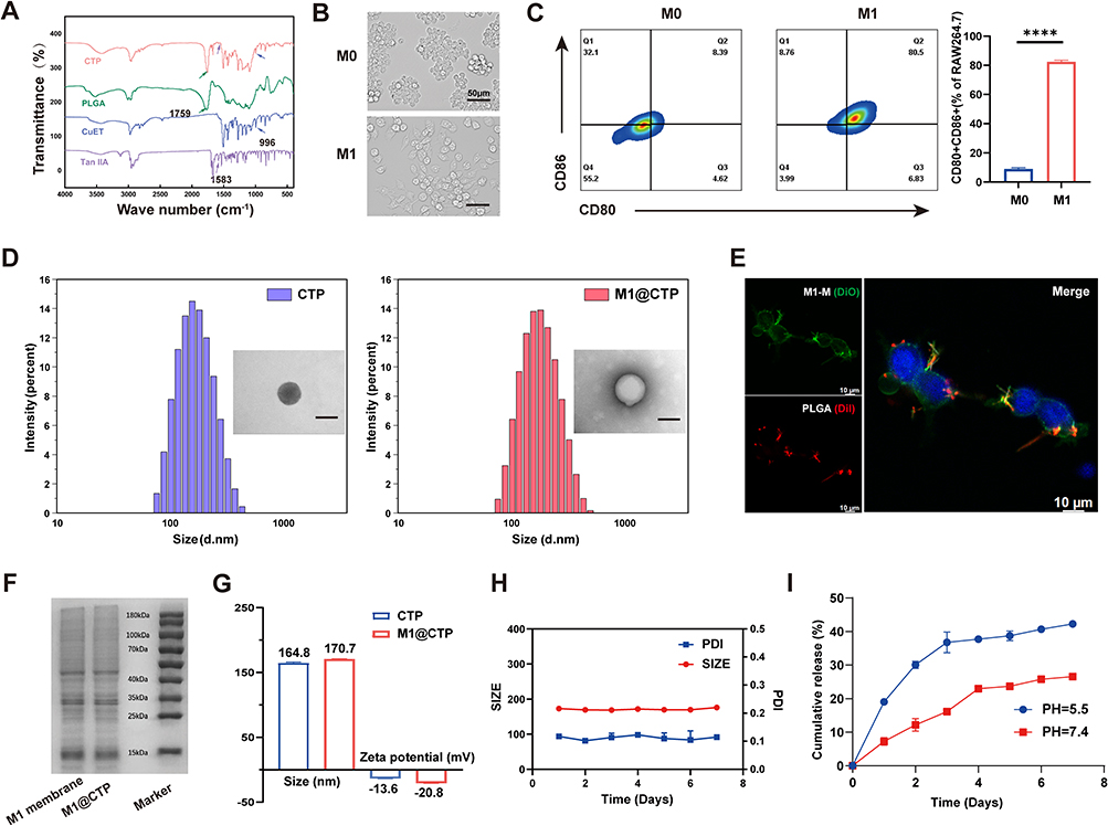 |
Figure 2 Characterization of M1@CTP NPs. (A) Fourier-Transform Infrared Spectroscopy (FTIR) spectra of Tan IIA, CuET, PLGA and CTP NPs. (B) Microscopic images of RAW264.7 cells before and after treating with LPS and IFN-γ. Scale bar: 50 μm. (C) Representative images and percentages of M1 phenotype (CD80+ CD86+) macrophages in RAW264.7 cells before and after treating with LPS and IFN-γ. (D) Size distribution and TEM image of CTP and M1@CTP NPs. Scale bar: 100 nm. (E) Confocal fluorescence images of colocalization between DiO-labeled M1 membrane and Dil-labeled PLGA. Scale bar: 10 μm. (F) SDS-PAGE protein analysis of M1membrane and M1@CTP with Coomassie blue staining. (G) Particle size and zeta potential of CTP and M1@CTP NPs. (H) Size and PDI of M1@CTP NPs in PBS over 7 days. (I) Cumulative Tan IIA release from M1@CTP NPs under different pH conditions for 7 days. ****P < 0.0001. |
RAW264.7 macrophages were labeled as M0 macrophages and polarized into M1-phenotype macrophages by stimulation with 100ng/mL LPS and 50ng/mL IFN-γ. The morphological differences of the stimulated macrophages were observed under the microscope (Figure 2B). The expression of M1 markers CD80 and CD86 was measured by flow cytometry, and the results showed that the expression of CD86+CD80+ on stimulated macrophages was significantly increased to more than 80%, while the expression of CD86+CD80+ on M0 macrophages was less than 10% (Figure 2C). These results indicated that the majority of M0 macrophages were successfully polarized to the M1 phenotype. To verify the successful coating of the M1 membrane and construction of the M1@CTP, we systematically characterized its morphology, physicochemical properties, and environmental stability. The morphologies of the CTP and M1@CTP NPs were determined through transmission electron microscopy (TEM), the particles were nearly spherical and uniformly dispersed, Notably, a distinct membrane coating was observed on M1@CTP, confirming the successful formation of a core-shell structure (Figure S1A), diameter profiling by dynamic light scattering (DLS) revealed distributed around 173.3 nm (Figure 2D). Dil and Dio were labeled PLGA NPs and M1 membrane, respectively, the merged red and green fluorescence further validated the successful coating by confocal laser scanning microscopy (CLSM) (Figure 2E). We confirmed the retention of membrane proteins during NPs manufacturing by analyzing proteins on M1@CTP and M1 membrane using SDS-PAGE (Figure 2F). Additionally, CCR2 and integrin α4, which are associated with macrophage migration and tumor-homing behavior, as well as the M1 marker iNOS, were all retained in M1@CTP after membrane coating (Figure S1B). Membrane coating increased particle size from approximately 164.8 nm to about 170.7 nm. Zeta potential shifted from around −13.6 mV to approximately −20.8 mV, consistent with the expected contribution from the negatively charged M1 membrane, which further confirms the successful coating (Figures 2G and S1C).
The size and PDI of M1@CTP NPs, monitored by DLS in PBS over 7 days, remained stable, indicating good nanoparticle stability (Figure 2H). Subsequently, we investigated the in vitro release profile of Tan IIA from M1@CTP NPs under different pH conditions, NPs released only less than 10% of the drug in the first 24 h at pH 7.4, whereas a significant increase in Tan IIA release was observed at pH 6.5 (Figure 2I), a condition mimicking the acidic TME. This pH-dependent release profile is attributed to the acid-sensitive nature of PLGA.40 Thus, M1@CTP NPs constitute a promising drug nanocarrier platform for targeting the slightly acidic TME.
In vitro Uptake and Cytotoxicity
Given that we verified the retention of M1 membrane proteins on M1@CTP, we therefore performed in vitro cellular uptake assays to assess the resulting immune escape and tumor targeting ability conferred by M1 membrane. In order to visualize the uptake of NPs by cells, Dil was used to label NPs and DAPI was used to label cell nucleus. CT26 cells showed more effective internalization of PLGA/Dil than M1@PLGA/Dil NPs at the same incubation time, as observed by CLSM. In addition, Compared with PLGA/Dil, M1@PLGA/Dil is less susceptible to phagocytosis by macrophage cell line RAW264.7 cells, with reduced internalization of fluorescent material (Figure 3A). Collectively, these results demonstrate that the homologous M1 membrane encapsulation plays a crucial role in reducing immunogenicity and achieving specific targeting.
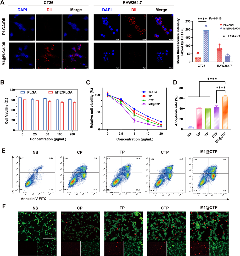 |
Figure 3 In vitro antitumor therapeutic efficacy of M1@CTP. (A) CLSM images and quantification of the cellular uptake of Dil-labelled nanoparticles in CT26 tumor cells and RAW264.7 macrophage cells. Cell nuclei were stained with DAPI. Scale bar = 10 μm. (B) Cell viability of CT26 cells under different conditions for 24 h (n = 3). (C) The anticancer efficacy of various treatments against CT26 cells (n = 3). (D and E) Detection of apoptosis in CT26 cells treated with NS, CuET/PLGA (CP), Tan IIA/PLGA (TP), CuET/Tan IIA/PLGA (CTP) or M1@CTP after 24 hours by flow cytometry. (F) Fluorescence images of calcein-AM /PI co-stained CT26 cells treated with various formulations. The green and red fluorescence represented live cells and dead cells, respectively. Scale bar: 200 µm. *P < 0.05;****P < 0.0001. |
CCK-8 assays were performed to evaluate the cytotoxicity of the nanocarrier PLGA and the M1 membrane-coated M1@PLGA. The results showed that even at a concentration of 200 µg/mL of PLGA, the cell viability remained above 80%, demonstrating the favorable biocompatibility of both the M1 membrane and the nanocarrier (Figure 3B).
Previous studies have reported that Tan IIA exhibits antitumor activity. We verified by CCK-8 assay that the modified M1@CTP retained the cytotoxicity of Tan IIA on tumor cells and shown to be dose-dependent. As shown in Figure 3C, the TP nanoparticles encapsulating Tan IIA with PLGA exhibited greater cytotoxicity than free Tan IIA. This may be attributed to the nano-encapsulation overcoming the intrinsic solubility limitations of the free drug. The CTP group demonstrated enhanced cytotoxicity over TP alone, suggesting a stronger synergistic effect between the loaded agents. Notably, a significant enhancement in cytotoxicity was observed in the M1@CTP group, surpassing the CTP group. This can be attributed to the M1 membrane coating, which further augmented the tumor cell killing effect by enhancing cellular uptake.
To quantify the therapeutic efficacy of M1@CTP, the apoptosis of CT26 cells was quantitatively analyzed via flow cytometry. While the TP and CP single-treatment groups showed certain pro-apoptotic activity, the M1@CTP treatment induced the highest level of apoptosis (62.4%) compared to all other groups (Figure 3D and E). To visually assess the in vitro antitumor activity, CT26 cells were co-stained with calcein-AM and PI. CT26 cells treated with CTP NPs and M1@CTP NPs showed stronger red fluorescence than those treated with TP or CP, whlie M1@CTP inducing the most significant cytotoxicity and antiproliferative effects (Figures 3F and S2). This underscores the precise tumor targeting and the synergistic effect of the loaded agents, validating the feasibility and efficacy of this nanoplaform.
Induction of ICD and DC Maturation in vitro
Immunogenic cell death (ICD) describes the conversion of tumor cells into an immunogenic form, whereby dying under specific external stimuli can initiate a potent anti-tumor immune response.41 CuET has been reported to induce ICD in tumor cells and promote the expression and release of damage-associated molecular patterns (DAMPs).10,14,15 These DAMPs include calreticulin (CRT) translocation to the cell membrane and high mobility group box 1 (HMGB1) secretion, which are recognized by DCs as endogenous danger signals and promote their maturation. The mature DCs subsequently activate T cells effectively, initiating downstream immune responses (Figure 4A).7,41,42 The effect of M1@CTP in triggering ICD was verified by evaluating the exposure and release of DAMPs after different treatments with CT26 tumor cells. CLSM images showed that both CP and M1@CTP treatments increased the exposure of CRT on the surface of CT26 cells (Figures 4B and S3). In addition, the extracellular release of HMGB1 in the CP and M1@CTP treatment groups was observed by immunofluorescence, while HMGB1 was mainly localized in the nucleus in the NS control group (Figure 4C). Notably, M1@CTP NPs induced the most significant translocation of CRT to the cell membrane and HMGB1 release, this superior effect can be attributed to the co-delivery of CuET and Tan IIA likely induces complementary cell death pathways, leading to amplified DAMP release. Additionally, the PLGA nanocarrier combined with the active targeting of the M1 membrane ensures highly efficient and tumor-specific drug delivery, maximizing the local ICD trigger.
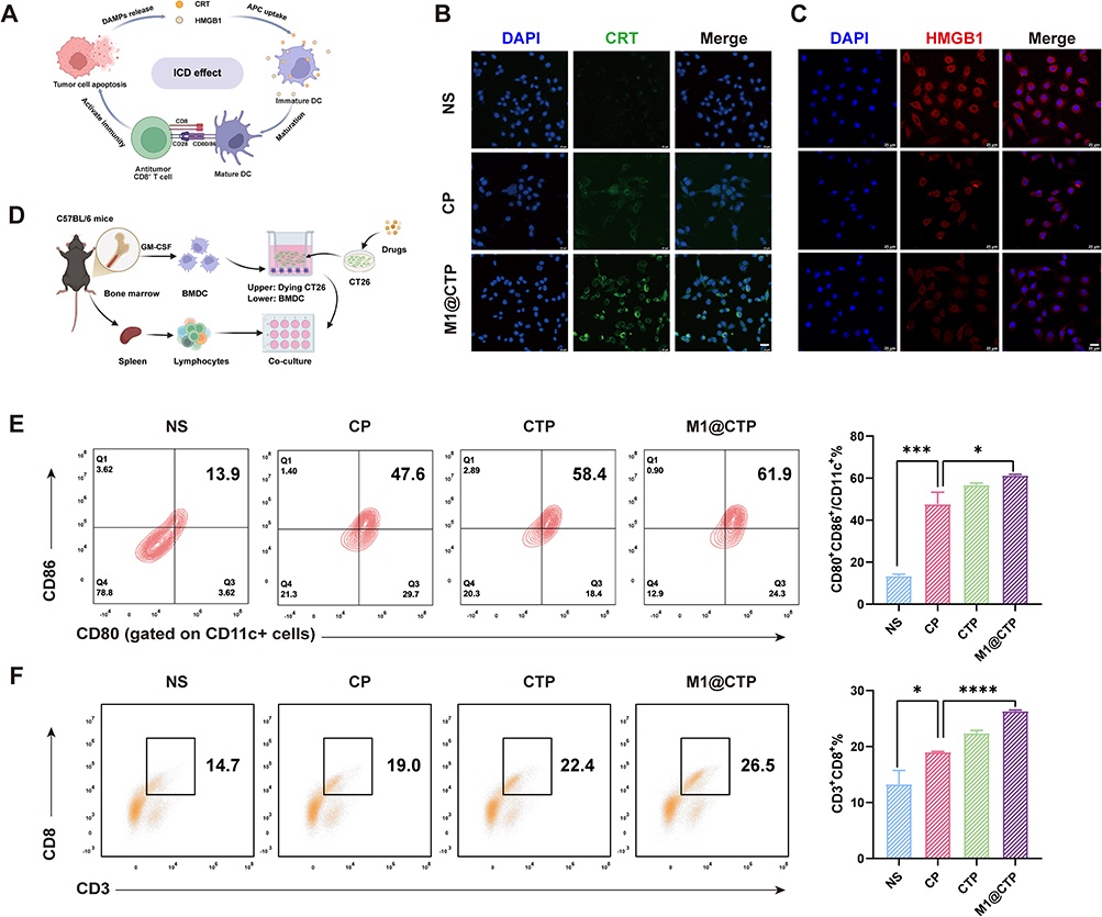 |
Figure 4 Immunogenic cell death (ICD) induced by M1@CTP NPs to activate the immune response. (A) Schematic diagram of the mechanism of induced ICD. (B) Confocal images of CRT expression of CT26 cells after incubating with NS, CP NPs and M1@CTP NPs. Scale bar: 25 μm. (C) The release of HMGB1 in CT26 cells was visualized through fluorescence microscopy after different treatments. Scale bar: 25 μm. (D) A schematic illustration of the M1@CTP stimulated maturation of BMDCs and activated T cells immunity. (E and F) Flow cytometry plots and quantitative analysis of mature BMDCs and CD3⁺CD8⁺ T lymphocytes following their co-culture with CT26 cells treated with different agents (n = 3). *P < 0.05;***P < 0.001; ****P < 0.0001. |
DCs are widely recognized as the most potent antigen-presenting cells and mature DCs play a key role in ICD-induced anti-tumor immune responses. To evaluate downstream immune activation, we co-cultured bone marrow-derived dendritic cells (BMDCs) with CT26 cells treated with NS, CP, CTP or M1@CTP, and evaluated the maturation of DC by measuring the expression of the costimulatory molecules CD80 and CD86 via flow cytometry (Figure 4D). The proportion of mature BMDCs in the CP, CTP or M1@CTP groups were upregulated to 47.6%, 58.4%, and 61.9%, respectively (Figure 4E). This indicates that CuET induces DC maturation in the co-culture system by triggering ICD in tumor cells. The combination of Tan IIA and the M1 membrane synergized with CuET to further promote BMDC maturation, likely due to amplified DAMPs release, apoptotic tumor cell debris, and the immunostimulatory contribution of the M1 membrane collectively. The M1@CTP group displayed the highest proportion of mature DCs, which is consistent with its superior efficacy in inducing ICD.
CD8+ T cells play a critical role in directly eliminating tumor cells. To better simulate DC-mediated T cell activation, we co-cultured the above mature, antigen-loaded DCs with mouse spleen lymphocytes. Flow cytometric analysis revealed a significant increase in the proportion of CD3+ CD8+ T cells in the M1@CTP group (Figure 4F). Together, these findings suggest that M1@CTP induces and amplifies ICD in tumor cells through the synergistic action of its components, which in turn induces DC maturation and robust antitumor immune responses.
In vivo Biodistribution
We injected the DiR-labeled preparation intravenously into CT26 tumor-bearing mice, the biodistribution of two groups NPs (PLGA/DiR and M1@PLGA/DiR) was detected via Near-infrared (NIR) fluorescence imaging.
The nanoparticles continued to accumulate at the tumor site two hours post-injection. Notably, the fluorescence intensity of M1@PLGA/DiR remained higher than that of PLGA/DiR at subsequent time points. The fluorescence intensity of both groups peaked at 48 hours, where the intensity of M1@PLGA/DiR was 1.3-fold higher than that of PLGA/DiR (Figure 5A).
 |
Figure 5 In vivo biodistribution of M1@CTP. (A) Representative real-time near-infrared (NIR) fluorescence imaging of CT26 tumor-bearing mice after intravenous injection of PLGA/DiR NPs and M1@PLGA/DiR NPs, Tumor regions were circled for clarity. (B and C) Ex vivo NIR images of major organs and tumors harvested from mice at 48 hours post-injection of the two different NPs. *P < 0.05. |
At 48 hours post-injection, major organs and tumors were harvested and subjected to ex vivo imaging. As shown in Figure 5B and C, the fluorescence intensity in tumors from the M1@PLGA/DiR group was higher than that in PLGA/DiR, and there was not much accumulation in non-target organs such as liver. This indicates that M1 membrane-derived encapsulation effectively accumulate in tumor tissues with small off-target distribution, confirming the good biodistribution and significant tumor selectivity of CCM-LDH.
Immunotherapeutic Effects of M1@CTP in vivo
Based on the anti-tumor efficacy in vitro, we sought to evaluate the performance of M1@CTP against solid tumors in vivo. We selected CRC, a highly immunosuppressive tumor, and established a CT26 subcutaneous tumor model in BALB/c mice. Mice were injected intravenously with with NS, TP, CP, CTP or M1@CTP once every four days for a total of three doses (n=5) (Figure 6A). Treatment with M1@CTP significantly delayed tumor growth compared to all other groups (Figure 6B–D). In addition, no significant differences in body weight were observed among the treatment groups, indicating that NPs did not induce significant in vivo toxicity (Figure 6E). These results suggest that targeted and efficient drug delivery to tumors can enhance antitumor efficacy.
 |
Figure 6 Antitumor efficacy and immune response following treatment with M1@CTP. (A) Schematic diagram of the treatment protocol used in CT26 tumor-bearing mice. (B and C) Subcutaneous CT26 tumors in BALB/c mice were treated with NS, TP, CP, CTP or M1@CTP, and tumor growth curves are shown (5 mice per group). (D) Tumor weight from each group after treatment. (E) Body weight of CT26 tumor-bearing mice in the five treatment groups during the treatment period (n = 5). (F) Percentages of mature dendritic cell (mDC), CD8+ T cell, regulatory T cell (Treg), and and the M1/M2 macrophage ratio in tumors (n=5). (G) Percentages of mDC, CD8+ T cell in tumor-draining lymph node (TDLN) (n=5). (H) Percentages of Treg and central memory T cell (Tcm) in spleens (n=5). (I) Representative immunofluorescence images of CD8+ T-cell infiltration in tumor sections after different treatments. (J and K) Representative images of TUNEL immunofluorescence and Ki67 immunostaining of tumor sections. Scale bar: 100μm. ns, not significant;*P < 0.01; ***P < 0.001; ****P < 0.0001. |
To investigate the immunomodulatory mechanism of tumor suppression, we collected tumors, spleens, and draining lymph nodes (TDLNs) on day 3 after the last treatment for flow cytometry analysis. Results showed that M1@CTP treatment increased the proportion of mature DC and CD8+ T cells within tumors, while reducing the proportion of immunosuppressive Tregs (Figure 6F). These results demonstrated that M1@CTP effectively promoted the infiltration of lymphocytes into tumors, suggesting enhanced antigen presentation and improved T cell activating. Analysis of tumor-associated macrophages (TAM) suggested that M1@CTP with M1 membrane coating significantly increased the ratio of anti-tumor M1 macrophages to pro-tumor M2 macrophages compared with the CTP group, this M2-to-M1 phenotype shift suggested that M1@CTP could activate antitumor immunity by modulating macrophage polarization and relieving local immune suppression.43,44 M1@CTP treatment similarly increased the infiltration of mature DC and CD8+T cells in TDLNs, indicating effective activation of systemic immune responses (Figure 6G). Furthermore, M1@CTP treatment group showed a decreased proportion of Tregs and an elevated proportion of central memory T cells (Tcm) in spleens (Figure 6H), underscoring the establishment of long-term antitumor immunity. Moreover, immunofluorescence staining of tumor sections revealed markedly enhanced CD8+ T-cell infiltration in the M1@CTP group, further confirming the robust recruitment of cytotoxic lymphocytes into the TME (Figures 6I and S4A). The remodeling of the TME achieved by M1@CTP significantly induced tumor cell apoptosis and inhibited proliferation (Figures 6J, K and S4B, C).
In summary, these results demonstrate that the synergistic action of components within M1@CTP drives anti-tumor immunity by initiating immune responses in the lymph nodes, relieving immunosuppressive TME and enhancing cytotoxic activity in the tumors, and inducing systemic immune memory.
Biosafety Assessment
To assess the translational potential of M1@CTP in immunotherapy applications, we performed in vivo biosafety assessments by histopathological examination and serum biochemical assays. The main organs were collected three days after the final treatment for H&E staining. The structure of heart, liver, spleen, lung, and kidney in each treatment group was intact without abnormal pathological changes (Figure 7A), and the serum biochemical markers of liver and kidney were maintained at normal levels comparable to the NS group (Figure 7B). These findings confirm that the administration of M1@CTP does not cause liver or kidney injury and exhibits excellent biocompatibility and systemic safety, supporting its use as a promising nanoplatform for further clinical development.
 |
Figure 7 In vivo Biosafety evaluation of M1@CTP. (A) Representative hematoxylin and eosin (H&E) staining images of major organs excised from CT26 tumor-bearing mice after different treatments. Scale bar: 50 μm. (B) Serum biochemical analysis of major function indicators of mice (n=5). The data are shown as the means with SEMs. Abbreviations: ALT, Alanine Aminotransferase; AST, Aspartate Aminotransferase; AKP, alkaline phosphatase; BUN, Blood Urea Nitrogen; CRE, Creatinine; ns, not significant. |
Combined Therapy with αPD-1
Given the demonstrated capacity of M1@CTP treatment to recruited CTLs to tumors, we next examined its potential to enhance the response to ICI. We evaluated the therapeutic potential of M1@CTP in combination with αPD-1 in a CT26 subcutaneous tumor model (Figure 8A). While M1@CTP monotherapy exhibited good antitumor efficacy, the combination with αPD-1 resulted in the most significant tumor suppression and survival benefit (Figure 8B–E), without inducing any significant change in body weight (Figure 8F).
 |
Figure 8 M1@CTP enhanced anti-PD-1 immunotherapy in a CRC model. (A) Schematic diagram of the treatment schedule for M1@CTP and αPD1 in BALB/c mice bearing CT26 tumors. (B and C) Subcutaneous CT26 tumors in BALB/c mice were treated with NS, M1@CTP, αPD-1, or M1@CTP+αPD-1, and tumor growth curves of each mouse in four groups (n=5). (D) Tumor weight from each group after treatment. (E) Survival curves of different treatment groups for 60 days (n = 5). (F) Body weight of CT26 tumor-bearing mice in the five treatment groups during the treatment period (n = 5). (G–I) Representative images and percentages of mature dendritic cell, CD8+ T and Treg in tumors from BALB/c mice 3 days after the final injection in different groups (n=5). (J) Representative images and percentages of M2 macrophage and M1macrophage in CT26 tumors (n=5). **P < 0.01; ****P < 0.0001. |
To determine whether T cell recruitment by M1@CTP underlies the enhanced efficacy of combination therapy with αPD-1, we analyzed the proportion of immune cells within TME. Flow cytometry analysis showed that the combination of M1@CTP and αPD-1 significantly increased the number of activated DC and CD8+ T cell in both tumors and draining lymph nodes (Figures 8G, H and S5), while the immunosuppressive Treg in the TME was decreased compared with αPD-1 alone (Figure 8I). In addition, the combination therapy significantly reshaped the TME into an immune-active one, characterized by a downregulation of M2 macrophages and Tregs and an upregulation of M1 macrophages (Figure 8J). These results confirmed that our nanoplatform can effectively recruit CTL and reshape the tumor immune landscape, thereby sensitizing CRC to αPD-1 treatment and presenting a promising approach for combination therapy in CRC.
Discussion
Nano-drug delivery system represents advanced drug delivery platform that utilizes various nanocarriers such as lipids, polymers, inorganic materials, and biomimetic structures to achieve targeted and controlled delivery of therapeutic agents.45,46 FTIR, TEM, CLSM, DLS, and SDS-PAGE techniques were employed to characterize the NPs in this study. Collectively, the results confirmed the successful M1 membrane camouflage with retention of functional immune-related proteins, a uniform core-shell architecture, and excellent stability. These properties endow the NPs with the capacity for immune evasion, tumor homing, and pH-responsive drug release, thereby establishing a solid foundation for subsequent immune modulation within the TME. Furthermore, in vivo biodistribution further confirmed that M1 membrane-camouflaged NPs exhibited significantly higher accumulation at the tumor site compared to uncoated counterparts, with relatively lower distribution in non-target organs such as the liver. This enhanced tumor-targeting ability facilitates efficient intratumoral drug delivery while minimizing adverse effects. It should be noted that two-dimensional fluorescence imaging is inherently semi-quantitative and limited by tissue penetration depth, therefore, future studies integrating three-dimensional quantitative imaging or radioisotope tracer technique will enable accurate acquisition of pharmacokinetic parameters and absolute organ-specific quantification.
ICD is a unique form of regulated cell death that not only eliminates cells but also actively stimulates an immune response against the released antigens from dying cells, acting as a bridge between dying cells and the immune system.7,47 However, due to the highly immunosuppressive TME, ICD-mediated immune activation is often insufficient to generate robust anti-tumor effects alone. Herein, we co-delivered the ICD inducer CuET and the natural anti-cancer compound Tan IIA via a PLGA nanocarrier, further coated with M1 membrane, achieving synergistic efficacy at the tumor site. This synergistic immune activation can be attributed to the following: CuET-triggered ICD provides abundant tumor antigens and danger signals; Tan IIA enhances the sensitivity of tumor cells to immune attacks through multiple pharmacological pathways; and the co-stimulatory molecules inherently carried by the M1 membrane further amplify antigen presentation and T-cell priming signals. Notably, this immune activation exhibits systemic and durable characteristics that extend far beyond the local tumor. In TDLNs, the M1@CTP treatment significantly increased the proportion of mature DCs and CD8⁺ T cells, indicating that antigen signals generated at the tumor site were effectively transmitted to secondary lymphoid organs, initiating a systemic adaptive immune response. In the spleen, M1@CTP treatment not only reduced the proportion of immunosuppressive Tregs but also significantly elevated the levels of central Tcm, marking the establishment of long-term immune memory. Collectively, the co-delivery of CuET and Tan IIA synergistically enhances ICD and immune activation, achieving efficient immunogenic killing locally, and extending this response into a systemic, durable anti-tumor immunity via TDLNs and the spleen, providing an immunological basis for preventing tumor recurrence. Future studies will include tumor rechallenge and metastasis models to validate long-term immune memory and systemic efficacy.
ICIs exhibit limited efficacy in “cold” tumors, which is primarily attributed to insufficient T‑cell infiltration and an immunosuppressive TME.48,49 In this study, M1@CTP monotherapy significantly increased intratumoral CD8⁺ T cell numbers and alleviated immunosuppressive barriers including Tregs and M2 macrophages. Building upon this, when the PD-1/PD-L1 inhibitory axis was subsequently blocked by αPD-1, these CD8⁺ T cells could fully restore their effector functions, generating a potent synergistic anti-tumor immune response. This combination strategy demonstrates that nanoparticle-mediated TME remodeling can overcome the primary resistance to ICIs, providing a rational and highly effective approach for expanding immunotherapy to traditionally “cold” tumors like colorectal cancer.
While emphasizing anti-tumor efficacy, the biosafety of M1@CTP is critical for its clinical translation. Our biosafety assessment, combining histological analysis and serum biochemical tests, confirmed that M1@CTP effectively inhibited tumor growth without causing obvious organ toxicity, highlighting its favorable short-term safety profile. Further long-term toxicity and pharmacokinetic experiments will be necessary to comprehensively confirm its safety for clinical use. In fact, achieving the optimal balance between maximizing anticancer efficacy and minimizing side effects on normal tissues remains a challenge in cancer treatment. Pioneering studies have successfully addressed this by demonstrating that the biocompatibility and tumor-selective cytotoxicity of inorganic nanomaterials can be significantly enhanced through sophisticated green synthesis and surface tailoring.50–52 These innovative approaches provide powerful tools for overcoming drug resistance and reducing non-specific toxicity via precise physicochemical engineering. Alongside these approaches, our M1@CTP platform employs a biological paradigm by leveraging the natural affinity of M1 membranes to achieve tumor targeting, alongside a biodegradable PLGA core. Despite the differing technological pathways, both strategies underscore the central role of rational surface design in improving biocompatibility. Together, these approaches advance the development of safe and potent next-generation cancer immunotherapies.
Conclusions
This study reports a biomimetic nanoplatform M1@CTP based on M1 macrophage membrane coating. This system actively delivers Tan IIA and CuET to tumor sites. The biomimetic design avoids off-target issues, demonstrating excellent tumor accumulation and retention. The components within the platform work synergistically to induce ICD, successfully remodel the immunosuppressive TME, and promote the infiltration of CTLs, thereby triggering a potent immune response and achieving effective tumor suppression, while maintaining a favorable biosafety profile. In addition, M1@CTP sensitizes CRC to αPD-1 treatment, the synergistic antitumor effect of this combination provides a new strategy for extending immunotherapy to cold tumors. These findings suggest that M1@CTP is a novel low-toxicity and efficient nanoplatform that can elicit systemic and robust antitumor immunity.
Data Sharing Statement
Data are available from the corresponding authors, Qun Zhang ([email protected]) and Xiaoping Qian ([email protected]), upon reasonable request.
Ethical Approval
All the animal experiments were conducted in accordance with the Guide for the Care and Use of Laboratory Animals and approved by the Animal Ethics Committee of Drum Tower Hospital (2025AE01061). The ARRIVE guidelines were used for the reporting of the in vivo experiments.
Author Contributions
Xueying Bai and Xingzhi Han should be considered as co-first authors. All authors made a significant contribution to the work reported, whether that is in the conception, study design, execution, acquisition of data, analysis and interpretation, or in all these areas; took part in drafting, revising or critically reviewing the article; gave final approval of the version to be published; have agreed on the journal to which the article has been submitted; and agree to be accountable for all aspects of the work.
Funding
The authors are grateful to the Jiangsu Health International Exchange Program for its support. This research was supported by the National Natural Science Foundation of China (82303970), the Nanjing Health Science and Technology Development Key Program (ZKX21028), the Jiangsu Scientific and Technological Development of Traditional Chinese Medicine Key projects (ZD202227) and the Provincial Natural Science Foundation of Jiangsu (BK20211007).
Disclosure
No potential conflict interest was reported by the authors.
References
1. Finck A, Gill SI, June CH. Cancer immunotherapy comes of age and looks for maturity. Nat Commun. 2020;11(1):3325. doi:10.1038/s41467-020-17140-5
2. Ochoa de Olza M, Navarro Rodrigo B, Zimmermann S, Coukos G. Turning up the heat on non-immunoreactive tumours: opportunities for clinical development. Lancet Oncol. 2020;21(9):e419–19. doi:10.1016/S1470-2045(20)30234-5
3. Sharma P, Hu-Lieskovan S, Wargo JA, Ribas A. Primary, adaptive, and acquired resistance to cancer immunotherapy. Cell. 2017;168(4):707–723. doi:10.1016/j.cell.2017.01.017
4. Galon J, Bruni D. Approaches to treat immune hot, altered and cold tumours with combination immunotherapies. Nat Rev Drug Discov. 2019;18(3):197–218. doi:10.1038/s41573-018-0007-y
5. O’Donnell JS, Teng MWL, Smyth MJ. Cancer immunoediting and resistance to T cell-based immunotherapy. Nat Rev Clin Oncol. 2019;16(3):151–167. doi:10.1038/s41571-018-0142-8
6. Rabinovich GA, Gabrilovich D, Sotomayor EM. Immunosuppressive strategies that are mediated by tumor cells. Annu Rev Immunol. 2007;25:267–296. doi:10.1146/annurev.immunol.25.022106.141609
7. Krysko DV, Garg AD, Kaczmarek A, Krysko O, Agostinis P, Vandenabeele P. Immunogenic cell death and DAMPs in cancer therapy. Nat Rev Cancer. 2012;12(12):860–875. doi:10.1038/nrc3380
8. Hayashi K, Nikolos F, Lee YC, et al. Tipping the immunostimulatory and inhibitory DAMP balance to harness immunogenic cell death. Nat Commun. 2020;11(1):6299. doi:10.1038/s41467-020-19970-9
9. Garg AD, Agostinis P. Cell death and immunity in cancer: from danger signals to mimicry of pathogen defense responses. Immunol Rev. 2017;280(1):126–148. doi:10.1111/imr.12574
10. Xiao C, Wang X, Li S, et al. A cuproptosis-based nanomedicine suppresses triple negative breast cancers by regulating tumor microenvironment and eliminating cancer stem cells. Biomaterials. 2024;313:122763. doi:10.1016/j.biomaterials.2024.122763
11. Tsvetkov P, Coy S, Petrova B, et al. Copper induces cell death by targeting lipoylated TCA cycle proteins. Science. 2022;375(6586):1254–1261. doi:10.1126/science.abf0529
12. Qiao L, Zhu G, Jiang T, et al. Self-destructive copper carriers induce pyroptosis and cuproptosis for efficient tumor immunotherapy against dormant and recurrent tumors. Adv Mater. 2023;36(8):e2308241. doi:10.1002/adma.202308241
13. Tang D, Chen X, Kroemer G. Cuproptosis: a copper-triggered modality of mitochondrial cell death. Cell Res. 2022;32(5):417–418. doi:10.1038/s41422-022-00653-7
14. Li Q, Chao Y, Liu B, et al. Disulfiram loaded calcium phosphate nanoparticles for enhanced cancer immunotherapy. Biomaterials. 2022;291:121880. doi:10.1016/j.biomaterials.2022.121880
15. Li T, Zhang Y, Zhu J, et al. A pH-activatable copper-biomineralized proenzyme for synergistic chemodynamic/chemo-immunotherapy against aggressive cancers. Adv Mater. 2023;35(14):e2210201. doi:10.1002/adma.202210201
16. Kelley KC, Grossman KF, Brittain-Blankenship M, et al. A Phase 1 dose-escalation study of disulfiram and copper gluconate in patients with advanced solid tumors involving the liver using S-glutathionylation as a biomarker. BMC Cancer. 2021;21(1):510. doi:10.1186/s12885-021-08242-4
17. Lu K, Psakhye I, Jentsch S. Autophagic clearance of polyQ proteins mediated by ubiquitin-Atg8 adaptors of the conserved CUET protein family. Cell. 2014;158(3):549–563. doi:10.1016/j.cell.2014.05.048
18. Kanellis DC, Zisi A, Skrott Z, et al. Actionable cancer vulnerability due to translational arrest, p53 aggregation and ribosome biogenesis stress evoked by the disulfiram metabolite CuET. Cell Death Differ. 2023;30(7):1666–1678. doi:10.1038/s41418-023-01167-4
19. Li R, He Y, Zhang S, Qin J, Wang J. Cell membrane-based nanoparticles: a new biomimetic platform for tumor diagnosis and treatment. Acta Pharm Sin B. 2017;8(1):14–22. doi:10.1016/j.apsb.2017.11.009
20. Zhen X, Cheng P, Pu K. Recent advances in cell membrane-camouflaged nanoparticles for cancer phototherapy. Small. 2018;15(1):e1804105. doi:10.1002/smll.201804105
21. Fang RH, Hu C-MJ, Luk BT, et al. Cancer cell membrane-coated nanoparticles for anticancer vaccination and drug delivery. Nano Lett. 2014;14(4):2181–2188. doi:10.1021/nl500618u
22. Zhang Y, Cai K, Li C, et al. Macrophage-membrane-coated nanoparticles for tumor-targeted chemotherapy. Nano Lett. 2018;18(3):1908–1915. doi:10.1021/acs.nanolett.7b05263
23. Liu R, An Y, Jia W, et al. Macrophage-mimic shape changeable nanomedicine retained in tumor for multimodal therapy of breast cancer. J Control Release. 2020;321:589–601. doi:10.1016/j.jconrel.2020.02.043
24. Biswas SK, Mantovani A. Macrophage plasticity and interaction with lymphocyte subsets: cancer as a paradigm. Nat Immunol. 2010;11(10):889–896. doi:10.1038/ni.1937
25. Lewis CE, Pollard JW. Distinct role of macrophages in different tumor microenvironments. Cancer Res. 2006;66(2):605–612. doi:10.1158/0008-5472.CAN-05-4005
26. Li H, Guan X, Li S, et al. Bioinspired black phosphorus delivers histone deacetylase inhibitor-induced synergistic therapy for lung cancer. J Colloid Interface Sci. 2024;680(Pt B):298–310. doi:10.1016/j.jcis.2024.11.033
27. Song X, Qian R, Li T, et al. Imaging-guided biomimetic M1 macrophage membrane-camouflaged magnetic nanorobots for photothermal immunotargeting cancer therapy. ACS Appl Mater Interfaces. 2022;14(51):56548–56559. doi:10.1021/acsami.2c16457
28. Chen S, Ma T, Wang J, et al. Macrophage-derived biomimetic nanoparticles enhanced SDT combined with immunotherapy inhibited tumor growth and metastasis. Biomaterials. 2024;305:122456. doi:10.1016/j.biomaterials.2023.122456
29. Fontana F, Raimondi M, Marzagalli M, Di Domizio A, Limonta P. The emerging role of paraptosis in tumor cell biology: perspectives for cancer prevention and therapy with natural compounds. Biochim Biophys Acta Rev Cancer. 2020;1873(2):188338. doi:10.1016/j.bbcan.2020.188338
30. Garg N, Luzzatto-Knaan T, Melnik AV, et al. Natural products as mediators of disease. Nat Prod Rep. 2016;34(2):194–219. doi:10.1039/C6NP00063K
31. Xie J, Liu J, Liu H, et al. The antitumor effect of tanshinone IIA on anti-proliferation and decreasing VEGF/VEGFR2 expression on the human non-small cell lung cancer A549 cell line. Acta Pharm Sin B. 2015;5(6):554–563. doi:10.1016/j.apsb.2015.07.008
32. Guan Z, Chen J, Li X, Dong N. Tanshinone IIA induces ferroptosis in gastric cancer cells through p53-mediated SLC7A11 down-regulation. Biosci Rep. 2020;40(8). doi:10.1042/BSR20201807
33. Ren T, Wang J, Ma Y, et al. Preparation of pH-responsive Tanshinone IIA-loaded calcium alginate nanoparticles and their anticancer mechanisms. Pharmaceutics. 2025;17(1):66. doi:10.3390/pharmaceutics17010066
34. Ge T, Li H, Xiang P, Yang D, Zhou J, Zhang Y. Tanshinone IIA induces ferroptosis in colorectal cancer cells through the suppression of SLC7A11 expression via the PI3K/AKT/mTOR pathway. Eur J Med Res. 2025;30(1):576. doi:10.1186/s40001-025-02842-7
35. Cao Y, Tang H, Wang G, et al. Targeting survivin with Tanshinone IIA inhibits tumor growth and overcomes chemoresistance in colorectal cancer. Cell Death Discov. 2023;9(1):351. doi:10.1038/s41420-023-01622-8
36. Zhang Y-Z, Lai H-L, Huang C, et al. Tanshinone IIA induces ER stress and JNK activation to inhibit tumor growth and enhance anti-PD-1 immunotherapy in non-small cell lung cancer. Phytomedicine. 2024;128:155431. doi:10.1016/j.phymed.2024.155431
37. Huang F, Zhang Q, Xiao J, et al. Cancer cell membrane-coated gambogic acid nanoparticles for effective anticancer vaccination by activating dendritic cells. Int J Nanomed. 2023;18:2261–2273. doi:10.2147/IJN.S408521
38. Kang X, Zhang Y, Song J, et al. A photo-triggered self-accelerated nanoplatform for multifunctional image-guided combination cancer immunotherapy. Nat Commun. 2023;14(1):5216. doi:10.1038/s41467-023-40996-2
39. Chen Q, Chen J, Yang Z, et al. Nanoparticle-enhanced radiotherapy to trigger robust cancer immunotherapy. Adv Mater. 2019;31(10):e1802228. doi:10.1002/adma.201802228
40. Su Y, Zhang B, Sun R, et al. PLGA-based biodegradable microspheres in drug delivery: recent advances in research and application. Drug Deliv. 2021;28(1):1397–1418. doi:10.1080/10717544.2021.1938756
41. Kroemer G, Galluzzi L, Kepp O, Zitvogel L. Immunogenic cell death in cancer therapy. Annu Rev Immunol. 2012;31:51–72. doi:10.1146/annurev-immunol-032712-100008
42. Galluzzi L, Buqué A, Kepp O, Zitvogel L, Kroemer G. Immunogenic cell death in cancer and infectious disease. Nat Rev Immunol. 2016;17(2):1.
43. Ahirwar DK, Charan M, Mishra S, et al. Slit2 inhibits breast cancer metastasis by activating M1-like phagocytic and antifibrotic macrophages. Cancer Res. 2021;81(20):5255–5267. doi:10.1158/0008-5472.CAN-20-3909
44. Gordon SR, Maute RL, Dulken BW, et al. PD-1 expression by tumour-associated macrophages inhibits phagocytosis and tumour immunity. Nature. 2017;545(7655):495–499. doi:10.1038/nature22396
45. Liang G, Cao W, Tang D, et al. Nanomedomics. ACS Nano. 2024;18(17):10979–11024. doi:10.1021/acsnano.3c11154
46. Azevedo C, Macedo MH, Sarmento B. Strategies for the enhanced intracellular delivery of nanomaterials. Drug Discov Today. 2017;23(5):944–959. doi:10.1016/j.drudis.2017.08.011
47. Kroemer G, Galassi C, Zitvogel L, Galluzzi L. Immunogenic cell stress and death. Nat Immunol. 2022;23(4):487–500. doi:10.1038/s41590-022-01132-2
48. Wu B, Zhang B, Li B, Wu H, Jiang M. Cold and hot tumors: from molecular mechanisms to targeted therapy. Signal Transduct Target Ther. 2024;9(1):274. doi:10.1038/s41392-024-01979-x
49. Zhang Y, Zhang Z. The history and advances in cancer immunotherapy: understanding the characteristics of tumor-infiltrating immune cells and their therapeutic implications. Cell Mol Immunol. 2020;17(8):807–821. doi:10.1038/s41423-020-0488-6
50. Akhtar MJ, Alhadlaq HA, Alshamsan A, Majeed Khan MA, Ahamed M. Aluminum doping tunes band gap energy level as well as oxidative stress-mediated cytotoxicity of ZnO nanoparticles in MCF-7 cells. Sci Rep. 2015;5:13876. doi:10.1038/srep13876
51. Akhtar MJ, Ahamed M, Kumar S, Khan MM, Ahmad J, Alrokayan SA. Zinc oxide nanoparticles selectively induce apoptosis in human cancer cells through reactive oxygen species. Int J Nanomed. 2012;7:845–857. doi:10.2147/IJN.S29129
52. Ahamed M, Javed Akhtar M, Majeed Khan MA, Alhadlaq HA. Facile green synthesis of ZnO-RGO nanocomposites with enhanced anticancer efficacy. Methods. 2021;199:28–36. doi:10.1016/j.ymeth.2021.04.020
 © 2026 The Author(s). This work is published and licensed by Dove Medical Press Limited. The
full terms of this license are available at https://www.dovepress.com/terms
and incorporate the Creative Commons Attribution
- Non Commercial (unported, 4.0) License.
By accessing the work you hereby accept the Terms. Non-commercial uses of the work are permitted
without any further permission from Dove Medical Press Limited, provided the work is properly
attributed. For permission for commercial use of this work, please see paragraphs 4.2 and 5 of our Terms.
© 2026 The Author(s). This work is published and licensed by Dove Medical Press Limited. The
full terms of this license are available at https://www.dovepress.com/terms
and incorporate the Creative Commons Attribution
- Non Commercial (unported, 4.0) License.
By accessing the work you hereby accept the Terms. Non-commercial uses of the work are permitted
without any further permission from Dove Medical Press Limited, provided the work is properly
attributed. For permission for commercial use of this work, please see paragraphs 4.2 and 5 of our Terms.
Recommended articles
Advances of Drug-Loaded Microsphere Technology for Targeted Immunotherapy Against Prostate Cancer
Feng W
International Journal of Nanomedicine 2025, 20:11479-11489
Published Date: 20 September 2025
Tumor Microenvironment Reprogramming via Copper-Enriched Black Phosphorus Nanoplatform for Cuproptosis-Sensitized Low-Dose Radioimmunotherapy
Chen Y, Wang J, Zheng D, Zhang W
International Journal of Nanomedicine 2025, 20:14551-14569
Published Date: 5 December 2025
Hafnium-Oxide Based Nanoplatform with Tumor Microenvironment Responsive Drug Release for Enhanced Radio-Chemotherapy
Liu H, Chen L, Liu M, Liu Y, Jia R, Liu H, Liang J, Li X, Fauzia RP, Yu X, Bai Y, Wang M
International Journal of Nanomedicine 2026, 21:612052
Published Date: 8 July 2026