
Back to Journals » Journal of Pain Research » Volume 19
Ge-Gen Decoction Modulates the HSP90/AKT Signaling Pathway and the NLRP3 Inflammasome to Alleviate Primary Dysmenorrhea

Received 1 April 2026
Accepted for publication 19 June 2026
Published 8 July 2026 Volume 2026:19 612129
DOI https://doi.org/10.2147/JPR.S612129
Checked for plagiarism Yes
Review by Single anonymous peer review
Peer reviewer comments 2
Editor who approved publication: Professor Wendy Imlach
Yazhen Xie,1 Limei Xu,2 Yijuan He3
1Department of Gynaecology, Taicang Hospital of Traditional Chinese Medicine, Taicang TCM Hospital Affiliated to Nanjing University of Chinese Medicine, Taicang, Jiangsu, People’s Republic of China; 2Department of Traditional Chinese Medicine, Taicang Hospital of Traditional Chinese Medicine, Taicang TCM Hospital Affiliated to Nanjing University of Chinese Medicine, Taicang, Jiangsu, People’s Republic of China; 3Department of Science and Education, Taicang Hospital of Traditional Chinese Medicine, Taicang TCM Hospital Affiliated to Nanjing University of Chinese Medicine, Taicang, Jiangsu, People’s Republic of China
Correspondence: Yazhen Xie, Department of Gynaecology, Taicang Hospital of Traditional Chinese Medicine, No. 140 Renmin South Road, Taicang, Jiangsu, 215400, People’s Republic of China, Tel + 86 0512 53728366, Email [email protected]
Objective: Ge-Gen Decoction (GGD) is a traditional Chinese medicinal formula composed of seven herbs (including Pueraria lobata, Ephedra sinica, and Cinnamomum cassia), commonly used for the treatment of primary dysmenorrhea (PDM). However, its mechanism is unclear. This animal study investigated whether GGD alleviates cold-damp stagnation-type PDM by modulating the HSP90/AKT signaling pathway and NLRP3 inflammasome.
Methods: Female Wistar rats were subjected to cold stimulation plus estradiol benzoate and oxytocin to establish a PDM model. Rats were divided into five groups (n = 6 each): control, PDM model, GGD (1.8 g/kg), GGD plus Terazosin (an HSP90 agonist, 0.08 g/kg), and ibuprofen (0.06 g/kg). Treatments were given orally once daily. PDM severity was assessed by writhing frequency and latency. Uterine histopathology, serum prostaglandins (PGE2, PGF2α) and inflammatory cytokines (IL-1β, IL-18, IL-6) were measured. Uterine expression of HSP90, p-AKT, NLRP3, Caspase-1, and IL-1β was examined by immunohistochemistry.
Results: Compared to controls, the model group showed increased writhing frequency, elevated PGF2α and PGF2α/PGE2 ratio, severe uterine damage, and upregulation of HSP90, p-AKT, NLRP3, Caspase-1, and IL-1β. GGD treatment significantly attenuated all these changes, with effects comparable to ibuprofen. Notably, co-administration of Terazosin (HSP90 inhibitor) reversed the protective effects of GGD. GGD also reduced serum IL-1β, IL-18, and IL-6, and this suppression was antagonized by Terazosin.
Conclusion: GGD alleviates cold-damp stagnation-type PDM in a rat model. The findings suggest that its mechanism involves modulation of the HSP90/AKT pathway, thereby suppressing NLRP3 inflammasome activation and subsequent pro-inflammatory cytokine release.
Keywords: primary dysmenorrhea, Ge-Gen Decoction, NLRP3 inflammasome, inflammatory factors
Introduction
Primary dysmenorrhea (PDM) is defined as menstrual pain resulting from excessive uterine contractions in the absence of pelvic anatomical abnormalities. Affecting approximately 40–50% of young women, PDM significantly impairs physical and mental health and has emerged as a major public health concern among adolescents and women of reproductive age.1–3 Current clinical management of PDM primarily relies on oral contraceptives, analgesics, and non-steroidal anti-inflammatory drugs (NSAIDs). Although these agents can provide rapid pain relief, their use is often limited by short-lasting effects, notable adverse reactions, inability to achieve a radical cure, and the requirement for long-term administration.2,3 Traditional Chinese medicine (TCM) offers therapeutic efficacy comparable to that of conventional drugs for PDM, with a more favorable safety profile.4 Among the common TCM patterns of PDM, the Cold-Damp Stagnation type is particularly prevalent.
Ge-Gen Decoction (GGD), a renowned classical formula in TCM, has garnered increasing attention in recent years for the treatment of Cold-Damp Stagnation type PDM. GGD is composed of seven crude herbs: Pueraria lobata (Gegen), Ephedra sinica (Mahuang), Cinnamomum cassia (Guizhi), Paeonia lactiflora (Baishao), Zingiber officinale (Shengjiang), Glycyrrhiza uralensis (Zhi Gancao), and Ziziphus jujuba (Dazao), which are typically decocted in water to produce the final extract. Originating from Zhang Zhongjing’s Treatise on Cold Damage Disorders (Shang Han Lun), this formula exhibits marked therapeutic effects on conditions involving abnormal muscle tension and spasm, particularly those induced or exacerbated by wind-cold factors. Notably, Cold-Damp Stagnation type PDM shares a common pathogenic factor with externally-contracted wind-cold disorders, namely, “Cold pathogen”. Cold impairs Yang Qi, leading to sluggishness and stagnation of Qi and blood circulation, which culminates in the classic TCM principle of “stagnation leading to pain”. Several clinical studies have demonstrated that GGD produces sustained therapeutic benefits in PDM, with effects persisting for months or even years after treatment discontinuation.5,6 Nevertheless, the specific molecular mechanisms underlying its therapeutic action remain elusive.
Heat shock protein 90 (HSP90), a molecular chaperone, participates in the activation and stabilization of numerous client proteins and is implicated in various human diseases, including cancer, inflammatory conditions, and disorders associated with protein misfolding.7,8 Our previous research revealed that GGD could reduce HSP90 levels in uterine tissue of a PDM rat model.9 Furthermore, this effect was significantly antagonized by Terazosin, an HSP90 agonist,10 suggesting that HSP90 may mediate the therapeutic action of GGD in PDM. However, the downstream signaling pathways downstream of HSP90 involved in this process have not been fully characterized.
In this study, we aimed to elucidate the therapeutic mechanism of GGD by investigating its effects on the HSP90/AKT signaling pathway in uterine tissue of a rat model of Cold-Damp Stagnation type PDM, thereby providing an experimental basis for its clinical application in treating this condition.
Materials and Methods
Experimental Animals
A total of 24 healthy, non-pregnant female specific pathogen-free (SPF) Wistar rats, aged 7–8 weeks and weighing 200 ± 20 g, were purchased from Jiangsu Huachuang Xinuo Pharmaceutical Technology Co., Ltd. (license No. SCXK (Su) 2020–0009). The animals were housed under controlled conditions: temperature 23 ± 1°C, humidity 45 ± 5%, with free access to food and water. This study was approved by the Experimental Animal Ethics Committee of Taicang Hospital of Traditional Chinese Medicine (approval No. 2021–031) and followed the guidelines for the ethical review of laboratory animal welfare issued by Regulations of Jiangsu Laboratory Animal Management.
Preparation of GGD
GGD was prepared from the following crude herbs: 20 g of Pueraria lobata (Gegen), 5 g of Ephedra sinica (Mahuang), 15 g of Cinnamomum cassia (Guizhi), 15 g of stir-fried Paeonia lactiflora (Baishao), 10 g of Zingiber officinale (Shengjiang), 10 g of honey-fried Glycyrrhiza uralensis (Zhi Gancao), and 20 g of Ziziphus jujuba (Dazao).
All crude herbal materials were purchased from Suzhou Tianling Traditional Chinese Medicine Pieces Co., Ltd (Suzhou, China). The herbs were immersed in water for 30 min. Pueraria lobata was decocted for 20 min before the addition of the other herbs, and the mixture was subsequently decocted twice (30 min each time). The resulting decoctions were combined, coarsely filtered through gauze to remove residues, and concentrated to obtain the aqueous extract. The extract was concentrated to a final concentration equivalent to 1.8 g/kg of crude drug, sterilized, and stored for subsequent use.
Animal Model, Grouping, and Drug Administration
After one week of acclimatization, rats were randomly divided into five groups (n = 6 per group) using a random number table generated by SPSS: control group, primary dysmenorrhea (PDM) model group, Gegen Tang group, and ibuprofen group (positive control). Allocation concealment and blinding were applied: the investigator performing gavage, the observer recording writhing responses, and the pathologist scoring histology were blinded to group assignment.
Cold-damp stagnation-type PDM was induced as previously described.10 Except for the control group, all rats received daily cold stimulation (immersion of hind limbs and lower abdomen in ice-water mixture, 0±1°C, 20 min) plus subcutaneous injections of estradiol benzoate (2 mg/kg on days 1 and 10; 0.8 mg/kg on days 2–9). On day 11, oxytocin (2 U/rat, i.p.) was injected. The control group received equal volumes of saline.
Drugs were administered by gavage from day 3 to day 9. Doses were converted from human equivalent doses based on body surface area (human/rat conversion factor = 6.17). The GGD group received 1.8 g/kg of GGD aqueous extract. The ibuprofen group received 0.06 g/kg ibuprofen. The GGD + Terazosin group received GGD (1.8 g/kg) plus Terazosin (0.08 g/kg), an agonist of HSP90.10,11 The model group received distilled water (2 mL). The control group underwent the same handling without any drug.
Euthanasia and Tissue Collection
All rats were deeply anesthetized by intraperitoneal injection of 10% chloral hydrate (Proteinbio Biotechnology Co., Ltd., Nanjing, China). The depth of anesthesia was confirmed by the absence of a pedal withdrawal reflex and the loss of consciousness. Subsequently, the rats were euthanized by cervical dislocation while still under deep anesthesia. All procedures were performed in accordance with the institutional animal care and ethical guidelines. Uteri were collected, rinsed with PBS, and either fixed in 4% paraformaldehyde for histology or snap-frozen in liquid nitrogen for protein analysis.
Outcome Parameters
Primary Outcomes
(1) writhing frequency and latency after oxytocin injection: Within 30 min of intraperitoneal oxytocin injection on day 11, the frequency and latency of writhing responses were observed and recorded for each group. Writhing was defined as the occurrence of abdominal wall adhesion, inward depression of both sides of the abdomen, elevated hips, and body twisting. The latency period was defined as the time from oxytocin injection to the first writhing event. (2) uterine histopathological score: After euthanasia, uterine tissues were collected and fixed in 4% paraformaldehyde. Following paraffin embedding, sections were stained with hematoxylin and eosin (HE) for histopathological evaluation under a light microscope. Uterine tissue damage was scored based on previously reported criteria:5 0, normal uterus; 1, endometrial degeneration and necrosis; 2, edema in the lamina propria; 3, increased glands in the lamina propria; 4, inflammatory cell infiltration in the lamina propria; 5, inflammation in the myometrium.
Secondary Outcomes
(1) serum levels of PGF2α, PGE2, IL-1β, IL-18, and IL-6 (ELISA): After the final administration, rats were anesthetized with an intraperitoneal injection of 2% sodium pentobarbital (2 mL/kg). Blood samples were collected from the abdominal aorta and allowed to stand at room temperature for 20 min. Serum was obtained by centrifugation at 3000 rpm for 15 min. The levels of PGF2α, PGE2, IL-1β, IL-18, and IL-6 in serum were measured using enzyme-linked immunosorbent assay (ELISA) kits according to the manufacturer’s instructions. (2) uterine expression of HSP90, p-AKT, NLRP3, Caspase-1, and IL-1β by immunohistochemistry: Uterine tissues were sectioned and dewaxed using graded ethanol. After three washes with PBS, antigen retrieval was performed by heating the sections in sodium citrate buffer in a microwave for 10 min. Sections were blocked with calf serum at room temperature for 1 h and incubated with primary antibodies overnight at 4°C. Following three PBS washes, sections were incubated with secondary antibodies, visualized using DAB, and counterstained with hematoxylin. The sections were then mounted with neutral balsam. The dilution ratios for primary antibodies were as follows: HSP90 (1:200), p-AKT (1:200), NLRP3 (1:100), Caspase-1 (1:200), and IL-1β (1:100). Optical density values were measured using IPP 6.0 image analysis software, and the mean optical density was calculated for each sample.
Statistical Analysis
Results are expressed as mean ± SD from at least three independent replicates. GraphPad Prism 10 was used. Normality was tested with Shapiro–Wilk. For two-group comparisons (eg, Sham vs. LPS), Student’s t-test was used. For comparisons involving three or more groups, one-way ANOVA followed by Tukey’s post hoc test was applied. For non-normal distributions, Mann–Whitney U (two groups) or Kruskal–Wallis with Dunn’s post hoc (≥3 groups) was used. A p-value <0.05 was considered significant.
Results
Effects of Terazosin on Writhing Latency and Frequency in Rats Following GGD Intervention
As shown in Figure 1, the PDM model group exhibited a marked increase in writhing frequency (mean 21.83 ± 4.17 writhes/30 min) compared to the control group (0 writhes; p < 0.001). GGD treatment significantly reduced writhing frequency to 11.00 ± 3.34 writhes/30 min (p < 0.001 vs. model) and increased writhing latency to 334.50 ± 68.23 s (p < 0.001 vs. model), an effect comparable to ibuprofen (11.33 ± 2.66 writhes/30 min, p = 0.68 vs. GGD; 315.33 ± 34.25 s, p = 0.45 vs. GGD). Co-administration of the HSP90 agonist Terazosin partially reversed the effect of GGD, increasing writhing frequency to 20.17 ± 1.83 writhes/30 min (p < 0.01 vs. GGD alone), decreasing writhing latency to 190.33 ± 17.31 s (p < 0.01 vs. GGD alone).
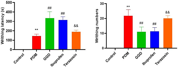 |
Figure 1 Inhibitory effects of Terazosin on the anti-PD effect of GGD. Comparison of writhing responses among rat groups. Data are presented as the means ± SD. **P < 0.01 vs. control group; ##P < 0.01 vs. PDM group; &&P < 0.01 vs. GGD group. n=6. |
Effects of Terazosin on Uterine Morphology and Pathological Injury Scores in Rats Following GGD Intervention
HE staining (Figure 2A–E) and the pathological scoring system (Figure 2F) showed that the PDM group had a significantly higher uterine injury score (4.00 ± 0.63) compared to controls (1.33 ± 0.52; p < 0.001). GGD reduced the score to 1.50 ± 0.58 (p < 0.001 vs. model), while Terazosin co-treatment increased the score to 3.83 ± 0.75 (p < 0.01 vs. GGD).
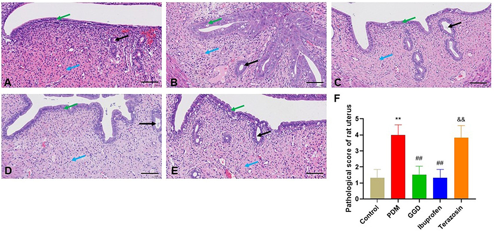 |
Figure 2 Comparison of rat uterine histomorphology among groups. Pathological changes in the uterine tissue of rats in each group by H&E staining. (A) Control group; (B) Primary dysmenorrhea model (PDM) group; (C) GGD control group; (D) Ibuprofen group; (E) Terazosin group. Scale bar = 50 μm. Black arrows indicate uterine glands; Green arrows indicate mucosal epithelium; Blue arrows indicate connective tissue. (F) Pathological score of rat uterus in each group. **P < 0.01 vs. control group; ##P < 0.01 vs. PDM group; &&P < 0.01 vs. GGD group. |
Effects of Terazosin on Serum PGF2α Levels and PGF2α/PGE2 Ratio in Rats Following GGD Intervention
As shown in Figure 3, the PDM model group exhibited a marked increase in serum PGF2α levels (mean 235.83 ± 23.36 pg/mL vs. control 167.17 ± 12.61 pg/mL; p < 0.01) and PGF2α/PGE2 ratio (mean 0.88 ± 0.07 vs. control 0.38 ± 0.03; p < 0.01) compared to the control group. Both GGD and ibuprofen treatments significantly reduced these parameters relative to the PDM group (GGD: PGF2α 162.00 ± 10.16 pg/mL, p < 0.01; ratio 0.35 ± 0.03, p < 0.01; ibuprofen: PGF2α 161.00 ± 21.67 pg/mL, p < 0.01; ratio 0.36 ± 0.06, p < 0.01), with no statistically significant differences between the two groups (PGF2α: p = 0.94; ratio: p = 0.88). However, co-administration of Terazosin significantly increased serum PGF2α levels to 201.00 ± 11.98 pg/mL (p < 0.01 vs. GGD alone) and the PGF2α/PGE2 ratio to 0.61 ± 0.07 (p < 0.01 vs. GGD alone).
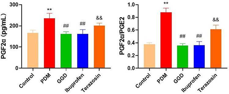 |
Figure 3 Comparison of serum PGF2α level and PGF2α/PGE2 ratio among groups. The levels of PGF2α and PGE2 in the serum of rats in each group were detected by ELISA and quantified. **P < 0.01 vs. control group; ##P < 0.01 vs. PDM group; &&P < 0.01 vs. GGD group. |
Effects of Terazosin on Serum Levels of Inflammatory Cytokines in Rats Following GGD Intervention
As shown in Figure 4, the PDM model group exhibited a marked increase in serum levels of IL-1β (mean 217.15 ± 33.07 pg/mL vs. control 59.75 ± 28.67 pg/mL; p < 0.01), IL-18 (57.90 ± 10.02 pg/mL vs. 111.24 ± 1.82 pg/mL; p < 0.01), and IL-6 (156.11 ± 25.60 pg/mL vs. 41.11 ± 10.69 pg/mL; p < 0.01) compared to the control group. GGD treatment significantly reduced these cytokines to 147.46 ± 26.64 pg/mL (IL-1β), 36.39 ± 4.83 pg/mL (IL-18), and 75.69 ± 18.77 pg/mL (IL-6), respectively (all p < 0.001 vs. model), an effect comparable to ibuprofen (IL-1β: 135.58 ± 25.54 pg/mL, p = 0.44 vs. GGD; IL-18: 32.67 ± 9.41 pg/mL, p = 0.41 vs. GGD; IL-6: 75.69 ± 18.77 pg/mL, p = 0.45 vs. GGD). Co-administration of the HSP90 agonist Terazosin partially reversed the effect of GGD, significantly increasing serum IL-1β to 200.00 ± 16.16 pg/mL (p < 0.01 vs. GGD alone), IL-18 to 58.66 ± 7.89 pg/mL (p < 0.01 vs. GGD alone), and IL-6 to 132.69 ± 19.63 pg/mL (p < 0.01 vs. GGD alone).
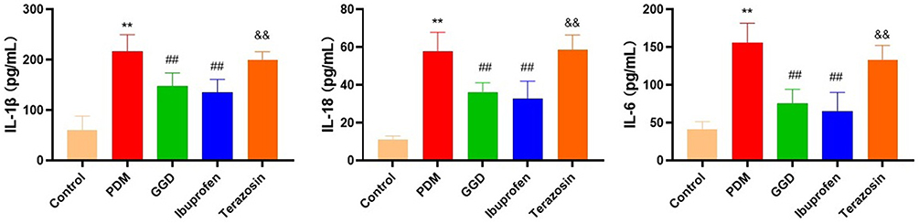 |
Figure 4 Comparison of serum IL-1β, IL-18, and IL-6 protein levels among groups. The levels of IL-1β, IL-18, and IL-6 in the serum of rats in each group were detected by ELISA and quantified. **P < 0.01 vs. control group; ##P < 0.01 vs. PDM group; &&P < 0.01 vs. GGD group. |
Effects of Terazosin on the Protein Expression of HSP90, p-AKT, NLRP3, Caspase-1, and IL-1β in Rat Uterine Tissue Following GGD Intervention
Immunohistochemistry (Figure 5) and optical density analysis revealed that the PDM model group exhibited a marked increase in uterine protein expression levels of HSP90 (mean 0.18 ± 0.01 vs. control 0.06 ± 0.01; p < 0.01), p-AKT (0.15 ± 0.02 vs. 0.05 ± 0.01; p < 0.01), NLRP3 (0.15 ± 0.01 vs. 0.05 ± 0.01; p < 0.01), Caspase-1 (0.27 ± 0.02 vs. 0.08 ± 0.01; p < 0.01), and IL-1β (0.29 ± 0.01 vs. 0.03 ± 0.02; p < 0.01) compared to the control group. GGD treatment significantly reduced these protein levels (HSP90: 0.10 ± 0.01, p < 0.01 vs. model; p-AKT: 0.08 ± 0.01, p < 0.01; NLRP3: 0.08 ± 0.01, p < 0.01; Caspase-1: 0.11 ± 0.01, p < 0.01; IL-1β: 0.10 ± 0.01, p < 0.01), an effect comparable to ibuprofen (p-AKT: 0.08 ± 0.01, p = 0.93; NLRP3: 0.09 ± 0.01, p = 0.37; Caspase-1: 0.11 ± 0.01, p = 0.73; IL-1β: 0.11 ± 0.02, p = 0.65). Co-administration of the HSP90 agonist Terazosin partially reversed the effect of GGD, significantly increasing HSP90 to 0.116 ± 0.01 (p < 0.01 vs. GGD alone), p-AKT to 0.15 ± 0.01 (p < 0.01), NLRP3 to 0.13 ± 0.01 (p < 0.01), Caspase-1 to 0.19 ± 0.02 (p < 0.01), and IL-1β to 0.16 ± 0.02 (p < 0.01).
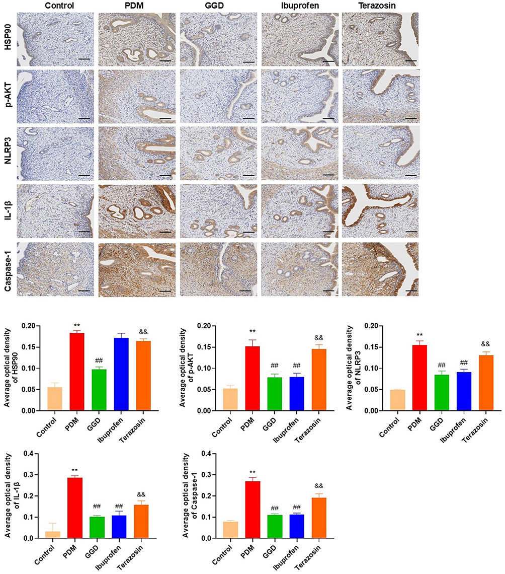 |
Figure 5 Comparison of NLRP3, Caspase-1, and IL-1β protein levels in rat uterine tissue among groups. The protein levels of NLRP3, Caspase-1, and IL-1β in the uterine tissue of rats in each group were detected by IHC and quantified. Scale bar = 50 μm. Data are presented as the means ± SD. **P < 0.01 vs. control group; ##P < 0.01 vs. PDM group; &&P < 0.01 vs. GGD group. n=6. |
Discussion
Primary dysmenorrhea (PDM), particularly the Cold-Damp Stagnation subtype, is common in clinical practice. Ge-Gen Decoction (GGD), a classical Chinese formula, has shown advantages in treating this subtype, including low recurrence rates and sustained effects.5 However, its mechanism of action has been unclear. In this study, we established a rat model of Cold-Damp Stagnation PDM using cold stimulation plus estradiol benzoate and oxytocin. We found that GGD significantly reduced writhing responses, shortened writhing latency, and ameliorated uterine histopathological damage. Notably, co-administration of the HSP90 agonist Terazosin partially reversed these effects, indicating that HSP90 mediates the therapeutic action of GGD.
NLRP3 inflammasome-mediated inflammation is a key event in PDM12,13 The ischemic, hypoxic uterine environment triggers local inflammation and releases cytokines such as IL-1β, IL-18, and IL-6. These factors cause tissue damage and directly participate in pain signaling.14–16 The NLRP3 inflammasome is activated in two stages: priming (transcriptional upregulation) and activation (complex assembly).17 Activated NLRP3 oligomerizes and recruits ASC, which then activates Caspase-1. This protease matures and releases IL-1β and IL-18, amplifying inflammation.17–19 Our data support this framework: compared to controls, the PDM model had higher uterine expression of NLRP3, Caspase-1, and IL-1β, as well as elevated serum IL-18, IL-1β, and IL-6. These results confirm the role of the NLRP3 inflammasome in PDM.
Several limitations should be acknowledged. First, this study was conducted only at the whole-animal level. We did not perform in vitro experiments (eg, isolated uterine strips or primary cell cultures) to directly validate the molecular targets of GGD. Second, we measured only protein expression of HSP90, p-AKT, NLRP3, Caspase-1, and IL-1β. The effects of GGD at the transcriptional or post-translational modification levels remain unknown. Third, we used the whole GGD formula without analyzing individual active components (eg, puerarin, glycyrrhizic acid, or ephedrine). Therefore, we cannot attribute the observed effects to any single herb or compound. Fourth, our PDM model is a rat model of cold-damp stagnation; human confirmation is needed, Finally, PDM is a multifactorial condition involving hormonal, inflammatory, neurovascular, and psychosocial components; therefore, while our findings highlight the HSP90/AKT/NLRP3 axis as one contributing mechanism, further studies are needed to explore other potential pathways and their interactions in the pathogenesis of cold-damp stagnation PDM.
Despite these limitations, our findings have several implications. First, GGD showed comparable efficacy to ibuprofen in this animal model, suggesting it may serve as an alternative or complementary therapy for PDM, especially for patients who cannot tolerate NSAIDs. Second, the reversal of GGD’s effects by an HSP90 agonist provides strong evidence that the HSP90/AKT/NLRP3 axis is a key mechanism. Third, future studies should identify which compounds in GGD are responsible for HSP90 inhibition. For example, puerarin from Pueraria lobata has been reported to modulate AKT signaling.20,21 Isolated component testing and structure-activity relationship studies are warranted. Fourth, we recommend in vitro validation using uterine smooth muscle cells or macrophages stimulated with lipopolysaccharide/ATP to confirm that GGD or its active compounds directly suppress NLRP3 inflammasome activation. Fifth, clinical trials are necessary to evaluate the efficacy and safety of GGD in women with cold-damp stagnation PDM. Until then, our findings should be considered preclinical evidence that may inform translational research.
Conclusion
In conclusion, this study, conducted in a rat model of PDM, suggests that GGD alleviates PDM-related symptoms and uterine pathology. The observed effects are associated with inhibition of the HSP90/AKT signaling pathway in uterine tissue, which in turn downregulates NLRP3 inflammasome activation and reduces the release of pro-inflammatory cytokines (IL-1β, IL-18, IL-6). These findings indicate a potential mechanism by which GGD may act in this specific experimental model. However, further studies are required to confirm whether similar mechanisms operate in humans, and to explore the translational relevance of these observations. Within the limitations of this animal study, our results provide a basis for future mechanistic and preclinical investigations of GGD.
Abbreviations
GGD, Ge-Gen decoction; PDM, primary dysmenorrhea; NLRP3, nucleotide-binding oligomerization domain-like receptor protein 3; NSAIDs, nonsteroidal anti-inflammatory drugs; DEPs, differentially expressed proteins. TCM, Traditional Chinese medicine; HSP90, Heat shock protein 90; PGs, prostaglandins; COX, cyclooxygenase.
Data Sharing Statement
The datasets used and/or analyzed during the study are available from the corresponding author upon reasonable request.
Author Contributions
All authors made a significant contribution to the work reported, whether that is in the conception, study design, execution, acquisition of data, analysis and interpretation, or in all these areas; took part in drafting, revising or critically reviewing the article; gave final approval of the version to be published; have agreed on the journal to which the article has been submitted; and agree to be accountable for all aspects of the work.
Funding
This work was supported by the Project of Taicang Science and Technology (TC2024JCYL07) and Taicang Health Talent Project (TCWS202502).
Disclosure
The authors declare that there are no competing interests.
References
1. Panova DI, Boshku AA, Stankovic ZB. Primary dysmenorrhea in adolescents. Best Pract Res Clin Obstet Gynaecol. 2026;106:102717. doi:10.1016/j.bpobgyn.2026.102717
2. Tu F, Hellman K. Primary dysmenorrhea: diagnosis and therapy. Obstet Gynecol. 2021;137(4):752. doi:10.1097/AOG.0000000000004341
3. Itani R, Soubra L, Karout S, Rahme D, Karout L, Khojah HMJ. Primary dysmenorrhea: pathophysiology, diagnosis, and treatment updates. Korean J Fam Med. 2022;43(2):101–9. doi:10.4082/kjfm.21.0103
4. Duan W, Chen D, Li D, et al. Traditional Chinese medicine treatment strategies for primary dysmenorrhea. Front Endocrinol. 2025;16:1580051. doi:10.3389/fendo.2025.1580051
5. Chai C, Hong F, Yan Y, et al. Effect of traditional Chinese medicine formula GeGen decoction on primary dysmenorrhea: a randomized controlled trial study. J Ethnopharmacol. 2020;261:113053. doi:10.1016/j.jep.2020.113053
6. Yang L, Chai CZ, Yue XY, et al. Ge-Gen Decoction attenuates oxytocin-induced uterine contraction and writhing response: potential application in primary dysmenorrhea therapy. Chin J Nat Med. 2016;14(2):124–132. doi:10.1016/S1875-5364(16)60005-5
7. Sumi MP, Ghosh A. Hsp90 in human diseases: molecular mechanisms to therapeutic approaches. Cells. 2022;11(6). doi:10.3390/cells11060976
8. Singh MK, Ranbhise JS, Fu M, et al. Beyond folding: expanding the functional landscape of Hsp90 chaperone machinery in health and disease. Int J Mol Sci. 2025;26(21). doi:10.3390/ijms262110279
9. Xie Y, Xu H, Gu Z. Ge-gen decoction alleviates primary dysmenorrhoea symptoms in a rat model. J Obstet Gynaecol. 2024;44(1):2337691. doi:10.1080/01443615.2024.2337691
10. Xie Y, Qian J. Ge-Gen Decoction exerts an anti-primary dysmenorrhea effect in rats by inactivating the HSP90/NLRP3/NF-kappaB/COX-2 pathway. J Inflamm Res. 2023;16:1571–1580. doi:10.2147/JIR.S400545
11. Tang ZR, Deng SL, Lian ZX, Yu K. Terazosin reduces steroidogenic factor 1 and upregulates heat shock protein 90 expression in LH-induced bovine ovarian theca cells. Free Radic Biol Med. 2021;163:190–195. doi:10.1016/j.freeradbiomed.2020.12.016
12. Yang X, Tian Y, Liu J, et al. Peony pollen protects against primary dysmenorrhea in mice by inhibiting inflammatory response and regulating the COX2/PGE2 pathway. Int J Mol Sci. 2023;24(24). doi:10.3390/ijms242417245
13. Tang B, Liu D, Chen L, Liu Y. NLRP3 inflammasome inhibitor MCC950 attenuates primary dysmenorrhea in mice via the NF-kappaB/COX-2/PG pathway. J Inflamm. 2020;17:22. doi:10.1186/s12950-020-00251-7
14. Gajtko A, Bakk E, Hegedus K, Ducza L, Hollo K. IL-1beta induced cytokine expression by spinal astrocytes can play a role in the maintenance of chronic inflammatory pain. Front Physiol. 2020;11:543331. doi:10.3389/fphys.2020.543331
15. Jo EK, Kim JK, Shin DM, Sasakawa C. Molecular mechanisms regulating NLRP3 inflammasome activation. Cell Mol Immunol. 2016;13(2):148–159. doi:10.1038/cmi.2015.95
16. Pilat D, Piotrowska A, Rojewska E, et al. Blockade of IL-18 signaling diminished neuropathic pain and enhanced the efficacy of morphine and buprenorphine. Mol Cell Neurosci. 2016;71:114–124. doi:10.1016/j.mcn.2015.12.013
17. Swanson KV, Deng M, Ting JP. The NLRP3 inflammasome: molecular activation and regulation to therapeutics. Nat Rev Immunol. 2019;19(8):477–489. doi:10.1038/s41577-019-0165-0
18. Xue X, Wang S, Li J, et al. Electroacupuncture and parecoxib reduce inflammatory injury in a primary dysmenorrhea rat model: investigating the role of the COX-2/NF-kappaB/NLRP3 pathway. J Pain Res. 2025;18:3573–3592. doi:10.2147/JPR.S512243
19. Fu J, Wu H. Structural mechanisms of NLRP3 inflammasome assembly and activation. Annu Rev Immunol. 2023;41:301–316. doi:10.1146/annurev-immunol-081022-021207
20. Wang K, Xu C, Wang H, et al. Puerarin protects against renal ischemia-reperfusion injury by restoring mitochondrial function and modulating the PI3K/AKT/NF-k B pathway and suppressing inflammatory responses. Cell Signal. 2026;139:112350. doi:10.1016/j.cellsig.2025.112350
21. Xu G, Zhang J, Shao C, et al. Puerarin alleviates cerebral ischemia/reperfusion (CIR)-induced neurocyte oxidative stress and apoptosis via DNA demethylation-mediated PI3K/Akt activation. Phytomedicine. 2025;145:157094. doi:10.1016/j.phymed.2025.157094
 © 2026 The Author(s). This work is published and licensed by Dove Medical Press Limited. The
full terms of this license are available at https://www.dovepress.com/terms
and incorporate the Creative Commons Attribution
- Non Commercial (unported, 4.0) License.
By accessing the work you hereby accept the Terms. Non-commercial uses of the work are permitted
without any further permission from Dove Medical Press Limited, provided the work is properly
attributed. For permission for commercial use of this work, please see paragraphs 4.2 and 5 of our Terms.
© 2026 The Author(s). This work is published and licensed by Dove Medical Press Limited. The
full terms of this license are available at https://www.dovepress.com/terms
and incorporate the Creative Commons Attribution
- Non Commercial (unported, 4.0) License.
By accessing the work you hereby accept the Terms. Non-commercial uses of the work are permitted
without any further permission from Dove Medical Press Limited, provided the work is properly
attributed. For permission for commercial use of this work, please see paragraphs 4.2 and 5 of our Terms.