
Back to Journals » Journal of Inflammation Research » Volume 19
First-Line Guselkumab Combined with Exclusive Enteral Nutrition in Biologic-Naïve Young‑Onset Crohn’s Disease: A Three-Case Series from China
Authors Sha S, Xu B, Qiao C, Wang K, Liu L  , Wu J, Ma W, Shi A, Liu X
Received 2 December 2025
Accepted for publication 27 February 2026
Published 13 March 2026 Volume 2026:19 584416
DOI https://doi.org/10.2147/JIR.S584416
Checked for plagiarism Yes
Review by Single anonymous peer review
Peer reviewer comments 3
Editor who approved publication: Dr Nadia Andrea Andreani
Sumei Sha,1 Bin Xu,2 Chenyang Qiao,1 Kairuo Wang,1 Le Liu,3 Jie Wu,4 Wenqi Ma,5 Ameng Shi,5 Xin Liu1
1Department of Gastroenterology, The Second Affiliated Hospital of Xi’an Jiaotong University, Xi’an, Shaanxi, People’s Republic of China; 2Department of Radiotherapy, Tangdu Hospital of the Air Force Medical University, Xi’an, Shaanxi, People’s Republic of China; 3Department of Radiology, The Second Affiliated Hospital of Xi’an Jiaotong University, Xi’an, Shaanxi, People’s Republic of China; 4Department of Pathology, The Second Affiliated Hospital of Xi’an Jiaotong University, Xi’an, Shaanxi, People’s Republic of China; 5Department of Ultrasound, The Second Affiliated Hospital of Xi’an Jiaotong University, Xi’an, Shaanxi, People’s Republic of China
Correspondence: Xin Liu, Department of Gastroenterology, The Second Affiliated Hospital of Xi’an Jiaotong University, Xi’an, Shaanxi, 710004, People’s Republic of China, Email [email protected]
Background: Guselkumab, a monoclonal antibody targeting the p19 subunit of interleukin‑23 (IL‑23), is a treatment option in inflammatory bowel disease (IBD). Evidence in young‑onset Crohn’s disease (CD) in the first‑line setting remains scarce.
Objective: To describe short-term clinical, biochemical, endoscopic and imaging outcomes with first-line guselkumab combined with exclusive enteral nutrition (EEN) in biologic-naïve young adults with CD.
Methods: We report a case series of three biologic‑naïve young adults with moderate‑to‑severe CD confirmed by clinical, endoscopic, and imaging criteria. All received guselkumab combined with exclusive enteral nutrition as initial therapy and were followed for up to 20 weeks. Outcomes were assessed across clinical, endoscopic, and cross‑sectional imaging (intestinal ultrasound, computed tomography enterography, or magnetic resonance enterography) domains. Safety was monitored throughout.
Results: All three patients achieved a clinical response and clinical remission by week 4. C‑reactive protein, fecal calprotectin, and bowel wall thickness decreased markedly after therapy. Computed tomography enterography and intestinal ultrasound suggested marked transmural improvement in all cases. Endoscopic assessment demonstrated endoscopic improvement or remission in all three patients. Diets were gradually liberalized in all patients. Guselkumab was generally well tolerated. Two patients exhibited mild, asymptomatic elevations of liver transaminases prior to the second or third dose, which were managed successfully with hepatoprotective therapy while continuing guselkumab. No serious adverse events occurred during follow‑up up to 20 weeks.
Conclusion: In this three-case real-world series from China, first‑line guselkumab plus EEN was associated with rapid short-term improvement across clinical, biochemical, endoscopic and imaging domains and was generally well tolerated. These observations are preliminary and hypothesis-generating and require confirmation in larger prospective cohorts.
Keywords: guselkumab, crohn’s disease, exclusive enteral nutrition, young-onset, case series, IL 23
Introduction
Crohn’s disease (CD) is a chronic, relapsing, and progressive inflammatory disorder of the gastrointestinal tract that commonly affects individuals during early adulthood. Young‑onset CD, defined as disease onset before the age of 25 years, is often associated with a more aggressive phenotype, greater inflammatory burden, and increased risk of complications compared to late‑onset disease. Early optimal management is crucial in this high‑risk population to prevent irreversible structural damage and long‑term disability.1 Current treatment guidelines advocate the early introduction of effective biologic therapy for patients with moderate‑to‑severe CD, particularly those with poor prognostic factors. Anti‑tumor necrosis factor (TNF) agents have historically been the cornerstone of biologic treatment; however, up to one‑third of patients exhibit primary non‑response, while others experience secondary loss of response or develop adverse events.2–4
Interleukin‑23 (IL‑23) plays a pivotal role in the pathogenesis of inflammatory bowel disease by promoting the differentiation and maintenance of pathogenic Th17 cells and their downstream pro‑inflammatory cytokines.5 Guselkumab, a selective monoclonal antibody targeting the IL‑23 p19 subunit, has demonstrated efficacy in immune‑mediated diseases and is now included in the latest American College of Gastroenterology (ACG) guidelines for moderate‑to‑severe CD.6 This rationale is supported by emerging clinical trial evidence: in the Phase 3 GALAXI-2 and GALAXI-3 programs, guselkumab demonstrated significant efficacy in inducing and maintaining clinical and endoscopic remission in patients with moderate-to-severe CD, including those who were biologic-naïve. Nevertheless, real‑world data in Chinese patients remain limited, as guselkumab was only approved for CD in China in 2025. Evidence is particularly sparse for biologic‑naïve, young‑onset CD patients receiving guselkumab as first‑line therapy. Furthermore, exclusive enteral nutrition (EEN) is a well-established induction therapy with direct anti-inflammatory effects, and its potential synergy with biologic agents represents an area of growing clinical interest. Here, we describe a case series of three biologic‑naïve young adults with newly diagnosed, moderate‑to‑severe CD treated with guselkumab plus EEN as initial therapy. We aimed to characterize their clinical, biochemical, imaging, and endoscopic outcomes and to discuss the potential role of early IL‑23 blockade in modifying disease progression in this challenging subgroup. To our knowledge, this represents the first case series from China describing the combined use of first-line guselkumab and EEN in biologic-naïve young adults with moderate-to-severe CD.
Interventions
Young-onset CD was defined as diagnosis at or before 25 years of age. All three patients in this series met this criterion. Diagnosis was based on standard clinical, endoscopic, histologic, and cross-sectional imaging criteria aligned with the European Crohn’s and Colitis Organisation (ECCO) diagnostic guidelines. All patients received guselkumab induction (intravenous 200 mg at weeks 0, 4 and 8) followed by subcutaneous maintenance (100 mg every 4 or 8 weeks). Maintenance dosing with subcutaneous 100 mg was tailored, with intervals (every 4 or 8 weeks) determined by ongoing clinical assessment and tolerability, reflecting a real-world, patient-centered approach. All three patients received a concomitant EEN protocol as part of the induction therapy. A whole-protein formula was administered via a nasojejunal tube. The daily caloric target was set at 1500–2000 kcal, adjusted to meet individual nutritional requirements. EEN was maintained as the sole source of nutrition until the completion of the scheduled endoscopic and cross-sectional imaging re-evaluations. Following these assessments, a gradual transition to a normal oral diet was initiated under dietary supervision. No concomitant corticosteroids, immunomodulators (thiopurines, methotrexate), proton-pump inhibitors, probiotics or other biologics were used during the study period. The use of other medications, if any, is noted in the individual case descriptions.
Outcome Assessment and Definitions
Clinical, endoscopic, and transmural outcomes were evaluated using predefined criteria. Clinical activity was measured by the Crohn’s Disease Activity Index (CDAI), with clinical response defined as a reduction from baseline by ≥100 points and clinical remission as a CDAI score <150. Endoscopic activity was assessed using the Simple Endoscopic Score for Crohn’s Disease (SES-CD). Endoscopic response was defined as a reduction in SES-CD by >50% from baseline. Endoscopic remission was defined as an absolute SES-CD of ≤2 with complete absence of ulcers (ulcerated surface subscore of 0 in all segments). Transmural inflammation was evaluated by intestinal ultrasound (IUS) and/or cross-sectional imaging (magnetic resonance enterography [MRE] or computed tomography enterography [CTE]), with key parameters including bowel wall thickness (BWT), wall stratification, and vascularization. Given the lack of universally standardized criteria, we adopted conservative terminology: marked transmural improvement described substantial resolution of inflammation (eg, BWT reduction ≥25% alongside improved stratification and hyperemia), while transmural healing was reserved for cases achieving complete normalization of all parameters (BWT ≤3 mm, normal wall stratification, and absence of hyperemia on Doppler). Corresponding individual patient data are detailed in the case descriptions. Safety assessments, including clinical evaluation and laboratory tests (complete blood count, liver and renal function panels), were conducted at baseline, prior to each guselkumab administration during the induction phase, and at routine intervals (every 4–8 weeks) during maintenance therapy.
Case Presentation
Case 1
An 18‑year‑old male presented with a 13‑month history of abdominal pain, diarrhea (three times daily), and unintentional weight loss of 7 kg. Laboratory evaluation revealed elevated C‑reactive protein (CRP, 43.9 mg/L) and fecal calprotectin (FC, 658.7 µg/g). Ileocolonoscopy demonstrated deep ulcers in the terminal ileum, cecum, and ascending colon (Figure 1A–C). Gastroscopy showed mild mucosal hypertrophy of the gastric fundus and corpus, presenting a “bamboo‑like” appearance (Figure 1D). CTE confirmed ileocolonic inflammation without evidence of strictures or fistulas (Figure 1E). The CDAI was 256 and the SES-CD was 13. Given the presence of high‑risk features and absence of prior biologic therapy, guselkumab was initiated at a dose of 200 mg by intravenous infusion at weeks 0, 4, and 8. Due to the emergence of asymptomatic liver enzyme elevation, the maintenance dose of guselkumab was adjusted to 100 mg subcutaneously every 8 weeks to prioritize long-term safety and tolerability.
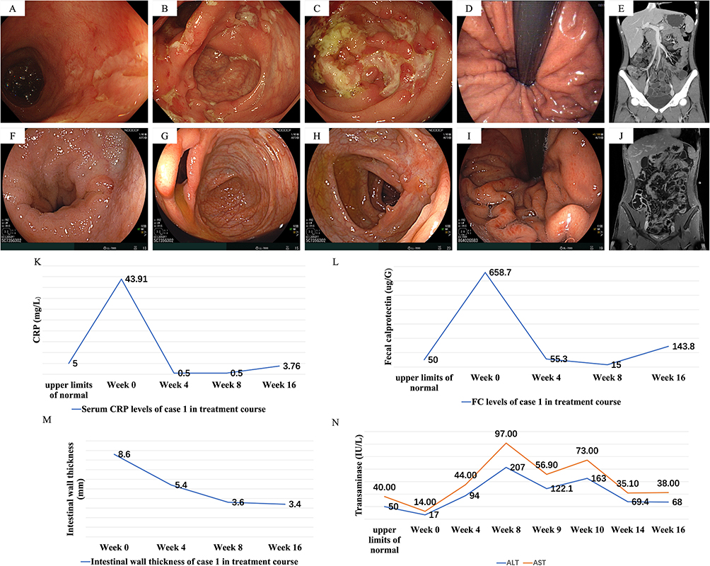 |
Figure 1 Clinical, endoscopic, and imaging evolution in Case 1 at baseline and following guselkumab induction. Time points correspond to weeks after initiation of guselkumab therapy. (A and F) Terminal ileum at baseline (A) and week 10 (F), showing transition from deep ulcers to scarring. (B and G) Ileocecal valve and cecum at baseline (B) and week 10 (G) showing resolution of shallow ulcers. (C and H) Ascending and partial transverse colon at baseline (C) and week 10 (H) demonstrating mucosal healing. (D and I) Gastric fundus and corpus at baseline (D) and week 10 (I) showing alleviation of the “bamboo‑like” appearance. (E and J) Computed tomography enterography (CTE) at baseline (E) and Magnetic resonance enterography (MRE) at week 10 (J), showing reduced bowel wall thickening in the ileocecal region. (K–N) Longitudinal changes in C‑reactive protein (K, CRP), fecal calprotectin (L, FC), terminal ileum bowel wall thickness (M, BWT), and transaminase levels (N) from baseline to week 16. |
The patient simultaneously received EEN via a nasogastric tube. Guselkumab was well tolerated; no infusion reactions, serious infections, or other major adverse events occurred during treatment.
By week 4, the patient had achieved clinical remission (CDAI < 150) with normalization of CRP and a decline in FC to 55.3 µg/g. At week 8, stool occult blood became negative, and both CRP and FC levels remained within normal ranges. Follow‑up colonoscopy at week 10 demonstrated endoscopic remission (Figure 1F–I), with the SES-CD reduced to 1. IUS and MRE (Figure 1J) suggested significant improvement in intestinal inflammation. Changes in CRP, FC and BWT over the treatment course are illustrated in Figure 1K–M. These data demonstrated a significant reduction in BWT (from 8.6mm to 3.6mm by week 8) along with improved wall stratification and decreased hyperemia, meeting the criteria for marked transmural improvement. After the week‑10 assessment, the nasojejunal tube was removed, and the patient gradually resumed oral feeding. He remained symptom‑free at the 20‑week follow‑up.
During therapy, mild elevations in liver transaminases were detected on routine laboratory monitoring before the second guselkumab dose (week 4, ALT/AST increased from 17/14 U/L at baseline to 94/44 U/L, peaking at 207/97 U/L). The patient was clinically asymptomatic, and bilirubin levels remained within normal limits. Hepatoprotective therapy was initiated, and guselkumab was continued. Liver function tests gradually normalized without recurrence during follow‑up (week 20, Figure 1N). No other clinically significant laboratory abnormalities were noted.
Case 2
An 18‑year‑old male with ileocolonic‑type CD and a 2‑year history of myasthenia gravis was referred to our hospital. The patient reported a 2‑month history of intermittent abdominal pain and frequent diarrhea (approximately 7 times daily). He had undergone surgical treatment for a perianal abscess and thymoma 1 year prior to presentation. There was no family history of CD and no prior use of immunomodulators, corticosteroids, or biologics. Laboratory evaluation revealed elevated CRP (16.57 mg/L) and FC (598.0 µg/g). Ileocolonoscopy demonstrated longitudinal deep ulcers in the terminal ileum, shallow ulcers at the ileocecal valve and cecum, and multiple superficial aphthous ulcers throughout the colon (Figure 2A–C). CTE revealed diffuse edema and multifocal bowel wall thickening in the distal ileum and ileocecal region (Figure 2D). CD was diagnosed based on endoscopic and histologic evidence with a CDAI of 194 and SES-CD of 12. A top‑down therapeutic approach with a biologic was considered. Given its therapeutic efficacy and safety profile, guselkumab was initiated as the first‑line biologic, and the patient simultaneously received EEN via a nasogastric tube. Guselkumab was initiated at a dose of 200 mg by intravenous infusion at weeks 0, 4, and 8, followed by 100 mg subcutaneously every 4 weeks thereafter.
 |
Figure 2 Clinical, endoscopic, and imaging evolution in Case 2 at baseline and following guselkumab induction. Time points correspond to weeks after initiation of guselkumab therapy. (A and E) Terminal ileum at baseline (A) and week 10 (E), showing transition from deep ulcers to scarring. (B and F) Ileocecal valve and cecum at baseline (B) and week 10 (F) showing resolution of shallow ulcers. (C and G) Representative colonic segment at baseline (C) and week 10 (G) demonstrating mucosal healing. (D and H) Computed tomography enterography (CTE) at baseline (D) and follow-up (H) showing reduced bowel wall thickening and lymphadenopathy in the ileocecal region. (I–K) Longitudinal changes in C‑reactive protein (I) fecal calprotectin (J) and terminal ileum bowel wall thickness (K) from baseline to week 16. |
By week 4 after induction, symptoms had markedly improved; the patient passed soft stools once or twice daily. Clinical remission (CDAI < 150) was achieved, with normalization of CRP and FC levels, and a negative stool occult blood test. Changes in CRP, FC and BWT during follow‑up are shown in Figure 2I–K. At week 10, follow‑up colonoscopy revealed inconspicuous deep longitudinal ulcers in the terminal ileum, consistent with the scarring stage (Figure 2E). Ulcerated areas at the ileocecal valve and cecum had flattened, decreased in size, and resolved (Figure 2F). Superficial aphthous ulcers demonstrated endoscopic remission (Figure 2G), and the SES-CD reduced to 0. IUS and CTE (Figure 2H) showed a marked reduction in inflammatory activity. Changes in CRP, FC and BWT over the treatment course are illustrated in Figure 2I–K. These data demonstrated a marked transmural improvement at week 12 (BWT reduced from 5.6 mm to 4.2 mm) and transmural healing by week 16 (BWT=2.8 mm). Following endoscopic and imaging assessments, the patient gradually resumed oral intake.
Guselkumab was generally well tolerated and was continued at a maintenance dose of 100 mg subcutaneously every 4 weeks. No infusion reactions, serious infections, or other major adverse events occurred during the 20‑week follow‑up period.
Case 3
A 22‑year‑old male presented with a six‑month history of recurrent abdominal pain and progressive weight loss of 6 kg. Over the preceding month, the abdominal pain had worsened significantly, prompting further evaluation. Laboratory investigations revealed markedly elevated CRP (17.42 mg/L) and FC (540.8 µg/g), indicating active intestinal inflammation. Colonoscopy revealed protruding lesions and ulcerations in both the ileocecal region and the sigmoid colon, with notable luminal narrowing at the ileocecal area. Additional mucosal erythema and superficial erosions were noted in other segments of the colon (Figure 3A–C), the SES-CD was 14. IUS and CTE further demonstrated clustered small bowel loops in the pelvic cavity with localized adhesions forming a “petal‑like” configuration, suggestive of complex intestinal fistulas (Figure 3D). Based on these findings, CD was diagnosed. The CDAI was 289, consistent with severe disease activity. Stool testing was positive for Clostridioides difficile toxin, confirming concomitant infection. Considering the patient’s young age and high‑risk disease features—including a stricturing phenotype, suspected fistula formation, and a high inflammatory burden—guselkumab was selected as first‑line therapy, in combination with targeted antimicrobial therapy for C. difficile infection (a standard 14-day course of oral vancomycin, 125 mg four times daily). EEN and Guselkumab were initiated at a dose of 200 mg by intravenous infusion at weeks 0, 4, and 8, followed by 100 mg subcutaneously every 4 weeks thereafter.
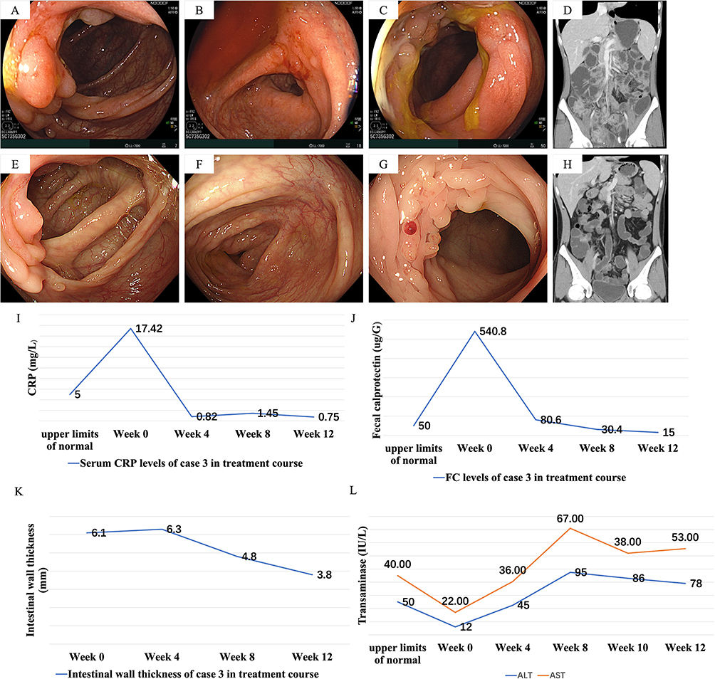 |
Figure 3 Clinical, endoscopic, and imaging evolution in Case 3 at baseline and following guselkumab induction. Time points correspond to weeks after initiation of guselkumab therapy. (A and E) Ileocecal region at baseline (A) and week 12 (E) showing protruding lesions and luminal narrowing at baseline, with marked reduction of surrounding edema at follow-up. (B and F) Ascending colon at baseline (B) and week 12 (F) showing transition from mucosal erythema and erosions to scar formation. (C and G) Sigmoid colon at baseline (C) and week 12 (G) showing transition from protruding ulcerations to proliferative changes. (D and H) Computed tomography enterography (CTE) at baseline (D) and week 12 (H) showing resolution of the inflammatory “petal‑like” configuration and no definitive fistula. (I–L) Longitudinal changes in C‑reactive protein (I, CRP), fecal calprotectin (J, FC), terminal ileum bowel wall thickness (K, BWT), and transaminase levels (L) from baseline to week 12. |
A marked reduction in abdominal pain was noted within 72 hours of initiating vancomycin, which occurred before the administration of the second induction dose of guselkumab. Similarly, the elevated CRP level reduced from 17.42 to 9.84 within one week of starting anti-infective therapy. At week 4, the patient experienced less frequent abdominal pain. Changes in CRP, FC, and BWT during follow‑up are shown in Figure 3I–K. At week 12, follow‑up colonoscopy was performed to evaluate treatment response. The patient was asymptomatic with complete resolution of abdominal pain. Although the endoscope could not pass through the ileocecal area, surrounding edema was markedly reduced, with no purulent exudates or ulcers observed; only proliferative changes were noted. The previously affected area in the ascending colon showed evidence of scar formation (Figure 3E–G), the SES-CD was reduced to 3. CTE revealed a significant reduction in inflammatory extent and no definitive evidence of intestinal fistulas (Figure 3H), demonstrated a marked transmural improvement at week 12 (BWT reduced from 6.1 mm to 3.8 mm).
Routine laboratory monitoring after the second dose showed mild, asymptomatic elevation of liver transaminases (ALT/AST increased from 12/22 U/L at baseline to a peaking of 95/67 U/L at week 8), while bilirubin levels remained within normal limits. The patient remained asymptomatic. Hepatoprotective therapy was initiated, and guselkumab was continued. Liver function tests remained stable throughout follow‑up (Figure 3L, normalized at week 20). No other clinically significant laboratory abnormalities were noted. By the end of observation, the patient had experienced significant symptomatic improvement and gained more than 10 kg in weight, with no serious adverse events reported during the follow‑up period.
Discussion
Early-onset CD is often characterized by a more aggressive clinical course, extensive intestinal involvement, and a higher frequency of complications compared with late-onset disease. Early introduction of effective biologic therapies has been recommended to achieve sustained disease control and to prevent irreversible bowel damage.7 Anti-tumor necrosis factor (TNF) monoclonal antibodies have long been the mainstay of treatment in this setting. However, up to one-third of patients exhibit primary non-response, and many experience secondary loss of response over time. In addition, young patients with CD may face decades of cumulative biologic exposure, emphasizing the need to explore alternative treatment strategies with distinct mechanisms of action.8 Interleukin-23 (IL-23) plays a central role in the immunopathogenesis of CD by promoting the differentiation, activation, and maintenance of Th17 cells, as well as the secretion of downstream pro-inflammatory cytokines that perpetuate mucosal inflammation. Guselkumab, a highly selective monoclonal antibody targeting the IL-23 p19 subunit, precisely inhibits this crucial pathway while preserving IL-12–mediated immune responses. The efficacy and safety of guselkumab have been well established in immune-mediated diseases such as psoriasis and psoriatic arthritis, and accumulating evidence supports its potential role in inflammatory bowel diseases.9 Phase II and III clinical trials in ulcerative colitis have shown significant improvements in clinical remission and endoscopic healing rates compared with placebo, reinforcing the therapeutic relevance of IL-23 blockade in intestinal inflammation.10,11 Emerging data support the efficacy of guselkumab in moderate-to-severe CD, with benefits observed in both induction and maintenance phases.12 The latest ACG guideline has recommended guselkumab as a treatment option for CD,6 reflecting its proven benefit in controlled trials. However, guselkumab will not be officially approved for CD in China until 2025, and local clinical experience remains limited. Consequently, real-world evidence in Chinese populations is scarce.
In the present real-world case series, three biologic-naïve young adults with newly diagnosed moderate-to-severe CD achieved rapid and sustained remission with first-line guselkumab therapy combined with EEN. All patients demonstrated marked improvements in clinical symptoms, inflammatory biomarkers, and endoscopic improvement/remission on follow-up endoscopy. One notable aspect of case 3 was the presence of an intestinal fistula at the time of diagnosis, a complication associated with more aggressive disease behavior and poorer prognosis in CD. Remarkably, follow-up assessment after induction therapy demonstrated substantial improvement of the intestinal fistula, as evidenced by both clinical symptoms and imaging findings. This outcome underscores the potential of early guselkumab use not only to control luminal inflammation but also to promote fistula healing, aligning with emerging evidence that timely initiation of advanced therapy in high-risk CD may improve long-term disease outcomes.13 Another notable strength of our study is the use of a comprehensive, multidimensional assessment to evaluate treatment outcomes. Beyond conventional clinical indices such as the CDAI, we incorporated biochemical biomarkers (CRP and FC), endoscopic evaluation using the SES-CD, IUS parameters (including bowel wall thickness and vascularity) and CTE/MRE. Following first-line guselkumab therapy, all three patients exhibited consistent improvement across these multiple dimensions: rapid symptom relief, significant reductions in inflammatory biomarkers, mucosal healing or substantial endoscopic improvement, and decreased bowel wall inflammation and vascularity on ultrasound. This concordance between objective and subjective outcome measures provides robust real-world evidence for the efficacy of guselkumab in young-onset CD and underscores its potential to deliver not only disease control but also tangible benefits for patients’ daily living.
Notably, the QUASAR phase 3 study14 demonstrated that induction efficacy was greater when guselkumab was used as first-line therapy compared to second- or later-line use. Our present findings align with these results. In this first reported Chinese case series, guselkumab used as first-line biologic therapy in biologic-naïve young-onset CD demonstrated rapid and sustained clinical remission, biochemical normalization, and endoscopic improvement/remission. Our case series offers preliminary real-world support for frontline guselkumab in young, biologic-naïve CD patients, potentially bolstering clinical confidence in newly approving regions. The observed responses suggest a potential benefit of early IL-23 inhibition in aggressive phenotypes. Notably, this use, including our tailored maintenance dosing, was off-label in China, highlighting the gap between clinical need and formal guidelines, and necessitating individualized treatment decisions. Furthermore, in Case 3, the concurrent Clostridioides difficile infection and its treatment represent a significant confounding variable. The rapid clinical and biochemical improvement observed coincided closely with antimicrobial therapy, making it challenging to definitively attribute the early response solely to guselkumab in this patient. This underscores a common challenge in real-world practice—disentangling treatment effects from the resolution of intercurrent illnesses.
Combination therapy with EEN may have contributed to the favorable outcomes observed in our cohort. EEN provides complete nutritional support while modulating intestinal microbiota, mucosal barrier integrity, and local immune responses. When used concurrently with biologics, it may augment mucosal healing and induce deeper remission by promoting intestinal rest and reducing antigenic stimulation. Previous pediatric studies have shown that concomitant EEN enhances biologic efficacy and accelerates clinical improvement in severe CD.15 The present observations suggest that similar synergy may be achievable in young adult populations. Nevertheless, the interpretation of our findings must be tempered by the recognition that all patients received combination therapy. Although the standardized EEN protocol likely contributed to the observed clinical and transmural responses, its effect is inextricably intertwined with that of guselkumab in this study design. The notion of synergy between biologic therapy and EEN is biologically plausible and clinically appealing, as they may target inflammation through complementary pathways. However, the present case series cannot quantify the magnitude of this potential synergy nor delineate the individual efficacy of each agent. This inherent limitation underscores the necessity for future controlled studies designed specifically to disentangle their respective contributions.
The safety profile observed in our small series is consistent with previous reports of guselkumab in immune-mediated diseases.13,16 Guselkumab was well tolerated in all cases, with no reported infectious or immunological complications over the 20-week follow-up. In our series, two patients (case 1 and case 3) experienced mild, asymptomatic transaminase elevation after 4w or 8w of guselkumab administration, which resolved with hepatoprotective therapy while continuing treatment. This parallels the benign course of hepatic laboratory abnormalities described in prior guselkumab studies. In the Phase II GALAXI-1 trial evaluating guselkumab in moderate-to-severe CD, adverse events were generally mild to moderate, with the most frequent being headache and nasopharyngitis. Elevations in liver enzymes were infrequent (≤3% of patients) and typically transient without necessitating drug discontinuation.17,18 In long-term psoriasis studies (VOYAGE 1 and 2), occasional asymptomatic transaminase elevations have been observed without progression to serious hepatic injury, and these typically resolved spontaneously or with standard supportive measures, with no apparent increase in serious hepatic events over long-term follow-up.19,20 The clinical course mirrored the benign pattern previously reported, suggesting such events are manageable and need not preclude continuation of therapy in the absence of symptoms or significant laboratory derangements. In this case series, the observed asymptomatic, transient transaminase elevations resolved shortly and are consistent with the established safety profile of guselkumab. Nonetheless, proactive laboratory monitoring remains advisable, particularly in young patients receiving combination therapy with EEN.
Conclusion
To our knowledge, this case series provides the first preliminary real-world data from China on the use of first-line guselkumab combined with EEN in young, biologic-naïve patients with CD. All patients achieved rapid clinical remission, biochemical normalization, radiologic improvement, and marked transmural improvement with excellent tolerability. These findings suggest that early IL-23 blockade, particularly when integrated with nutritional therapy, may offer a promising treatment paradigm capable of achieving early and short-term remission in high-risk, young-onset CD. Limitations of this report include the small sample size, short follow-up duration, lack of comparator, and potential selection and reporting biases, inherent to case series design. Therefore, our findings should be viewed as preliminary signals supporting feasibility, and they warrant confirmation in larger, prospective studies. Such future studies should employ standardized EEN protocols, predefined imaging criteria, and assess longer-term outcomes like steroid-free clinical remission and durable transmural response. Given these limitations, our observations should be interpreted cautiously, as they may not be generalizable to all populations. Ultimately, larger, prospective, multicenter trials—including those focusing on Asian cohorts—are needed to validate these results and define the optimal positioning of guselkumab in CD treatment algorithms.
Data Sharing Statement
The datasets generated and/or analyzed during the current study are not publicly available due to patient privacy and ethical restrictions but are available from the corresponding author on reasonable request.
Ethical Approval and Consent to Participate
The study was approved by the Ethics Committee of the Second Affiliated Hospital of Xi’an Jiaotong University (No.: 2025YS708). This study was conducted in accordance with the declaration of Helsinki. This case series was prepared in accordance with the CARE (CAse REport) guidelines to ensure transparency and completeness of reporting. Written informed consent was obtained from the patients for the publication of all the images and data included in this article. The patients have consented to the submission of the report to the journal. All individual patient identifiers have been removed to preserve anonymity.
Acknowledgments
We would like to acknowledge our patients for their consent to publish this case series.
Author Contributions
Conceptualization: XL, CQ. Methodology: XL, CQ, KW. Investigation/Clinical management: SS, XL, KW. Data curation: SS, JW, WM, AS. Visualization (endoscopy/imaging/IUS): BX, LL, JW, WM. Formal analysis: SS, LL, JW. Writing – original draft: SS, XL. Writing – review & editing: XL, CQ, BX, KW, LL, JW, WM, AS. Supervision: XL. All authors contributed to data analysis, drafting or revising the article, have agreed on the journal to which the article will be submitted, gave final approval of the version to be published, and agree to be accountable for all aspects of the work.
Funding
This study was supported by grants from the National Natural Science Foundation of China (No. 82400642) and the youth program of Shaanxi Natural Science Foundation (No. 2022SF-195).
Disclosure
No potential conflict of interest was reported by the author(s).
References
1. Fernandes SR, Rodrigues IC, Neves ADS, et al. Early Top-Down treatment with biologics improves the rates of transmural remission in crohn’s Disease-A Risk-Adjusted propensity score matched analysis. Inflamm Bowel Dis. 2025. doi:10.1093/ibd/izaf112
2. Kapizioni C, Desoki R, Lam D, et al. Biologic therapy for inflammatory bowel disease: real-world comparative effectiveness and impact of drug sequencing in 13 222 patients within the UK IBD bioresource. J Crohns Colitis. 2024;18(6):790–10. doi:10.1093/ecco-jcc/jjad203
3. Krugliak Cleveland N, Ghosh S, Chastek B, et al. Real-World persistence of successive biologics in patients with inflammatory bowel disease: findings from ROTARY. Inflamm Bowel Dis. 2024;30(10):1776–1787. doi:10.1093/ibd/izad245
4. Hanauer SB, Duk Ye B, Cross RK, Danese S, D’Haens G, Jung J. The position of anti-tumor necrosis factor agents for the treatment of adult patients with Crohn’s disease. Expert Rev Gastroenterol Hepatol. 2025;19(7):725–743. doi:10.1080/17474124.2025.2511148
5. Verstockt B, Salas A, Sands BE, Abraham C, Leibovitzh H, Neurath MF. IL-12 and IL-23 pathway inhibition in inflammatory bowel disease. Nat Rev Gastroenterol Hepatol. 2023;20(7):433–446. doi:10.1038/s41575-023-00768-1
6. Lichtenstein GR, Loftus EV, Afzali A, et al. ACG clinical guideline: management of crohn’s disease in adults. Am J Gastroenterol. 2025;120(6):1225–1264. doi:10.14309/ajg.0000000000003465
7. Otake H, Matsumoto S, Mashima H. Long-term clinical and real-world experience with Crohn’s disease treated with anti-tumor necrosis factor-alpha antibodies. Intest Res. 2022;20(4):464–474. doi:10.5217/ir.2021.00139
8. Zhao M, Larsen L, Dige A, et al. Clinical outcomes after First-Line Anti-Tumor-Necrosis-Factor treatment of patients with inflammatory bowel Disease-A prospective multicenter cohort study. J Crohns Colitis. 2025;19(5). doi:10.1093/ecco-jcc/jjae192
9. Sbidian E, Chaimani A, Garcia-Doval I, et al. Systemic pharmacological treatments for chronic plaque psoriasis: a network meta-analysis. Cochrane Database Syst Rev. 2021;4(4):CD011535. doi:10.1002/14651858.CD011535.pub4
10. Peyrin-Biroulet L, Allegretti JR, Rubin DT, et al. Guselkumab in patients with moderately to severely active ulcerative colitis: QUASAR phase 2b induction study. Gastroenterology. 2023;165(6):1443–1457. doi:10.1053/j.gastro.2023.08.038
11. The efficacy and safety of guselkumab as maintenance therapy in patients with moderately to severely active ulcerative colitis: results from the phase 3 QUASAR maintenance study. Gastroenterol Hepatol. 2024;20(7 Suppl 6):8–9.
12. Hart A, Panaccione R, Steinwurz F, et al. Efficacy and safety of guselkumab subcutaneous induction and maintenance in participants with moderately to severely active crohn’s disease: results from the phase 3 GRAVITI study. Gastroenterology. 2025;169(2):308–325. doi:10.1053/j.gastro.2025.02.033
13. Panaccione R, Feagan BG, Afzali A, et al. Efficacy and safety of intravenous induction and subcutaneous maintenance therapy with guselkumab for patients with Crohn’s disease (GALAXI-2 and GALAXI-3): 48-week results from two phase 3, randomised, placebo and active comparator-controlled, double-blind, triple-dummy trials. Lancet. 2025;406(10501):358–375. doi:10.1016/S0140-6736(25)00681-6
14. Rubin DT, Allegretti JR, Panes J, et al. Guselkumab in patients with moderately to severely active ulcerative colitis (QUASAR): phase 3 double-blind, randomised, placebo-controlled induction and maintenance studies. Lancet. 2025;405(10472):33–49. doi:10.1016/S0140-6736(24)01927-5
15. Mitrev N, Huang H, Hannah B, Kariyawasam VC. Review of exclusive enteral therapy in adult Crohn’s disease. BMJ Open Gastroenterol. 2021;8(1). doi:10.1136/bmjgast-2021-000745
16. Lebwohl MG, Merola JF, Rowland K, et al. Safety of guselkumab treatment for up to 5 years in patients with moderate-to-severe psoriasis: pooled analyses across seven clinical trials with more than 8600 patient-years of exposure. Br J Dermatol. 2023;189(1):42–52. doi:10.1093/bjd/ljad115
17. Danese S, Panaccione R, Feagan BG, et al. Efficacy and safety of 48 weeks of guselkumab for patients with Crohn’s disease: maintenance results from the phase 2, randomised, double-blind GALAXI-1 trial. Lancet Gastroenterol Hepatol. 2024;9(2):133–146. doi:10.1016/S2468-1253(23)00318-7
18. Efficacy and safety of guselkumab maintenance therapy among guselkumab induction week 24 clinical responders: results from the phase 3 QUASAR maintenance study. Gastroenterol Hepatol. 2024;20(12 Suppl 10):2–3.
19. Megna M, Potestio L, Fabbrocini G, Ruggiero A. Long-term efficacy and safety of guselkumab for moderate to severe psoriasis: a 3-Year real-life retrospective study. Psoriasis. 2022;12:205–212. doi:10.2147/PTT.S372262
20. Kim BS, Jo SJ, Youn S, et al. Five-year maintenance of clinical response and consistent safety profile for guselkumab in asian patients with psoriasis from VOYAGE 1 and VOYAGE 2. Dermatol Ther. 2023;13(11):2721–2737. doi:10.1007/s13555-023-01026-7
 © 2026 The Author(s). This work is published and licensed by Dove Medical Press Limited. The
full terms of this license are available at https://www.dovepress.com/terms
and incorporate the Creative Commons Attribution
- Non Commercial (unported, 4.0) License.
By accessing the work you hereby accept the Terms. Non-commercial uses of the work are permitted
without any further permission from Dove Medical Press Limited, provided the work is properly
attributed. For permission for commercial use of this work, please see paragraphs 4.2 and 5 of our Terms.
© 2026 The Author(s). This work is published and licensed by Dove Medical Press Limited. The
full terms of this license are available at https://www.dovepress.com/terms
and incorporate the Creative Commons Attribution
- Non Commercial (unported, 4.0) License.
By accessing the work you hereby accept the Terms. Non-commercial uses of the work are permitted
without any further permission from Dove Medical Press Limited, provided the work is properly
attributed. For permission for commercial use of this work, please see paragraphs 4.2 and 5 of our Terms.
Recommended articles
Prediction of the Short-Term Effectiveness of Ustekinumab in Patients with Moderate to Severe Crohn’s Disease
Su T, Liu L, Meng F, Wu H, Liu T, Deng J, Peng X, Zhi M, Yao J
Journal of Inflammation Research 2024, 17:9181-9191
Published Date: 20 November 2024

Positioning Guselkumab in The Treatment Algorithm of Patients with Crohn’s Disease
D’Amico F, Bencardino S, Magro F, Dignass A, Gutiérrez Casbas A, Verstockt B, Hart A, Armuzzi A, Peyrin-Biroulet L, Danese S
Biologics: Targets and Therapy 2025, 19:351-363
Published Date: 31 May 2025