Back to Journals » International Journal of Nanomedicine » Volume 21
Ferroptosis-Like Death: An Emerging Innovative Antibacterial Strategy
Authors Luo C, Chen J, Duan Q, Zhao H, Fu N, Deng Q, Li Y ![]()
Received 5 December 2025
Accepted for publication 18 February 2026
Published 24 February 2026 Volume 2026:21 586869
DOI https://doi.org/10.2147/IJN.S586869
Checked for plagiarism Yes
Review by Single anonymous peer review
Peer reviewer comments 3
Editor who approved publication: Dr RDK Misra
Cheng Luo,1 Jialin Chen,1,2 Qifei Duan,1,3 Haibo Zhao,1,2 Nanyan Fu,1 Qin Deng,1 Yan Li1
1School of Basic Medical Sciences, Yichun University, Yichun, Jiangxi, People’s Republic of China; 2School of Clinical Medicine, Yichun University, Yichun, Jiangxi, People’s Republic of China; 3School of Pharmaceutical Sciences, Yichun University, Yichun, Jiangxi, People’s Republic of China
Correspondence: Yan Li, School of Basic Medical Sciences, Yichun University, Yichun, Jiangxi, People’s Republic of China, Email [email protected]
Abstract: The increasing global crisis of antibiotic resistance underscores the imperative for innovative antibacterial strategies that transcend conventional mechanisms. Ferroptosis, an iron-dependent form of regulated cell death characterized by lethal lipid peroxidation, is a promising therapeutic approach. This review systematically explores ferroptosis-like death as an emerging antibacterial paradigm. The core mechanism involves exogenous interventions that result in intracellular iron overload, the incorporation of polyunsaturated fatty acids (PUFAs), and the disruption of bacterial antioxidant defenses. A comprehensive evaluation of three key strategies is provided: host-directed approach, in which immune cells are programmed to eliminate intracellular pathogens; small molecule-induced pathway, in which iron agents, PUFAs, and others are used to directly trigger bacterial death; and nanomaterial-mediated precision therapy, in which functionalized nanosystems are employed for synergistic and intelligent targeting. Despite the challenges in mechanistic understanding and biosafety, future advancements through multiomics, intelligent nanosystems, and synergistic cell death pathways are anticipated to propel this field. This strategy indicates a possible transformation in anti-infective therapy from broad-spectrum killing to precision regulation. This shift offers a potentially effective solution to address drug-resistant bacterial infections.
Keywords: ferroptosis-like death, antibiotic resistance, lipid peroxidation, nanomedicine, reactive oxygen species
Antibiotic Resistance
Antibiotic resistance is defined as the capacity of bacteria to resist antibiotics, resulting in failed standard treatment, persistent or recurrent infections, and a heightened risk of pathogen transmission.1 Since the advent of penicillin, antibiotics have constituted a cornerstone of modern medicine and significantly reduce the incidence and mortality associated with infectious diseases. However, the extensive and indiscriminate applications of antibiotics in human medicine and animal agriculture have resulted in potent selective pressure. This pressure drives microbes to continuously evolve resistant phenotypes through mechanisms like genetic mutation, efflux pump expression, and biofilm formation.2 The World Health Organization has formally acknowledged the serious harm of antibiotic resistance, classifying it as one of the most pressing concerns to global public health. In 2019, approximately 4.95 million deaths worldwide were associated with antibiotic resistance, with 1.27 million of these deaths being directly attributable to it. This figure exceeds the combined mortality from HIV/AIDS and malaria.1 In the absence of effective interventions, the number of annual deaths attributable to antibiotic resistance is projected to exceed 1.9 million by 2050, with particularly devastating consequences in regions with limited healthcare resources.3 The challenges posed by antibiotic resistance have extended beyond health to economic domains, thereby triggering significant global socioeconomic challenges.4 The fundamental reason for the emergence of antibiotic resistance crisis is the critical disconnection between the rapid evolution of pathogens and the stagnation of the antimicrobial development. The development of novel antibiotics is confronted by multiple challenges, including the rapid evolution of bacterial resistance, prolonged development cycles coupled with high costs and failure rates. Therefore, there is an urgent need for novel antibacterial paradigms that function independently of conventional antibiotic mechanisms.
Numerous alternative antimicrobial strategies with distinct mechanisms have been explored. Reactive oxygen species (ROS)-based antimicrobial approaches, such as photodynamic therapy (PDT) and hyperbaric oxygen therapy, primarily induce nonspecific oxidative damage to bacterial lipids, proteins, and nucleic acids.5 Metal-based antimicrobial strategies, including silver nanoparticles, copper-based coatings, and zinc oxide materials, utilize the intrinsic toxicity and catalytic properties of metal ions or their nanostructured derivatives.6 Strategies targeting regulated bacterial death involve modulating specific programmed cell death pathways in bacteria, such as apoptosis-like death, pyroptosis, immunogenic cell death, and NETosis.7 Recently, ferroptosis-like death, which involves the targeting of bacterial iron metabolism and redox homeostasis, has garnered significant attention as a highly promising innovative antibacterial pathway.8 This strategy is less prone to inducing drug resistance and easily combines with nanocarriers for targeted delivery, significantly reducing off-target damage to host cells. Therefore, it offers a highly promising new direction for mitigating antibiotic resistance crisis.
To date, there are only few reviews addressing ferroptosis-like antibacterial strategies, with primary focus on elucidating ferroptosis mechanisms and its dual role in host–pathogen interactions.9–11 This review firstly compares the ferroptosis in eukaryotes with ferroptosis-like death in bacteria. The core mechanisms driving bacterial ferroptosis-like death is then outlined, and the strategies utilized are categorized into three types: host-directed, small molecule–mediated, and nanomaterial-mediated approaches. In particular, the nanomaterial-mediated section, a major focus of current research, is discussed in detail covering mechanisms, multimodal synergy, intelligent responsiveness, and application scenarios. Finally, future directions and clinical translation prospects for ferroptosis-like antibacterial strategies are discussed.
Antibacterial Potential of Ferroptosis
Ferroptosis
Ferroptosis is an iron dependent and nonapoptotic form of regulated cell death formally defined in 2012.12 It is morphologically characterized by mitochondrial shrinkage and the reduction or disappearance of cristae. The nuclear structure remains intact and lacks typical apoptotic features. Biochemically, this process is characterized by iron-dependent peroxidation of phospholipids on cell membranes, consequently resulting in membrane system failure.
The initiation of ferroptosis is contingent upon the dysregulation and synergistic interaction of three fundamental elements: the catalytic center (iron), the reaction substrate (polyunsaturated fatty acid (PUFA)-containing phospholipids, PUFA-PLs), and the defense system (antioxidant pathways).13 Intracellular labile ferrous ions (Fe2+) function as the primary catalysts for the Fenton reaction and initiate ferroptosis. The Fenton reaction is a chemical process that involves the conversion of hydrogen peroxide (H2O2) into highly reactive hydroxyl radicals (•OH). These radicals subsequently initiate lipid peroxidation, which damages cell membranes and other biological molecules. This process can also be specifically catalyzed by iron-dependent enzymes (eg, lipoxygenases and cytochrome P450 oxidoreductases). The complex formed by ALOX15 and phosphatidylethanolamine-binding protein 1 plays a critical role in phosphatidylethanolamine oxidation and ferroptosis signal transduction.14 Long-chain PUFA-PLs in the cell membrane, particularly phosphatidylethanolamine, function as the principal fuel for lipid peroxidation. The bisallylic hydrogen atoms of PUFAs are highly susceptible to radical attack and peroxidation chain reactions. The enzymatic esterification of PUFAs, a process facilitated by acyl-CoA synthetase long-chain family member 4 (ACSL4) and lysophosphatidylcholine acyltransferase 3 (LPCAT3), has been shown to promote the generation and incorporation of oxidizable phospholipids into membranes. Consequently, this results in increased vulnerability to ferroptosis. Conversely, monounsaturated fatty acids have been demonstrated to impede the propagation of lipid peroxidation through two distinct mechanisms: competitive esterification via ACSL3 and membrane fluidity alteration. This results in resistance to ferroptosis. Furthermore, lipid droplets play a pivotal role in providing protection by sequestering PUFAs, thereby reducing their availability for incorporation into membrane phospholipids.15 Cells are equipped with a sophisticated and multitiered defense system to regulate ferroptosis, thereby preventing the lethal accumulation of lipid peroxides. The Xc− cystine/glutamate antiporter (consisting of the SLC7A11 and SLC3A2 subunits) is responsible for cystine uptake, which is the rate-limiting step for de novo glutathione (GSH) synthesis. GSH plays a pivotal role as a reducing cofactor, facilitating the action of GSH peroxidase 4 (GPX4). This process entails the reduction of membrane lipid hydroperoxides to nontoxic lipid alcohols. In addition to the GPX4-GSH system, the FSP1-CoQ10-NAD(P)H, GCH1-BH4, and DHODH-CoQH2 systems have been identified as crucial independent defense systems. Calcium-independent phospholipase A2 beta which catalyzes the hydrolysis of oxidized phospholipids, and the endosomal sorting complex required for transport-III, which facilitates plasma membrane repair, also contribute to delaying the ferroptosis process.16
The susceptibility of cells to ferroptosis is precisely regulated by intricate molecular networks. NRF2 is the master regulator of the antioxidant response. It transcriptionally upregulates genes such as SLC7A11, ferritin heavy chain, and FSP1, thereby providing a comprehensive enhancement of cellular defense capabilities. On the other hand, p53 has been shown to play a context-dependent dual role. It can promote ferroptosis by transcriptionally repressing SLC7A11 or exert protective effects by inducing genes such as p21. The hypoxia-inducible factor pathway exerts a regulatory influence on ferroptosis by modulating iron and lipid metabolism. The specific outcomes resulting from this regulatory process vary depending on the particular context. Furthermore, epigenetic mechanisms, including histone modifications and DNA methylation, have been shown to play regulatory roles in the expression of key ferroptosis-related genes, such as GPX4 and ACSL4.17,18
Potential of Ferroptosis-Like Death as Antibacterial Mechanism
A similar phenomenon to ferroptosis has been observed in microorganisms, which is also accompanied by hallmark features such as an iron overload-driven Fenton reaction, impairment of the antioxidant system, and membrane lipid peroxidation.9 This process can be reversed by ferroptosis-specific inhibitors, such as ferrostatin-1 and liproxstatin-1. However, bacteria lack the core molecular machinery functioned in eukaryotic cells in regard to ferroptosis (eg, ACSL4 and GPX4), and their cell membranes usually do not contain many long-chain PUFAs, which can be easily damaged by peroxides. For these reasons, it is defined as ferroptosis-like death rather than canonical ferroptosis (Table 1). Therefore, bacterial ferroptosis-like death should be defined as an iron-dependent cell death driven primarily by the abnormal accumulation of intracellular lipid peroxidation (Figure 1).
 |
Table 1 Ferroptosis vs Ferroptosis-Like Death |
 |
Figure 1 Mechanism of bacterial ferroptosis-like death. This cellular process is driven by iron-dependent ROS generation, predominantly via the Fenton reaction between intracellular Fe2+ and H2O2. Highly reactive •OH is generated, which triggers membrane lipid peroxidation and simultaneously depletes intracellular GSH. Collectively, the synergistic action of these biochemical cascades culminates in ferroptosis-like death in bacterial cells. |
The core biochemical milieu of ferroptosis-like death includes the iron overload occurrence, PUFA incorporation, and compromise of the antioxidant defense system.
Iron Overload Occurrence
Direct administration of Fe2+ has been identified as the most effective strategy to initiate an ROS burst and subsequent damage in bacteria. The administration of FeSO4 results in a significant increase in intracellular ROS levels within Staphylococcus aureus (S. aureus).19 In addition to Fe2+, other metal ions such as Fe3+ and Cu2+ can also lead to the accumulation of intracellular Fe2+, either directly or by reprogramming bacterial iron metabolism (eg, upregulating iron uptake genes), thereby priming the cell for the Fenton reaction.20,21
PUFA Incorporation
The native bacterial membrane is an unsuitable substrate for extensive lipid peroxidation. Consequently, the provision of exogenous PUFAs, such as arachidonic acid (AA) and docosahexaenoic acid (DHA), is often necessary. Specifically, certain microbes (eg, Vibrio vulnificus (V. vulnificus) and Saccharomyces cerevisiae) take up PUFAs from the environment and incorporate them into membrane phospholipids.8 Once integrated, PUFAs have the capacity to modify membrane properties, potentially resulting in membrane potential hyperpolarization, increased permeability, and even pore formation.22
Compromise of the Antioxidant Defense System
Bacteria possess a robust and multifaceted antioxidant defense network, which primarily relies on GSH, other low-molecular-weight thiols, and antioxidant enzymes (eg, catalase and alkyl hydroperoxide reductase). The depletion of intracellular GSH or the inhibition of key antioxidant enzymes is imperative for lowering the threshold for inducing ferroptosis-like death. Multiple nanomaterials can achieve ferroptosis-like antibacterial effects by disrupting the redox systems of bacteria. For example, gold nanoparticles can induce intracellular GSH depletion and inactivation of GPx-like enzymes.23 The release of polysulfides from diverse nanomaterials, such as iron sulfide (FeS2) and Fe2+Snaq, can directly oxidize and consume GSH. This results in a deleterious disruption of the bacterial GSH/GSSG ratio.24,25
Based on the elucidated mechanisms, the targeting of ferroptosis-like death strategies can effectively trigger bacterial death. The precise modulation of host immunometabolism can result in the strategic application of ferroptosis to eradicate intracellular bacteria. The use of small molecule compounds or prodrug has emerged as a promising approach for the exogenous delivery of key effectors, such as iron and PUFAs to induce ferroptosis-like death. In addition, functionalized nanomaterials can achieve both synergistic action and intelligent, targeted delivery. The explorations involving ferroptosis-like antibacterial strategy have considerable translational potential and will be discussed in more detail (Figure 2).
 |
Figure 2 Ferroptosis-like antibacterial strategies. The execution of these strategies is achieved through three primary approaches: the host-directed approach, which engenders a hostile environment; the small molecule-induced pathway, encompassing iron load, PUFA integration, and antioxidant defense system compromise; and the nanomaterial-mediated strategy, involving disruption of metabolic interference, redox dyshomeostasis, enzyme-mimetic activity and iron dyshomeostasis. |
Ferroptosis-Like Antibacterial Applications
Host-Directed Ferroptosis-Like Death
Host cells can transform the potentially self-damaging process of ferroptosis into an effective immune weapon for clearing intracellular pathogens by precisely regulating their own iron metabolism and lipid peroxidation pathways. By creating a harmful intracellular environment for pathogens, precise clearance can be achieved.
The controlled induction of ferroptosis in macrophages enhances the capacity of these cells to eliminate intracellular bacteria.26 Iron overload within the infection microenvironment can upregulate ACSL4 expression, thereby increasing the efficiency of PUFA incorporation into membranes. This consequently increases the susceptibility of macrophages to ferroptosis.27 Infection of macrophages with attenuated rough Brucella (strain RB14) results in a robust ferroptosis response, as demonstrated by significant decrease in GSH levels, accumulation of malondialdehyde (MDA), lipid ROS burst, and expansion of the labile Fe2+ pool. The administration of ferrostatin-1 substantially increased bacterial survival, indicating that in this specific scenario, ferroptosis induction might benefit the host by promoting the clearance of these defective pathogens. The host effectively transforms the damage instigated by the pathogens into a fatal countermeasure against them.28 p53 functions as a transcriptional repressor of SLC7A11, a critical component of System Xc−. The level of p53 protein rapidly increases in macrophages infected with the rough Brucella strain RB51, leading to decreased expression of SLC7A11 and its partner SLC3A2. This results in the inhibition of cystine uptake, leading to depletion of the cellular GSH pool. Consequently, this depletion inactivates GPX4, which ultimately triggers lethal lipid peroxidation and ferroptosis. Both ferroptosis inhibitors and p53 inhibitors can reverse the aforementioned pathway, thereby increasing intracellular bacterial survival. These observations further confirm the hypothesis that these mechanisms function as active host immune mechanisms that facilitate pathogen clearance.26
The functional repurposing of ferroportin (FPN) can target iron delivery. For example, infection of sea cucumber coelomocytes leads to downregulation of GPX4 and elevated levels of intracellular Fe2+ and MDA. The iron efflux protein AjFPN post-infection colocalizes with intracellular bacteria (up to 68.5%), delivering iron into bacterial vesicles and effectively “iron poisoning” the bacteria. The knockdown of AjFPN leads to significant reduction in Fe2+ and lipid ROS levels within bacteria, resulting in a substantial decrease in antibacterial efficiency.29 Macrophages exhibit a more sophisticated spatiotemporal control mechanism. Within 1 to 12 hours post-infection with S. aureus, macrophages enter a transient phase of ferroptotic stress, characterized by significant increase in Fe2+ and lipid ROS levels. Mechanistically, approximately 65% of FPN is internalized and enriched on the membranes of bacteria-containing vesicles in a specific manner. This results in a shift in its function from iron efflux channel to localized conduit for directional Fe2+ delivery into the bacterial microenvironment. This strategy significantly reduces the bacterial load. During the later infection stages (approximately 24 hours), as GPX4 expression recovers, macrophages effectively eliminate the accumulated lipid ROS without substantially compromising cell viability. This outcome exemplifies the programmed and self-contained utilization of ferroptosis by the host (Figure 3).30
 |
Figure 3 Host-directed vacuolar ferroptosis-like response eliminates bacteria. When bacteria are ingested into the cellular vacuoles by the host cell, NCOA4-dependent ferritinophagy releases cytosolic Fe2+, which is transported into the pathogen-containing vacuole via ferroportin (FPN). Simultaneously, Nrf2 translocates to the nucleus and upregulates HO-1, further modulating cytosolic iron homeostasis. Within the vacuole, elevated Fe2+ levels promote GSH oxidation to GSSG, inactivating GPX4 and triggering localized lipid peroxidation. This vacuole-restricted ferroptosis-like response results in bacterial death, ensuring pathogen clearance. Reproduced with permission from Ref.30 Copyright © 2022 by the authors, Ivyspring International Publisher. |
Small Molecule-Mediated Ferroptosis-Like Death
Small molecule-mediated strategies are designed to create the necessary conditions for triggering ferroptosis-like death of pathogens. This approach is distinct from host-directed ferroptosis stress strategies. The primary benefit of this approach lies in its substantial design flexibility and combinability. This methodology has been established through the implementation of individual or synergistic actions that disrupt pathogen iron homeostasis, introduce lipid peroxidation substrates (PUFAs), and disrupt antioxidant defenses (Table 2).
 |
Table 2 Small Molecule-Mediated Bacterial Ferroptosis-Like Death |
Iron Overload
The most straightforward approach to induce bacterial ferroptosis-like death is direct administration of Fe2+. The efficacy of this treatment has been demonstrated across a variety of bacterial species, although its specific manifestations can vary. FeSO4 can induce rapid (within 15 min) and dramatic increases in ROS levels (12.3-fold), depletion of GSH (from 25 μmol/L to 8 μmol/L), membrane potential collapse (68% decrease), significant MDA accumulation, and membrane rupture in S. aureus.19 Ferrous gluconate efficiently kills Escherichia coli (E. coli) through a multifaceted mechanism involving the combined processes of lipid peroxidation and DNA damage.36 Nevertheless, the bactericidal effect of FeSO4 on Vibrio parahaemolyticus (V. parahaemolyticus) does not involve a substantial increase in ROS, which are categorized as atypical ferroptosis-like death.45
A combined strategy has been formulated to increase iron utilization efficiency and achieve synergistic sterilization. Ultrasound treatment generates cavitation effects, thereby increasing membrane permeability. When combined with FeSO4, ultrasound can promote the influx of Fe2+ and subsequently intensifies the Fenton reaction.34 The combination of FeSO4 with a cinnamaldehyde nanoemulsion synergistically inhibits E. coli O157:H7 and its biofilms through concurrent ferroptosis-like death and direct membrane disruption.35 This formulation has been developed into active packaging films for food preservation.46
The advent of delivery systems has been driven by the need to address two fundamental challenges: the low bioavailability of Fe2+ and the associated host toxicity. The ferrous sulfide in glycyrrhizic acid (FeS/GA) hydrogel facilitated the concurrent release of Fe2+ and H2S, thereby achieving a substantial reduction in bacterial ATP levels. This treatment enhanced wound healing rates in diabetic mice, with a documented improvement of up to 78%. Additionally, the FeS/GA hydrogel promoted the polarization of macrophages toward the reparative M2 phenotype, which increased from 18% to 52%.37 PEGylated liposomes (P/Fe@L-P) successfully codeliver polymyxin B and Fe2+, achieving a high release rate (78%) in the acidic infection microenvironment (pH 5.5). The survival rates in murine pneumonia model increased from 45% (free drug group) to 85% while the nephrotoxicity is reduced.38
Other metal ions and compounds can also induce similar iron dysregulation and oxidative damage. FeCl3 can effectively kills Pseudomonas aeruginosa (P. aeruginosa) and induces classic ferroptosis markers.20 Catechol-type flavonoids (eg, 7,8-dihydroxyflavone) can reduce Fe3+ to Fe2+ and inhibit bacterial two-component systems, potently reversing colistin resistance.33 Fe3+-salophene complex exhibits high levels of bacterial uptake. The bactericidal activity is similar to that of ciprofloxacin and can be reversed via ferrostatin-1.47 The phenolic compound phloroglucinol can form stable complexes with Fe3+ and markedly increases the efficiency of the Fenton reaction, inducing GSH depletion and lipid peroxidation (Figure 4).39 CuSO4 upregulates the iron-responsive regulator Fur in S. aureus and initiates iron metabolism reprogramming which predisposes bacteria to ferroptosis.21 Vitamin B6 promotes Fe2+ accumulation by inhibiting potassium transport and increases colistin efficacy.40 In the presence of oxygen and iron, vitamin C drives the Fenton reaction by reducing Fe3+ and completely eradicate Mycobacterium tuberculosis.44 The natural product thymol directly promotes ferritin-mediated Fe2+ release and induces ferroptosis-like death in V. parahaemolyticus.42 Photosensitizer precursor 5-aminolevulinic acid can be metabolized by bacteria into protoporphyrin IX. Subsequent to light exposure, the production of ROS is initiated, in addition to the upregulation of heme oxygenase. This results in the accumulation of Fe2+ and the exacerbation of ferroptosis-like death.31
 |
Figure 4 Phloroglucinol–Fe3+ complexes induce ferroptosis-like bacterial death. Phloroglucinol (PG) forms stable, redox-active complexes with Fe3+. This complex can drive Fenton-like reactions, producing highly reactive HO•. HO• causes direct peroxidation of membrane lipids, leading to irreversible membrane damage and depletion of cellular reducing power. Collectively, these oxidative disruptions overwhelm bacterial cellular homeostasis, resulting in ferroptosis-like death. Reproduced with permission from Ref.39 Copyright © 2024 by the Authors, Springer Nature. |
PUFA Integration
The properties of bacterial membranes can be altered by the absorption and incorporation of exogenous PUFAs into phospholipids. Low concentrations of AA induce hyperpolarization of the membrane potential and increase permeability in Streptococcus mutans. At the minimum inhibitory concentration (MIC), more than 75% of the cells exhibited ATP and K+ leakage. Electron microscopy revealed the formation of 20–50 nm pores in the cell membrane, and this damage could be reversed by α-tocopherol.48 DHA and eicosapentaenoic acid also cause bacterial shrinkage and localized membrane dissolution.49
However, there are still significant challenges in direct integration of PUFAs into bacterial membranes. Bacteria have evolved multiple mechanisms to withstand such stress. The murophospates of gram-positive bacteria and the lipopolysaccharides of gram-negative bacteria form a physical barrier and impede the transport of hydrophobic PUFAs to the cell membrane. S. aureus employs the FarE efflux pump system to expel PUFAs that have entered the cell. P. aeruginosa secretes 15-lipoxygenase and utilizes host AAs to generate immune regulatory mediators, which may help regulate host responses. E. coli integrates exogenous PUFAs into membrane phospholipids, which unexpectedly increases resistance to antibiotics such as polymyxin B and promoting biofilm formation.50 Consequently, the development of combination therapies is imperative. Sorafenib derivative SC5005 combined with DHA can rapidly eradicate both planktonic and persister methicillin-resistance S. aureus (MRSA) and is also effective against biofilms.51 Triple therapy comprising AA, triclosan, and fluoride significantly inhibits oral biofilms.32
Antioxidant Defense System Compromise
ROS generation is considered the primary cause of lipid peroxidation which further leads to ferroptosis. The ruthenium complex Ru2 under light irradiation has been shown to generate elevated levels of ROS, which leads to redox imbalance, lipid peroxidation, and a ferroptosis-like bacterial death.41 Innate immune components, including antimicrobial peptides, directly compromise membrane integrity and disrupt bacterial iron and redox homeostasis. This, in turn, renders bacteria susceptible to ferroptosis-like death. Urechistachykinin I elicits increased levels of ROS in V. vulnificus, culminating in a disruption of the GSH/GSSG ratio and lipid peroxidation. The observed reversal by ferrostatin-1 substantiated the involvement of a ferroptosis-like death pathway.43
Nanomaterial-Mediated Ferroptosis-Like Death
The implementation of small molecule-mediated strategies is encumbered by considerable challenges, particularly concerning issues related to targeting specificity and stability. Nanomaterials, with high specific surface area, adaptable physicochemical characteristics, and potential for multifunctional integration, provide an optimal foundation for the precise and efficient induction of ferroptosis-like death. These nanomaterials not only function as efficient carriers but also act as intrinsic catalytic centers or active substance donors. Lethal lipid peroxidation storm is synergistically induced by nanomaterials at the site of infection through various mechanisms, including iron ions overload, biocatalytic activation, and disruption of redox homeostasis. The subsequent sections methodically delineate the fundamental mechanisms, multimodal synergistic approaches, intelligent responsive systems, and applications in specific infection scenarios of nanomaterial-induced ferroptosis-like death (Table 3).
 |
Table 3 Nanomaterial-Mediated Bacterial Ferroptosis-Like Death |
Core Mechanisms
Iron Dyshomeostasis
Metal sulfides are highly effective iron ion carriers. These nanomaterials exhibit responsiveness within the infection microenvironment, leading to the release of high concentrations of Fe2+. These ions serve as a crucial catalyst for the Fenton reaction, which is fundamental in the overall antimicrobial response. Biogenic FeS2 releases approximately 120 μmol/L Fe2+ under acidic conditions (pH 5.0), which is significantly greater than other iron sulfides (~45 μmol/L). Concurrently released dimeric sulfur species oxidize and consume GSH, and increased MDA levels approximately 3.5-fold. Bio-FeS2 remains the initial antibacterial activity against the resistant E. coli even after ten passages.24 Water-soluble ferrous polysulfides (Fe2+Snaq) rapidly kill 99% of planktonic S. aureus within 5 min at 50 μg/mL via an oxygen‒sulfur exchange reaction. The concomitant release of hydrogen persulfide and Fe2+ results in the formation of a “nanodecoction” that rapidly depletes GSH (85% decrease) and induces lipid peroxidation.25 Fe3S4 demonstrated Gram-dependent activity. The MIC for Gardnerella vaginalis and Lactobacillus is 25 μg/mL and greater than 500 μg/mL, respectively. The released polysulfides can penetrate thin-walled bacteria with greater efficacy, thereby inhibiting glucokinase (~80%) and reducing ATP (~65%). This process occurs synergistic with Fe2+, resulting in the selective killing of bacteria.59
Iron-doped carbon dots have been shown to achieve >99.999% bactericidal efficacy against E. coli. This efficacy is achieved through a multifaceted mechanism, including membrane disruption, ROS bursts, GSH depletion, and DNA damage. Subsequent analysis via transcriptomics has substantiated the impacts on membrane stress, iron homeostasis, and energy metabolism.67
Enzyme-Mimetic Activity
Many nanomaterials possess intrinsic enzyme-like activities (nanozyme) that are capable of triggering endogenous ROS storms. CeO2@Mn3O4 nanorods exhibit notable peroxidase (POD)-like and GPX-like catalytic activity when exposed to H2O2. These activities result in the eradication of >99.9% of both MRSA and E. coli.75 Combined with visible light irradiation or H2O2, CuFeS2 nanozymes can result in lipid peroxidation through expediting ROS generation, depleting intracellular GSH, and interfering with respiratory metabolisms.52 The CFp/HPDA@BNN6 nanozyme not only releases Cu+ and Fe2+ but also initiates cascades generation of •OH, NO and O2 in the infection microenvironment. It can effectively eliminate bacteria and the associated biofilm via the NO strengthened bacterial ferroptosis-like death, with MICs of 8 μg/mL against planktonic E. coli and S. aureus. Meanwhile, NO and O2 can synergistically promote the wound healing.53
Single-atom catalysts (SACs) maximize the exposure of catalytic sites and achieve uniform properties by anchoring metal active centers as individual atoms within ordered porous frameworks. The Ir and Ru SACs on sp2c-COF achieve >99.9% killing of MRSA under light. The mechanism involves the generation of •OH/O2•− by Ir via electron transfer, and the production of 1O2 by Ru via energy transfer. Besides, the SACs disturb the nitrogen and respiratory metabolisms, leading to ferroptotic damage.78
The CuSA-COF material employs a light-controlled proton self-supply strategy to address the issue of reduced nanozyme activity in neutral physiological environments. The local microenvironment pH decreases from 7.4 to 6.2 within a span of 10 minutes upon exposure to 635 nm laser light. This decrease in pH facilitates the maintenance of 67.4% peroxidase-like activity, even at a relatively neutral pH of 7.5. Consequently, CuSA-COF induces ROS surge and disrupts metabolic pathways, ultimately leading to lipid peroxidation-driven ferroptotic damage.76
Disruption of Intracellular Redox Homeostasis
The survival of bacteria is contingent on their intracellular redox homeostasis, particularly antioxidant molecules such as GSH. Nanomaterials have the potential to disrupt the defense system via multiple pathways. Materials such as FeS2, Fe2+Snaq, and CeO2@Mn3O4 can efficiently deplete GSH via reactive sulfur species or catalytic activity.24,25,75
Metabolic Interference
Nanomaterials have also been shown to disrupt quorum sensing and core metabolism, thereby promoting ferroptosis-like death indirectly. Fe3S4 polysulfides can inhibit glucokinase, a key enzyme of glycolysis.59 Ga/Cu-MOF nanozymes continuously release Ga3+ and Cu2+ ions, which independently inhibit NO production and promote the decomposition of S-nitrosothiols. This nanozyme has the unique capacity to induce both ferroptosis-like and cuproptosis-like death concurrently. When vancomycin is loaded on the nanozyme, the rate of MRSA death is 98.66%.82
Multimodal Synergistic Strategies
Multifunctional integrated nanosystems that combine ferroptosis with other physical therapies, gas treatments, and immunomodulatory strategies have been shown to generate significant synergistic effects, facilitating the efficient eradication of refractory infections.
Photothermal Therapy
Photothermal therapy (PTT) is a minimally invasive therapeutic modality that employs photothermal agents to convert light energy, typically near-infrared (NIR) radiation, into localized heat. The induced hyperthermia leads to the selective ablation of target cells or tissues, such as cancer cells or pathogenic bacteria. The integration of PTT with ferroptosis-like antibacterial mechanisms represents a powerful and rationally designed synergistic strategy. PTT may further enhance the ferroptosis-like antibacterial efficacy by compromising the bacterial antioxidant defense system, increasing iron bioavailability and Fenton reaction catalytic efficiency, as well as promoting lipid peroxidation. CuFeS2 nanozymes exhibit PTT and catalytic activity, achieving bactericidal rates >99.99% under NIR irradiation.52 FGO@MN microneedle containing Fe3O4 nanoparticles can catalyze •OH generation in biofilm microenvironment, which can disrupt the bacterial biofilm heat-shock response. When combined with mild PTT, ferroptosis-like bacterial death is induced in biofilms due to iron overload. Meanwhile, neutrophils can acquire iron ions to restore the antibiofilm function.61 Metal‒phenolic nanoparticles (E-Au NPs) achieve 96.62% MRSA killing and >90% biofilm disruption when exposed to NIR irradiation, even with mild heating (to 36°C).74 The development of a self-sustaining H2O2 system has been engineered to overcome the limitations posed by insufficient H2O2 in the infection microenvironment. CuFeOx/IR825@PCM exhibits bacterial infection microenvironment/NIR dual-responsive antibacterial efficacy. Under laser irradiation, the hyperthermia effect generated by IR825 induces the rapid release of CuFeOx. CuFeOx decomposes to release a substantial amount of H2O2 and metal ions, which not only consumes GSH but also promotes the generation of •OH. This nanomaterial could effectively destroy the bacterial structure and induce bacterial inactivation with ignored side toxicity.60
PDT
PDT employs light-activated photosensitizers to produce ROS, primarily singlet oxygen (1O2), leading to the destruction of target cells. Emerging research highlights a potent synergistic relationship between PDT and ferroptosis-like antibacterial pathways, offering a promising combinatorial strategy. Fe-doped ZnO nanoparticles exhibit enhanced photocatalytic activity. This phenomenon can be attributed to the effect of Fe doping, which reduces the bandgap of the material from approximately 3.37 eV to 3.18–3.22 eV. When subjected to ultraviolet irradiation, the MIC against E. coli and S. aureus were determined to be 0.20 and 0.15 mg/mL, respectively. The bactericidal activity was primarily achieved through the generation of 1O2 for PDT and the release of Zn2+/Fe2+ for lipid peroxidation.68
PTT + PDT
The concurrent or sequential application of PTT, PDT, and ferroptosis-like antibacterial pathways represents a sophisticated multimodal strategy designed to maximize antibacterial efficacy through complementary and mutually reinforcing mechanisms. This tripartite combination holds significant promise for addressing complex infections, particularly those involving drug-resistant bacteria and biofilms. Microneedles loaded with heterojunctions (MoS2/FeS2, MXenes/CuS) penetrate the skin (~0.15 N/needle) with minimal discomfort. When exposed to NIR irradiation, these microneedles can generate enhanced PTT and PDT effects, while concurrently releasing metal ions to induce ferroptosis-like death. These effects result in a substantial disruption of mature biofilms.77,83 Engineered bioheterojunctions (F-bio-HJs) under NIR irradiation can disrupt membranes via PTT/PDT, promote Fe2+ influx, and induce ferroptosis in both extra and intracellular bacteria (99% antibacterial rate), thus promoting the healing in diabetic wounds.69 The metal‒polyphenol platform (ICG@Fe-Qu) disrupts membranes through PTT and enhance Fe2+ influx in conjunction with PDT/chemodynamic therapy to augment ROS, thereby initiating lipid peroxidation and circumventing the limitations associated with monotherapy.54
Gas
As previously discussed, NO can enhance •OH induced ferroptosis-like death, resulting in a synergy index of 2.8.53 The FeS@Au nanozymes exhibit glucose oxidase (GOx)-like and POD-like activities. The GOx-like activity catalyzes glucose into gluconic acid and H2O2, which further enhance the POD-like activity to generate •OH and induce ferroptosis-like death in drug-resistant bacteria. Furthermore, H2S is released from nanozymes in the diabetic wound microenvironment, which not only upregulates hypoxia-inducible factor-1α and vascular endothelial growth factor but also reduces the damage to endothelial cells caused by excessive ROS. This, in turn, promotes angiogenesis and combines bactericidal and tissue repair functions.55
Immune Modulation
When bacteria undergo ferroptosis-like death, the released bacterial antigens and damage-associated molecular patterns can serve as potent endogenous adjuvants to activate the host’s adaptive immune response. Some nanoplatforms possess the capacity to modulate the polarization of macrophages. The piezoelectric signals generated by oxygen vacancy-rich (BiFe)0.9(BaTi)0.1O3−x (BFBT) nanoreactor have been shown to induce M2 polarization.79 The Fe-POM@HA hydrogel exhibits chronologically adaptive functionality. During the acidic infection phase, it induces bacterial ferroptosis through ROS generation. Whereas in the new tissue proliferation stage, it can promote wound angiogenesis through by modulating inflammation and polarization of M2 (from ~15% to ~65%). These processes have been shown to achieve nearly complete healing within 14 days in diabetic rats.62 In addition to inducing of bacterial ferroptosis-like death, the emodin-conjugated and Mn-doped titanium dioxide (TOMPE) platform can also temporally modulate M1 and M2 polarization of macrophages to promote osteogenic differentiation.80 The biomimetic Fe-G@MM (which utilizes trained macrophage membranes) has been demonstrated to achieve active targeting and immune regulation, thereby reducing the bacterial load and inflammation while increasing the number of repair factors in models of pneumonia and osteomyelitis.70 The iron-coordinated glycopeptide hydrogel (Fe-GP) facilitates a three-stage repair process for drug-resistant bacteria-infected chronic wounds. First, rapid release of TA/Fe nanocomplexes induces bacterial ferroptosis, eliminating over 98% of MRSA bacteria. Second, sustained release of glucomannan promotes M2 polarization, resulting in a 5-fold increase within 48 h. Third, the 3D peptide nanofibers framework facilitates extracellular remodeling.71 The Hemin@ER-IR808 system can activate the ferroptosis-like stress of intracellular bacteria under NIR laser. Besides, the system can engenders M1 polarization via glycolysis enhancement and protect the macrophage from ferroptosis, thereby impeding bone loss and promoting repair.84
Intelligent Responsive Nanosystems
The advent of intelligent systems has facilitated the optimization of ferroptosis-like antibacterial strategies. This approach achieves a fundamental shift from always-on to on-demand precision targeting. These strategies are initiated by specific internal or external signals. Exogenous control systems provide a remote control for treatment, whereas endogenous response systems endow nanomaterials with autonomous intelligence to sense and adapt to the disease environment.
Exogenous Stimulus-Responsive Systems
These systems facilitate remote, real-time, and controllable antimicrobial treatment through external physical signals such as light and ultrasound, thereby achieving remarkably high temporal and spatial precision. Light-responsive systems typically trigger reactions through PTT and PDT, which has been discussed in section “multimodal synergistic strategies”. The use of ultrasound as an external trigger to induce bacterial ferroptosis-like death represents an emerging and innovative antibacterial strategy. The BFBT nanoreactor self-generates H2O2 under ultrasound and facilitates ROS generation, resulting in the clearance of more than 85% of mature biofilms.79 The TOMPE platform (with the bandgap reduced from 3.05 eV to 2.76 eV via emodin/Mn doping) increased the amount of sonocatalytic ROS (•OH/O2•− increased ~3.5/2.8-fold). The generated ROS disrupts the bacterial cell membrane and facilitates the uptake of Mn ions, ultimately triggers bacterial ferroptosis-like death in MRSA.80 Carrier-free nanosideromycin is prepared through self-assembly of siderophore-sonosensitizer conjugate and Fe3+. Upon ultrasound irradiation, sonodynamic therapy and sono-Fenton catalysis are simultaneously triggered, resulting in an explosive ROS burst and ferroptosis-like bacterial death.72 Nanoamplifier is prepared through supramolecular co-assembly of tirapazamine and sonosensitizer purpurin 18. Under ultrasound stimulation, purpurin 18 is activated, resulting in the production of ROS and hypoxia. Consequently, this results in the activation of tirapazamine, which in turn induces ferroptosis cascade via ROS overproduction and extracellular Fe2+ influx enhancement.81
Endogenous Microenvironment-Responsive Systems
Endogenous response systems possess the capacity to autonomously activate in specific physiological or pathological characteristics at the infection site, such as pH, enzyme, and metabolite levels.
pH
The infected site is slightly acidic. The release of Fe2+ from biogenic FeS2 at pH 5.0 is significantly greater than pH 7.4.24 CuFeOx/IR825@PCM decomposes and supplies H2O2 at pH 6.0 while remaining inert at a pH 7.4.60 Pt@FeMOF exhibits considerable POD/GOx-like activity at pH 5.5 and converts into CAT-like activity at pH 8.0. This feature enables it to modulate activity in response to changes in wound pH.56
GSH
GSH in bacteria serves as an essential target for response mechanisms. The Fe-POM@HA hydrogels deplete more than 90% of the GSH in a 10 mmol/L GSH environment.62 Polysulfides released from Fe2+Snaq efficiently oxidize GSH.25 OVT@ADM nanoreactors leverage high intracellular GSH concentrations to reduce Fe3+ to Fe2+, thereby triggering structural dissociation and controlled doxorubicin release. The generated H2O2 undergoes a self-sustaining Fenton reaction with Fe2+, achieving an inhibition rate of up to 99.3% against S. aureus.85
Enzyme/Glucose
These systems are designed to target enzymes that are specific to bacteria, or metabolites associated with infection. Phenothiazine-ZnO QDs respond to bacterial amidases, resulting in approximately 6.5-fold enhanced bactericidal activity.86 FeS@Au nanozymes induce a cascade reaction under conditions of elevated glucose levels. The GOx-like activity consumes glucose and produce H2O2. H2O2 is subsequently catalyzed by POD-like activity to generate •OH. This leads to reduction of glucose concentration (approximately 45% in local wound glucose levels), highly efficient antibacterial activity (4-log reduction in bacterial load), and promotion of angiogenesis (approximately 2.8-fold increase in CD31-positive vessels). This process achieves synergistic bactericidal effects in diabetic infection microenvironments.55
Applications in Specific Infection Scenarios
The ultimate validation of ferroptosis-like antibacterial strategies lies in their effectiveness against specific infections in complex physiological settings. These methods have shown significant potential across diverse scenarios.
Skin and Wound Infections
Chronic wounds, such as diabetic ulcers, pose significant challenges due to the complex microenvironment, drug resistance, and the formation of biofilms. The advent of ferroptosis strategies has emerged as a promising avenue for addressing these challenges, leveraging a multifaceted approach encompassing sterilization, biofilm disruption, and immunomodulation. The Fe-POM@HA hydrogel, when utilized in a diabetic MRSA wound model, enables nearly complete wound closure (99.75% healing rate) within 14 days, which is ~60% faster than the control group.62 The FeS@Au nanozymes can achieve synergistic effects in diabetic rat models through a multistep intervention strategy. The 14-day healing rate exhibited an exceeded of 90%.55 F-bio-HJ and ICG@Fe-Qu have also demonstrated effective antibacterial and healing properties in diabetic wounds.54,69 Nano-iron sulfide integrated with erythrocyte-templated nanozyme (ETN@Fe7S8) promotes comprehensive healing via multistage regulation.63
Deep Tissue and Implant-Associated Infections
These infections are difficult to treat because of biofilms and poor drug penetration. Nanosystems capable of generating high levels of ROS or possessing physical targeting functionalities demonstrate significant advantages in this field. BFBT piezoelectric nanoreactors, when utilized in conjunction with ultrasound, have demonstrated a remarkable capacity to eradicate bacteria by approximately 3.8 logs, significantly promoting the formation of new bone tissue in vivo.79 The “Restauro” strategy has been demonstrated to be highly efficacious in the eradication of biofilms in an artificial joint infection model, with a reported reduction in viable bacteria of approximately 99.7%. This approach has the potential to obviate the need for implant removal, thus facilitating implant-preserving treatment.73 In infection microenvironment, SP-PFe implants can generate S2O82− and release Fe2+, effectively killing bacteria through formation of •SO4−/•OH and Fe2+ triggered ferroptosis-like death. Meanwhile, the concomitant release of SO42− promotes osteogenesis via calcium signaling, thereby underscoring the potential biological significance of this process.57 Nanoswords of Fe-doped titanite can increase the environmental pH to decrease the ATP synthesis in bacteria. Ferroptosis-like bacterial death is triggered by the accelerated influx of Fe2+ ions. In addition, the nanoswords can improve osteoblast behavior and bone regeneration.64 The ultrasmall Prussian blue nanoparticle-mesoporous calcium-silicate nanoparticle composites elicit a Fenton reaction, thereby instigating ferroptosis-like death in E. faecalis. Concurrently, these nanoparticles can disrupt mature biofilms by up to 4.5 logs, thereby ensuring optimal root canal disinfection.65
Systemic and Organ-Specific Infections
The management of systemic and organ-specific infections requires enhanced targeting specificity and systemic biological safety. The administration of a single intravenous injection of Fe2+Snaq (5 mg/kg) resulted in a significant increase in survival from 0% to 80% and the modulation of systemic cytokines in a sepsis model.25 Hybrid biomimetic membrane particles and the biomimetic Fe-G@MM nanocage showed excellent targeting ability and safety in acute MRSA pneumonia.66,70 The sonosensitizer purpurin 18-tirapazamine nanosystem enables ferroptosis-immune regulation in bacterial pneumonia.81
The MoS2/Fe@mercaptophenylboronic acid@hyaluronic acid nanoflowers administered at low doses via a single intravitreal injection exhibited potent bactericidal activity against S. aureus. This efficacy is analogous to that of vancomycin, and no retinal toxicity was observed (Figure 5).58 The orally available nanosecoy-lipopeptide nanospecies (CF-Dab/PLGA@RBCNPS) is formed via pH-responsive self-assembly. This nanospecies has been shown to penetrate membranes, resist enzymes, clear luminal and tissue pathogens, alleviate host cell ferroptosis by scavenging lipid peroxides, effectively clear bacteria, and reduce colon inflammation in a mouse model.87
 |
Figure 5 MFBH induces bacterial ferroptosis-like death to cure bacterial endophthalmitis. Following intravitreal administration, MFBH anchors to bacterial surfaces via its peptidoglycan-binding MBA moieties. This platform subsequently triggers a surge in ROS production, driven by sulfur vacancies in the MoS2 framework and a sustained Mo6+/Mo4+ redox cycle to regenerate Fe2+. Concurrently, it promotes the accumulation of labile Fe2⁺ inside bacterial cells, leading to iron overload. The resulting •OH initiates membrane lipid peroxidation, which causes irreversible structural damage to bacterial membranes. Meanwhile, intracellular GSH is oxidized to GSSG, depleting key antioxidant reserves. The bacterial respiratory chain is inhibited, which disrupts the ATP synthesis. These synergistic oxidative and metabolic disturbances exacerbate bacterial ferroptosis-like death, directly eliminating pathogens and ultimately resolving the infection to cure bacterial endophthalmitis. Reproduced with permission from Ref.58 Copyright © 2025 by the Authors, John Wiley and Sons. |
Nanomaterials mediated ferroptosis-like antibacterial strategies hold enormous clinical potential owing to their distinctive advantages, such as targeted bactericidal activity via intelligent responsiveness, and enhanced antibacterial and therapeutic efficacy through the integration of multiple functions (eg, PTT/PDT model, immunomodulation and tissue repair). Yet, this field still faces several critical limitations. Most studies lack long-term (more than 14 days) in vivo safety evaluation. The detailed biodistribution profiles, metabolic pathways and clearance mechanisms of nanomaterials remain poorly characterized. Furthermore, the bacterial strains examined in most studies are largely restricted to common pathogenic bacteria (eg, S. aureus, E. coli). To effectively advance the translation of these strategies from bench to bedside, it is imperative to clarify the applicable scenarios of different nanoformulations and conduct multidimensional long-term safety evaluations.
For a more comprehensive comparison, the advantages, limitations and development stages of the three strategies discussed above are summarized in Table 4.
 |
Table 4 Summary Comparison of Three Strategies |
Perspectives
The research into ferroptosis-like antibacterial mechanisms is transitioning from phenomenological discovery to clinical translation. It is imperative to acknowledge the potential inherent in this phenomenon. This section proposes a strategic framework by analyzing the core challenges and outlining forward-looking development paths.
Bridging the Cognitive Gap
From Homology Search to Prokaryotic Specificity
Fundamental questions regarding the existence of ferroptosis homologs in prokaryotes has yet to be elucidated. Future research needs to move beyond simplistic analogies of mammalian ferroptosis and commit to elucidating the distinctive molecular underpinnings of bacterial ferroptosis-like death. The integration of multiomics technologies (eg, lipidomics and metabolomics) with large-scale genetic screens (eg, CRISPR-based knockout libraries) can be used to map key molecular networks and specific signaling pathways. It is imperative to elucidate the following critical areas: the failure mechanisms of bacteria-specific antioxidant systems; the pathways of lipid peroxidation in the absence of canonical executors such as GPX4; and the question whether this process is subject to endogenous programmed regulation.
Elucidating the Double-Edged Sword Effect and Developing Specific Tools
The role of ferroptosis in infection exhibits a complex double-edged sword effect. On the one hand, the host can utilize ferroptosis as a defense mechanism to clear intracellular bacteria.30 On the other hand, pathogens can also exploit ferroptosis to damage host cells (eg, P. aeruginosa secretes pLoxA to trigger host ferroptosis).88 Therefore, particular emphasis should be placed on the biosafety and selectivity of host-directed strategies. Uncontrolled regulation of this strategy may cause multiple damages due to off-target activation or metabolic disorders, including abnormal activation of ferroptosis in normal host cells, systemic iron homeostasis disorder and organ iron deposition, immune function inhibition and risk of secondary infection.
To enhance the selectivity of this strategy and minimize off-target effects, the following three aspects may be considered. 1) Targeted delivery. Nanocarriers or similar technologies can be utilized to deliver pro-ferroptotic agents specifically to infected cells or to subcellular compartments harboring bacteria, thereby sparing normal tissues. 2) Conditional activation. Stimuli-responsive inducers or genetically engineered circuits that are activated exclusively in the infection microenvironment (eg, specific pH, enzymes, or bacterial metabolites) can enable precise, on-demand induction of ferroptosis. 3) Combined regulation. Integrated approaches that simultaneously exploit synergistic cytotoxicity and bolster host protection are recommended. Examples include co-administering ferroptosis inducers with endogenous antioxidants or rapidly engaging cellular repair pathways after pathogen clearance, which can widen the therapeutic window and constrain excessive tissue damage.
Developing Spatiotemporally Precise Regulation Platforms
Therapeutic strategies are evolving from static drug administration towards dynamic regulation. The field encompasses the design of dual- or multi-responsive nanosystems that are activated exclusively upon the fulfillment of predetermined conditions to ensure maximal specificity.56 The use of synthetic biology tools to engineer smart live therapeutics is another key element of this approach. A notable example is the utilization of probiotics that are engineered to synthesize and secrete ferroptosis inducers exclusively at the site of infection.89 Furthermore, it can combine exogenous physical fields (eg, ultrasound, specific wavelength light) for remote and real-time manipulation, enabling on-demand activation.90
Overcoming Technical Bottlenecks
Networked Integration of Death Pathways
Antibacterial therapies should not be restricted to a single death pathway. The integration of ferroptosis-like death with other regulated cell death pathways, such as cuproptosis-like death, represents a promising frontier for overcoming resistance. For example, the Ga/Cu-MOF nanozyme has been demonstrated to modulate pivotal proteins implicated in ferroptosis-like and cuproptosis-like death, consequently attaining substantial bactericidal efficacy against MRSA by inducing parallel death networks.82
Passive Carriers to Active Homing
Contemporary delivery systems are undergoing a transformation from basic drug delivery vehicles to more sophisticated and advanced solutions. Biomimetic nanosystems, such as macrophage membrane-camouflaged nanocages, possess inherent active targeting and immunomodulatory capabilities.70 Novel formulations, including microneedle-based transdermal delivery and oral targeted nanoformulations, offer diverse solutions for different infection sites.83,87 These systems comprise an intelligent medical arsenal capable of sensing the environment, actively homing, and executing controlled release.
Anti-Infection to Ecological Regulators
Materials integrating pathogen eradication and tissue regeneration possess enhanced therapeutic potential. The development of stage-adaptive materials (eg, the Fe-POM@HA hydrogel) that induce bacterial ferroptosis-like death during the acute infection phase and transition to an antioxidant, pro-regenerative mode (eg, promoting macrophage M2 polarization) during the resolution phase is a promising avenue for further research.62 This temporal therapy elevates the anti-infection strategy from mere destruction to more sophisticated reconstruction, which guides tissue back to homeostasis while concurrently addressing off-target effects and biosafety concerns.
Accelerating Clinical Translation
Standardization of Material Platforms
Addressing the synthetic reproducibility and batch-to-batch consistency for complex nanomaterials, especially advanced structures such as single-atom nanozymes, is a prerequisite for scalable manufacturing.78 To address these challenges, standardized and modular material platforms must be established, which is expected to result in a reduction in production cost and an increase in translation efficiency.
Upgrade of Evaluation Systems
A more comprehensive evaluation system, encompassing antibacterial efficacy, immunomodulatory effects, tissue repair capacity, and long-term safety, must be implemented in more physiologically relevant models, such as large animal models and human organoids. A systematic evaluation of the long-term in vivo fate of nanomaterials, potential organ accumulation, and broader impact on the host microbiome and systemic iron homeostasis is imperative.
Personalization of Treatment Strategies
To ensure optimal patient care, it is imperative to incorporate personalized medicine principles. The integration of artificial intelligence and multiomics data has enabled the development of precision treatment regimens tailored to the pathogen profile, host immune status, and individual iron metabolism. Meanwhile, expanding the antibacterial spectrum to encompass clinically challenging pathogens (eg, fungi and mycobacteria) and actively exploring the synergistic combinations with conventional antibiotics will provide tailored solutions for a range of clinical scenarios.
Summary
Ferroptosis-like death is evolving profoundly as an emerging antibacterial strategy, transitioning from conceptual analogy to mechanistic dissection and technological innovation. Research has established a foundational framework for triggering bacterial lipid peroxidation and disrupting redox homeostasis via exogenous interventions. The strategy can be implemented through multiple approaches, including host-directed, small molecule-induced, and nanomaterial-mediated approaches. The advent of intelligent and responsive nanosystems, in particular, has enabled spatiotemporally precise induction of ferroptosis-like death. Multifunctional composite materials can achieve the integration of synergistic sterilization, immune regulation, and tissue repair.
However, this field still faces significant challenges, including the inadequate definition of the molecular basis of bacterial ferroptosis-like death and the urgent need to clarify its interactions with prokaryote-specific metabolic networks. The intricacies of pathogen defense mechanisms, the potential hazards associated with the viable but non-culturable state, and the translational viability of nanomaterials from laboratory to clinical practice are also pressing concerns. In the future, three strategic transitions are imperative. From a scientific perspective, the research focus should shift from morphological analogy to in-depth exploration of the mechanistic essence. This transition requires the utilization of multiomics and gene-editing tools to elucidate its distinctive characteristics. Technological development should advance from single-function to intelligent and synergistic integrated platforms, fostering innovations such as logic-gated nanosystems and live therapeutics. From a therapeutic standpoint, it is imperative to transition from broad-spectrum killing to precision regulation. This transformation involves the development of temporal treatment strategies that integrate sterilization, immunomodulation, and repair mechanisms.
Ferroptosis-like antibacterial strategies, characterized by profound interdisciplinary integration and systematic innovation, have the capacity to induce a strategic transformation in anti-infective therapy. Developing this mode of cell death into a regulable and on-demand antibacterial tool represents a fundamental strategic innovation in addressing the global antibiotic resistance crisis. Despite the challenges that remain, sustained conceptual iteration and technological convergence will continue to drive the application of this strategy in future anti-infective therapy.
Acknowledgments
We greatly acknowledge the financial support of the Jiangxi Provincial Natural Science Foundation (No.20242BAB25547), and the Jiangxi College Students Innovation and Entrepreneurship Training Program (S202510417026).
Disclosure
The authors declare that they have no known competing financial interests or personal relationships that could have appeared to influence the work reported in this paper.
References
1. Ho CS, Wong CTH, Aung TT, et al. Antimicrobial resistance: a concise update. Lancet Microbe. 2025;6(1):100947. doi:10.1016/j.lanmic.2024.07.010
2. Niu H, Gu J, Zhang Y. Bacterial persisters: molecular mechanisms and therapeutic development. Signal Transduct Target Ther. 2024;9(1):174. doi:10.1038/s41392-024-01866-5
3. Oliveira LMA, Costa NS, Mestrovic T, Jauneikaite E, Pinto TCA. The battle against antimicrobial resistance is more important now than ever: time to educate, advocate and act. Int J Infect Dis. 2025;150:107301. doi:10.1016/j.ijid.2024.107301
4. Innes GK, Randad PR, Korinek A, et al. External societal costs of antimicrobial resistance in humans attributable to antimicrobial use in livestock. Annu Rev Public Health. 2020;41:141–25. doi:10.1146/annurev-publhealth-040218-043954
5. Alfei S, Schito GC, Schito AM, Zuccari G. Reactive Oxygen Species (ROS)-Mediated antibacterial oxidative therapies: available methods to generate ROS and a novel option proposal. Int J Mol Sci. 2024;25(13):7182. doi:10.3390/ijms25137182
6. Yu Y, Zhu S, Hu B, Feng Y, Pan G. Metal-Based nanocatalysts: exploring new frontiers in antibacterial therapy. Adv Funct Mater. 2025;35(51):e11530. doi:10.1002/adfm.202511530
7. Hu Y, Shao J, Dong H, Yang D, Dong X. Bacterial programmed cell death induced by nanotherapeutic strategies. ACS Mater Lett. 2024;6(9):4209–4229. doi:10.1021/acsmaterialslett.4c01165
8. Kwun MS, Lee DG. Ferroptosis-Like death in microorganisms: a novel programmed cell death following lipid peroxidation. J Microbiol Biotechnol. 2023;33(8):992–997. doi:10.4014/jmb.2307.07002
9. Guo R, Fang X, Shang K, Wen J, Ding K. Induction of ferroptosis: a new strategy for the control of bacterial infections. Microbiol Res. 2024;284:127728. doi:10.1016/j.micres.2024.127728
10. Gao J, Wang Q, Tang YD, Zhai J, Hu W, Zheng C. When ferroptosis meets pathogenic infections. Trends Microbiol. 2023;31(5):468–479. doi:10.1016/j.tim.2022.11.006
11. Xiao L, Huang H, Fan S, et al. Ferroptosis: a mixed blessing for infectious diseases. Front Pharmacol. 2022;13:992734. doi:10.3389/fphar.2022.992734
12. Dixon SJ, Lemberg KM, Lamprecht MR, et al. Ferroptosis: an iron-dependent form of nonapoptotic cell death. Cell. 2012;149(5):1060–1072. doi:10.1016/j.cell.2012.03.042
13. Stockwell BR. Ferroptosis turns 10: emerging mechanisms, physiological functions, and therapeutic applications. Cell. 2022;185(14):2401–2421. doi:10.1016/j.cell.2022.06.003
14. Ru Q, Li Y, Chen L, Wu Y, Min J, Wang F. Iron homeostasis and ferroptosis in human diseases: mechanisms and therapeutic prospects. Signal Transduct Target Ther. 2024;9(1):271. doi:10.1038/s41392-024-01969-z
15. Pope LE, Dixon SJ. Regulation of ferroptosis by lipid metabolism. Trends Cell Biol. 2023;33(12):1077–1087. doi:10.1016/j.tcb.2023.05.003
16. Ding X, Cui L, Mi Y, et al. Ferroptosis in cancer: revealing the multifaceted functions of mitochondria. Cell Mol Life Sci. 2025;82(1):277. doi:10.1007/s00018-025-05812-8
17. Dixon SJ, Olzmann JA. The cell biology of ferroptosis. Nat Rev Mol Cell Biol. 2024;25(6):424–442. doi:10.1038/s41580-024-00703-5
18. Zhou Q, Meng Y, Le J, et al. Ferroptosis: mechanisms and therapeutic targets. MedComm. 2024;5(12):e70010. doi:10.1002/mco2.70010
19. Wang Z, Li H, Zhou W, et al. Ferrous sulfate-loaded hydrogel cures Staphylococcus aureus infection via facilitating a ferroptosis-like bacterial cell death in a mouse keratitis model. Biomaterials. 2022;290:121842. doi:10.1016/j.biomaterials.2022.121842
20. Huang M, Wang Z, Yao L, et al. Ferric chloride induces ferroptosis in Pseudomonas aeruginosa and heals wound infection in a mouse model. Int J Antimicrob Agents. 2023;61(5):106794. doi:10.1016/j.ijantimicag.2023.106794
21. Zhao L, Li H, Liu Z, et al. Copper ions induces ferroptosis in Staphylococcus aureus and promotes healing of MRSA-induced wound infections. Microbiol Res. 2025;296:128122. doi:10.1016/j.micres.2025.128122
22. Fischer CL. Antimicrobial activity of host-derived lipids. Antibiotics. 2020;9(2):75. doi:10.3390/antibiotics9020075
23. Kwun MS, Lee DG. Ferroptosis-Like death induction in Saccharomyces cerevisiae by gold nanoparticles. J Microbiol Biotechnol. 2025;35(4):1–12. doi:10.4014/jmb.2501.01029
24. He S, Chen J, Zhao J, Wang Z, Wu R, Zhang Y. Highly efficient sterilization of biogenic FeS2 nanoparticles: mechanism and inhibition of antibiotic resistance. Chem Eng J. 2025;509:160975. doi:10.1016/j.cej.2025.160975
25. Shen X, Ma R, Huang Y, et al. Nano-decocted ferrous polysulfide coordinates ferroptosis-like death in bacteria for anti-infection therapy. Nano Today. 2020;35:100981. doi:10.1016/j.nantod.2020.100981
26. Hu H, Zhang G, Tian M, et al. Brucella rough RB51 infection activates P53-Slc7a11-Gpx4/GSH pathway to induce ferroptosis to attenuate the intracellular survival on macrophages. Vet Microbiol. 2024;298:110224. doi:10.1016/j.vetmic.2024.110224
27. Doll S, Proneth B, Tyurina YY, et al. ACSL4 dictates ferroptosis sensitivity by shaping cellular lipid composition. Nat Chem Biol. 2017;13(1):91–98. doi:10.1038/nchembio.2239
28. Hu H, Zhang G, Tian M, et al. Brucella abortus Rough-Type mutant induces ferroptosis and more oxidative stress in infected macrophages. Pathogens. 2023;12(10):1189. doi:10.3390/pathogens12101189
29. Wang C, Xiang Y, Shao Y, Li C. Ferroptosis resists intracellular Vibrio splendidus AJ01 mediated by ferroportin in sea cucumber apostichopus japonicus. Fish Shellfish Immunol. 2024;149:109585. doi:10.1016/j.fsi.2024.109585
30. Ma R, Fang L, Chen L, Wang X, Jiang J, Gao L. Ferroptotic stress promotes macrophages against intracellular bacteria. Theranostics. 2022;12(5):2266–2289. doi:10.7150/thno.66663
31. Wang X, Wan M, Zhang L, et al. ALA_PDT promotes Ferroptosis-Like death of mycobacterium abscessus and antibiotic sterilization via oxidative stress. Antioxidants. 2022;11(3):546. doi:10.3390/antiox11030546
32. Melkam A, Sionov RV, Shalish M, Steinberg D. Enhanced Anti-Bacterial activity of arachidonic acid against the cariogenic bacterium Streptococcus mutans in combination with triclosan and fluoride. Antibiotics. 2024;13(6):540. doi:10.3390/antibiotics13060540
33. Zhong Z, Zhou S, Liang Y, et al. Natural flavonoids disrupt bacterial iron homeostasis to potentiate colistin efficacy. Sci Adv. 2023;9(23):eadg4205. doi:10.1126/sciadv.adg4205
34. Peng S, Yao L, Zhu X, et al. Ultrasound combined with FeSO4 facilitated the occurrence of ferroptosis in Vibrio parahaemolyticus. Ultrason Sonochem. 2024;111:107080. doi:10.1016/j.ultsonch.2024.107080
35. Sun J, Shen Q, Pan J, Zheng X, Yu T, Zhou W. Ferrous sulfate combined with ultrasound emulsified cinnamaldehyde nanoemulsion to cause ferroptosis in Escherichia coli O157:H7. Ultrason Sonochem. 2024;106:106884. doi:10.1016/j.ultsonch.2024.106884
36. Jing W, Guo R, Zhu X, et al. Ferrous gluconate triggers ferroptosis in Escherichia coli: implications of lipid peroxidation and DNA damage. Microbiol Res. 2024;284:127711. doi:10.1016/j.micres.2024.127711
37. Xu Z, Xu Z, Gu J, et al. In situ formation of ferrous sulfide in glycyrrhizic acid hydrogels to promote healing of multi-drug resistant Staphylococcus aureus-infected diabetic wounds. J Colloid Interface Sci. 2023;650:1918–1929. doi:10.1016/j.jcis.2023.07.141
38. Wei X, Cao X, Xu C, et al. Revolutionizing antibiotic therapy: polymyxin B and Fe2+-enriched liposomal carrier harness novel bacterial ferroptosis mechanism to combat resistant infections. J Pharm Anal. 2025;15(11):101293. doi:10.1016/j.jpha.2025.101293
39. Sui X, Wang J, Zhao Z, et al. Phenolic compounds induce ferroptosis-like death by promoting hydroxyl radical generation in the Fenton reaction. Commun Biol. 2024;7(1):199. doi:10.1038/s42003-024-05903-5
40. Xu T, Fang D, Li F, Wang Z, Liu Y. Vitamin B6 resensitizes mcr-carrying gram-negative bacteria to colistin. Commun Biol. 2025;8(1):459. doi:10.1038/s42003-025-07911-5
41. Chen J, Gao J, Hao L, et al. Polypyridyl biguanide ruthenium complex induces photodynamic membrane damage, ferroptosis-like bacterial death, and “bubbling cell death”. J Inorg Biochem. 2026;274:113110. doi:10.1016/j.jinorgbio.2025.113110
42. Zhang J, Meng X, Zhu X, et al. Thymol induces fenton-reaction-dependent ferroptosis in Vibrio parahemolyticus. J Agric Food Chem. 2024;72(25):14337–14348. doi:10.1021/acs.jafc.4c01584
43. Han G, Lee DG. Urechistachykinin I induced ferroptosis by accumulating reactive oxygen species in Vibrio vulnificus. Appl Microbiol Biotechnol. 2023;107(24):7571–7580. doi:10.1007/s00253-023-12802-y
44. Vilchèze C, Hartman T, Weinrick B, Jacobs WR. Mycobacterium tuberculosis is extraordinarily sensitive to killing by a vitamin C-induced fenton reaction. Nat Commun. 2013;4(1):1881. doi:10.1038/ncomms2898
45. Zhao Y, Kang X, Zhou W, et al. Ferrous sulfate efficiently kills Vibrio parahaemolyticus and protects salmon sashimi from its contamination. Int J Food Microbiol. 2022;382:109929. doi:10.1016/j.ijfoodmicro.2022.109929
46. Sun J, Shen H, Pan J, Yu T, Zhou W. Ferrous sulfate/carboxymethyl chitosan agar-based film triggers ferroptosis in Pseudomonas aeruginosa planktonic and biofilm cells for antibacterial preservation of fruits and vegetables. Int J Biol Macromol. 2025;308:142697. doi:10.1016/j.ijbiomac.2025.142697
47. Baecker D, Sesli Ö, Knabl L, Huber S, Orth-Höller D, Gust R. Investigating the antibacterial activity of salen/salophene metal complexes: induction of ferroptosis as part of the mode of action. Eur J Med Chem. 2021;209:112907. doi:10.1016/j.ejmech.2020.112907
48. Chamlagain M, Hu J, Sionov RV, Steinberg D. Anti-bacterial and anti-biofilm activities of arachidonic acid against the cariogenic bacterium Streptococcus mutans. Front Microbiol. 2024;15:1333274. doi:10.3389/fmicb.2024.1333274
49. Sun M, Dong J, Xia Y, Shu R. Antibacterial activities of docosahexaenoic acid (DHA) and eicosapentaenoic acid (EPA) against planktonic and biofilm growing Streptococcus mutans. Microb Pathog. 2017;107:212–218. doi:10.1016/j.micpath.2017.03.040
50. Kengmo Tchoupa A, Eijkelkamp BA, Peschel A. Bacterial adaptation strategies to host-derived fatty acids. Trends Microbiol. 2022;30(3):241–253. doi:10.1016/j.tim.2021.06.002
51. Yeo H, Shiau C, Jao Y, Wan T, Chiu H. Rapid bactericidal activity of SC5005 combined with docosahexaenoic acid against multidrug-resistant Staphylococcus aureus persisters and biofilms. Antimicrob Agents Chemother. 2022;66(12):e00803–22. doi:10.1128/aac.00803-22
52. Wang H, Guo J, Yang Y, et al. CuFeS2 nanozyme regulating ROS/GSH redox induces ferroptosis-like death in bacteria for robust anti-infection therapy. Mater Des. 2024;239:112809. doi:10.1016/j.matdes.2024.112809
53. Liu W, Fang Y, Xu P, et al. Cu-Fe bimetallic peroxide-based nanozyme with microenvironment-triggered cascade catalysis for synergistic hydroxyl radical, nitric oxide, and oxygen generation in trimodal wound infection therapy. Mater Today Bio. 2025;32:101912. doi:10.1016/j.mtbio.2025.101912
54. Liu S, Feng Y, Meng Q, et al. Metal-polyphenol nanoplatform facilitates the healing of infected wounds through a combination of bacterial phototherapy and ferroptosis. Chem Eng J. 2025;503:158108. doi:10.1016/j.cej.2024.158108
55. Yin Y, Guo W, Chen Q, et al. A Single H2S-releasing nanozyme for comprehensive diabetic wound healing through multistep intervention. ACS Appl Mater Interfaces. 2025;17(12):18134–18149. doi:10.1021/acsami.5c00889
56. Li F, Du Y, Zheng Y, et al. Microenvironment-responsive MOF nanozymes armored cryogels promoted wound healing via rapid hemostasis, infection elimination and angiogenesis. J Control Release. 2025;384:113838. doi:10.1016/j.jconrel.2025.113838
57. Wang Z, Huang Y, He S, et al. Oxygen-Independent sulfate radical and Fe2+-Modified implants for fast sterilization and osseointegration of infectious bone defects. ACS Nano. 2025;19(19):18804–18823. doi:10.1021/acsnano.5c04147
58. Sun C, Jiang Y, Zhang S, et al. A tailored artificial biocatalyst for bacterial endophthalmitis therapy via enhanced Ferroptosis-Like death. Adv Sci. 2025;12(33):e04601. doi:10.1002/advs.202504601
59. Fang L, Ma R, Gao XJ, et al. Metastable iron sulfides Gram-Dependently counteract resistant Gardnerella vaginalis for bacterial vaginosis treatment. Adv Sci. 2022;9(10):2104341. doi:10.1002/advs.202104341
60. Zhao L, Chen Y, Wei Q, et al. H2O2 self-supplied CuFeOx nanosystem as fenton-like reaction agents for endogenous/exogenous responsive synergetic antibacterial therapy. Chem Eng J. 2024;492:152265. doi:10.1016/j.cej.2024.152265
61. Zhu W, Mei J, Zhang X, et al. Photothermal Nanozyme-Based microneedle patch against refractory bacterial biofilm infection via Iron-Actuated janus ion therapy. Adv Mater. 2022;34(51):2207961. doi:10.1002/adma.202207961
62. Liu C, Lv M, Xu Q, et al. Chronological adaptive polyoxometalate-based hydrogel for diabetic chronic wounds through synchronous bacterial ferroptosis death and immunomodulation. Nano Today. 2024;58:102415. doi:10.1016/j.nantod.2024.102415
63. Chen M, Liu T, Wang X, et al. Comprehensive wound healing using ETN@Fe7S8 complex by positively regulating multiple programmed phases. JJ Nanobiotechnol. 2025;23(1):342. doi:10.1186/s12951-025-03396-w
64. Xue Y, Zhang L, Liu F, et al. Alkaline “Nanoswords” coordinate ferroptosis-like bacterial death for antibiosis and osseointegration. ACS Nano. 2023;17(3):2711–2724. doi:10.1021/acsnano.2c10960
65. Zhao X, Wang Y, Zhu T, et al. Mesoporous calcium-silicate nanoparticles loaded with prussian blue promotes Enterococcus faecalis ferroptosis-like death by regulating bacterial redox pathway ROS/GSH. Int J Nanomed. 2022;17:5187–5205. doi:10.2147/IJN.S382928
66. Hu H, Hua SY, Lin X, et al. Hybrid biomimetic membrane coated particles-mediated bacterial ferroptosis for acute MRSA pneumonia. ACS Nano. 2023;17(12):11692–11712. doi:10.1021/acsnano.3c02365
67. Huang C, Duan M, Shi Y, et al. Insights into the antibacterial mechanism of iron doped carbon dots. J Colloid Interface Sci. 2023;645:933–942. doi:10.1016/j.jcis.2023.04.149
68. Sun X, Yu J, Li X, Chen H, Gao Y. Synthesis, characterization and antibacterial mechanism study of small water-soluble iron-doped zinc oxide nanoparticles. Colloids Surf a Physicochem Eng Asp. 2024;686:133421. doi:10.1016/j.colsurfa.2024.133421
69. Dai W, Shu R, Yang F, et al. Engineered bio-heterojunction confers extra- and intracellular bacterial ferroptosis and hunger-triggered cell protection for diabetic wound repair. Adv Mater. 2024;36(9):2305277. doi:10.1002/adma.202305277
70. Lu F, Hu H, Wu X, et al. Trained membrane-decorated smart nanocage: a broad-spectrum antibacterial system inducing bacterial ferroptosis via metabolic reprogramming and ROS cascade reaction. Chem Eng J. 2025;522:167360. doi:10.1016/j.cej.2025.167360
71. Liu S, Ge Z, Liu Y, et al. Iron homeostasis regulating glycopeptide hydrogel reprograms the healing process of diabetic wounds infected with MRSA. Adv Funct Mater. 2025;35(51):e09677. doi:10.1002/adfm.202509677
72. Pang X, Zhang C, Xiao Q, et al. Sonotheranostic nanosideromycin eradicates bacterial biofilm infections via ultrasound-detonated ROS generation and ferroptosis-like death. Bioactive Mater. 2026;55:241–256. doi:10.1016/j.bioactmat.2025.09.020
73. Cui W, Zou J, Li C, et al. “Restauro” strategy: siderophore-like antibiofilm coating combats prosthetic joint infection and preserves implants via bacterial ferroptosis-like death. Biomaterials. 2026;327:123756. doi:10.1016/j.biomaterials.2025.123756
74. Ye Y, Zheng Q, Wang Z, et al. Metal-phenolic nanoparticles enhance low temperature photothermal therapy for bacterial biofilm in superficial infections. J Nanobiotechnol. 2024;22(1):713. doi:10.1186/s12951-024-02985-5
75. Huo D, Wang F, Yang F, et al. Medical titanium surface-modified coatings with antibacterial and anti-adhesive properties for the prevention of implant-associated infections. J Mater Sci Technol. 2024;179:208–223. doi:10.1016/j.jmst.2023.09.016
76. Wu T, Han F, Mei J, et al. Photoactive metal-covalent organic framework nanozymes with enhanced peroxidase-mimicking activity for eliminating drug-resistant bacterial infections. J Colloid Interface Sci. 2025;699:138178. doi:10.1016/j.jcis.2025.138178
77. Wang W, Wang G, Li S, et al. Biomolecular microneedles loaded with MXenes/CuS heterojunction improve biofilm management in chronic wounds via activating nanozyme-like reactions and bacterial ferroptosis. Chem Eng J. 2025;518:164490. doi:10.1016/j.cej.2025.164490
78. Sun B, Wang X, Ye Z, et al. Designing single-atom active sites on sp2-Carbon linked covalent organic frameworks to induce bacterial ferroptosis-like for robust anti-infection therapy. Adv Sci. 2023;10(13):2207507. doi:10.1002/advs.202207507
79. Zheng F, Wan X, Zhang Y, et al. A multimodal defect-rich nanoreactor triggers sono-piezoelectric tandem catalysis and iron metabolism disruption for implant infections. Sci Adv. 2025;11(11):eads8694. doi:10.1126/sciadv.ads8694
80. Li Z, Lu Y, Song J, et al. An emodin-mediated multifunctional nanoplatform with augmented sonodynamic and immunoregulation for osteomyelitis therapy. J Colloid Interface Sci. 2025;684:122–137. doi:10.1016/j.jcis.2025.01.094
81. Wang Z, Bei R, Xu H, et al. Sono-triggered nanoamplifier mediates ferroptosis-immune regulation against bacterial pneumonia. J Control Release. 2025;387:114234. doi:10.1016/j.jconrel.2025.114234
82. Wang M, Li R, Sheng S, et al. MOF nanozyme mediated bacterial metabolic regulation to intervene MRSA antibiotic tolerance for enhanced antimicrobial efficacy. Nano Today. 2025;63:102753. doi:10.1016/j.nantod.2025.102753
83. You W, Cai Z, Xiao F, et al. Local delivery of MoS2/FeS2 heterojunction by biomolecular microneedles for multimodal therapy of infected wounds. Chem Eng J. 2024;498:155722. doi:10.1016/j.cej.2024.155722
84. Qiu X, Wang W, Shen C, et al. NIR-responsive bio-system with sequential antibacterial and immunomodulatory effects for the treatment of periodontitis. Bioactive Mater. 2025;51:512–530. doi:10.1016/j.bioactmat.2025.05.009
85. Zhuo Z, Yin C, Zhang Z, et al. Nano-Reactors based on ovotransferrin organic skeleton through a ferroptosis-like strategy efficiently enhance antibacterial activity. J Funct Biomater. 2024;15(8):205. doi:10.3390/jfb15080205
86. Shao W, Luo R, Huang Y, et al. Construction of phenothiazine-decorated ZnO quantum dots with intelligent response to bacterial pH/amidase microenvironment for inducing bacterial ferroptosis-like death. Chem Eng J. 2025;509:161352. doi:10.1016/j.cej.2025.161352
87. Fang Y, Yan J, Yu T, et al. Boosting ferroptosis-based therapy for intestinal pathogens: co-assembled decoy nanoparticle-lipopeptide nano-spear with membrane-penetrating capacity and enzymatic resistance. Chem Eng J. 2025;521:166081. doi:10.1016/j.cej.2025.166081
88. Dar HH, Tyurina YY, Mikulska-Ruminska K, et al. Pseudomonas aeruginosa utilizes host polyunsaturated phosphatidylethanolamines to trigger theft-ferroptosis in bronchial epithelium. J Clin Investig. 2018;128(10):4639–4653. doi:10.1172/JCI99490
89. Gao C, Ma J, Yu Y, et al. The interplay between gut microbiota-derived metabolites and ferroptosis: implications for intestinal health and disease. J Agric Food Chem. 2025;73(23):14129–14143. doi:10.1021/acs.jafc.5c00130
90. Zhu X, Wang Y, Peng S, et al. Physical fields reverse FeSO4-induced VBNC state in listeria monocytogenes and facilitate ferroptosis. Food Microbiol. 2025;131:104796. doi:10.1016/j.fm.2025.104796
 © 2026 The Author(s). This work is published and licensed by Dove Medical Press Limited. The
full terms of this license are available at https://www.dovepress.com/terms
and incorporate the Creative Commons Attribution
- Non Commercial (unported, 4.0) License.
By accessing the work you hereby accept the Terms. Non-commercial uses of the work are permitted
without any further permission from Dove Medical Press Limited, provided the work is properly
attributed. For permission for commercial use of this work, please see paragraphs 4.2 and 5 of our Terms.
© 2026 The Author(s). This work is published and licensed by Dove Medical Press Limited. The
full terms of this license are available at https://www.dovepress.com/terms
and incorporate the Creative Commons Attribution
- Non Commercial (unported, 4.0) License.
By accessing the work you hereby accept the Terms. Non-commercial uses of the work are permitted
without any further permission from Dove Medical Press Limited, provided the work is properly
attributed. For permission for commercial use of this work, please see paragraphs 4.2 and 5 of our Terms.
Recommended articles
An Oxygen-Sufficient Nanoplatform for Enhanced Imaging-Guided Microwave Dynamic Therapy Against Hypoxic Tumors
Yang R, Huang J, Liao M, Huang J, Gao B, Zhang H, Zhou J, Xu J, Lu Q
International Journal of Nanomedicine 2022, 17:5525-5545
Published Date: 21 November 2022
The Emerging Roles of Ferroptosis in Neonatal Diseases
Chen W, Zheng D, Yang C
Journal of Inflammation Research 2023, 16:2661-2674
Published Date: 26 June 2023
Acid-Unlocked Two-Layer Ca-Loaded Nanoplatform to Interfere With Mitochondria for Synergistic Tumor Therapy
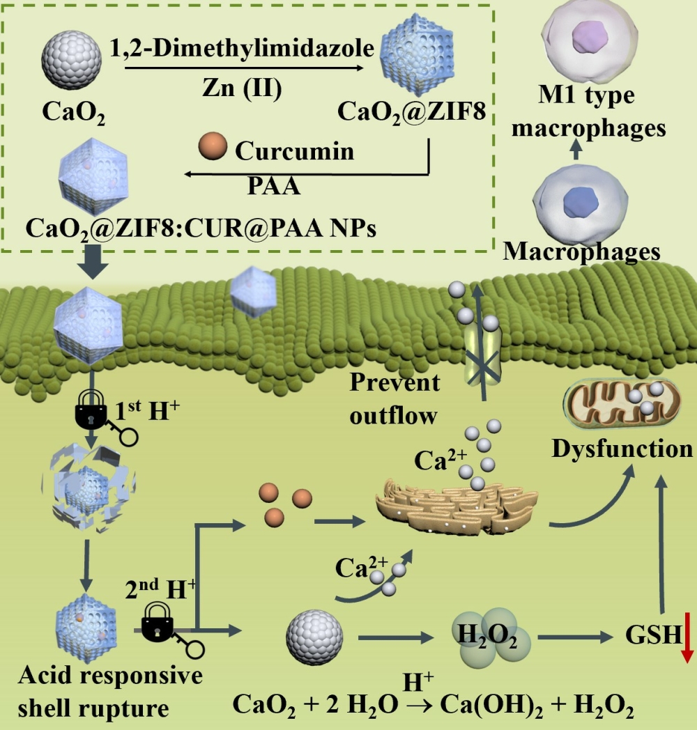
Zheng Y, Williams GR, Hu R, Tong S, Xu J, Wang T, Zhang Y, Wu J, Li F, Cai Y, Zhu LM
International Journal of Nanomedicine 2025, 20:1899-1920
Published Date: 12 February 2025
Exploring the Potential Role of Manganese-Based Zeolitic Imidazolate Framework Nanoparticles in Cancer Therapy: In vitro Studies Using Lung Cancer Cells
Ivasiv V, Neves IC, Baltazar F, Moreira JN, Bañobre López M, Larios J, Duruz J, Balog S, Vanhecke D, Lee WS, Keshavan S, Petri-Fink A, Rothen-Rutishauser B
International Journal of Nanomedicine 2026, 21:578869
Published Date: 20 March 2026