
Back to Journals » International Journal of Chronic Obstructive Pulmonary Disease » Volume 20
Evaluation of the Efficacy of Jiangqi Dingchuan Pill Based on Network Pharmacology Analysis and Cigarette Smoke and Lipopolysaccharide Induced Chronic Obstructive Pulmonary Disease Rat Model
Authors Zhou J, Song J, Peng D, Zheng Y, Ning N  , Chen G, Huang Q, Li Y
Received 6 September 2024
Accepted for publication 21 March 2025
Published 2 April 2025 Volume 2025:20 Pages 929—941
DOI https://doi.org/10.2147/COPD.S489696
Checked for plagiarism Yes
Review by Single anonymous peer review
Peer reviewer comments 2
Editor who approved publication: Prof. Dr. Richard Russell
Jiewen Zhou,1,* Jinbin Song,2,* Danting Peng,3 Yanqiu Zheng,2 Na Ning,4 Guirong Chen,2 Qiuling Huang,4 Yanwu Li2
1The First Clinical College, Guangzhou University of Chinese Medicine, Guangzhou, Guangdong, People’s Republic of China; 2Science and Technology Innovation Center, Guangzhou University of Chinese Medicine, Guangzhou, Guangdong, People’s Republic of China; 3Clinical Research Center, Guangzhou Baiyunshan Qixing Pharmaceutical Co. Ltd, Guangzhou, Guangdong, People’s Republic of China; 4Clinical Research Center, Guangzhou Baiyunshan Zhongyi Pharmaceutical Co. Ltd, Guangzhou, Guangdong, People’s Republic of China
*These authors contributed equally to this work
Correspondence: Qiuling Huang, Email [email protected] Yanwu Li, Email [email protected]
Background: Jiangqi Dingchuan Pill (JDP) is a patent Chinese medicine in the treatment of asthma. JDP consists of six herbal drugs, namely, Ephedrae Herba, Mori Cortex, Citri Reticulatae Pericarpium, Perillae Fructus, Descurainiae Semen, Sinapis Semen.
Objective: To employ the tools of network pharmacology and in vivo experiments, exploring the possible mechanism of JDP in treating chronic obstructive pulmonary disease (COPD).
Materials and Methods: Chemical constituents of JDP, collection of targets of COPD, target prediction were conducted, and then network pharmacological analysis was performed based on protein–protein interaction (PPI). The cigarette smoke and lipopolysaccharide-induced COPD model was applied to assess the effects of JDP. Rats were randomly divided into five groups (n = 8), ie, a sham group, a COPD-control group, two COPD groups treated with different doses of JDP (1.26 and 2.52 g/kg/d, respectively), and one COPD group treated with aminophylline (54 mg/kg/d). Pulmonary functions were assessed. The inflammatory cytokines in bronchial alveolar lavage fluid (BALF) were quantified using enzyme-linked immunosorbent assay (ELISA). The expression of matrix metalloprotein-9 (MMP-9) was quantified using Western blot.
Results: A total of 108 genes were found to be the main target genes regulated by JDP in the treatment of COPD, according to PPI analysis. Compared with the COPD-control group, rats in the JDP group exhibited amelioration in lung function, including 20 ms forced expiratory volume/forced vital capacity, maximal mid-expiratory flow curve, and airway resistance (all p < 0.05). A reduction of IL-1β and TNF-α expressions in BALF was also observed (both p < 0.05). Compared with the COPD-control group, the expression of MMP-9 in lung tissue was down-regulated in the JDP group (p < 0.05).
Conclusion: This study explored the effects and its mechanisms of JDP in COPD treatment. JDP exhibited therapeutic potential as a COPD intervention drug.
Keywords: Traditional Chinese Medicine, network pharmacology, chronic obstructive pulmonary disease, Jiangqi Dingchuan Pill
Graphical Abstract:

Introduction
Chronic obstructive pulmonary disease (COPD) is the most common chronic respiratory disease. It is estimated that the global prevalence of COPD among people aged 30–79 years was 10.3% in the Global Initiative for Chronic Obstructive Lung Disease (GOLD) report.1 The mortality rate of COPD ranks third in China, only behind the cerebrovascular disease and ischaemic heart disease.2 The COPD incidence in China will continue to increase over the next two decades due to several factors, including aging population, air pollution, and the high smoking rate.3 There exist great need for the research and development of therapeutic medications for COPD.4
Recently, it is generally acknowledged that the pathogenesis of COPD is intricately linked to inflammation, oxidative stress, and protease induced-tissue degradation.5 Owing to the influence of inflammatory cytokines, there exists an imbalance between endogenous proteases responsible for degrading connective tissue within the lungs and the antiproteases that counteract this process in COPD patients.6 The MMP superfamily plays an important role in the damage and repair of the extracellular matrix.7
The formula of Jiangqi Dingchuan Pill (JDP) is composed of six herbal medicines, including Ephedrae Herba, Mori Cortex, Citri Reticulatae Pericarpium, Perillae Fructus, Descurainiae Semen and Sinapis Semen. Several reports had revealed that the main constituents of the above-mentioned herbal medicine possess explicit efficacy in treating COPD. For example, the main constituent of Citri Reticulatae Pericarpium, hesperetin could ameliorate the lung function of COPD animal model.8 The main constituent of Ephedrae Herba, ephedrine ameliorates COPD through restraining endoplasmic reticulum stress both in vitro and in vivo models.9 More importantly, both ephedrine and pseudoephedrine are extensively employed as sympathomimetic agents in clinical practice, exerting agonistic action on β2 receptors.10,11 However, the complexity of the constituents in traditional Chinese medicine (TCM) had made it difficult to decipher the drug actions. Nowadays, researchers have come to be aware that TCM should be interpreted in a systemic manner, adopting tools like network pharmacology, in order to provide deeper insight into the treatment of complex diseases.12
In brief, network pharmacology analysis combined with in vivo model was applied to understand the underlying mechanisms of JDP treating COPD. In addition, in vivo model was applied to validate the effects of JDP on experimental COPD model.
Materials and Methods
Materials and Reagent
Jiangqi Dingchuan Pill were manufactured by Guangzhou Baiyunshan Pharmaceuticals Co. Ltd. (Lot No.: A01001M, A01002M, A01003M), complying to the legislations of China’s Drug Administration (Approval No. Z44020241). The detailed formula of JDP was shown in Table 1. Specimens of JDP were deposited in Guangzhou University of Chinese Medicine.
 |
Table 1 Formula of JDP, Origins of Natural Medicines and the Amounts of Crude Drugs Administered in Daily Dosage |
Lipopolysaccharides (LPS) (L2880) was purchased from Sigma-Aldrich Co. (St. Louis, MO, USA). Commercial cigarette (each containing nicotine 1.0 mg and CO 11 mg) was manufactured by China Tobacco Anhui Industrial. Co. Ltd. (Bangbu, China). Aminophylline was produced by Taiyuan Pharmaceuticals Co. Ltd. (Taiyuan, China). Enzyme-linked immunosorbent assay (ELISA) kits for tumor necrosis factor alpha (TNF-α, catalog No.: JL13202), interleukin 1 beta (IL-1β, catalog No.: JL20884) were obtained from Jianglaibio Co. Ltd. (Shanghai, China).
Anti-MMP9 (ab76003), Goat Anti-Mouse IgG H&L (HRP) (ab6789) were purchased from Abcam Inc. (Cambridge, UK). β-actin (#3700) was purchased from CST Inc. (Beverly, MA, USA).
Collection of Chemical Constituents
In the Traditional Chinese Medicine Systems Pharmacology Database and Analysis Platform (TCMSP) database, the chemical constituents of the herbal drugs are available.13 In this study, six herbal drugs along with their components were searched and tabulated. For all constituents, oral bioavailability (OB) and drug-likeness (DL) are two main indicators for the screening of bioactive components. The compound information for JDP was retrieved from TCMSP, with the criteria of OB ≥ 30% and DL ≥ 0.18.14 In addition, for other components with OB <30% or DL <0.18, if previous reports exhibited potential role in the treatment of COPD, the components as well as their targets were supplemented as well. A detailed list of molecular IDs, molecular names, and chemical structures of the chemical compounds was shown in Supplementary Materials (Table S1). The distribution was shown in (Supplementary Materials Table S2).
Their Therapeutic Target Collection in JDP
TCMSP and PharmMapper were utilized to obtain target information of each constituent.13,15 Text mining to establish a target information for the chemical constituents of JDP.
Collection of COPD Related Genes and Therapeutic Targets
The retrieval of COPD-related genes and target interactions was conducted from the following five databases: DrugBank Database (http://www.drugbank.ca/), Therapeutic Target Database (TTD) (https://db.idrblab.org/ttd/), GeneCards Database (www.genecards.org), DisGeNET Database (www.disgenet.com).16–19 In all databases, the term “chronic obstructive pulmonary disease” was searched. In the DrugBank and Therapeutic Target Database, all targets in the search results were collected. In addition, genes from the GeneCards database were selected with twice the median value of the relevance score.20 Genes from the DisGeNET database were selected with gene–disease association (GDA) score ≥0.4 (according to the definition of GDA, a score above 0.4 indicated that at least one curated source supporting a GDA).18 The protein names obtained are cross-referenced with gene names in the UniProt protein database (https://www.uniprot.org/) for verification. A detailed list was shown in Supplementary Materials Table S3.
Construction and Analysis of Protein–Protein Interaction (PPI) Network
The PPI network was constructed using the STRING database (http://string-db.org/cgi/input.pl). This database defines a confidence score (ranging from 0 to 1) for each interaction. A higher score indicates greater confidence in the PPI. The STRING database defines PPIs with a combined score above 0.9 as “very high confidence” and those above 0.4 as “medium confidence”.21 Genes and therapeutic targets collected in 2.3 and 2.4 were uploaded to the STRING database, generating two sets of PPI network (combined score ≥ 0.4). The PPI networks were merged, screening interaction with combined score ≥0.9, showing the central PPI network of JDP in the treatment of COPD.
GO Gene Enrichment Analysis and KEGG Pathway Annotation
The Database for Annotation, Visualization and Integrated Discovery (DAVID) was applied in GO annotation and KEGG pathway analysis. The false discovery rate (FDR) calculated in DAVID was used to show the enrichment significance.22
Network Visualization Tool
To visualize the PPI network and the compound-target-pathway network, Cytoscape 3.2.1 is utilized for data analysis.
Animal and Public Database Ethic
The SD rats were procured from the Guangdong Medical Laboratory Animal Center (No. SCXK(Yue)2018–0002). These rats were housed in the SPF-grade animal facility at the Experimental Animal Center of Guangzhou University of Chinese Medicine (No. SYXK(Yue)2018–0001). The animal experimentation was conducted following approval from the Ethics Committee for Experimental Animals at Guangzhou University of Chinese Medicine (No. 20220304011), complying with the Regulations on the Administration of Experimental Animals of Guangdong Province (dated November 29, 2019).
The study involved database relating to human data, including DrugBank Database, Therapeutic Target Database, GeneCards Database, DisGeNET Database and STRING database. The study is exempt from approval as the data were retrieved from the publicly available data, based on Measures for Ethical Review of Life Science and Medical Research Involving Human Subjects (China, February 18, 2023), according to Item 1 and 2 of Article 32.
Establishment of COPD Model
The rats were acclimated for 3 days. COPD model was established through LPS challenge combined with cigarette smoke exposure.23 On the first day of modeling, rats were anesthetized using isoflurane anesthesia in animal anesthesia machines (R550I, RWD Life Science, Shenzhen, China). Neck hair was removed, and the area was disinfected. The skin and muscles along the midline of the neck were carefully incised, exposing the trachea. A syringe was inserted into the upper end of the second tracheal cartilage, injecting 0.1 mL of LPS saline solution. The rat’s head was immediately raised vertically for 10–20 seconds to ensure the distribution of LPS down the trachea. The neck muscles and skin were then sutured. After gaining consciousness, the rat was placed back into the cage for normal feeding. On the 15th day, a second round of LPS tracheal instillation was performed using the same procedure. The sham group underwent the same procedure, except for injecting 0.1 mL of saline solution.
On the 16th day, smoke exposure was initiated. Rats were placed in a smoke chamber (40 × 30 × 25 cm), 10 rats at a time. Five cigarettes were ignited, and the extracting pump was used to draw smoke into the chamber. Smoke exposure was conducted for 30 min. The smoke exposure process was conducted for two times per day, lasting for 6 weeks.24
Intervention
After 4 weeks of modeling, rats were randomly divided into five groups: sham group, COPD-control group, JDP low-dose group JDP high-dose group and aminophylline group. The intragastric administration for each group started in the 5th week. Aminophylline was administered at a dose of 0.054 g/kg, while JDP was administered at doses of 1.26 g/kg and 2.52 g/kg, once daily, with a gastric volume of 10 mL/kg. The COPD-control group and sham group received distilled water in an equivalent volume. Administration continued for 4 weeks.
Pulmonary Function Testing and Sample Collection
At the end of the 8-week experiment, rats were anesthetized with 20% urethane (7 mL/kg) via intraperitoneal injection.25 Tracheal intubation was then performed, and pulmonary function assessment was conducted using the rat pulmonary function testing system (Buxco PFT, DSI INC., USA). Blood collection was performed from the abdominal aorta. The rats were sacrificed after completing pulmonary function testing and blood collection. The bronchoalveolar lavage fluid (BALF) was collected for further analysis. Briefly, the right lung root was ligated, and the left lung was washed with saline for twice (2 mL per time). ELISA kit was applied for the measurement of TNF-α and IL-1β in BALF, in accordance to the instructions.
Western Blot Analysis
The lung tissues were homogenated, lysed and centrifugated, collecting the supernatant. The expressions of MMP-9 were quantified. Loading control was β-actin. Samples were subjected to electrophoresis on gels, then transferred onto membranes, subsequently incubated with the antibody overnight at 4°C. Blots were washed followed by secondary antibody treatment. Analysis was carried out on Gel Imaging System (Bio-Rad, USA).
Statistical Analysis
Data were analyzed using SPSS 25.0. For continuous variables that follow a normal distribution, one-way ANOVA was employed for comparing means among multiple samples, and post hoc pairwise comparisons were conducted using the LSD method. Data are presented as mean ± standard deviation. For variables not complying to normal distribution, the Mann–Whitney test was used to compare differences between groups. Data were presented as median with interquartile range (IQR). p < 0.05 was considered statistically significant.
Results
Target Collection of JDP in the Treatment of COPD
A total of 116 chemical constituents were collected from six herbs in JDP. The number of chemical constituents in each herb was 37 from Ephedrae Herba, 39 from Mori Cortex, 14 from Citri Reticulatae Pericarpium, 11 from Perillae Fructus, 20 from Descurainiae Semen and 19 from Sinapis Semen. The specific names of the compounds and their distribution are detailed in Supplementary Materials Table S1.
The targets of compounds in JDP were collected. After removing duplicates, the number of targets in each herb was 582 for Ephedrae Herba, 574 for Mori Cortex, 390 for Citri Reticulatae Pericarpium, 491 for Perillae Fructus, 438 for Descurainiae Semen and 283 for Sinapis Semen (Supplementary Materials Table S2). After removing duplicates and converting to official gene symbols, a total of 512 COPD-related genes and targets were collected (Supplementary Materials Table S3).
The gene lists of JDP targets were submitted to the STRING server to build PPI networks, displaying all high confident PPI relationships (combined score ≥ 0.9). The network comprised 635 genes and 4984 PPI connections. The results are depicted in Figure 1A. The data pertaining to COPD-related genes (Supplementary Materials Table S3) and therapeutic targets were uploaded, generating another network comprising 290 targets and 1100 PPI connections (Figure 1B).
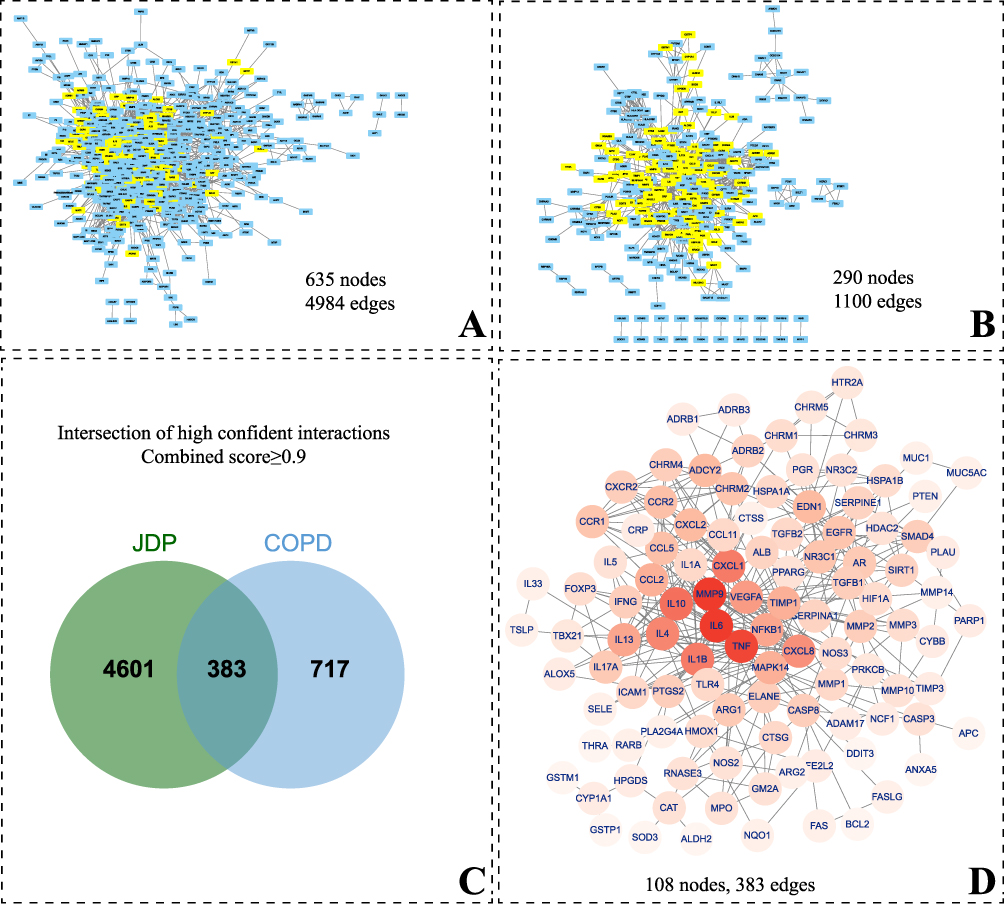 |
Figure 1 PPI network and network intersection. (A) PPI network of JDP with 635 nodes and 4984 edges. (B) PPI network of COPD with 290 nodes and 1100 edges. (C) Venn diagram of high confident interaction of PPI network between JDP and COPD. (D) Visualization of central PPI network of JDP treating COPD. In (A) and (B), the yellow nodes denoted the intersected PPI network nodes between JDP and COPD, while the blue are those not intersected. Abbreviations: PPI, protein–protein interaction. JDP, Jiangqi Dingchuan Pill. COPD, chronic obstructive pulmonary disease. |
To elucidate the mechanism underlying the therapeutic effect of the JDP on COPD, we identified common nodes between the two PPI networks as shown in Figure 1C and D. This resulted in a network consisting of 108 nodes and 383 edges (Figure 1C). As depicted in the Figure 1D, the genes IL6, MMP9, TNF, IL10, IL1B, and CXCL1 had the top six degree-values. These genes represent key nodes in the network, highlighting their significance in the therapeutic effects of the JDP in COPD treatment.
Gene Ontology (GO) Annotation and KEGG Pathway Analysis
The GO annotation and enrichment analysis were performed on the 108 central PPI network genes (Figure 2). The enriched results were based on -log10 false discovery rate (FDR) (all p < 0.01). The top 5 GO annotation included inflammatory response, response to hypoxia, response to lipopolysaccharide, immune response, and negative regulation of apoptosis process (Figure 2A).
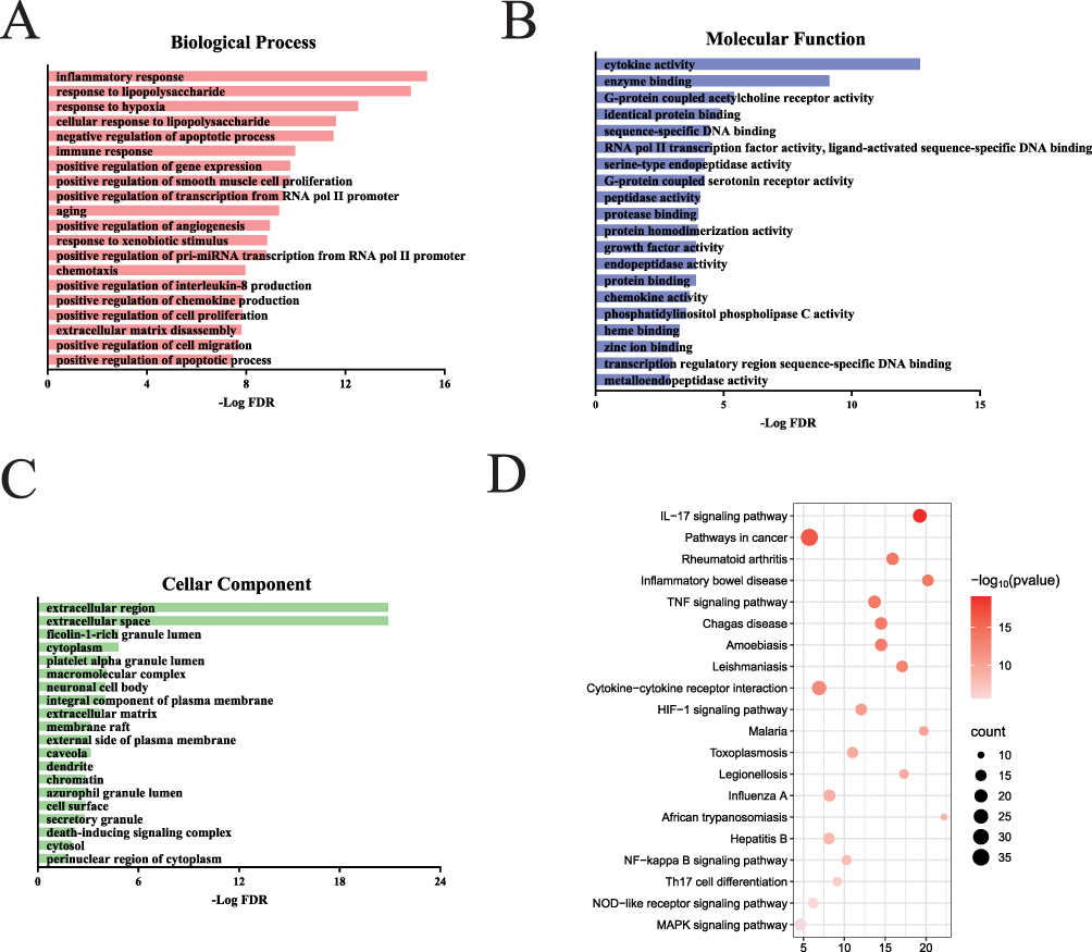 |
Figure 2 GO functional annotation and KEGG analysis of central network genes of JDP treating COPD. Biological process (A), molecular function (B), cellar components (C), and KEGG pathways (D) were sorted according to − log FDR (all p < 0.01). Abbreviations: GO, Gene Ontology; KEGG, Kyoto Encyclopedia of Genes and Genomes; JDP, Jiangqi Dingchuan Pill. COPD, chronic obstructive pulmonary disease. |
The top 5 molecular functions included cytokine activity, enzyme binding, G-protein coupled acetylcholine receptor activity, identical protein binding, and sequence-specific DNA binding (Figure 2B). The top 5 cellar components included extracellular space, extracellular region, ficolin-1-rich granule lumen, cytoplasm, and platelet alpha granule lumen (Figure 2C).
To provide further insight into the functions of these target genes, KEGG pathway analysis was performed on the aforementioned 108 genes. Several pathways potentially related to COPD were included, such as IL-17 signaling pathway, tumor necrosis factor signaling pathway, MAPK signaling pathway, HIF-1 signaling pathway, nucleotide binding oligomerization domain-like receptor signaling pathway, and hypoxia-induced factor-1 signaling pathway, NF-kappa B signaling pathway, Th17 cell differentiation. The JDP’s potential therapeutic effect on COPD may be achieved through the modulation of these signaling pathways (Figure 2D).
Construction of Component-Target-Pathway Network
By integrating the data from pathway enrichment, the component-target-pathway network was developed, illustrating the mechanism of the JDP’s therapeutic action in COPD treatment (Figure 3). The component-target-pathway diagram provides a comprehensive overview of the primary mechanisms underlying the JDP’s role in treating COPD. The major active ingredients in the JDP include resveratrol, ursolic acid, rutin, luteolin, rosmarinic acid, etc. The key genes regulated by the JDP include MMP9, CASP3, MAPK14, MMP2, MMP3, IL1B, with some overlap with the results of PPI network analysis.
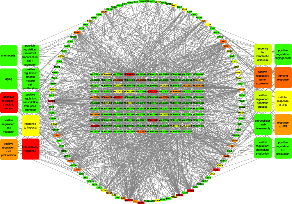 |
Figure 3 Component-target-pathway network of JDP in the treatment of COPD. In the diagram, nodes are represented by a gradient of red to green coloration. Nodes in red are considered as important (with higher degree value), and the green nodes are considered as less important (with lower degree value). The inner grid denotes the major chemical components of the compound formula. The ellipses within the circles represent the main target genes regulated by the JDP. The outer rectangles at both sides denote the key biological processes regulated by the JDP. Abbreviations: JDP, Jiangqi Dingchuan Pill. COPD, chronic obstructive pulmonary disease. |
As can be seen in Figure 3, the following biological processes have the most genes involved, including inflammatory response, negative regulation of apoptotic process, immune response, positive regulation of gene expression, positive regulation of cell proliferation.
Rat Weight Changes
The study flowchart of modeling and intervention is shown in Figure 4A. Compared to the sham group, the body weight of COPD-control rats and rats in each intervention group showed a significant decrease starting from the 2nd week (all p < 0.05), persisting until the 8th week. When compared to the COPD-control group, there were no significant differences observed in all intervention groups except in 7th week of the JDP-low group (Figure 4B).
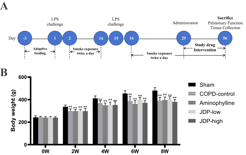 |
Figure 4 Experimental flow chart and animal weight changes. (A) Experimental flow chart. (B) animal weight changes. n=8. *p < 0.05, versus sham group. Data are presented as mean±standard deviation. One-way ANOVA was employed for comparing means among multiple samples, and post hoc pairwise comparisons were conducted using the LSD method. Abbreviations: LPS, lipopolysaccharide. JDP, Jiangqi Dingchuan Pill. COPD, chronic obstructive pulmonary disease. |
Pulmonary Function Assessment
Compared to the sham group, significant changes in lung function were observed in the COPD-control group, including a significant decrease in 20 ms forced expiratory volume (FEV20), 20 ms forced expiratory volume/forced vital capacity (FVC), and maximal mid-expiratory flow (MMEF), accompanied by a significant increase in airway resistance (RI) (all p < 0.05). These changes suggest the presence of typical characteristics of COPD such as ventilation impairment and airflow limitation. After intervention with low and high doses of the JDP, there were significant improvements in FEV20, FEV20/FVC, and MMEF (all p < 0.05). The aminophylline group also showed significant improvement in MMEF. In addition, it was observed that in FEV20 and FEV20/FVC, JDP-high exhibited significant improvement compared with the aminophylline group.
These findings suggest that the JDP formula partially ameliorated the pulmonary function impairment in the COPD rat model (Table 2).
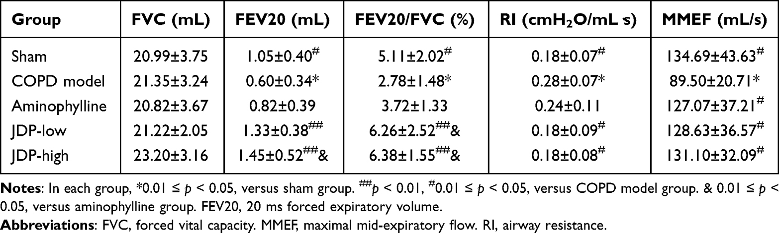 |
Table 2 Lung Functions of Rats After 8 weeks of Treatment |
Changes in Cytokine Levels in Lung Lavage Fluid
Compared to the sham group, the BALF of the COPD-control group showed a significant increase in the levels of IL-1β and TNF-α. There was a notable decrease in the levels of IL-1β and TNF-α in the JDP-low group, JDP-high group and the aminophylline group compared with the COPD-control group (as shown in Figure 5).
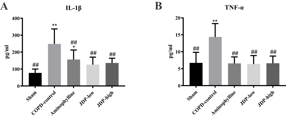 |
Figure 5 Effects of JDP on BALF cytokines, n=8. (A) IL-1β expression in BALF. (B) TNF-α expression in BALF. **p < 0.01, *p < 0.05, versus sham group. ##p < 0.01, versus COPD-control group. Data are presented as mean±standard deviation. One-way ANOVA was employed for comparing means among multiple samples, and post hoc pairwise comparisons were conducted using the LSD method. Abbreviations: IL-1β, Interleukin 1β; TNF-α, tumor necrosis factor α; BALF, bronchial alveolar lavage fluid; JDP, Jiangqi Dingchuan Pill. |
Changes in MMP9 Protein Expression in Lung Tissue
Compared to the sham group, the expression of MMP9 protein in the lung tissue of the COPD-control group was significantly increased. There was a significant decrease in MMP9 expression in the lung tissue in the JDP-low group, JDP-high group and the aminophylline group compared with the COPD-control group (as shown in Figure 6).
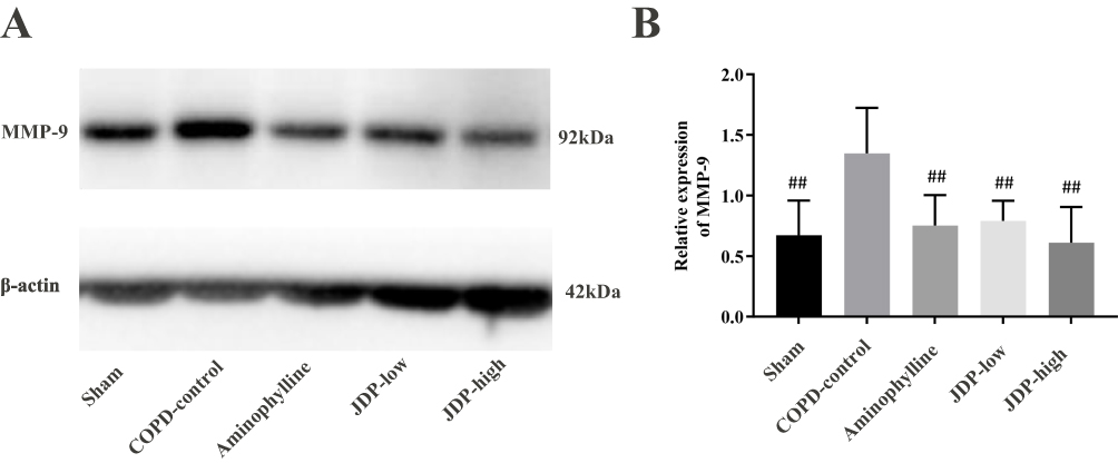 |
Figure 6 Effects of JDP on MMP9 protein expression in the lung. (A) Representative Western blotting for MMP9. (B) Quantitative analysis of Western blotting of MMP9. In each group, n=3. ##p < 0.01, versus COPD-control group. Data are presented as mean±standard deviation. One-way ANOVA was employed for comparing means among multiple samples, and post hoc pairwise comparisons were conducted using the LSD method. Abbreviations: MMP-9, matrix metalloprotein-9; JDP, Jiangqi Dingchuan Pill. |
Discussion
In this study, we employed database searches, text mining, and target fishing to collect the target genes affected by the JDP. By applying PPI network enrichment, we extracted 108 target genes related to the pathology of COPD influenced by the JDP. Building upon this, we further extracted the core network and key target genes, and correlated them with the compounds in the JDP. This provides a general understanding of the key components and targets of the JDP in treating COPD.
Network pharmacology research identified the 10 compounds with the highest number of target interactions among the 116 main components. The top 10 constituents included one stilbene (resveratrol), two triterpenoids (ursolic acid and oleanolic acid), five flavonoids (rutin, hesperidin, luteolin, apigenin, quercetin), two phenylpropanoid (eugenol, rosmarinic acid). A detailed list of these components is available in Supplementary Material, Table S4.
Plants of the Moraceae family are rich in stilbenes, with resveratrol being a representative compound.26 Previous clinical trial had proved that resveratrol inhibits inflammatory cytokine release from alveolar macrophages in COPD.27 Resveratrol alleviates airway inflammation and remodeling through the various pathways, for example, reducing IL-1β, IL-10, and TNF-α through HMGB1/TLR4/NF-κB signaling pathway, reducing STAT3 expression and inhibits the release of MMP-3 and MMP-9, etc. However, in a randomized controlled study, it was found that resveratrol administration for 4 weeks did not have any positive effects on muscle mitochondrial function, adipose tissue inflammatory and metabolic gene expression, and systemic inflammation in COPD patients.28
Two triterpenoids were also listed as the key components treating COPD, including ursolic acid and oleanolic acid. Both constituents derived from the Mori Cortex, and exhibited therapeutic potential in the treatment of COPD.29,30 It is also observed that in JDP, the presence and effects of flavonoids should also be noted. Among these flavonoid constituents, several are extensively distributed among diverse plant taxa, such as rutin, luteolin, quercetin, apigenin, and tectorigenin.31 The aforementioned compounds collectively exhibit well-defined anti-inflammatory and antioxidative effects in experimental and small-scale clinical trials.32
By comparing the top 10 target genes identified in Figures 1 and 3, an intersection of three genes was found: matrix metalloproteinase-9 (MMP9), tumor necrosis factor (TNF), interleukin-1 beta (IL-1β).
MMP-9, a member of the MMP superfamily, possesses enzymatic activity capable of acting on elastin, proteoglycans, and type IV collagen.33 This is closely linked to extracellular matrix damage and repair. MMP-9 can digest elastin and other structural components on alveolar walls, which is one of the key factor in causing emphysema. Furthermore, studies indicate that lung tissue expression of MMP-9 is associated with smoking and negatively correlates with FEV1.34 JDP can down-regulate the expressions of MMP-9 in the lung tissues, thus might restore the balance of protease-antiprotease balance and lung function. A previous report had proved that IL-1β and TNF-α significantly increased the release of MMP-9 in the alveolar macrophage of smokers.
IL-1β can elevate neutrophil aggregation and, through protease production, disrupt the balance of protease/anti-protease systems, causing airway and lung tissue damage.35 TNF-α, produced by various inflammatory cells like activated monocytes, macrophages, and T lymphocytes, is a highly biologically active inflammatory cytokine. Healthy TNF-α levels contribute to immune system regulation and pathogen defense, while excessive and prolonged presence can lead to tissue damage.36 In our present COPD model, the levels of both IL-1β and TNF-α in BALF were reduced after JDP intervention (Figure 5).
In this study, the target genes involved in the main components of JDP were screened by network pharmacology, and the potential mechanism of JDP in the treatment of COPD was further predicted by enrichment analysis, functional annotation and pathway analysis. Subsequently, a rat model of COPD was used for experimental verification, and the results showed that JDP may play a role in the treatment of COPD by anti-inflammatory effect and reducing MMP-9 damage to the alveolar wall. This study explores the potential therapeutic mechanism of JDP in COPD and provides basic research evidence for its clinical application. Intriguingly, we found that the intervention of JDP exhibited significant improvement compared with the aminophylline group in FEV20 and FEV20/FVC. However, significant changes were not observed in other parameters, including inflammatory markers in BALF or MMP-9 levels in the lung tissue. A previous report demonstrated that aminophylline exhibited anti-inflammatory effects in COPD model and could also down-regulate the level of MMP-9.37 Our results were consistent with this finding. However, the current study was insufficient to elucidate the reason for improvement of pulmonary functions of JDP in comparison to the aminophylline.
Several limitations should be addressed in this study. At first, in the network pharmacological analysis, human targets and genes were taken into consideration. However, the therapeutic effects were assessed on rats. The network pharmacological analysis in human could not be simply generalized to rat. Second, immunological status is a important part of the pathological process of COPD. The change of immune cells, both in the respiratory tract and the whole body, should also be assessed in further study to evaluate the effect of JDP.
In conclusion, this study explored the effects and its mechanisms of JDP in COPD treatment. JDP exhibited therapeutic potential as a COPD intervention drug.
Data Sharing Statement
The data used to support the findings of this study are included within the article and the supplementary materials.
Author Contributions
All authors made a significant contribution to the work reported, whether that is in the conception, study design, execution, acquisition of data, analysis and interpretation, or in all these areas; took part in drafting, revising or critically reviewing the article; gave final approval of the version to be published; have agreed on the journal to which the article has been submitted; and agree to be accountable for all aspects of the work.
Supplementary Materials
Supplementary materials relating to this manuscript are attached.
Funding
This work was financially supported by Research and Development Plan Project in Key Area of Guangdong Province (2020B1111110004).
Disclosure
Authors all declare that there is no existing conflict of interest.
References
1. Adeloye D, Song P, Zhu Y. et al. Global, regional, and national prevalence of, and risk factors for, chronic obstructive pulmonary disease (COPD) in 2019: a systematic review and modelling analysis. Lancet Respir Med. 2022;10(5):447–458. doi:10.1016/S2213-2600(21)00511-7
2. Zhou M, Wang H, Zhu J, et al. Cause-specific mortality for 240 causes in China during 1990-2013: a systematic subnational analysis for the global burden of disease study 2013. Lancet. 2016;387(10015):251–272. doi:10.1016/S0140-6736(15)00551-6
3. Hu W, Fang L, Zhang H, Ni R, Pan G. Global disease burden of COPD from 1990 to 2019 and prediction of future disease burden trend in China. Public Health. 2022;208:89–97. doi:10.1016/j.puhe.2022.04.015
4. Xian M, Xu J, Zheng Y, et al. Network pharmacology and experimental verification reveal the regulatory mechanism of chuanbeimu in treating chronic obstructive pulmonary disease. Int J Chron Obstruct Pulmon Dis. 2024;19:799–813. doi:10.2147/COPD.S442191
5. Christenson SA, Smith BM, Bafadhel M, Putcha N. Chronic obstructive pulmonary disease. Lancet. 2022;399(10342):2227–2242. doi:10.1016/S0140-6736(22)00470-6
6. Elkington PTG, Friedland JS. Matrix metalloproteinases in destructive pulmonary pathology. Thorax. 2006;61(3):259–266. doi:10.1136/thx.2005.051979
7. Belvisi MG, Bottomley KM. The role of matrix metalloproteinases (MMPs) in the pathophysiology of chronic obstructive pulmonary disease (COPD): a therapeutic role for inhibitors of MMPs? Inflammation Res. 2003;52(3):95–100. doi:10.1007/s000110300020
8. Zhou L, Gu W, Kui F, et al. The mechanism and candidate compounds of aged citrus peel (chenpi) preventing chronic obstructive pulmonary disease and its progression to lung cancer. Food Nutr Res. 2021;65. doi:10.29219/fnr.v65.7526
9. Wang H, Chen F, Wu L. Ephedrine ameliorates chronic obstructive pulmonary disease (COPD) through restraining endoplasmic reticulum (ER) stress in vitro and in vivo. Int Immunopharmacol. 2022;103:107842. doi:10.1016/j.intimp.2021.107842
10. Petty TL. COPD in perspective. Chest. 2002;121(5 Suppl):116S–120S. doi:10.1378/chest.121.5_suppl.116S
11. Shekelle PG, Hardy ML, Morton SC, et al. Efficacy and safety of ephedra and ephedrine for weight loss and athletic performance: a meta-analysis. JAMA. 2003;289(12):1537–1545. doi:10.1001/jama.289.12.1537
12. Wang Y, Yang H, Chen L, Jafari M, Tang J. Network-based modeling of herb combinations in traditional Chinese medicine. Brief Bioinform. 2021;22(5). doi:10.1093/bib/bbab106
13. Ru J, Li P, Wang J, et al. TCMSP: a database of systems pharmacology for drug discovery from herbal medicines. J Cheminform. 2014;6:13. doi:10.1186/1758-2946-6-13
14. Zhang W, Chen Y, Jiang H, et al. Integrated strategy for accurately screening biomarkers based on metabolomics coupled with network pharmacology. Talanta. 2020;211:120710. doi:10.1016/j.talanta.2020.120710
15. Liu X, Ouyang S, Yu B, et al. PharmMapper server: a web server for potential drug target identification using pharmacophore mapping approach. Nucleic Acids Res. 2010;38:W609–W614. doi:10.1093/nar/gkq300
16. Safran M, Dalah I, Alexander J, et al. GeneCards version 3: the human gene integrator. Database. 2010;2010:baq020. doi:10.1093/database/baq020
17. Wishart DS, Feunang YD, Guo AC, et al. DrugBank 5.0: a major update to the DrugBank database for 2018. Nucleic Acids Res. 2018;46(D1):D1074–D1082. doi:10.1093/nar/gkx1037
18. Piñero J, Ramírez-Anguita JM, Saüch-Pitarch J, et al. The DisGeNET knowledge platform for disease genomics: 2019 update. Nucleic Acids Res. 2020;48(D1):D845–D855. doi:10.1093/nar/gkz1021
19. Zhou Y, Zhang Y, Lian X, et al. Therapeutic target database update 2022: facilitating drug discovery with enriched comparative data of targeted agents. Nucleic Acids Res. 2022;50(D1):D1398–D1407. doi:10.1093/nar/gkab953
20. Xie X, Zhang L, Lin Y, et al. Liangxue Jiedu formula improves imiquimod-induced psoriasiform dermatitis with circadian desynchrony by regulating Th17 cell differentiation based on network pharmacological analysis. J Ethnopharmacol. 2023;317:116807. doi:10.1016/j.jep.2023.116807
21. Szklarczyk D, Gable AL, Nastou KC, et al. The STRING database in 2021: customizable protein-protein networks, and functional characterization of user-uploaded gene/measurement sets. Nucleic Acids Res. 2021;49(D1):D605–D612. doi:10.1093/nar/gkaa1074
22. Dennis G, Sherman BT, Hosack DA, et al. DAVID: database for annotation, visualization, and integrated discovery. Genome Biol. 2003;4(5):P3. doi:10.1186/gb-2003-4-9-r60
23. Nie Y, Wu H, Li P, et al. Characteristic comparison of three rat models induced by cigarette smoke or combined with LPS: to establish a suitable model for study of airway mucus hypersecretion in chronic obstructive pulmonary disease. Pulm Pharmacol Ther. 2012;25(5):349–356. doi:10.1016/j.pupt.2012.06.004
24. Wang X, Li T, Li J, Miao S, Xiao X. The effects of resveratrol on inflammation and oxidative stress in a rat model of chronic obstructive pulmonary disease. Molecules. 2017;22(9). doi:10.3390/molecules22091529
25. Hara K, Harris RA. The anesthetic mechanism of urethane: the effects on neurotransmitter-gated ion channels. Anesth Analg. 2002;94(2):313–318. doi:10.1213/00000539-200202000-00015
26. Burlando B, Clericuzio M, Cornara L. Moraceae plants with tyrosinase inhibitory activity: a review. Mini Rev Med Chem. 2017;17(2):108–121. doi:10.2174/1389557516666160609071854
27. Culpitt SV, Rogers DF, Fenwick PS, et al. Inhibition by red wine extract, resveratrol, of cytokine release by alveolar macrophages in COPD. Thorax. 2003;58(11):942–946. doi:10.1136/thorax.58.11.942
28. Beijers RJ, Gosker HR, Sanders KJ, et al. Resveratrol and metabolic health in COPD: a proof-of-concept randomized controlled trial. Clin Nutr. 2020;39(10):2989–2997. doi:10.1016/j.clnu.2020.01.002
29. Song GR, Choi YJ, Park SJ, et al. Root bark of Morus alba L. and its bioactive ingredient, ursolic acid, suppress the proliferation of multiple myeloma cells by inhibiting Wnt/β-catenin pathway. J Microbiol Biotechnol. 2021;31(11):1559–1567. doi:10.4014/jmb.2109.09002
30. Ma X, Liu A, Liu W, et al. Analyze and identify peiminine target EGFR improve lung function and alleviate pulmonary fibrosis to prevent exacerbation of chronic obstructive pulmonary disease by phosphoproteomics analysis. Front Pharmacol. 2019;10:737. doi:10.3389/fphar.2019.00737
31. Fu Y, Kang N, Yu Y, et al. Polyphenols, flavonoids and inflammasomes: the role of cigarette smoke in COPD. Eur Respir Rev. 2022;31(164):220028. doi:10.1183/16000617.0028-2022
32. Han MK, Barreto TA, Martinez FJ, Comstock AT, Sajjan US. Randomised clinical trial to determine the safety of quercetin supplementation in patients with chronic obstructive pulmonary disease. BMJ Open Respir Res. 2020;7(1). doi:10.1136/bmjresp-2018-000392
33. Lim S, Roche N, Oliver BG, et al. Balance of matrix metalloprotease-9 and tissue inhibitor of metalloprotease-1 from alveolar macrophages in cigarette smokers. regulation by interleukin-10. Am J Respir Crit Care Med. 2000;162(4):1355–1360. doi:10.1164/ajrccm.162.4.9910097
34. Dimic-Janjic S, Hoda MA, Milenkovic B, et al. The usefulness of MMP-9, TIMP-1 and MMP-9/TIMP-1 ratio for diagnosis and assessment of COPD severity. Eur J Med Res. 2023;28(1):127. doi:10.1186/s40001-023-01094-7
35. Guo P, Li R, Piao TH, et al. Pathological mechanism and targeted drugs of COPD. Int J Chron Obstruct Pulmon Dis. 2022;17:1565–1575. doi:10.2147/COPD.S366126
36. Matera MG, Calzetta L, Cazzola M. TNF-alpha inhibitors in asthma and COPD: we must not throw the baby out with the bath water. Pulm Pharmacol Ther. 2010;23(2):121–128. doi:10.1016/j.pupt.2009.10.007
37. Zhang L, Tian Y, Zhao P, et al. Network pharmacology analysis uncovers the effect on apoptotic pathway by Bu-Fei formula for COPD treatment. J Ethnopharmacol. 2022;289:115022. doi:10.1016/j.jep.2022.115022
 © 2025 The Author(s). This work is published and licensed by Dove Medical Press Limited. The
full terms of this license are available at https://www.dovepress.com/terms
and incorporate the Creative Commons Attribution
- Non Commercial (unported, 4.0) License.
By accessing the work you hereby accept the Terms. Non-commercial uses of the work are permitted
without any further permission from Dove Medical Press Limited, provided the work is properly
attributed. For permission for commercial use of this work, please see paragraphs 4.2 and 5 of our Terms.
© 2025 The Author(s). This work is published and licensed by Dove Medical Press Limited. The
full terms of this license are available at https://www.dovepress.com/terms
and incorporate the Creative Commons Attribution
- Non Commercial (unported, 4.0) License.
By accessing the work you hereby accept the Terms. Non-commercial uses of the work are permitted
without any further permission from Dove Medical Press Limited, provided the work is properly
attributed. For permission for commercial use of this work, please see paragraphs 4.2 and 5 of our Terms.