")
Back to Journals » Infection and Drug Resistance » Volume 16
Evaluation of the Effect and Mechanism of Sanhuang Ointment on MRSA Infection in the Skin and Soft Tissue via Network Pharmacology
Authors Pan H , Wang T, Che Y, Li X, Cui Y , Chen Q, Wu Z, Yi J, Wang B
Received 7 June 2023
Accepted for publication 27 October 2023
Published 7 November 2023 Volume 2023:16 Pages 7071—7095
DOI https://doi.org/10.2147/IDR.S424746
Checked for plagiarism Yes
Review by Single anonymous peer review
Peer reviewer comments 6
Editor who approved publication: Prof. Dr. Héctor Mora-Montes
Haibang Pan,1,* Tianming Wang,1,2,* Ying Che,3,4 Xiaoli Li,1,5 Yan Cui,1 Quanxin Chen,1 Zhihang Wu,1 Jianfeng Yi,1 Bo Wang3
1First School of Clinical Medicine, Gansu University of Chinese Medicine, Lanzhou, People’s Republic of China; 2Gansu Provincial Key Laboratory of Traditional Chinese Medicine Recipe Mining and Innovation Transformation, Gansu Province New Production of Traditional Chinese Medicine Product Creation Engineering Laboratory, Lanzhou, People’s Republic of China; 3School of Nursing, Gansu University of Chinese Medicine, Lanzhou, People’s Republic of China; 4Research Ward, Gansu Provincial People’s Hospital, Lanzhou, People’s Republic of China; 5Gansu Provincial Maternity and Child-Care Hospital, Lanzhou, People’s Republic of China
*These authors contributed equally to this work
Correspondence: Bo Wang; Jianfeng Yi, Email [email protected]; [email protected]
Introduction: Skin and soft tissue infection (SSTI) is a frequently encountered clinical disease, and Sanhuang ointment, a traditional Chinese medicine, is used to treat it. However, the pharmacological effect of Sanhuang ointment on SSTI and its underlying mechanism remains unclear. Here, we investigate the protective effect of Sanhuang ointment on Methicillin-resistant Staphylococcus aureus (MRSA) infection in the skin and soft tissues and the underlying mechanism by network pharmacological analysis, followed by in vivo experimental validation.
Methods: Via network pharmacology, the active components and disease targets of Sanhuang ointment were screened and intersected for Gene Ontology (GO) and Kyoto Encyclopedia of Genes and Genomes (KEGG) enrichment analysis. A rat model of skin and soft tissue infection was established, and pathological features were observed. Large, medium, and small-dose groups (1 g, 0.5 g, and 0.25 g/animal, with the total amount of Vaseline, dispensed 1 g/animal) of Sanhuang ointment were prepared and Mupirocin ointment was used as a positive control (0.5 g/animal, with the total amount of Vaseline, dispensed 1 g/animal). The expressions of key proteins of the IL-17/NF-κB signaling pathway and downstream inflammatory factors were analyzed by histomorphological analysis, enzyme-linked immunosorbent assay, polymerase chain reaction, and Western blotting.
Results: In all, 119 active components and 275 target genes of Sanhuang ointment were identified and intersected with MRSA infection-related genes via network pharmacology analysis, and 34 target genes of Sanhuang ointment were found to be involved in skin and soft tissue infections with MRSA. Sanhuang ointment (1 g/mouse) could effectively ameliorate histopathological changes and significantly inhibit the expression of key proteins involved in the IL-17/NF-κB signaling pathway and downstream inflammatory factors (p < 0.05).
Conclusion: Sanhuang ointment has a protective effect on MRSA infection and inhibits inflammation by inhibiting the IL-17/NF-κB signaling pathway. Our findings are important for the secondary development and new drug development of Sanhuang ointment.
Plain Language Summary: Sanhuang ointment can significantly inhibit inflammatory response after skin and soft tissue infection with MRSA.
Sanhuang ointment may inhibit the inflammatory response induced by MRSA in the skin and soft tissue infections by targeting the IL-17/NF-κB signaling pathway.
We study the active components and mechanism of action of Sanhuang ointment on MRSA infection through network pharmacology.
Keywords: Sanhuang ointment, MRSA, SSTI, network pharmacology, IL-17, NF-κB
Introduction
Skin and soft tissue infections (SSTIs) are common clinical diseases.1 Most soft tissue infections are inflammatory reactions mainly caused by pathogenic invasion and reproduction. Bacterial infections (most commonly caused by Staphylococcus aureus and Hemolytic Streptococci) are common problems faced while treating soft tissue infections on the body surface.2 Methicillin-resistant Staphylococcus aureus (MRSA) is the most commonly encountered drug-resistant Staphylococcus aureus, with increasing detection rates in body surface-infected tissues, secretions, and pus. MRSA has become bacteria with the highest incidence of nosocomial infections worldwide.3 According to the Global Surveillance System for Antibiotic Resistance and Use (GLASS) report survey statistics in 2020, approximately 700,000 deaths resulting from antibiotic-resistant bacterial infections are reported each year worldwide, and MRSA is one of its major risk factors.4 At present, vancomycin is the first-line antibiotic used for the clinical treatment of MRSA infections, but the increasing number of vancomycin-resistant strains has increased the complexity of clinical anti-MRSA infection treatment5,6 Although some progress has been made in the research and development of new antibiotics for treating MRSA infections in recent years, it is important to find new treatment strategies to prevent the emergence of new antibiotic-resistant strains.
“Sanhuang ointment” (in-hospital preparation of the Affiliated Hospital of Gansu University of Traditional Chinese Medicine, Ganyao Zhunzi Z04010878). Huangqin, Huanglian, Huangbai, Borneol, sesame oil, and Vaseline, among others are prepared in specific proportions. Huangqin, the dried rhizome of Scutellariae Radix. Huanglian, the dried rhizome of Coptidis Rhizoma. Huangbai, the dried rhizome of Phellodendri Chinensis Cortex. The plant name corresponds to the latest revision in “World Flora Online” (www.worldfloraonline.org). It has been clinically used for more than 35 years, with significant curative effects and few side effects, especially for boils, carbuncles, erysipelas, abscesses, and other acute and chronic purulent infections, burns, soft tissue injuries, fluid extravasation, body surface ulcers, banding caused by infusion and chemotherapy, Herpes and other infectious diseases and has a significant curative effect.7–9 Chemical analysis of Sanhuang ointment by ultra-performance liquid chromatography-mass spectrometry (UPLCQ-MS/MS) found that the main components of Coptidis Rhizoma were alkaloids, among which magnolia, berberine, coptisine, jatrorrhizine, palmatine, and Berberine inhibit Staphylococcus aureus.10 The main components of Phellodendri Chinensis Cortex are alkaloids (such as berberine, palmatine, and Phellodendron), which are known as anti-inflammatory agents,11,12 and work by inhibiting nuclear factor (NF) activation of kappa-B (κB) and mitogen-activated protein kinase (MAPK) and downregulation of nitrogen monoxide (NO) and inducible nitric oxide synthase (iNOS) for anti-inflammatory purposes.13 The main active components of Scutellariae Radix are flavonoids such as baicalin, baicalein, wogonin, and oroxylin A.14–16 Antioxidant and anti-inflammatory effects have been demonstrated in various disease models, including diabetes, cardiovascular disease, inflammatory bowel disease, gout, rheumatoid arthritis, asthma, neurodegenerative diseases, liver and kidney disease, cerebrospinal inflammation, and cancer.17 Previous studies have found that Sanhuang ointment can treat the inflammatory response caused by methicillin-resistant Staphylococcus aureus infection in rat subcutaneous soft tissue by inhibiting the key proteins of the TLR2/NF-κB signaling pathway and its downstream inflammatory factors.18
Network pharmacology is an emerging pharmacological research field integrating traditional pharmacology, bioinformatics, chemoinformatics, and network biology,19,20 and it can systematically determine the active ingredients and potential mechanisms of action of traditional Chinese medicines.21,22 We used network pharmacology to predict whether Sanhuang ointment treats skin and soft tissue infections caused by MRSA via the IL-17/NF-κB signaling pathway. A rat model of skin and soft tissue infection caused by MRSA was developed by a subcutaneous injection of MRSA bacterial suspension. After intervention with Sanhuang ointment, histopathological analysis was performed to determine the chances of infection. The expression levels of IL-17, TRAF6, TAK1, TAB1, IKKβ, NF-κB p65, and inflammatory cytokines IL-1β, IL-4, IL-5, IL-6, TNF-α, and IFN-γ, which are key proteins in the IL-17/NF-κB signaling pathway, were also determined, thus confirming the role of Sanhuang ointment in the treatment of MRSA infection on the skin and soft tissue and the involvement of IL-17/NF-κB signaling pathway.
Materials and Methods
Preparation of Sanhuang Ointment and Collection, Screening, and Target Prediction of Chemical Constituents
Sanhuang ointment is composed of Coptidis Rhizoma, Scutellariae Radix, and Phellodendri Chinensis Cortex, according to the Chinese Pharmacopoeia 2020 edition. All medicinal materials were purchased from Lanzhou Foci Pharmaceutical Industry Development Group Co., Ltd. These medicinal materials were identified by Professor Guotai Wu of Gansu University of Traditional Chinese Medicine and stored in the pharmacy of the Affiliated Hospital of Gansu University of Traditional Chinese Medicine. Preparation of Sanhuang plaster powder: 60 g of Coptidis Rhizoma, 30 g of Phellodendri Chinensis Cortex, and 30 g of Scutellariae Radix were washed with water and dried. The aforementioned drugs were then mixed, crushed, passed through a 100-mesh sieve, dispensed, and sterilized. Preparation of Sanhuang ointment: 36 g sesame oil was placed in a stainless steel pot and brought to a boil; 36 g petrolatum was added, and after melting, Sanhuang ointment powder was added and mixed thoroughly by stirring until completely solidified. Dispense, then.
Upon referring to the Systematic Pharmacology Database of Traditional Chinese Medicine (TCMSP, https://tcmspw.com/TCMSP.php),23 the active ingredients of Sanhuang ointment were predicted. Ingredients from Sanhuang ointment were filtered by drug-likeness (DL). DL is a qualitative concept used in drug design for an estimate on how “drug-like” a prospective compound is. This vital property is used as a selection criterion for the “drug-like” compounds in the traditional Chinese herbs and it helps to optimize pharmacokinetic and pharmaceutical properties. A drug similarity (DL) of ≥0.18 with the screening threshold was indicative of the active ingredient of Sanhuang ointment. Finally, all target names were converted into standard gene symbols using the UniProt database (https://www.UniProt.org/).24 Because Sanhuang ointment is a topical formulation, its first-pass effect is not related to liver and kidney metabolism, but it rather acts directly on the target organs. Accordingly, oral bioavailability cannot be used for screening.
MRSA Infection Target Search
The keyword “methicillin-resistant Staphylococcus aureus infection” was used to screen for relevant targets in the Gene Cards database (https://www.genecards.org/), Drug Bank database (https://go.drugbank.com/), TTD database (https://db.idrblab.net/ttd/), the DisGeNET database (https://www.disgenet.org/), and the OMIM database (https://omim.org/),25 removing duplicate values after retrieval.
Network Construction and Analysis
The active ingredient of Sanhuang ointment and the corresponding targets were introduced into Cytoscape 3.7.2 (https://Cytoscape.org/index.html) software to construct an “active component-target” network diagram. Subsequently, using the Venny platform (https://bioinfogp.cnb.csic.es/tools/venny/index.html), the target genes of Sanhuang ointment and the disease intersected. The intersected genes were selected as potential targets for Sanhuang ointment intervention during MRSA infection.
Construction and Analysis of Interaction Network
After introducing Sanhuang ointment and common targets of the disease into the STRING26 database (https://string-db.org/cgi/input.pl), the protein interaction network was constructed. The species was selected as “Homo sapiens” with a minimum interaction score set as “0.900” and PPI network maps were exported after hiding free points. The active ingredients and corresponding targets of Sanhuang ointment were imported into Cytoscape 3.7.2 to construct the “active component-target” network diagram.
Gene Ontology (GO) and Kyoto Encyclopedia of Genes and Genomes (KEGG) Pathway Enrichment Analysis
Based on the Sanhuang ointment target gene set and the methicillin-resistant Staphylococcus aureus infection-related gene set, a compound‐target network is constructed by means of Cytoscape version 3.8.0. Enrichment analysis, including gene ontology (GO) and Kyoto Encyclopedia of Genes and Genomes (KEGG) pathway analysis, was performed to reveal the underlying mechanism through biological processes, cellular components, molecular function, and key signalling pathways.
Phytochemical Analysis of Sanhuang Ointment
Sample Preparation and Extraction
For sample preparation, 50 mg of the mixed sample was taken and placed in a 2 mL Eppendorf tube. Subsequently, 1200 μL 70% methanol internal standard extract solution was added and Vortexed for 15 min. This was followed by centrifugation (12,000 r/min, 4°C) for 10 min; the supernatant was filtered with a microporous filter membrane (0.22 μm) and stored in an injection bottle for liquid chromatography-tandem mass spectrometry (LC-MS/MS) test.
UPLC Conditions
The sample extracts were analyzed using a rapid and sensitive ultra-performance liquid chromatography electrospray ionization mass spectrometry (UPLC-ESI-MS/MS) system (UPLC, ExionLC™ AD, https://sciex.com.cn/; MS, Applied Biosystems 6500 Q TRAP, https://sciex.com.cn/). The analysis conditions were as follows: UPLC column, Agilent SB-C18 (1.8 µm, 2.1 mm * 100 mm); the mobile phase comprised solvent A, pure water with 0.1% formic acid, and solvent B, acetonitrile with 0.1% formic acid. Sample measurements were performed with a gradient program employing the starting conditions of 95% A, 5% B. Within 9 min, a linear gradient to 5% A, 95% B was programmed, and the composition of 5% A, 95% B was maintained for 1 min. Subsequently, a composition of 95% A, 5% B was adjusted within 1.1 min and maintained for 2.9 min. The flow velocity was set to 0.35 mL per minute. The column oven was set to 40°C, and the injection volume was 2 μL. The effluent was alternatively connected to an ESI-triple quadrupole-linear ion trap (Q TRAP)-MS.
Triple Quadrupole-Linear Ion Trap Mass Spectrometer
The ESI source operation parameters were as follows: source temperature 500°C; ion spray voltage (IS) 5500 V (positive ion mode)/-4500 V (negative ion mode); ion source gas I (GSI), gas II (GSII), and curtain gas (CUR) were set at 50, 60, and 25 psi, respectively; the collision-activated dissociation (CAD) was high. QQQ scans were acquired as multiple reaction monitoring (MRM) experiments with collision gas (nitrogen) set to medium. The declustering potential (DP) and collision energy (CE) for individual MRM transitions were evaluated with further DP and CE optimization. A specific set of MRM transitions was monitored for each period according to the metabolites eluted within the period.
Experimental Animals
In all, 96 Specific pathogen Free (SPF) healthy Wistar rats, in 1:1 male:female ratio, weighing 280 ± 10 g, were provided by the Medical Experimental Center of Gansu University of Traditional Chinese Medicine with animal license number SCXK (Gan) 2015-0002. The rats were housed in the SPF laboratory of the Scientific Research Experimental Center, Gansu University of Traditional Chinese Medicine. Experimental animals were handled keeping in mind all animal ethical principles. The study was approved by Ethics Committee of Institutional Committee for the Protection and Use of Animals at Gansu University of Chinese Medicine (2022–545), and all experiments were conducted in accordance with the relevant guidelines and regulations. This study was performed in compliance with the ARRIVE guidelines. All animal experiments were carried out in accordance with the EU Directive 2010/63/EU. All ARRIVE guidelines were adhered to, and the checklist was supplied.
Main Reagents
The following reagents were used in the study: Sanhuang Ointment, provided by Affiliated Hospital of Gansu University of Traditional Chinese Medicine, batch number (210329); Mupirocin Ointment, Sino-American Tianjin Shi Ke Pharmaceutical Co., Ltd., batch number 3L4K; MRSA (ATCC 25923), gifted by Clinical Medical Translation Center of Gansu Provincial People’s Hospital; IL-4, IL-5, IL-17, TNF-α, IL-1β, IL-6, and IFN-γ ELISA kits (article numbers JL13252, JL13268, JL13282, JL13202, JL20884, JL20897, and JL207308, respectively, Shanghai Future Industrial Co., Ltd.; TRAF6, TAK1, TAB1, IKKβ, and NF-κB p65 antibodies (batch numbers GR3277367-3, GR190324-31, GR3273233-1, GR117080-30, and GR32932611, respectively), GeneTex, USA; TRlzol (batch No. 152104), Ambion, USA; reverse transcription kit and RT-qPCR kit (batch numbers AI40704A and AI61180A, respectively), TaKaRa, Japan; and GAPDH antibody (batch No. B1501), ImmunoWay, USA.
Animal Grouping, Model Making, and Intervention
The 96 Wistar rats were divided into blank, model, Mupirocin Ointment, and Sanhuang ointment high-, medium-, and low-dose groups according to a random number table after 1 week of adaptive feeding, with 16 rats in each group. According to the method designed by Malachowa et al27 each rat was depilated in a 2×2 cm area marked with a signature pen near the cervical side of the back, considering the spine as the midline. The blank group did not receive any treatment, and the other groups of rats were subcutaneously injected with 1 mL MRSA suspension at a concentration of 6×10⁸ CFU/mL, with purulent infection foci in the depilated area indicating successful modeling. On the day after model establishment, the external application was initiated. Mupirocin ointment was applied at a strength of 0.5 g/mouse in the Mupirocin ointment group (the total amount of ointment adjusted with Vaseline was 1 g/mouse). Further, the concentrations in the Sanhuang ointment high-, medium-, and low-dose groups were 1, 0.5, and 0.25 g/mouse, respectively (the total amount of ointment adjusted with Vaseline was 1 g/mouse), and the same amount of Vaseline was applied twice daily for 7 days in the blank and model groups. The general condition and soft tissue infection in rats were observed daily. Changes in skin tissue before and after modeling are shown in Figure 1.
 |
Figure 1 Images comparing the skin tissue before and after modeling. |
Animal Handling and Tissue Collection
After 7 days of drug intervention in each observation group, 3% sodium pentobarbital was injected intraperitoneally (1mL/1000 g) to anesthetize the rats. 10–15 mL of whole blood was collected from the abdominal aorta, and finally the rats were killed by cervical dislocation. The surface infected tissues were separated, and some infected soft tissues were subjected to pathological detection; some tissues were routinely homogenized and ELISA was used to detect cytokine content. The other two parts of tissue were rinsed with ice-cold 0.9% sodium chloride, placed in cryopreservation tubes, and immediately placed in liquid nitrogen for storage. Real-time fluorescent quantitative PCR and immunohistochemistry were used to detect infected tissues on the surface of each group.
Hematoxylin-Eosin (HE) Staining for the Histopathological Detection of the Infection
Infected tissues were stained via HE staining. The procedure involved the following steps: the tissues were fixed in 4% paraformaldehyde, embedded in paraffin, sectioned, deparaffinized in xylene, dehydrated in graded ethanol, stained with hematoxylin, differentiated in hydrochloric acid in alcohol, dehydrated in graded ethanol, stained with eosin, cleared in xylene, mounted in neutral gum, and observed under a light microscope.
Detection of Infected Tissue and Serum by Enzyme-Linked Immunosorbent Assay
According to the instructions mentioned in the Enzyme-linked Immunosorbent Assay kit, the absorbance at 450 nm was measured using a microplate reader. The standard curve was drawn, and the contents of IL-1β, IL-4, IL-5, IL-6, IL-17, TNF-α, and IFN-γ in the serum and infected tissue samples were calculated.
Detection of IL-1β, IL-4, IL-5, IL-6, IL-17, TNF-α, TRAF6, TAK1, TAB1, IKKβ, and NF-κB p65 mRNA Levels in the Serum and Infected Tissues by qRT-PCR
Total RNA from infected tissues was extracted by the Trizol method.28 The cDNA was synthesized by reverse transcription using RNA as a template, and the reaction system and parameters were set according to the qRT-PCR kit instructions for PCR. The relative mRNA expression was calculated by the 2−ΔΔCt method using β-actin as an internal reference. Primers were synthesized by Bao Biological Engineering (Dalian) Co., Ltd. The primer sequences are shown in Table 1.
 |
Table 1 PCR Primer Sequences of Each Gene |
Detection of TRAF6, TAK1, TAB1, IKKβ, and NF-κB p65 Protein Expression in the Infected Tissue by Immunocytochemistry
Infected paraffin sections were placed in a constant temperature oven at 72°C for 1 h and then placed in a machine for deparaffinization. Antigen retrieval and blocking sections were placed in a citrate buffer at pH 6.0 and then placed in a microwave oven for antigen retrieval. Antibody incubation decant the blocking solution on the sections, and approximately 80 μL of corresponding primary antibodies were added (TRAF6, TAK1, TAB1, IKKβ, and NF-κB p65) dropwise to each section. PBS was discarded, and 80 μL of secondary antibody conjugated with horseradish peroxidase (HRP) was added dropwise, followed by incubation at room temperature for 30 min. A fresh DAB chromogenic solution (850 μL deionized water + 150 μL working solution) was prepared. The slide was placed under the microscope while developing the color, and approximately 60 μL of color developing solution was added dropwise. The color change was observed under a light microscope, and the reaction time was controlled depending on the color change. The sections were immediately placed in tap water to terminate the reaction. All sections were rinsed with tap water for 10 min after color development and then counterstained in hematoxylin for 10–20 s (staining time was determined according to the freshness of the hematoxylin preparation, and overstaining was not allowed). After counterstaining, the sections were quickly placed in tap water to terminate the reaction, and rinsed for 10 min. The sections were allowed to naturally dry, and a drop of 10% neutral gum was added dropwise to cover the slides.
Statistical Analysis
Comparison analysis was performed via a one-way analysis of variance (ANOVA) using GraphPad Prism 9 software to compare differences between groups and to compare the least significant difference (LSD). Significant difference was indicated by α = 0.05 and p < 0.05 at the two-sided test levels.
Results
Construction and Analysis of the “Active Ingredient-Target” Network of Sanhuang Ointment
In TCMSP, a total of 151 effective active compounds of Sanhuang ointment were screened based on DL, including 72 kinds of Scutellariae Radix, 31 kinds of Coptidis Rhizoma, and 70 kinds of Phellodendri Chinensis Cortex (compound details are shown in Table 2). Among them, Coptidis Rhizoma, Phellodendri Chinensis Cortex, and Scutellaria Radix share components of jatrorrhizine and coptisine. Coptidis Rhizoma and Phellodendri Chinensis Cortex share the ingredients berberine, columbamine, magnoflorine, berberrubine, phellodendrine, magnograndiolide, palmatine, quercetin, and Worenine. The common component of Coptidis Rhizoma and Scutellariae Radix is epiberberine; Phellodendri Chinensis Cortex and Scutellariae Radix common components are Sitogluside, beta-sitosterol, and Stigmasterol. The targets of effective active compounds were searched in the TCMSP database, and after removing duplicate data, 275 targets related to components were obtained. The active ingredients and corresponding targets of Sanhuang ointment were imported into Cytoscape 3.7.2 software to construct the “active component-target” network diagram (Figure 2). The network consists of 382 nodes and 2116 edges. Of these, Scutellariae Radix (49) is marked with a blue regular octagon, Coptidis Rhizoma (9) is marked with a pink regular octagon, and Phellodendri Chinensis Cortex (29) is marked with a green regular octagon. The chemical composition and attribution of Chinese medicines represented by the letters in the figure are shown in Table 3.
 |
Table 2 Main Chemical Constituents of Sanhuang Ointment |
 |
Table 3 Drug Ingredients Represented by Letters and Assignments |
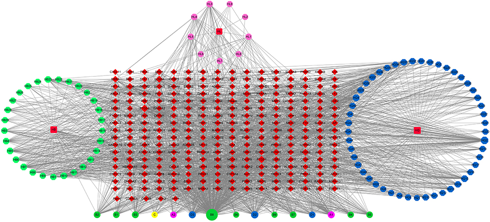 |
Figure 2 Network diagram of active ingredients and targets of Sanhuang ointment. The size of the node represents the degree value. The typical ingredients of Scutellariae Radix baicalensis, Coptidis Rhizoma, and Phellodendri Chinensis Cortex are marked with purple circles. The common elements between Coptidis Rhizoma and Phellodendri Chinensis Cortex are marked with green circles, Coptidis Rhizoma and Scutellariae Radix baicalensis are marked with a yellow circle, and Phellodendri Chinensis Cortex and Scutellariae Radix baicalensis are marked with a blue circle. The target of the active ingredient (275) are marked with red diamonds. |
The edges between nodes and nodes indicate the interaction relationship between active components and targets, and larger nodes are significant. From degree analysis, the top 10 compounds were quercetin, apigenin, beta-sitosterol, stigmasterol, magnoflorine, wogonin, columbamine, palmatine, baicalein, and isocorypalmine. Degree was 303, 79, 76, 64, 50, 46, 44, 40, 38, and 37, respectively.
Potential Targets for Sanhuang Ointment in the Treatment of MRSA Infections of the Skin and Soft Tissue
A total of 329 MRSA infection-related gene targets were identified using Gene cards, OMIM, TTD, Drug bank, and DisGeNET databases. The Venny tool was used to intersect the selected active ingredient targets of Sanhuang ointment with MRSA infection targets to obtain 34 common targets of Sanhuang ointment and MRSA infection.
Construction and Analysis of the Interaction Network
To comprehensively investigate the core pharmacological mechanism of Sanhuang ointment in treating skin and soft tissue MRSA infections, we constructed a protein–protein interaction (PPI) network using overlapping genes. The network has 22 nodes 3 3 edges, and the first three bases of the network diagram are TNF, IL-6, and IL-1β. The size and color of gene nodes are related to the degree value, and the thickness and color of the connecting lines are related to the correlation between nodes. The larger the node, the darker the color, the more important the node is in the network, and the greater the correlation between the nodes, the thicker the connection line, and the darker the color (Figure 3).
 |
Figure 3 Protein–Protein interaction network. (Matrix metalloproteinase9 (MMP9); 72 kDa type IV collagenase (MMP2); Amine oxidase [flavin-containing] B (MAOB); Amine oxidase [flavin-containing] A (MAOA); Cytochrome P450 3A43 (CYP3A4); Interleukin-8 (CXCL8); Nitric oxide synthase 2 (NOS2); NF-kappa-B essential modulator (IKBKG); Interferon gamma (IFNG); Tumor necrosis factor (TNF); Interleukin-6 (IL6); C-X-C motif chemokine 2 (CXCL2); Nitric oxide synthase 3 (NOS3); Intercellular adhesion molecule 1 (ICAM1); Interleukin-1 beta (IL1B); C-reactive protein (CRP); Protein kinase C delta type (PRKCD); C-C motif chemokine 2 (CCL2); Hypoxia-inducible factor 1-alpha (HIF1A); E-selectin (SELE); Cytochrome P450 1A2 (CYP1A2); Fibronectin (FN1)). |
Enrichment Analysis of Sanhuang Ointment for the Therapeutic Targets in Skin and Soft Tissue MRSA Infections
GO functional and KEGG pathway enrichment analysis of the 34 common targets was performed using the DAVID database to further understand the pharmacological mechanisms of Sanhuang ointment against MRSA in skin and soft tissue infections. The threshold was set at p < 0.05. We found that biological process functions were mainly related to inflammatory responses, responses to xenogeneic stimuli, and defense responses to bacteria. Cellular components were mainly associated with membrane rafts, membrane microdomains, and plasma membrane rafts. The molecular function was mainly related to cytokine activity, heme binding, and protein homodimer activity (Figure 4). The results of KEGG analysis showed that treatment with Sanhuang ointment for MRSA infection mainly targeted the inflammatory response. The top 10 pathways were the AGE-RAGE signaling pathway in diabetic complications, fluid shear stress and atherosclerosis, lipid and atherosclerosis, pathways in cancer, IL-17 signaling pathway, Chagas disease, TNF-α signaling pathway, malaria, and rheumatoid arthritis (Figure 5).
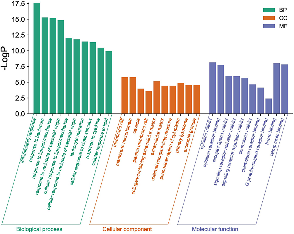 |
Figure 4 GO enrichment analysis of 34 crosspoint targets. The X-axis represents the top 10 significantly enriched biological processes, cellular components, and molecular functions. The Y-axis represents -log p-values. |
 |
Figure 5 KEGG enrichment analysis of 34 crosspoint targets. The Y-axis represents the top 20 significantly enriched pathways. The X-axis shows the ratio of enriched target genes to background genes. The size of the dots indicates the number of target genes in the pathway, and their color range reflects the different p-values. |
Screening of Bioactive Compounds via UPLC-MS/MS Analysis
Using UPLC-MS/MS analysis, we identified 743 bioactive compounds in Sanhuang ointment (Table 4). Of these, some important bioactive compounds were quercetin, apigenin, beta-sitosterol, stigmasterol, magnoflorine, wogonin, columbamine, palmatine, baicalein, and isocorypalmine.
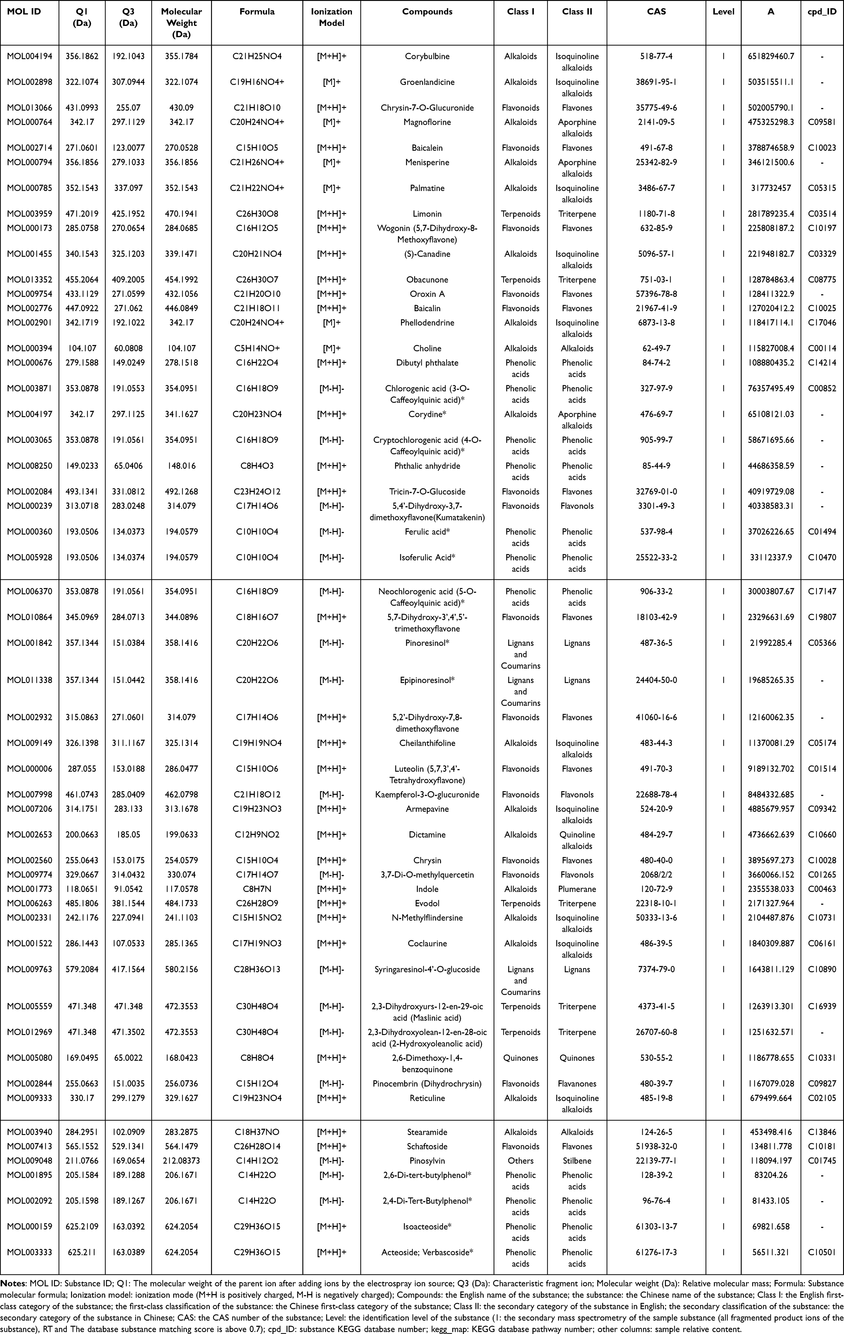 |
Table 4 The Chemical Constituents and Related Information of Sanhuang Ointment |
Pathological Changes in Infected Skin Tissues
The skin tissue structure of rats in the blank group was clear. In the model group, the squamous epithelium of the epidermal layer of the skin tissue was generally thickened. Further, the epidermal structure in the abscess area was destroyed and the level was unclear; the dermis layer became thinner and the staining deepened. The collagen fibers accumulate into sheets and clumps, and a large number of inflammatory cells infiltrated in each layer; the subcutaneous tissue structure is reduced. In the Sanhuang ointment high-dose group, the collagen fiber accumulation in the infected tissue was significantly reduced, the surrounding granulation tissue was filled, and new thin-walled capillaries were observed. In Sanhuang ointment low-dose group, the degree of injury of infected tissue was more serious compared with that in the high-dose group. Further, collagen fibers accumulated into cords and the necrotic layer was more prominent compared with that in the high-dose group. The granulation tissue filling was less than that seen in Sanhuang ointment high-dose group (Figure 6).
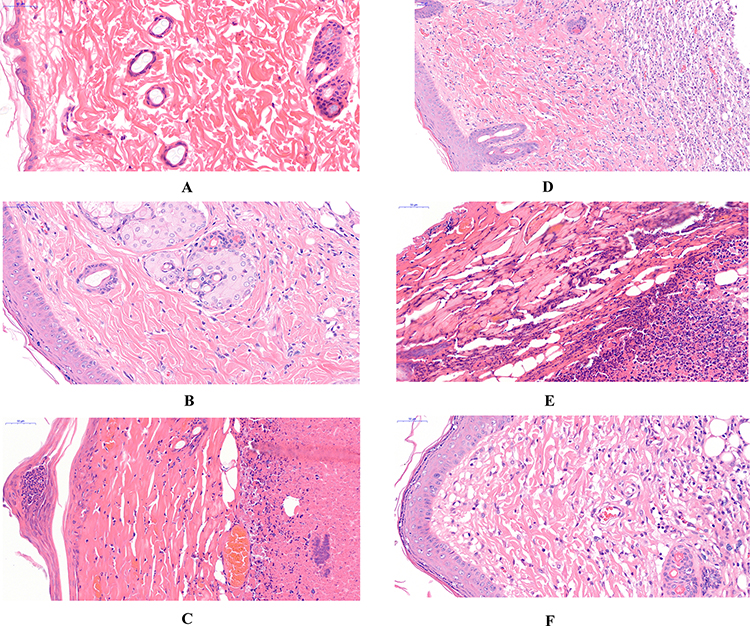 |
Figure 6 Histomorphology of rat skin in each group (HE staining, 40×). Notes: (A) Blank group; (B) Model group; (C) Mupirocin Ointment group; (D) Sanhuang Ointment high-dose group; (E) Sanhuang Ointment medium-dose group; (F) Sanhuang Ointment low-dose group. |
Determination of the Contents of Interleukin IL-1β, IL-4, IL-5, IL-6, IL-17, TNF-α, and IFN-γ in Serum and Skin Tissues of Rats by ELISA
Compared with the blank group, the model group showed significantly increased levels of IL-1β, IL-4, IL-5, IL-6, IL-6, IL-17, TNF-α, and IFN-γ in the serum and skin tissues (P < 0.01); compared with the model group, the Sanhuang ointment high- and medium-dose groups showed significantly reduced levels of IL-1β, IL-4, IL-5, IL-6, IL-6, IL-17, TNF-α, and IFN-γ in the serum and skin tissues (P < 0.05, P < 0.01). The results are shown in Figure 7.
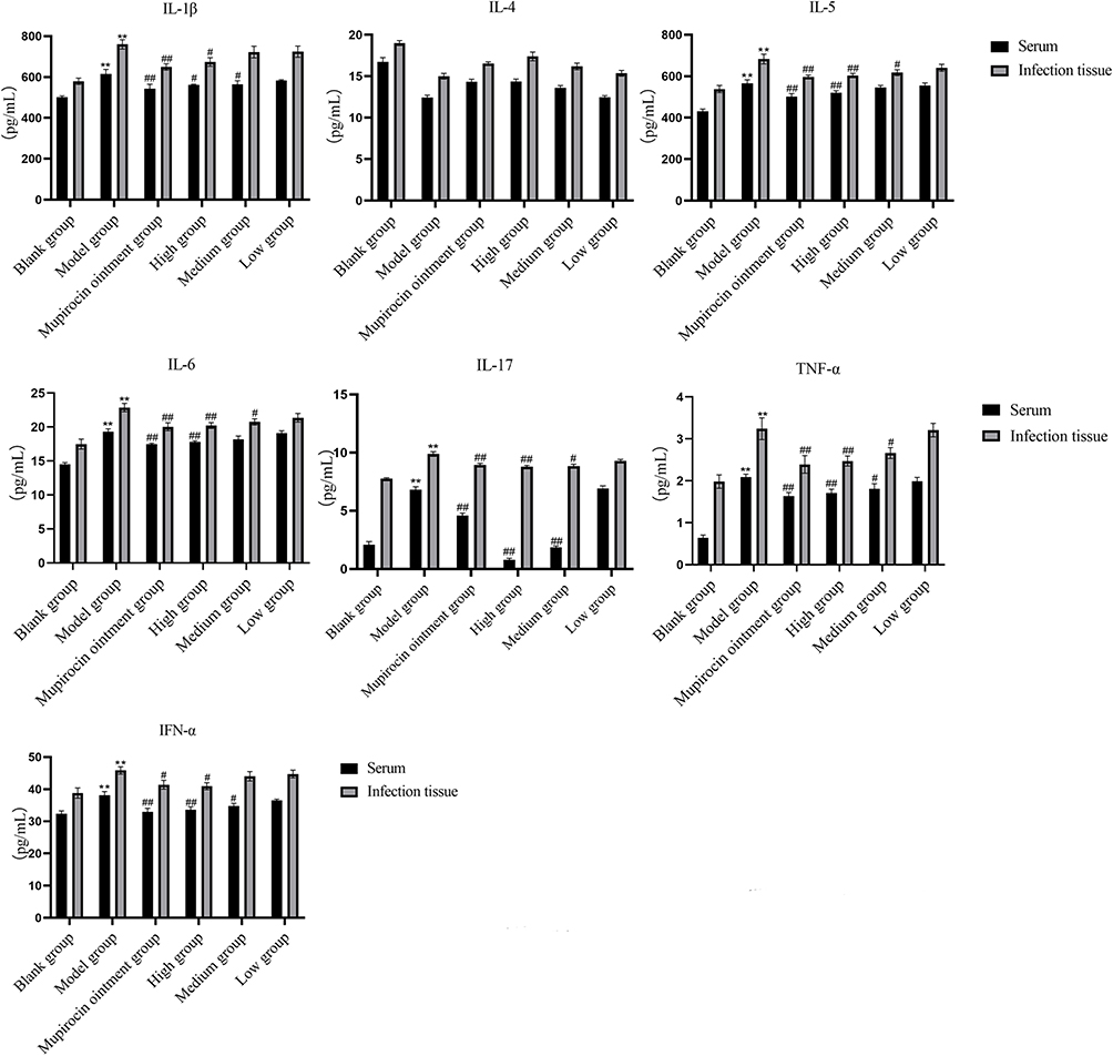 |
Figure 7 Comparison of IL-1β, IL-4, IL-5, IL-6, IL-17, TNF-α, and IFN-γ contents in the serum and skin tissues of rats in each group (x±s). Notes: Compared with the blank group, **P<0.01; compared with the model group, #P<0.05, ##P<0.01. |
Expression Levels of IL-1β, IL-4, IL-5, IL-6, IL-17, TNF-α, TRAF6, TAK1, TAB1, IKK β, NF-κB p65 mRNA in Rat Skin Tissues
Compared with the blank group, the model groups showed significantly increased levels of IL-1β, IL-4, IL-5, IL-6, IL-17, TNF-α, TRAF6, TAK1, TAB1, IKKβ, and NF-κB p65 mRNA in the skin tissue of rats (P < 0.01); compared with the model group, the Sanhuang ointment high- and medium-dose groups showed significantly decreased levels of IL-1β, IL-4, IL-5, IL-6, IL-17, TNF-α, TRAF6, TAK1, TAB1, IKKβ, and NF-κB p65 mRNA in the skin tissue of rats (P < 0.05, P < 0.01). Results are shown in Figure 8.
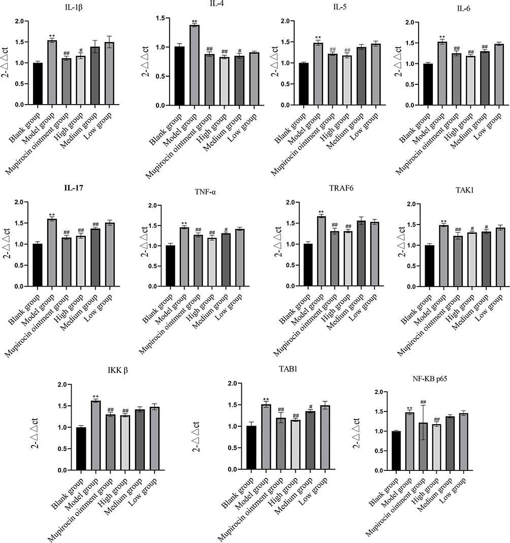 |
Figure 8 Comparison of IL-1β, IL-4, IL-5, IL-6, IL-17, TNF-α, IFN-γ, TRAF6, TAK1, TAB1, IKKβ, and NF-κB p65 mRNA expression levels in the skin tissue of rats in each group (x±s). Notes: Compared with the blank group, **P<0.01; compared with the model group, #P<0.05, ##P<0.01. |
TRAF6, TAB1, TAK1, IKK β, and NF-κB p65 Protein in Infected Rat Skin Tissues
Compared with the blank group, the model group showed significantly increased expression levels of TNF receptor-associated factor 6, transforming growth factor kinase 1, transforming growth factor kinase 1 binding protein 1, kappa B inhibitor kinase β, and NF-κB p65 protein in the skin tissue of rats (P < 0.01); further, compared with the model group, the Sanhuang ointment high- and medium-dose groups showed significantly decreased expression levels of NF-κB p65 protein in the skin tissue of rats (P < 0.05, P < 0.01). The results are shown in Figure 9.
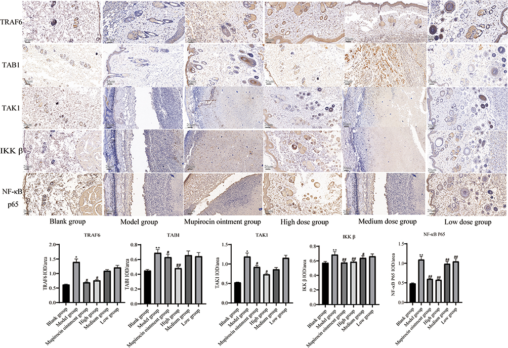 |
Figure 9 Comparison of TRAF6, TAK1, TAB1, IKKβ, and NF-κB p65 protein expressions in the skin tissues of rats in each group (x±s). Notes: Compared with the blank group, *P<0.05, **P<0.01; compared with the model group, #P<0.05, ##P<0.01. |
Discussion
According to the characteristics of clinical symptoms of local soft tissue infection on the body surface, which are often accompanied by “redness, swelling, heat, and pain”, it belongs to the category of yang syndrome of “swelling ulcer” in TCM and is mostly treated from the pathogenesis of “heat toxin”, with significant effect.
In this study, 275 potential targets of Sanhuang ointment were screened using a network pharmacology approach. In all, 34 common targets were identified on intersecting 329 MRSA infection targets. Enrichment analysis identified 488 biological processes, 32 molecular functions, 20 cellular components, and 76 signaling pathways. We found that the key compounds in the 254 active Sanhuang ointment immune system’s response against MRSA infection were as follows: quercetin, apigenin, beta-sitosterol, stigmasterol, magnoflorine, wogonin, columbamine, palmatine, baicalein, and isocorypalmine. By regulating NOS2, IL6, TNF-α, NOS3, CXCL8, IL1B, CCL2, IFNG, IKBKG, ICAM1, and other related target proteins against MRSA infection. These abovementioned components are also involved in regulating key biological pathways, such as inflammation and bacterial infection, and may treat MRSA infection via the IL-17/NF-κB signaling pathway.
IL-17 is an effector cytokine of the innate and adaptive immune systems involved in antimicrobial host defense and tissue integrity maintenance.29 Signaling through the IL-17RA/IL-17RC heterodimeric receptor complex triggers homotypic interactions of IL-17RA and IL-17RC chains with the TRAF3IP2 linker. This leads to downstream TRAF6-mediated activation of NF-κB and MAP kinase pathways, ultimately resulting in transcriptional activation of cytokines, chemokines, antimicrobial peptides, and interferon matrix metalloproteinases, accompanied by a potentially strong inflammatory response.29–34 Studies have confirmed that IL-17 expression levels are increased in infected tissues and sera of rats with skin and soft tissue MRSA infections, inducing an inflammatory response and thus the release of other inflammatory factors (ie, IL-1β, IL-4, IL-5, IL-6, TNF-α, and IFN-γ) in rat skin and soft tissue, thereby leading to imbalanced host inflammatory response.35 NF-κB has pleiotropic regulatory functions and can bind to multiple promoters and participate in the regulation of multiple inflammatory genes.36 When inflammatory changes occur in the skin and soft tissue, serum IL-17 levels rise, inducing NF-κB activity and promoting inflammation in the skin and soft tissue. Activated NF-κB can in turn reverse promote inflammatory factor expression and lead to further skin and soft tissue injury.37 TAB1 is an adaptor protein related to the N-terminal kinase domain of transforming growth factor β-activated kinase 1 (TAK1) and is an essential binding protein for sustained TAK1 activation.38 TAK1 is a key molecule in the IL-17/NF-κB signaling pathway that phosphorylates IκB kinase (IKK) upon binding with TAB1, ultimately leading to the nuclear translocation of the transcription factor NF-κB and promoting downstream inflammatory factor production.39 Mammalian NF-κB is formed by members of the Rel family of five related proteins that bind to form dimers, including p50 and p65, which contain major NF-κB transcriptional activity. NF-κB is responsible for the expression of a large number of pro-inflammatory mediators and promotes innate immune cell leukocyte recruitment.37 Accordingly, when the body is infected by microorganisms, an inflammatory response is initiated, inducing the gene expression of a large number of pro-inflammatory cytokines and chemokines through NF-κB activation.40 The pro-inflammatory cytokines including TNF-α and IL-1β can also directly activate the NF-κB pathway. This positive feedback effect contributes to the amplification of the inflammatory response, which persists at the site of infection and helps clear invading pathogens. In contrast, the recruited leukocytes (neutrophils) are quite important for regulating the inflammatory response. Given that neutrophils function in an unfavorable microenvironment, NF-κB regulates the survival of these neutrophils. Further, NF-κB regulates the transcription of many genes, and post-transcriptionally expressed proteins are widely involved in cell adhesion, differentiation, proliferation, angiogenesis, and apoptosis in addition to immune responses and inflammatory responses.41
In this study, a rat model of MRSA infection in the skin and soft tissue was designed to observe the effect of the external application of Sanhuang ointment on infected soft tissues. Furthermore, the anti-inflammatory mechanism of Sanhuang ointment was explored based on the pathological changes and the expressions of key molecules of signaling pathways in the serum and infected tissues. The results showed that compared with the blank group, the model group showed significantly increased levels of IL-1β, IL-4, IL-5, IL-6, TNF-α, IFN-γ, and IL-17 downstream inflammatory factors of IL-17/NF-κB signaling pathway in the serum and skin tissues of rats. Protein and mRNA expressions of TRAF6, TAK1, TAB1, IKK, and NF-κB p65 were significantly increased in the model group, suggesting that the skin tissues were significantly infected and an inflammatory response was aggravated. Compared with the model group, the Sanhuang ointment groups showed significantly improved general conditions with improvement in pathological changes in rats. The expression levels of the abovementioned factors were reduced to varying degrees, indicating that Sanhuang ointment external application can reduce MRSA-induced skin and soft tissue inflammation in rats; thus, Sanhuang ointment may play an anti-inflammatory role by inhibiting the expression of key factors of IL-17/NF-κB signaling pathway, thereby reducing the release of downstream pro-inflammatory factors. In this study, the targets of Sanhuang ointment and the involved signal transduction pathways were screened by network pharmacology and verified by animal experiments to reveal the mechanism of action of Sanhuang ointment in the treatment of MRSA infections in the skin and soft tissues, providing a scientific basis and further ideas for future research. The main chemical components of Sanhuang ointment were further verified by LC-MS/MS. The molecular mechanism of its network pharmacology-based anti-skin and soft tissue infection against MRSA was initially elucidated, and further experimental verification is needed.
Conclusion
Sanhuang ointment may inhibit the inflammatory response induced by MRSA in the skin and soft tissue infections by targeting the IL-17/NF-κB signaling pathway. We study the active components and mechanism of action of Sanhuang ointment on MRSA infection through network pharmacology.
Abbreviations
MRSA, Methicillin-resistant Staphylococcus aureus; SSTI, Skin and soft tissue infections; NF-κB, Nuclear factor kappa-B; MAPK, Mitogen-activated protein kinase; NO, Nitrogen monoxide; iNOS, inducible nitric oxide synthase; TLR2, Toll Like Receptor 2; IL-1β, Interleukin 1 beta; IL-4, Interleukin 4; IL-5, Interleukin 5; IL-6, Interleukin 6; IL-17, Interleukin 17; TNF-α, Tumor necrosis factor-alpha; IFN-γ, Interferon-gamma; TRAF6, TNF receptor associated factor 6; TAK1, Transforming growth factor kinase 1; TAB1, Transforming growth factor β-activated kinase 1 binding protein 1; IKKβ, Inhibitor of nuclear factor kappa-B kinase subunit beta.
Data Sharing Statement
All data generated or analyzed during this study are included in this article.
Author Contributions
All authors made a significant contribution to the work reported, whether that is in the conception, study design, execution, acquisition of data, analysis and interpretation, or in all these areas; took part in drafting, revising, or critically reviewing the article; gave final approval of the version to be published; have agreed on the journal to which the article has been submitted; and agree to be accountable for all aspects of the work.
Funding
This research was supported by the National Natural Science Foundation of China (81860850), the Gansu Provincial Department of Education Project (2021CXZX-734 and 2021CXZX-736), and the Gansu Provincial Higher Education Innovation Project (2023S-76).
Disclosure
Haibang Pan and Tianming Wang are co-first authors for this study. The authors declare that they have no known competing financial interests or personal relationships that could have appeared to influence the work reported in this paper.
References
1. Moffarah AS, Al Mohajer M, Hurwitz BL, Armstrong DG. Skin and soft tissue infections. Microbiol Spectr. 2016;4(4). doi:10.1128/microbiolspec.DMIH2-0014-2015
2. Lakhundi S, Zhang KY. Methicillin-resistant Staphylococcus aureus: molecular characterization, evolution, and epidemiology. Review. Clin Microbiol Rev. 2018;31(4):103. e00020–18. doi:10.1128/cmr.00020-18
3. Turner NA, Sharma-Kuinkel BK, Maskarinec SA, et al. Methicillin-resistant Staphylococcus aureus: an overview of basic and clinical research. Review. Nat Rev Microbiol. 2019;17(4):203–218. doi:10.1038/s41579-018-0147-4
4. Bateman A, Martin M-J, Orchard S. UniProt: the universal protein knowledgebase in 2021. Nucleic Acids Res. 2021;49(D1):D480–D489. doi:10.1093/nar/gkaa1100
5. Baek JY, Chung DR, Ko KS, et al. Genetic alterations responsible for reduced susceptibility to vancomycin in community-associated MRSA strains of ST72. J Antimicrob Chemother. 2017;72(9):2454–2460. doi:10.1093/jac/dkx175
6. Werth BJ, Jain R, Hahn A, et al. Emergence of dalbavancin non-susceptible, vancomycin-intermediate Staphylococcus aureus (VISA) after treatment of MRSA central line-associated bloodstream infection with a dalbavancin- and vancomycin-containing regimen. Clin Microbiol Infect. 2018;24(4):429.e1–429.e5. doi:10.1016/j.cmi.2017.07.028
7. Haibang P, Bo W, Xinping W, Guotai W, Hua Y. 2538 cases of common surgical diseases treated by external application of Sanhuang ointment. J External TreatTrad Chin Med. 2015;24(02):30–31.
8. Haibang P, Guotai W, Bo Y. Experimental study on anti-inflammatory and analgesic effects of different blends of Sanhuang Tablet. China Trad Chin Med Technol. 2015;22(05):502–503.
9. Haibang P, Guotai W, Jianfeng Y, Bo W, Xiaopeng D. Clinical observation of sanhuang ointment in treating local soft tissue infection. Chin J Exp Formulas. 2017;23(20):174–179. doi:10.13422/j.cnki.syfjx.2017200174
10. Hao Y, Huo J, Wang T, Sun G, Wang W. Chemical profiling of Coptis rootlet and screening of its bioactive compounds in inhibiting Staphylococcus aureus by UPLC-Q-TOF/MS. J Pharm Biomed Anal. 2020;180:113089. doi:10.1016/j.jpba.2019.113089
11. Hwang SJ, Lee HJ. Phenyl-beta-d-glucopyranoside exhibits anti-inflammatory activity in lipopolysaccharide-activated RAW 264.7 cells. Article. Inflammation. 2015;38(3):1071–1079. doi:10.1007/s10753-014-0072-2
12. Guven-Maiorov E, Keskin O, Gursoy A, Nussinov R. A structural view of negative regulation of the toll-like receptor-mediated inflammatory pathway. Biophys J. 2015;109(6):1214–1226. doi:10.1016/j.bpj.2015.06.048
13. Choi YY, Kim MH, Han JM, et al. The anti-inflammatory potential of Cortex Phellodendron in vivo and in vitro: down-regulation of NO and iNOS through suppression of NF-κB and MAPK activation. Int Immunopharmacol. 2014;19(2):214–220. doi:10.1016/j.intimp.2014.01.020
14. Hu ZW, Lin JH, Chen JT, et al. Overview of viral pneumonia associated with influenza virus, respiratory syncytial virus, and coronavirus, and therapeutics based on natural products of medicinal plants. Review. Front Pharmacol. 2021;12(21):630834. doi:10.3389/fphar.2021.630834
15. Dinda B, Dinda S, DasSharma S, Banik R, Chakraborty A, Dinda M. Therapeutic potentials of baicalin and its aglycone, baicalein against inflammatory disorders. Eur J Med Chem. 2017;131:68–80. doi:10.1016/j.ejmech.2017.03.004
16. Wang H-Z, Yu C-H, Gao J, Zhao G-R. HPLC分析比较炮制和提取方法对黄芩活性成分的影响 [Effects of processing and extracting methods on active components in Radix Scutellariae by HPLC analysis]. Zhongguo Zhong Yao Za Zhi. 2007;32(16):1637–1640. Chinese.
17. Liao H, Ye J, Gao L, Liu Y. The main bioactive compounds of Scutellaria baicalensis Georgi. for alleviation of inflammatory cytokines: a comprehensive review. Biomed Pharmacother. 2021;133:110917. doi:10.1016/j.biopha.2020.110917
18. Haibang P, Tianming W, Yan C, et al. Effects of sanhuang ointment on subcutaneous soft tissue inflammation and TLR2/ NF-κB signaling pathway in MRSA infection model rats. Chin J Inform Trad Chin Med. 2022;29(08):54–59. doi:10.19879/j.cnki.1005-5304.202112157
19. Li S, Zhang ZQ, Wu LJ, Zhang XG, Li YD, Wang YY. Understanding ZHENG in traditional Chinese medicine in the context of neuro-endocrine-immune network. IET Syst Biol. 2007;1(1):51–60. doi:10.1049/iet-syb:20060032
20. Li S, Zhang B. Traditional Chinese medicine network pharmacology: theory, methodology and application. Chin J Nat Med. 2013;11(2):110–120. doi:10.1016/S1875-5364(13)60037-0
21. Wang SL, Tang C, Zhao H, et al. Network pharmacological analysis and experimental validation of the mechanisms of action of Si-Ni-San against liver fibrosis. Article. Front Pharmacol. 2021;12(19):656115. doi:10.3389/fphar.2021.656115
22. Hopkins AL. Network pharmacology: the next paradigm in drug discovery. Nat Chem Biol. 2008;4(11):682–690. doi:10.1038/nchembio.118
23. Ru J, Li P, Wang J, et al. TCMSP: a database of systems pharmacology for drug discovery from herbal medicines. J Cheminform. 2014;6(1):13. doi:10.1186/1758-2946-6-13
24. Pichler K, Warner K, Magrane M. SPIN: submitting sequences determined at protein level to UniProt. Curr Protoc Bioinformatics. 2018;62(1):e52. doi:10.1002/cpbi.52
25. Amberger JS, Bocchini CA, Schiettecatte F, Scott AF, Hamosh A. OMIM.org: Online Mendelian Inheritance in Man (OMIM®), an online catalog of human genes and genetic disorders. Nucleic Acids Res. 2015;43(Database issue):D789–D798. doi:10.1093/nar/gku1205
26. Szklarczyk D, Gable AL, Lyon D, et al. STRING v11: protein-protein association networks with increased coverage, supporting functional discovery in genome-wide experimental datasets. Nucleic Acids Res. 2019;47(D1):D607–D613. doi:10.1093/nar/gky1131
27. Malachowa N, Kobayashi SD, Lovaglio J, DeLeo FR. Mouse model of Staphylococcus aureus skin infection. Methods Mol Biol. 2019;1960:139–147. doi:10.1007/978-1-4939-9167-9_12
28. Wang Z, Gao C, Zhang L, Sui R. Retraction notice to “Hesperidin methylchalcone (HMC) hinders amyloid-β induced Alzheimer’s disease by attenuating cholinesterase activity, macromolecular damages, oxidative stress and apoptosis via regulating NF-κB and Nrf2/HO-1 pathways” [Int. J. Biol. Macromol. 233 (2023) 123169]. Int J Biol Macromol. 2023;249:125762. doi:10.1016/j.ijbiomac.2023.125762
29. Boisson B, Wang C, Pedergnana V, et al. An ACT1 mutation selectively abolishes interleukin-17 responses in humans with chronic mucocutaneous candidiasis. Immunity. 2013;39(4):676–686. doi:10.1016/j.immuni.2013.09.002
30. Liu C, Qian W, Qian Y, et al. Act1, a U-box E3 ubiquitin ligase for IL-17 signaling. Sci Signal. 2009;2(92):ra63. doi:10.1126/scisignal.2000382
31. Puel A, Cypowyj S, Bustamante J, et al. Chronic mucocutaneous candidiasis in humans with inborn errors of interleukin-17 immunity. Science. 2011;332(6025):65–68. doi:10.1126/science.1200439
32. Kuestner RE, Taft DW, Haran A, et al. Identification of the IL-17 receptor related molecule IL-17RC as the receptor for IL-17F. J Immunol. 2007;179(8):5462–5473. doi:10.4049/jimmunol.179.8.5462
33. Wright JF, Bennett F, Li B, et al. The human IL-17F/IL-17A heterodimeric cytokine signals through the IL-17RA/IL-17RC receptor complex. J Immunol. 2008;181(4):2799–2805. doi:10.4049/jimmunol.181.4.2799
34. Fossiez F, Djossou O, Chomarat P, et al. T cell interleukin-17 induces stromal cells to produce proinflammatory and hematopoietic cytokines. J Exp Med. 1996;183(6):2593–2603. doi:10.1084/jem.183.6.2593
35. Veldhoen M. Interleukin 17 is a chief orchestrator of immunity. Nat Immunol. 2017;18(6):612–621. doi:10.1038/ni.3742
36. Taniguchi K, Karin M. NF-κB, inflammation, immunity and cancer: coming of age. Nat Rev Immunol. 2018;18(5):309–324. doi:10.1038/nri.2017.142
37. Mitchell JP, Carmody RJ. NF-κB and the Transcriptional Control of Inflammation. Int Rev Cell Mol Biol. 2018;335:41–84. doi:10.1016/bs.ircmb.2017.07.007
38. Y-R X, Lei C-Q. TAK1-TABs complex: a central signalosome in inflammatory responses. Front Immunol. 2020;11:608976. doi:10.3389/fimmu.2020.608976
39. Amatya N, Garg AV, Gaffen SL. IL-17 signaling: the Yin and the Yang. Trends Immunol. 2017;38(5):310–322. doi:10.1016/j.it.2017.01.006
40. Liu K, Ding T, Fang L, et al. Organic selenium ameliorates -induced mastitis in rats by inhibiting the activation of NF-κB and MAPK signaling pathways. Front Vet Sci. 2020;7:443. doi:10.3389/fvets.2020.00443
41. Liu T, Zhang L, Joo D, Sun S-C. NF-κB signaling in inflammation. Signal Transd Target Ther. 2017;2(1). doi:10.1038/sigtrans.2017.23
 © 2023 The Author(s). This work is published and licensed by Dove Medical Press Limited. The full terms of this license are available at https://www.dovepress.com/terms.php and incorporate the Creative Commons Attribution - Non Commercial (unported, v3.0) License.
By accessing the work you hereby accept the Terms. Non-commercial uses of the work are permitted without any further permission from Dove Medical Press Limited, provided the work is properly attributed. For permission for commercial use of this work, please see paragraphs 4.2 and 5 of our Terms.
© 2023 The Author(s). This work is published and licensed by Dove Medical Press Limited. The full terms of this license are available at https://www.dovepress.com/terms.php and incorporate the Creative Commons Attribution - Non Commercial (unported, v3.0) License.
By accessing the work you hereby accept the Terms. Non-commercial uses of the work are permitted without any further permission from Dove Medical Press Limited, provided the work is properly attributed. For permission for commercial use of this work, please see paragraphs 4.2 and 5 of our Terms.